
113
7 Mycobacterium
I. Olsen , R. G. Barletta , and C. O. Thoen
INTRODUCTION Mycobacteria belong to the Order Actinomycetales , Family Mycobacteriaceae . The genus Mycobac-terium includes the Mycobacterium tuberculosis and Mycobacterium avium complexes, other patho-genic mycobacteria, and numerous species of sap-rophytic microorganisms present in soil and water. The Mycobacterium tuberculosis complex includes M. tuberculosis , M. africanum , M. canettii , M. bovis , M. pinnipedii , M. caprae , and M. microti (fi g. 7.1 A ). The M. avium complex includes M. avium subsp. avium , M. avium subsp. hominissuis , M. avium subsp. paratuberculosis , and M. intracellu-lare (fi g. 7.1 B). Some other mycobacteria of clinical signifi cance are Mycobacterium chelonae , Myco-bacterium fortuitum , Mycobacterium kansasii , Myco bacterium leprae , Mycobacterium marinum , Mycobacterium ulcerans , and Mycobacterium scrofulaceum .
CHARACTERISTICS AND SOURCES OF THE ORGANISMS Mycobacteria are obligate aerobes, nonspore forming and nonmotile bacilli, and are 0.6 – 1.0 × 1.0 – 10 μ m in size. Their high cell wall lipid content excludes standard aniline dyes, so that once stained with special staining procedures, mycobac-teria are resistant to decolorization even by acid alcohol. This property is termed acid fastness, so that mycobacteria are commonly referred to as acid - fast bacilli. In contrast, these microorganisms are not readily stained with the Gram method and are considered weakly gram - positive. Growth rates
for mycobacteria are slow, with generation times ranging from 2 to more than 20 h. Based on different generation times, mycobacteria can be divided into slow and rapid growers. Slow growers require more than 7 days to form visible colonies on solid medium, whereas rapid growers form colonies within 7 days (Holt et al. 1994 ).
Tuberculosis remains the leading cause of death in humans caused by a single infectious agent, being responsible for nearly 2 million deaths annu-ally. It is caused primarily by M. tuberculosis ; M. africanum , M. bovis , and M. canettii account for less than 1% of tuberculosis in humans (Thoen and LoBue 2007; Thoen et al. 2009 ). In addition to the host adaptation of M. bovis largely to cattle, other host - adapted variants of M. bovis have been desig-nated (Hewinson et al. 2006 ), such as M. pinnipedii (seal - adapted) and M. caprae (goat - adapted). Interestingly, M. bovis bacille Calmette - G ü erin (BCG), attenuated by in vitro passages on potato slices, is used to vaccinate humans throughout the world. M. africanum and M. canettii are human pathogens. Mycobacterium microti has been iso-lated from humans, voles, and some other animals (Thoen et al. 2009 ). Pathogenic mycobacteria all produce granulomatous lesions in tissues of humans and a wide range of domestic and wild animals. Although the tubercle bacillus was discovered more than 120 years ago, defi nitive information on its pathogenesis is not yet available, although understanding is developing at a remarkable rate. Unex plained differences in susceptibility of differ-ent animals to various acid - fast bacilli occur (Thoen and Barletta 2004 ). M. tuberculosis , the human
Pathogenesis of Bacterial Infections in Animals, Fourth EditionEdited by C. L. Gyles, J. F. Prescott, J. G. Songer, and C. O. Thoen© 2010 Blackwell Publishing. ISBN: 978-0-813-81237-3

114 Chapter 7
Figure 7.1. Proposed evolution of the M. tuberculosis and M. avium complexes The tree diagrams for the proposed evolution of members of the M. tuberculosis (A) and M. avium (B) complexes are shown. See text for further details and citations.
M. tuberculosiscomplex
M. canettii(humans)
M. tuberculosis(humans)
M. africanum(humans)
M. microti(rodents)
M. bovis(humans,
ruminants)
M. pinnipedii(fin-footed
mammals)
M. caprae(goats)
(A)
M. aviumcomplex
M. avium subsp.
hominissuis(humans, porcine,
ruminants)
M. avium subsp.
paratuberculosis(ruminants)
Cattle strains Sheep strains
M. aviumsubsp.avium
(birds)
M. intracellulare(birds, humans)
(B)
tubercle bacillus, produces progressive generalized disease in nonhuman primates, dogs, swine, and guinea pigs, although cattle and cats are quite resis-tant (Thoen 2010 ). M. tuberculosis may induce tuberculin skin sensitivity in cattle and other ani-mals. M. bovis , the agent of bovine tuberculosis, is a slow - growing nonphotochromogenic organism that also causes disease in other domestic and wild animals, and has been reported in humans in several countries (Thoen et al. 2006 ; Thoen et al. 2009 ). Biochemical tests are available for differentiating bacteria of the M. tuberculosis complex, but molec-ular techniques are now widely used in reference
laboratories around the globe (Harris 2006 ; Thoen et al. 2009 ).
Mycobacterium leprae , the cause of leprosy (also known as Hansen ’ s disease), is a chronic granulo-matous disease. Leprosy is still considered a public health problem in countries in Africa and Southeast Asia, with a prevalence rate of more than 1 case per 10,000 individuals (Meima et al. 2004 ). M. leprae and leprosy has been identifi ed in armadillos in the United States. Mycobacterium lepraemurium has been isolated from leprosy - like lesions in cats, rats, and mice. Mycobacterium chelonei, M. intracellulare , M. marinum , M. nonchromo-
Mycobacterium 115
genicum , and certain other mycobacteria have been isolated from granulomatous lesions in cold - blooded animals.
Microorganisms of the M. avium complex have the widest host range among all mycobacteria. M. avium subsp. avium (serovars 1, 2, and 3) are iso-lated from tuberculous lesions in humans, birds, domestic, and wild animals (Thoen et al. 1981 ). In birds, disease is usually progressive, with lesions in the liver and spleen, whereas lesions in other animals are usually confi ned to lymph nodes asso-ciated with the intestinal tract. M. avium subsp. hominissuis is the subspecies most frequently cau-sing lesions in humans and swine, and M . intra-cellulare is widely distributed in the environment, causing granulomatous lesions mainly in cold - blooded animals (Thoen 2010 ). Rabbits are highly susceptible to experimental infection with M. avium subsp. avium , but relatively resistant to M. intracel-lulare. Interestingly, birds are susceptible to M. avium subsp. avium but resistant to infection by members of the M. tuberculosis complex (Thoen and Barletta 2004 ).
Mycobacterium avium subsp. paratuberculosis causes a transmissible intestinal disorder of rumi-nants commonly known as Johne ’ s disease that has a signifi cant economic impact on the livestock industry (Harris and Barletta 2001 ). Cattle, sheep, goats, and certain wild ruminants are susceptible. In addition, it has been suggested that this microorgan-ism may be the etiologic agent of Crohn ’ s disease, an infl ammatory bowel disease in humans (Chacon et al. 2004 ). However, this issue still remains controversial. A characteristic that is useful in differentiating this organism is its dependency on mycobactin, an iron - chelating agent, for in vitro growth. Mycobactin was initially extracted from Mycobacterium phlei , but later mycobactin J and certain extracellular iron - binding compounds were isolated from M. avium . Molecular techniques such as PCR and restriction endonuclease analysis have been developed for identifying M. avium subsp. paratuberculosis (Harris and Barletta 2001 ).
Mycobacterium ulcerans causes chronic skin ulcers in humans, termed Buruli ulcer or Bairnsdale ulcer. These lesions are caused by the effects of mycolactone, a polyketide - derived macrolide iso-lated from M. ulcerans (George et al. 1999 ). Studies have pointed to water insects from the family Naucoridae as a possible vector for the transmission of M. ulcerans (Marsollier et al. 2002 ). M. marinum
causes tuberculosis in fi sh and amphibians as well as cutaneous granulomatous disease in humans, known as swimming pool granuloma.
Other species of mycobacteria have been iso-lated from various animals (Thoen et al. 1981 ). Mycobacterium fortuitum , a rapid - growing, non-chromogenic organism has been isolated from humans and dogs with lung lesions, cattle with mastitis, and lymph nodes of slaughter cattle and swine. Mycobacterium chelonae, also a rapid grower, has been isolated from swine and humans. Granulomatous lesions in swine and cattle, which closely resemble lesions caused by M. bovis , have been reportedly caused by M. kansasii , a slow - growing, photochromogenic organism.
BACTERIAL VIRULENCE FACTORS In recent years, remarkable progress has been made in understanding the basis of virulence and the pathogenesis of mycobacterial infections particu-larly through the application of whole genome sequencing and comparative genomic analysis.
Genomics
The science of genomics has made possible the elu-cidation of the complete genetic blueprint of several mycobacterial species of importance in human and veterinary medicine, as well as environmental species. Complete genome sequences are now available for Mycobacterium abscessus , M. avium subsp. hominissuis, M. avium subsp. paratuberculo-sis , M. bovis, M. bovis BCG, Mycobacterium gilvum , M. leprae , M. marinum, Mycobacterium smegmatis , three strains of M. tuberculosis , M. ulcerans , Mycobacterium vanbaalenii , and four mycobacteria unclassifi ed at the species level These genomes possess high GC content ( ∼ 65%). Major fi ndings for two mycobacterial species of veterinary importance are reported below.
Sequencing and annotation of the M. avium subsp. paratuberculosis genome from strain K - 10, isolated from a cow with Johne ’ s disease, have been com-pleted (Li et al. 2005 ). This strain had a low number of in vitro passages, and a genetic system including transposon mutagenesis is available for the creation of mutant strains (Foley - Thomas et al. 1995 ; Harris et al. 1999 ). The K - 10 genome is a circular chromo-some of about 4.8 Mb encoding 4344 open reading frames (ORFs) with a 69.3% GC content. About 60% of the ORFs have known homologues in
116 Chapter 7
databases while 25% encode putative proteins of unknown functions. About 75% of the M. avium subsp. paratuberculosis genes have counterparts in M. tuberculosis , and although most genes have orthologs in M. avium subsp. hominissuis , there are 39 predicted proteins that are unique to M. avium subsp. paratuberculosis. ORFs are identifi ed by a location number following standard conventions (e.g., M. avium subsp. paratuberculosis 1152 signi-fi es ORF1152 from the ORF0001 — DnaA — in the clockwise direction; see also Wu et al. 2009 for corrections to the original assembly).
The genome possesses high redundancy because of gene duplication, especially for genes involved in lipid and redox metabolism. Nonetheless, differ-ences from other mycobacterial genomes are noted in the low abundance of PE and PPE families in M. avium subsp . paratuberculosis . In addition, the salicyl - MP ligase gene ( mbtA ) is truncated, which is likely the basis of its defect in mycobactin bio-synthesis. Analysis of genetic polymorphisms, espe-cially those including large sequences, indicates that M. avium subsp. paratuberculosis originated from M. avium subsp. hominissuis in a biphasic evolu-tionary process (Alexander et al. 2009 ). First, an original pathogenic clone of M. avium subsp. para-tuberculosis arose by acquisition of novel DNA and polymorphisms. Second, sheep and cattle strains arose from this ancestral clone by subsequent lineage - specifi c deletion events.
Genome sequencing demonstrated that the M. bovis genome (4 345 492 bp for the virulent bovine isolate AF2122/97) is a down - sized version of the genome of M. tuberculosis (4 411 532 bp for the human isolate H37Rv), with more than 99.95% identity and no new genetic material as compared to M. tuberculosis (Garnier et al. 2003 ). Thus, DNA deletions in M. bovis are the major contributors to these differences, which have been found to affect genes involved in transport, cell surface structures, and intermediary metabolism. These deletions may remove genes that are unnecessary for host adapta-tion and lead to a different and sometimes even wider host range. Point mutations also play a role in defi ning the phenotype, as it is the case for M. bovis resistance to pyrazimamide. In addition, sequence variations have been found in genes coding for cell wall and secreted proteins, such as the PE_PGRS and PPE protein families. Another notable change is a mutational event in the M. bovis pyruvate kinase gene that renders M. bovis unable
to use glycerol as a carbon source. Other sequence changes involve master regulatory genes controlling the expression of multiple gene families. The analy-sis of the M. bovis genome challenged the epidemio-logical hypothesis that M. tuberculosis was a human - adapted variety of M. bovis that was acquired from cattle. The irreversible loss of DNA material uncovered by the M. bovis genome sequencing and the systematic analysis of polymorphisms in a large panel of strains led to the new paradigm that M. canettii is likely the ancestral species of the M. tuberculosis complex. Successive DNA deletions, starting by the loss of region RD9 (RD stands for regions of difference), led to differentiation of M. africanum , M. bovis , and M. microti. Moreover, M. bovis BCG experienced further deletions during in vitro laboratory adaptation and its loss of region RD1 has been implicated as the mechanism of viru-lence attenuation. Thus, similarly to M. avium subsp . paratuberculosis , evolution of mycobacterial genomes involves a dominant process of reductive deletions. Based on comparative genomic studies, fi g. 7.1 depicts an overall tree diagram for the evolution of mycobacterial genomes (Brosch et al. 2002 ; Devulder et al. 2005 ; Mueller et al. 2008 ; Alexander et al. 2009 ).
Genome sequencing of mycobacteria has been enhanced by parallel developments in the genetic systems used to create defi ned mutants and to elu-cidate the function of each gene in the pathophysiol-ogy of mycobacterial infections (Braunstein et al. 2002 ). Both plasmid and mycobacteriophage vectors have been used extensively to create recombinant or mutant strains using all means of genetic exchange, including transformation, transduction, and con-jugation. A variety of reporter genes have been expressed in mycobacterial species including beta - galactosidase, fi refl y luciferase, and the green fl uorescent protein. In addition, several vectors for conditional or antisense expression in mycobacterial systems are now available. Mycobacteriophage vectors have been particularly useful in creating defi ned mutations in many mycobacterial species, including recent application of this technology to M. avium subsp . paratuberculosis , one of the slowest growing mycobacterial species (Park et al. 2008 ). Finally, the elegant technology of transposition - site hybridization (TraSH) mutagenesis has been devel-oped to defi ne genes essential under any desired condition (Sassetti et al. 2003; Sassetti and Rubin 2003 ).

Mycobacterium 117
Molecular Strategies to Identify Virulence Determinants
Several strategies are being followed to mine myco-bacterial genomes for virulence determinants or antigens of diagnostic importance. One approach seeks to identify M. avium subsp. paratuberculosis genes whose homologues in other mycobacteria, mostly M. tuberculosis , have been fl agged as candi-date virulence determinants. For example, a tyrosine phosphatase capable of interfering with macrophage activation has been identifi ed as M. avium subsp. paratuberculosis 1985 whose homologue in M. tuberculosis is Rv2232 (Bach et al. 2006 ). Com-parative genomic approaches have identifi ed a set of 39 genes unique to M. avium subsp. paratuber-culosis whose encoded proteins may have diagnos-tic signifi cance (Bannantine and Paustian 2006 ). Screening of a M. avium subsp. hominissuis trans-poson library led to the identifi cation of a patho-genicity island that carries important virulence determinants involved in bacterial uptake by mac-rophages (Danelishvili et al. 2007 ). Comparative genomic hybridizations identifi ed genomic islands in the M. avium subsp. paratuberculosis genome demonstrating a gene organization different from subsp. hominissuis (Wu et al. 2006 ). Another approach has been the screening of transposon mutant libraries. One such study directly sequenced strain ATCC 19698 transposon mutants and classi-fi ed them into functional groups (Shin et al. 2006 ). To ascertain a role in pathogenesis, 11 mutants were selected, based on bioinformatic analysis, and used in mouse virulence experiments. This analysis led to the identifi cation of the following genes: gcpE , pstA , kdpC , papA2 , impA , umaA1 , or fabG2_2 . Recent molecular approaches used to identify viru-lence determinants have involved transcriptomic analyses and TraSH mutagenesis (Talaat et al. 2000 ; Fisher et al. 2002 ; Stewart et al. 2002 ; Sassetti et al 2003 ; Sassetti and Rubin 2003 ; Wu et al. 2007 ; Tailleux et al. 2008 )
PATHOGENESIS Overview
Mycobacteria have evolved as pathogens to survive in macrophages and to overcome multifactorial hostile innate and acquired host immune mecha-nisms, so that they can survive and eventually mul-tiply and transmit to other animals. As successful pathogens, mycobacteria have developed strategies
to turn sets of genes on and off in response to the distinct environments encountered during different stages of infection. First, the microorganisms gain entry into host macrophages, where they survive and replicate intracellularly. For example, mycobac-teria use multiple cell surface receptors, including the mannose, complement, and Fc receptors to gain entry into macrophages (Schlesinger et al. 1990 ; Bartow and McMurray 1998 ). Subsequently, myco-bacteria reside within a membrane - bound vacuole and prevent the maturation of this compartment. This arrest involves a series of alterations in the phagosome, such as changes in the pattern of acqui-sition of Rab proteins and GTPases, exclusion of the proton ATPase from the phagosome, and retention of a phagosome coat protein that would normally be released from this compartment prior to phagolyso-some fusion (Sturgill - Koszycki et al. 1996 ; Ferrari et al. 1999 ; Russell et al. 2002 ; Deretic et al. 2004 ; Deretic et al. 2006 ). Macrophages may also use an autophagic process to bypass the blockage on phagosome maturation imposed by M. tuberculosis (Deretic 2008 ).
Pathogenicity of mycobacteria is a multifactorial phenomenon requiring the participation and cumu-lative effects of several components, including complex lipids and proteins in both the cell wall and the cytoplasm of tubercle bacilli (McNeil and Brennan 1991 ). Most studies of mycobacterial virulence determinants have been performed with M. tuberculosis rather than other mycobacteria. However, the presence of homologous genes in other mycobacteria and the close relationship between these microorganisms suggest the use of similar virulence determinants and mechanisms of pathogenicity.
Role of Complex Carbohydrates and Lipids
The cell wall core of mycobacteria is composed of three covalently attached molecules: peptidoglycan, arabinogalactan, and mycolic acid. Lipids and gly-colipid complexes present in the cell wall of virulent and attenuated strains of mycobacteria have been extensively examined to understand their signi-fi cance in granuloma formation (Ehrt and Sch-nappinger 2007 ; Jain et al. 2007 ). For example, the glycolipid trehalose - 6,6 ′ dimycolate endows M. tuberculosis with the ability to form cords when grown in liquid culture medium (Goren et al. 1979 ; Glickman et al. 2000 ). Mycobacteria differ in sulfur - containing glycolipids. Sulfolipids and
118 Chapter 7
sulfatides are present in M. tuberculosis but absent from M. bovis because the glycolipid sulfotransfer-ase and arylsulphatase genes are disrupted in the latter (Rivera - Marrero et al. 2002 ). This difference may also contribute to determining the host range and tissue tropism of M. bovis . Another glycolipid, lipoarabinomannan (LAM), may contribute to arresting phagosome maturation. In addition, LAM is a powerful scavenger of reactive oxygen (ROI) and nitrogen (RNI) intermediates (Chan et al. 1991 ). Moreover, M. tuberculosis and M. bovis contain mannosylated LAM (ManLAM), which provides a way for mycobacteria to enter phagocytes via mannose receptors (Schlesinger 1993 ).
Role of Proteins and Lipoproteins
Mycobacterial proteins and protein complexes (i.e., lipoproteins) also play important and diverse roles in pathogenesis. For example, M. avium subsp. paratuberculosis attachment and internalization of bacilli to the intestinal mucosa appears to be medi-ated by FAP - P, a protein of approximately 36 kDa made as a 54 - kDa precursor and located in the inner part of the cell envelope (Secott et al. 2001 ). A recombinant strain carrying a FAP - P antisense con-struct demonstrated reduced expression and attach-ment to fi bronectin (Secott et al. 2002 ). In addition, a 35 - kDa membrane protein that reacts with antisera from cattle with Johne ’ s disease has been shown to play a role in the invasion of bovine epithelial cells (Bannantine et al. 2003 ). Proteins encoded by the mce operons seem to play a role in the entry and survival of mycobacteria within phagocytic cells, and invasion of epithelial cells (Chitale et al. 2001 ; Kumar et al. 2003 ; Gioffre et al. 2005 ). There are four mce operons in M. tuberculosis , each encoding 5 to 6 proteins, while the mce3 operon is absent from M. bovis (Cole et al. 1998 ; Garnier et al. 2003 ).
The secreted proteins in the antigen 85 complex play an important role in the development of cell - mediated immunity (Andersen et al. 1991 ). These proteins also possess enzymatic activity and cata-lyze mycolyltransfer reactions involved in the fi nal stages of mycobacterial cell wall assembly (Belisle et al. 1997 ). Superoxide dismutases (SODs) are also released into culture supernatant fl uids by several mycobacterial pathogens (Harth and Horwitz 1999 ; Liu et al. 2001 ). M. bovis and M. tuberculosis possess a redundant system of SODs. The iron — manganese - dependent SOD (SodA) is secreted and seems the more critical enzyme for resistance
against ROI (Edwards et al. 2001 ). Another mem-brane – associated copper — zinc - dependent SOD (SodC) may play an additional role in protecting tubercle bacilli against the oxidative burst of acti-vated macrophages (Piddington et al. 2001 ). Protection against RNI is provided by two alkyl hydroperoxidases denominated AhpC and AhpD (Sherman et al. 1999 ; Olsen et al. 2000 ).
Virulence Gene Regulation
Successful infection is dependent on the ability of mycobacteria to adapt to different conditions such as exposure to ROI and RNI, hypoxia, low pH, nutrient starvation, and damage to the cell surface. Mycobacteria thus have several transcriptional reg-ulators that control expression of distinct sets of genes. The best described of these are sigma factors and two - component systems.
Two - component systems consist of a transmem-brane sensor histidine kinase that reacts to various stimuli and subsequently activate its transcriptional regulator. These systems have been studied in M. tuberculosis , but little is known about them in M. bovis and M. avium subsp . paratuberculosis . Genome sequencing has revealed that conserved two - component systems are present in these species and may have similar functions.
One two - component system that is important for virulence is PhoPR. An M. tuberculosis PhoP knockout mutant was more attenuated than BCG and conferred protective immunity against tubercu-losis (Martin et al. 2006 ). In addition, an outbreak of M. bovis in humans was caused by a strain that elicited increased expression of PhoP due to inser-tion of IS 6110 in the promoter region (Soto et al. 2004 ). Insertion of IS 6110 in the opposite direction and at a different site of the promoter has been demonstrated in some BCG strains (Leung et al. 2008 ). This may lead to diminished expression of PhoP and may explain some of the attenuation of these strains.
DosRS, or the dormancy regulon, is believed to be relevant for latent infections and is activated during hypoxia and by nitric oxide. It was required for full virulence, but the mutants showed a different attenuation pattern in the various animal models assessed (Converse et al. 2009 ). Interestingly, while the M. avium subsp . paratuberculosis DosR has 93% similarity to the M. tuberculosis/M. bovis ortholog on the amino acid level, the downstream sensor DosS histidine kinase only has a similarity of
Mycobacterium 119
60%. In comparison, a similarity of 80 – 95% is present between the other sensor histidine kinases of the conserved two - component systems. One could thus speculate that the dormancy genes may be dif-ferently regulated in M. avium subsp. para-tuberculosis compared with M. tuberculosis and M. bovis.
Other two - component systems such as PrrAB and SenX3 - RegX3 also appear to play a role in viru-lence, and MprAB is important for persistent infec-tions. The MprAB also regulates stress responses, including the sigma factors SigB and SigE (He et al. 2006 ). Sigma factors form a reversible complex with RNA polymerases that provide promoter rec-ognition and thus affect gene transcription. The primary sigma factor SigA and the primary - like SigB are conserved in mycobacteria and allow tran-scription of housekeeping genes (Manganelli et al. 2004 ). SigA is essential for growth, whereas SigB may serve as a backup mechanism since the subdo-mains responsible for promoter recognition are almost identical to those of SigA. SigA may be involved in virulence since a mutant with an amino acid substitution led to attenuated growth of M. bovis in guinea pigs. Mycobacteria also have several sigma factors (11 in M. bovis and 17 in M. avium subsp. paratuberculosis ) that are responsive to changes in the environment and belong to the group of extracellular function sigma factors (Manganelli et al 2004 ; Sechi et al. 2007 ). One of these is SigF, which in M. bovis BCG is induced after exposure to cold - shock, hypoxia and oxidative stress. Interestingly, this was not the case in M. tuberculo-sis . This demonstrates that care should be taken when extrapolating results from one mycobacterial species to another, especially when it comes to the complexity of gene regulation. SigE and SigH are also likely to be relevant for virulence since M. tuberculosis mutants showed altered virulence either in cell culture or in vivo experiments. For the SigH mutant the observed phenotype was rather subtle. No change in bacterial load was observed; however, there were differences in lung pathology with fewer granulomas and a decreased infl amma-tory response (Kaushal et al. 2002 ). The sigma factors are part of a tightly regulated network the complexity of which is illustrated by the presence of both anti - sigma factors and of anti - anti - sigma factors. Furthermore, sigma factors also regulate each other, and as already mentioned, can interact with two - component systems.
Lesions
The primary mycobacterial lesion is the granuloma, which is found in affected tissues and the draining lymph nodes. It consists of a core of infected mac-rophages and macrophage - derived giant cells sur-rounded by T cells, B cells, neutrophils, and fi brotic tissue. The structure of the granuloma contains the mycobacteria and prevents spread to other areas, but granulomas also provide a niche where the bacteria can hide from the immune system. Although the bacteria can lie quiescent for many years, the granu-loma appears to be a dynamic structure where a balanced cytokine milieu seems important to control the infection. In most cases the mycobacteria are contained within the granuloma and the infection is under control, often referred to in humans as “ latent TB. ” Whether latency is a feature of domestic live-stock infected with M. bovis is still open for debate. Clearly there are animals that are skin - test and/or IFN - γ positive but in which no lesions can be detected, and this might refl ect latency. Furthermore, during the Australian tuberculosis eradication program, previously negative animals developed infection years after the herd was declared as tuber-culosis free. Such animals can obviously represent a possible source of re - infection, whether they rep-resent true latency with reactivation or a slowly developing chronic infection. The exact mecha-nisms why some individuals lose control of the primary infectious foci are not clear but are funda-mental to understanding mycobacterial infections. Because of the complexity, it is likely that many different forms of immunosuppression can lead to disease progression. This is illustrated by HIV infection in humans where decreased levels of CD4+ T cells result in the reactivation of tubercu-losis. The reactivation of tuberculosis encountered in older people also suggests that a less effi cient immune response can lead to the loss of control of a dormant infection.
IMMUNITY The outcome of mycobacterial infections is depen-dent on a complex interplay between the invading bacteria and the immune responses of the host. The immune responses have been extensively studied in the M. tuberculosis complex infections using the murine or guinea pig model. Considerable informa-tion has become available, and well - documented theories for the interaction between the bacilli and
120 Chapter 7
the immune system of the infected host have been proposed. Nevertheless, when applying the hypoth-esis obtained from studies in inbred mice to an outbred human or livestock population, the outcome is more complex. The diffi culties in dissecting the roles of individual cell types and cytokines are partly due to the extensive redundancy in the immune system. The many overlapping functions of the components of the immune system are probably a great advantage for the host in the battle against mycobacterial infections, and, indeed, in the major-ity of infected individuals the host wins the battle. However, it is likely that there is also redundancy and complexity in the bacterial factors involved in virulence, making understanding of the intimate relationship between host and pathogen complex as well as fascinating.
Innate Immunity
Mycobacteria are intracellular bacteria that survive and replicate inside host cells. Both M. avium subsp. paratuberculosis and M. bovis elicit immune responses with similar characteristics. They invade dendritic cells and macrophages, but other phago-cytes and tissue cells can also be infected. Such nonprofessional phagocytes and tissue cells may provide a niche for the survival of the bacteria. After ingestion, M. avium subsp. paratuberculosis is probably transported by specialized M cells residing in the intestinal mucosa through the epithelial barrier where the bacteria are ingested by sub - epithelial macrophages (Momotani et al. 1988 ), whereas M. bovis is fi rst taken up by alveolar mac-rophages. Inside the macrophages, mycobacteria prevent the fusion of the lysosome with the phago-some and avoid lysosomal killing mechanisms. The bacteria are able to replicate inside these nonacti-vated macrophages, eventually leading to the death of the infected cells. The liberated bacteria are phagocytosed by freshly recruited macrophages activated by cytokines such as IFN - γ and TNF - α , which make them better equipped to kill the bacte-ria. TNF - α has the capacity to exert both benefi cial and detrimental effects within the lesions, illus-trating the dualism between protective immunity and immunopathology seen in tuberculosis. Lack of TNF - α in mice resulted in loss of granuloma for-mation and subsequent death (Chakravarty et al. 2008 ), whereas anti - TNF - α treatment of humans with Crohn ’ s disease gave enhanced susceptibility to tuberculosis (Keane et al. 2001 ). On the other
hand, excess TNF - α can be detrimental, especially when superimposed on lesions with a Th2 - biased cytokine pattern (Seah and Rook 2001 ).
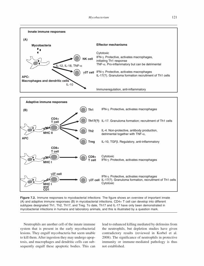
Several other cytokines are also important for controlling the infection. IL - 12 is produced by infected macrophages and is necessary for the development of a protective infl ammatory response. A major function of IL - 12 is to induce IFN - γ and hence tune the specifi c immune system onto a T helper 1 (Th1) pathway necessary for protective immunity. It thus represents a link between the innate and adaptive immune response. Several studies have demonstrated that antigen - presenting cells (APC) infected with mycobacteria secreted IL - 12 and IL - 18 (fi g. 7.2 A). However, there seem to be differences in cytokine production between the various APCs. This is also noted in cattle where bovine dendritic cells infected with virulent M. bovis produced IL - 12, TNF - α , and little IL - 10, whereas macrophages produced TNF - α , IL - 10, and little IL - 12 (Hope et al. 2004 ). Cytokines are pri-marily produced by dendritic cells and macro-phages in response to Toll - like receptor (TLR) signaling, and several molecules from the cell wall of mycobacteria have been shown to interact with the TLR.
IL - 12 acts in synergy with IL - 18 and induces IFN - γ secretion from natural killer (NK) cells, which are large granular lymphocytes belonging to the innate immune system (fi g. 7.2 A). These cells are believed to be involved in the fi rst - line defense against intracellular infections, and in addition to IFN - γ secretion they exhibit non - major histocompa-tability complex (MHC) restricted cytotoxic activity in response to mycobacteria (Katz et al. 1990 ). NK cells are likely to play a role in the initiation of a Th1 pathway and may function as a link between the innate and the adapted immune response. Al-though they are not believed to be essential for protection, they do seem to contribute to the protec-tive immune response. Human NK cells responded to extracellular BCG with proliferation, IFN - γ pro-duction and cytotoxicity (Esin et al. 2004 ), and op-timized CD8+ T cell effector function in response to M. tuberculosis (Vankayalapati et al. 2004 ). Similar results have been reported in cattle where NK cells can restrict intracellular growth of M. bovis and produce IFN - γ in response to both infected dendritic cells and macrophages stimulated with mycobacterial proteins like ESAT - 6 (Hope et al. 2002 ; Olsen et al. 2005 ).
Mycobacterium 121
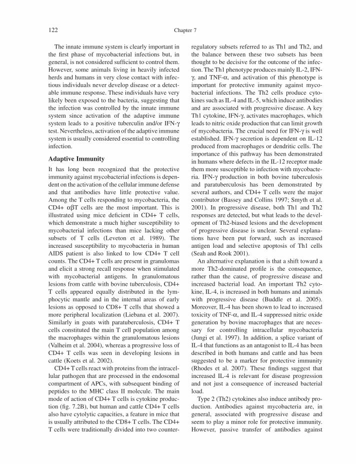
Figure 7.2. Immune responses to mycobacterial infections The fi gure shows an overview of important innate (A) and adaptive immune responses (B) in mycobacterial infections. CD4+ T cell can develop into different subtypes designated Th1, Th2, Th17, and Treg. To date, Th17 and IL - 17 have only been demonstrated in mycobacterial infections in humans and laboratory animals, and this is illustrated by a question mark.
CytotoxicCytotoxic
IFN-γ. Protective, activates macrophages,initiating Th1 response
IFN-γ. Protective, activates macrophages,initiating Th1 responseTNF-α. Pro-inflammatory but can be detrimentalTNF-α. Pro-inflammatory but can be detrimental
IFN-γ. Protective, activates macrophagesIFN-γ. Protective, activates macrophages
IL-17(?). Granuloma formation recruitment of Th1 cellsIL-17(?). Granuloma formation recruitment of Th1 cells
Immuneregulation, anti-inflammatoryImmuneregulation, anti-inflammatory
(A)
APC
CD4+ T cell
MHC IITh2
Th1
Th17(?)
Treg
IFN-γ. Protective, activates macrophagesIFN-γ. Protective, activates macrophages
IL-4. Non-protective, antibody production,IL-4. Non-protective, antibody production,
detrimental together with TNF-α,detrimental together with TNF-α,
IL-17. Granuloma formation; recruitment of Th1 cellsIL-17. Granuloma formation; recruitment of Th1 cells
IL-10, TGFβ. Regulatory, anti-inflammatoryIL-10, TGFβ. Regulatory, anti-inflammatory
CD8+ T cell
CD8+ T cell
MHC I
g dT cellIFN-γ. Protective, activates macrophagesIFN-γ. Protective, activates macrophagesIL-17(?). Granuloma formation, recruitment of Th1 cellsIL-17(?). Granuloma formation, recruitment of Th1 cellsCytotoxicCytotoxic
CytotoxicCytotoxicIFN-γ. Protective, activates macrophagesIFN-γ. Protective, activates macrophages
MHC IandCD1
g dT cell
Adaptive immune responsesAdaptive immune responses
(B)
Innate immune responses Innate immune responses
MycobacteriaMycobacteria
IL-12, IL-18, TNF-α IL-12, IL-18, TNF-α
IL-10
APC:
Macrophages and dendritic cells
g dT cell
NK cell
Effector mechanismsEffector mechanisms
Neutrophils are another cell of the innate immune system that is present in the early mycobacterial lesions. They engulf mycobacteria but seem unable to kill them. After ingestion they may undergo apop-tosis, and macrophages and dendritic cells can sub-sequently engulf these apoptotic bodies. This can
lead to enhanced killing mediated by defensins from the neutrophils, but depletion studies have given contradictory results (reviewed in Korbel et al. 2008 ). The signifi cance of neutrophils in protective immunity or immune - mediated pathology is thus not established.
122 Chapter 7
The innate immune system is clearly important in the fi rst phase of mycobacterial infections but, in general, is not considered suffi cient to control them. However, some animals living in heavily infected herds and humans in very close contact with infec-tious individuals never develop disease or a detect-able immune response. These individuals have very likely been exposed to the bacteria, suggesting that the infection was controlled by the innate immune system since activation of the adaptive immune system leads to a positive tuberculin and/or IFN - γ test. Nevertheless, activation of the adaptive immune system is usually considered essential to controlling infection.
Adaptive Immunity
It has long been recognized that the protective immunity against mycobacterial infections is depen-dent on the activation of the cellular immune defense and that antibodies have little protective value. Among the T cells responding to mycobacteria, the CD4+ α β T cells are the most important. This is illustrated using mice defi cient in CD4+ T cells, which demonstrate a much higher susceptibility to mycobacterial infections than mice lacking other subsets of T cells (Leveton et al. 1989 ). The increased susceptibility to mycobacteria in human AIDS patient is also linked to low CD4+ T cell counts. The CD4+ T cells are present in granulomas and elicit a strong recall response when stimulated with mycobacterial antigens. In granulomatous lesions from cattle with bovine tuberculosis, CD4+ T cells appeared equally distributed in the lym-phocytic mantle and in the internal areas of early lesions as opposed to CD8+ T cells that showed a more peripheral localization (Liebana et al. 2007 ). Similarly in goats with paratuberculosis, CD4+ T cells constituted the main T cell population among the macrophages within the granulomatous lesions (Valheim et al. 2004 ), whereas a progressive loss of CD4+ T cells was seen in developing lesions in cattle (Koets et al. 2002 ).
CD4+ T cells react with proteins from the intracel-lular pathogen that are processed in the endosomal compartment of APCs, with subsequent binding of peptides to the MHC class II molecule. The main mode of action of CD4+ T cells is cytokine produc-tion (fi g. 7.2 B), but human and cattle CD4+ T cells also have cytolytic capacities, a feature in mice that is usually attributed to the CD8+ T cells. The CD4+ T cells were traditionally divided into two counter -
regulatory subsets referred to as Th1 and Th2, and the balance between these two subsets has been thought to be decisive for the outcome of the infec-tion. The Th1 phenotype produces mainly IL - 2, IFN - γ , and TNF - α , and activation of this phenotype is important for protective immunity against myco-bacterial infections. The Th2 cells produce cyto-kines such as IL - 4 and IL - 5, which induce antibodies and are associated with progressive disease. A key Th1 cytokine, IFN - γ , activates macrophages, which leads to nitric oxide production that can limit growth of mycobacteria. The crucial need for IFN - γ is well established. IFN - γ secretion is dependent on IL - 12 produced from macrophages or dendritic cells. The importance of this pathway has been demonstrated in humans where defects in the IL - 12 receptor made them more susceptible to infection with mycobacte-ria. IFN - γ production in both bovine tuberculosis and paratuberculosis has been demonstrated by several authors, and CD4+ T cells were the major contributor (Bassey and Collins 1997 ; Smyth et al. 2001 ). In progressive disease, both Th1 and Th2 responses are detected, but what leads to the devel-opment of Th2 - biased lesions and the development of progressive disease is unclear. Several explana-tions have been put forward, such as increased antigen load and selective apoptosis of Th1 cells (Seah and Rook 2001 ).
An alternative explanation is that a shift toward a more Th2 - dominated profi le is the consequence, rather than the cause, of progressive disease and increased bacterial load. An important Th2 cyto-kine, IL - 4, is increased in both humans and animals with progressive disease (Buddle et al. 2005 ). Moreover, IL - 4 has been shown to lead to increased toxicity of TNF - α , and IL - 4 suppressed nitric oxide generation by bovine macrophages that are neces-sary for controlling intracellular mycobacteria (Jungi et al. 1997 ). In addition, a splice variant of IL - 4 that functions as an antagonist to IL - 4 has been described in both humans and cattle and has been suggested to be a marker for protective immunity (Rhodes et al. 2007 ). These fi ndings suggest that increased IL - 4 is relevant for disease progression and not just a consequence of increased bacterial load.
Type 2 (Th2) cytokines also induce antibody pro-duction. Antibodies against mycobacteria are, in general, associated with progressive disease and seem to play a minor role for protective immunity. However, passive transfer of antibodies against

Mycobacterium 123
LAM conferred partial protection against tuberculo-sis in mice (Teitelbaum et al. 1998 ). Such antibodies may infl uence the uptake of mycobacteria and stim-ulation of macrophages with subsequent modulation of the immune responses. Some protective effect of antibodies with certain specifi cities should therefore not be disregarded.
With the recognition of Th17 cells, it has become clear that the CD4+ T cell responses to tuberculosis is more complex than captured by the Th1 – Th2 paradigm of immunity. IL - 17 was initially described as important in several autoimmune diseases and extracellular infections, but cells that produce IL - 17 have also been described in both mice and humans with tuberculosis (Scriba et al. 2008 ). In mice, there appears to be a distinct lineage of CD4+ α β T cells called Th17 cells, whereas in humans there are often CD4+ T cells with a mixed Th1/Th17 profi le. Several cytokines such as TGF - β , IL - 6, and IL - 23 are involved in the differentiation of na ï ve T cells into Th17 and/or maintaining IL - 17 production from memory Th17 cells. IL - 23 is a dimer that shares the p40 subunit with IL - 12, implying that previous studies blocking the IL - 12 p40 subunit must be reevaluated in the light of the IL - 23/IL - 17 pathway. The role of IL - 23/IL - 17 in mycobacterial diseases is still elusive, but it does not appear to be necessary for protective immunity against tubercu-losis in mice. However, IL - 17 - producing cells that are reactive to mycobacterial antigens have been described, and depletion of IL - 17 leads to decreased recruitment of IFN - γ producing Th1 cells in the early stages of infection (Khader et al. 2007 ). It is possible that the cross - regulatory properties of IL - 12/IFN - γ and IL - 23/IL - 17 may be crucial to achieving the right balance between protection and immunopathology. Bovine IL - 17 has been identi-fi ed, but its role in mycobacterial infections remains to be established (Riollet et al. 2006 ).
The major role of CD4+ T cells in the protective immune response against mycobacteria is undispu-table, but the importance of CD8+ MHC class I restricted α β T cells has also been recognized. Early mouse studies using CD8 depletion or adoptive transfer of CD8+ T cells showed somewhat confl ict-ing evidence, but some protective effect of CD8+ T cells has been detected. However, one study deplet-ing CD8+ T cells in the bovine model led to decreased IFN - γ production but diminished pathol-ogy, suggesting a role for this cell type in immuno-pathology (Villarreal - Ramos et al. 2003 ). The CD8+
T cells recognize peptide antigens presented to their T cell receptor by classical MHC class I molecules. The importance of a functional MHC class I pre-senting pathway has been demonstrated in both β 2 - microglobulin and tapasin (TAP) knockout mice, which were more susceptible to intravenous M. tuberculosis infection than their wild - type coun-terparts. Following aerosol challenge of mice with M. tuberculosis, CD8+ T cells traffi cked to the lung as early as 2 weeks post infection, and CD8+ T cells are also present in bovine tuberculosis and paratu-berculosis lesions (Begara - McGorum et al. 1998 ; Liebana et al. 2007 ).
The function of CD8+ T cells is, in general, lysis of infected target cells. Cytolysis mediated by CD8+ T cells involves the release of granules containing perforin and granzyme, or cytolysis can be mediated through the Fas/Fas ligand molecules that result in programmed cell death of the target cell. Cytolytic responses in bovine tuberculosis have been demon-strated (Skinner et al. 2003b ). Furthermore, several studies have shown that while CD4+ T cells are the major IFN - γ producer in both bovine tuberculosis and paratuberculosis, CD8+ T cells can also elicit pronounced secretion of IFN - γ (Bassey and Collins 1997 ; Liebana et al. 1999 ). There is thus a redun-dancy in the functions of CD4+ and CD8+ T cells in that both cell - types have cytolytic capacities and can secrete cytokines (fi g. 7.2 B).
Cattle have a large proportion of T cells that use the γ δ T cell receptor instead of the α β chains. γ δ T cells constitute 30 – 80% of the T cells in peripheral blood, with the largest proportion in young calves. This is vastly different from mice and humans, where less than 10% of the peripheral blood lym-phocytes are of this subset. It is therefore not unlikely that these cells are more important for resistance to mycobacterial disease in ruminants compared with other hosts. Subsets of γ δ T cells in ruminants can be distinguished based on the surface marker WC1, and the prominent population in blood expresses WC1 but is negative for CD2, CD4, and CD8 (MacHugh et al. 1997 ). Another γ δ T popula-tion is negative for WC1 but expresses CD2 together with CD8, suggesting that these cells may be acti-vated via MHC class I. The latter population was found in large numbers in the spleen and intestines. It has also been demonstrated that the ruminant γ δ T cells express exceptionally diverse antigen recep-tors, and they have therefore been suggested to have a broader capacity to recognize diverse ligands than

124 Chapter 7
their counterparts in other species (Hein and Dudler 1997 ). In mice, γ δ T cells have been shown to expand in the fi rst phase of tuberculosis and have been sug-gested as a fi rst - line defense against mycobacterial infections, but these cells do not seem to be essential for resistance against tuberculosis (Ladel et al. 1995 ). However, they may play a role in granuloma formation (fi g. 7.2 B). In both cattle and humans, they accumulate early in lesion development, but depletion studies in cattle did not markedly change the course of disease (Kennedy et al. 2002 ).
The γ δ T cells are able to respond in both an innate and an adaptive fashion. They recognize both protein and nonprotein antigens in the context of classical MHC class I or CD1 respectively, but this cell type can also respond to self - derived stress mol-ecules as well as soluble factors induced by myco-bacterial antigens without the need for previous sensitization. Like α β T cells, γ δ T cells can secrete IFN - γ and have cytolytic capacities. They were able to directly kill both intracellular and extracellular mycobacteria through a granulysin - dependent mechanism (Dieli et al. 2001 ). Innate IFN - γ produc-tion by bovine γ δ T cells in response to mycobacte-rial cell wall antigens has also been detected, which was probably dependent on monocytes - derived cytokines (Vesosky et al. 2004 ). This was in accor-dance with another study that showed that bovine WC1+ γ δ T cells were synergistically stimulated by IL - 12 and IL - 18 to secrete large quantities of IFN - γ (Price et al. 2007 ).
A memory - type response has also been described with response to protein antigens whereas other studies did not fi nd signifi cant IFN - γ production in response to mycobacterial antigens in γ δ T cells (Bassey and Collins 1997 ; Smyth et al. 2001 ). In addition to IFN - γ it has been demonstrated that γ δ T cells from tuberculosis - infected mice can secrete IL - 17 in an innate fashion (Lockhart et al. 2006 ). This fi nding sheds light on results from the M. bovis model where γ δ T cells were recruited to the lungs early after infection. It is possible that innate IL - 17, in addition to IFN - γ production from γ δ T cells, plays a role in the early events of bovine tuberculosis.
Immune Regulatory Mechanisms
Protective immunity to mycobacterial infections is dependent on a strong Th1 response; however, without control, this response can also cause immune - mediated pathology with excessive infl am-mation. The right balance between protective
immunity and immunopathology is thus essential. Several immunoregulatory mechanisms have been described, and both cytokines IL - 10 and TGF - β have been shown to dampen the Th1 response. It is still not clear which cells are involved in the regula-tion of immune responses in mycobacterial infec-tions in ruminants. In mice and humans, a current major topic of research is the reawakening of T suppressor cells, now designated regulatory T cells (Treg). The most studied of these Treg cells are CD4+, CD25+, which express the transcription factor FOXp3. Treg cells have shown to be increased in humans with active tuberculosis, which may suggest that down - regulation of Th1 responses by Treg cells is responsible for reactivation of disease (Guyot - Revol et al. 2006 ). However, mice depleted of Treg cells did not have increased bacillary loads in a mouse model (Quinn et al. 2006 ). An immuno-suppressive role of IL - 10 has been demonstrated in both bovine tuberculosis and paratuberculosis, but a defi nite role of Treg cells was not established since the source of IL - 10 production was not identifi ed (Buza et al. 2004 ; Denis et al. 2007 ). In goats natu-rally infected with paratuberculosis , monocytes were the major producers of IL - 10 in peripheral blood, whereas little IL - 10 was produced by classi-cal CD4+ and CD25+ Treg cells (Lybeck et al. 2009 ). Similarly, only γ δ T cells and monocytes expressed IL - 10 and showed regulatory properties using sorted bovine blood cell subsets in a coculture suppression assay (Hoek et al. 2008 ). Furthermore, regulatory properties of γ δ T cells were also detected in bovine tuberculosis (Kennedy et al 2002 ). This does not exclude a role for Treg cells in myco-bacterial infections in ruminants, but the relative importance of these cells is not yet established.
Immunity and Disease Control
Mycobacterial diseases are slowly progressive infections with a large proportion of subclinically infected animals. Identifi cation of all these animals is diffi cult with available diagnostic assays. Further-more, the bacteria can survive for an extended time in the environment and several species of wildlife can be infected. These factors make bovine tuberculosis and paratuberculosis challenging dis-eases to control. Most developed countries have control programs for bovine tuberculosis due to the zoonotic risk of M. bovis . These programs are usually based on test - and - slaughter regimes with restriction of movement of animals from infected
Mycobacterium 125
herds. The most widely used test is the intradermal skin test, in which purifi ed protein derivative (PPD) from M. bovis is injected intracutaneously and measures an increase in skin thickness as an index of infection. Despite these control programs, several countries have failed to eradicate bovine tuberculo-sis. This is particularly true for countries that have a wildlife reservoir, such as the United Kingdom, Ireland, and New Zealand.
Mycobacterium bovis and M. avium subsp. para-tuberculosis can infect a wide range of domestic and wild animals. To what extent the wildlife con-stitutes a threat to the domestic animals depends on the ability of the species to act as a maintenance or as a spillover host. In the former, the infection can persist by intraspecies transmission alone, whereas in the latter, infection will not persist indefi nitely unless there is reinfection from another host. To control the infection in domestic livestock, it is important to focus on the wildlife that can act as maintenance hosts with less emphasis on spillover hosts. Species that are important for the spread of bovine tuberculosis are the badger in the United Kingdom and Ireland, the brush - tail possum in New Zealand, and the white - tail deer in Michigan. M. avium subsp. paratuberculosis has also been isolated from a wide range of species and most frequently from deer and rabbits. Paratuberculosis in farmed deer and in deer reserves is prevalent in some areas, and it seems clear that deer can maintain the infection in high - density populations (Balseiro et al. 2008 ). However, the signifi cance of wild deer and other wildlife for the continuation or spread of paratuberculosis in livestock is not clear.
An alternative strategy to wildlife depopulation in reducing the spread of tuberculosis to livestock is wildlife vaccination. To date, injectable vaccines give the best protective immunity but are not a real-istic alternative for purposes other than research trials. BCG, the human tuberculosis vaccine, deliv-ered in a lipid formulation as an oral vaccine, has been shown to reduce the severity of disease in an aerosol challenge model (Aldwell et al. 2003 ). The partial response achieved may lead to a reduced excretion of bacteria and hence diminished spread of infection to livestock. Vaccination of wildlife, together with a focus on improved management practices as a supplement to current control pro-grams, might reduce the transmission of bovine tuberculosis suffi ciently to control tuberculosis in livestock.
Another important cause for spread of mycobac-terial infections is movement and trading of animals. The long incubation time and slow progression of disease, together with suboptimal tests, makes it diffi cult to identify all infected animals. As a con-sequence, there will always be a risk when purchas-ing animals from a previously infected herd. It has been diffi cult to assess how much of the spread is caused by animal movement compared with infec-tion from wildlife and survival of bacteria in the environment, but discussion of this aspect, like that of management factors in controlling tubercu-losis and paratuberculosis, is beyond the scope of this book.
Vaccination and Immune Testing
Vaccination against mycobacterial diseases can reduce clinical signs and limit the spread of bacteria but is not suffi cient to eliminate the infection. For paratuberculosis, both commercial and in - house live attenuated and inactivated whole cell vaccines have been used. Since vaccination interferes with diag-nostic tests for paratuberculosis and also bovine tuberculosis, it cannot be used in countries that have control programs for bovine tuberculosis. Vaccines against bovine tuberculosis are currently not used for the same reason. The use of sophisticated vac-cines and diagnostic tests may circumvent this problem in the future. A strategy is to use different antigens in the vaccines and the diagnostic tests, a strategy denominated DIVA (Differentiation between Infected and Vaccinated Animals) strategy. This is already feasible for bovine tuberculosis with the traditional BCG vaccine. Mycobacterium bovis BCG lacks several genetic regions compared with virulent M. bovis , including the highly immuno-genic ESAT - 6 protein. A test measuring IFN - γ after in vitro stimulation with ESAT - 6 can be used to distinguish between infected and BCG - vaccinated animals (Buddle et al. 1999 ). However, before an IFN - γ test can be approved, comparable sensitivity and specifi city to the tuberculin skin test must be documented. To date, this test gives a lower speci-fi city, particularly in young animals, where false - positive results have been reported to be caused by IFN - γ production from NK cells (Olsen et al. 2005 ). This is of concern since the BCG vaccine gives better protection when given just after birth (Buddle et al. 2003 ), and it is thus important to be able to test these young animals. An IFN - γ test using specifi c antigens such as ESAT - 6 could also
126 Chapter 7
differentiate between M. bovis infected - animals and paratuberculosis - vaccinated animals and thereby allow paratuberculosis vaccination also in areas with bovine tuberculosis.
In addition to interfering with the skin test, the protective effect of BCG in cattle, as in humans, is variable. Although reduced lesions may be observed, the animals are still likely to be infected and a pos-sible source of further spread. Despite substantial effort, it has been challenging to develop second - generation vaccines that give a superior effect to that of BCG. The reason for this is likely that protec-tive immunity is dependent on a complex interaction between various immune cells with a range of anti - mycobacterial effector mechanisms. Several DNA vaccines and subunit protein vaccines have been tested in cattle, but used alone they generally showed lower effi cacy than BCG. The most promising alter-native is thus BCG combined with certain subunit vaccines, which have been reported to give enhanced protection compared with BCG (Skinner et al. 2003a ; Wedlock et al. 2008 ). These results show that a rational vaccine strategy seems feasible in the future but needs to be accompanied with the devel-opment of appropriate diagnostic tests.
GAPS IN KNOWLEDGE AND ANTICIPATED DEVELOPMENTS Considerable progress has been achieved by the application of the new molecular genetics and genomics methods, but there are still many issues remaining to be understood in mycobacterial patho-genesis. It is thus not surprising that effective vac-cines are still in a developmental phase, with only a few reaching the fi nal testing stages, most of them in the human tuberculosis fi eld. As discussed, the situation is further complicated in animal health as vaccines should not only protect animals but also avoid undesirable cross - species reactions.
Part of this challenge arises from the multifacto-rial nature of each step in the pathogenic process. Adhesion and invasion may involve a plethora of host cell receptors – microbial protein interactions. An array of antigens, whose expression requires specifi c timing and tissue expression, participate in the stimulation of the immune system. Mycobacteria possess redundant metabolic routes used to take maximum advantage of tissue microenvironments. Thus, in many experimental studies, a single muta-tion may not result in a discernible reduction in virulence, thus requiring the construction of multi-
ple knockouts. Complications then arise in the use of different selection markers or with strategies to create unmarked mutations. In some cases, different animal models (e.g., mice vs. guinea pigs) have led to different results regarding the virulence of mutant strains.
Challenges also arise at the interface between the new technologies and the classic acquired knowl-edge on the biology of these microorganisms. In many cases, this has resulted in the use of attenuated laboratory strains that have already lost the expres-sion of key virulence determinants because of muta-tions accumulated during in vitro passage. In other cases, contradictory fi ndings reported in the litera-ture may be tracked to differences in cultural condi-tions (media, growth phase, use of detergents to prevent clumping), insuffi cient analysis of the cor-relation between phenotypic and genotypic charac-terization of the strains used in each study (e.g., serotyping vs. spoligotyping), vaccine formulation, routes of inoculation, and so on. Continued progress will require close attention and standardization of these parameters to better correlate the various exciting new studies with known paradigms on the biology of these microorganisms.
Although much is known about protective immu-nity to mycobacteria, it is still not clear what mecha-nisms need to be enhanced to get a better effi cacy for new vaccines. Should a new vaccine generate stronger Th1 responses? Is it important to have Th1 response to a wider repertoire of antigens? Will a stronger CD8+ response with enhanced cytotoxicity give increased protection? Or is it stimulated Treg cells that can dampen a Th2 response? Perhaps all of the above is necessary to give the optimal result. The best alternative may be to develop a new live attenuated vaccine by deletion of genes from M. tuberculosis or M. bovis . Such a new attenuated strain of M. bovis appeared to provide greater protec-tion than BCG in cattle experimentally infected with M. bovis (Buddle et al. 2002 ). The fear that reversion to virulence by gene acquisition in vivo can occur makes it unlikely that such a vaccine will be approved in the near future. In humans, the ability of BCG to protect children from serious forms of the disease such as disseminated tuberculosis makes it ethically diffi cult to defend a withdrawal of the BCG vaccination program in developing countries. It is thus likely that a boost of the BCG vaccine or a postinfection vaccine will be the future in the human fi eld rather than a replacement.
Mycobacterium 127
REFERENCES Aldwell , F. E. , D. L. Keen , N. A. Parlane , M. A.
Skinner , G. W. de Lisle , and B. M. Buddle . 2003 . Oral vaccination with Mycobacterium bovis BCG in a lipid formulation induces resistance to pulmonary tuberculosis in brushtail possums . Vaccine 22 : 70 – 76 .
Alexander , D. C. , C. Y. Turenne , and M. A. Behr . 2009 . Insertion and deletion events that defi ne the pathogen Mycobacterium avium subsp. paratuber-culosis . J. Bacteriol. 191 : 1018 – 1025 .
Andersen , P. , D. Askgaard , L. Ljungqvist , J. Bennedsen , and I. Heron . 1991 . Proteins released from Mycobacterium tuberculosis during growth . Infect. Immun. 59 : 1905 – 1910 .
Bach , H. , J. Sun , Z. Hmama , and Y. Av - Gay . 2006 . Mycobacterium avium subsp. paratuberculosis PtpA is an endogenous tyrosine phosphatase secreted during infection . Infect. Immun. 74 : 6540 – 6546 .
Balseiro , A. , J. F. Garcia Marin , P. Solano , J. M. Garrido , and J. M. Prieto . 2008 . Histopathological classifi cation of lesions observed in natural cases of paratuberculosis in free — ranging fallow deer (Dama dama) . J. Comp. Pathol. 138 : 180 – 188 .
Bannantine , J. P. , J. F. Huntley , E. Miltner , J. R. Stabel , and L. E. Bermudez . 2003 . The Mycobacterium avium subsp. paratuberculosis 35 kDa protein plays a role in invasion of bovine epithelial cells . Microbiol. 149 : 2061 – 2069 .
Bannantine , J. P. , and M. L. Paustian . 2006 . Identifi cation of diagnostic proteins in Mycobacterium avium subspecies paratuberculosis by a whole genome analysis approach . Methods Mol. Biol. 345 : 185 – 196 .
Bartow , R. A. , and D. N. McMurray . 1998 . Lymphocytes expressing Fc gamma receptors suppress antigen - induced proliferation in cells from guinea pigs infected with virulent Mycobacterium tuberculosis . Cell Immunol. 184 : 51 – 57 .
Bassey , E. O. , and M. T. Collins . 1997 . Study of T - lymphocyte subsets of healthy and Mycobacterium avium subsp. paratuberculosis - infected cattle . Infect. Immun. 65 : 4869 – 4872 .
Begara - McGorum , I. , L. A. Wildblood , C. J. Clarke , K. M. Connor , K. Stevenson , C. J. McInnes , et al. 1998 . Early immunopathological events in experimental ovine paratuberculosis . Vet. Immunol. Immunopathol. 63 : 265 – 287 .
Belisle , J. T. , V. D. Vissa , T. Sievert , K. Takayama , P. J. Brennan , and G. S. Besra . 1997 . Role of the
major antigen of Mycobacterium tuberculosis in cell wall biogenesis . Science 276 : 1420 – 1422 .
Braunstein , M. , S. S. Bardarov , and W. R. Jr. Jacobs , 2002 . Genetic methods for deciphering virulence determinants of Mycobacterium tuberculosis . Methods Enzymol. 358 : 67 – 99 .
Brosch , R. , S. V. Gordon , M. Marmiesse , P. Brodin , C. Buchrieser , K. Eiglmeier , et al. 2002 . A new evolutionary scenario for the Mycobacterium tuberculosis complex . Proc. Natl. Acad. Sci. U S A 99 : 3684 – 3689 .
Buddle , B. M. , N. A. Parlane , D. L. Keen , F. E. Aldwell , J. M. Pollock , K. Lightbody , et al. 1999 . Differentiation between Mycobacterium bovis BCG - vaccinated and M. bovis - infected cattle by using recombinant mycobacterial antigens . Clin. Diagn. Lab. Immunol. 6 : 1 – 5 .
Buddle , B. M. , B. J. Wards , F. E. Aldwell , D. M. Collins , and G. W. de Lisle . 2002 . Infl uence of sensitisation to environmental mycobacteria on subsequent vaccination against bovine tuberculosis . Vaccine 20 : 1126 – 1133 .
Buddle , B. M. , D. N. Wedlock , M. Denis , and M. A. Skinner . 2005 . Identifi cation of immune response correlates for protection against bovine tuberculo-sis . Vet. Immunol. Immunopathol. 108 : 45 – 51 .
Buddle , B. M. , D. N. Wedlock , N. A. Parlane , L. A. Corner , G. W. de Lisle , and M. A. Skinner . 2003 . Revaccination of neonatal calves with Mycobacterium bovis BCG reduces the level of protection against bovine tuberculosis induced by a single vaccination . Infect. Immun. 71 : 6411 – 6419 .
Buza , J. J. , H. Hikono , Y. Mori , R. Nagata , S. Hirayama , Aodon - geril , et al. 2004 . Neutralization of interleukin - 10 signifi cantly enhances gamma interferon expression in peripheral blood by stimulation with Johnin purifi ed protein derivative and by infection with Mycobacterium avium subsp. paratuberculosis in experimentally infected cattle with paratuberculosis . Infect. Immun. 72 : 2425 – 2428 .
Chacon , O. , L. E. Bermudez , and R. G. Barletta . 2004 . Johne ’ s disease, infl ammatory bowel disease, and Mycobacterium paratuberculosis . Annu. Rev. Microbiol. 58 : 329 – 363 .
Chakravarty , S. D. , G. Zhu , M. C. Tsai , V. P. Mohan , S. Marino , D. E. Kirschner , et al. 2008 . Tumor necrosis factor blockade in chronic murine tuberculosis enhances granulomatous infl ammation and disorganizes granulomas in the lungs . Infect. Immun. 76 : 916 – 926 .
Chan , J. , X. D. Fan , S. W. Hunter , P. J. Brennan , and B. R. Bloom . 1991 . Lipoarabinomannan, a possible
128 Chapter 7
virulence factor involved in persistence of Mycobacterium tuberculosis within macrophages . Infect. Immun. 59 : 1755 – 1761 .
Chitale , S. , S. Ehrt , I. Kawamura , T. Fujimura , N. Shimono , N. Anand , et al. 2001 . Recombinant Mycobacterium tuberculosis protein associated with mammalian cell entry . Cell Microbiol. 3 : 247 – 254 .
Cole , S. T. , R. Brosch , J. Parkhill , T. Garnier , C. Churcher , D. Harris , et al. 1998 . Deciphering the biology of Mycobacterium tuberculosis from the complete genome sequence . Nature 393 : 537 – 544 .
Converse , P. J. , P. C. Karakousis , L. G. Klinkenberg , A. K. Kesavan , L. H. Ly , S. S. Allen , et al. 2009 . Role of the dosR - dosS two - component regulatory system in Mycobacterium tuberculosis virulence in three animal models . Infect. Immun. 77 : 1230 – 1237 .
Danelishvili , L. , M. Wu , B. Stang , M. Harriff , S. L. Cirillo , J. D. Cirillo , et al. 2007 . Identifi cation of Mycobacterium avium pathogenicity island important for macrophage and amoeba infection . Proc. Natl. Acad. Sci. U S A 104 : 11038 – 11043 .
Denis , M. , D. N. Wedlock , A. R. McCarthy , N. A. Parlane , P. J. Cockle , H. M. Vordermeier , et al. 2007 . Enhancement of the sensitivity of the whole - blood gamma interferon assay for diagnosis of Mycobacterium bovis infections in cattle . Clin. Vaccine Immunol. 14 : 1483 – 1489 .
Deretic , V. 2008 . Autophagy, an immunologic magic bullet: Mycobacterium tuberculosis phagosome maturation block and how to bypass it . Future Microbiol. 3 : 517 – 524 .
Deretic , V. , S. Singh , S. Master , J. Harris , E. Roberts , G. Kyei , et al. 2006 . Mycobacterium tuberculosis inhibition of phagolysosome biogen-esis and autophagy as a host defence mechanism . Cell Microbiol. 8 : 719 – 727 .
Deretic , V. , I. Vergne , J. Chua , S. Master , S. B. Singh , J. A. Fazio , et al. 2004 . Endosomal membrane traffi c: convergence point targeted by Mycobacterium tuberculosis and HIV . Cell Microbiol. 6 : 999 – 1009 .
Devulder , G. , M. Perouse de Montclos , and J. P. Flandrois . 2005 . A multigene approach to phyloge-netic analysis using the genus Mycobacterium as a model . Int. J. Syst. Evol. Microbiol. 55 : 293 – 302 .
Dieli , F. , M. Troye - Blomberg , J. Ivanyi , J. J. Fournie , A. M. Krensky , M. Bonneville , et al. 2001 . Granulysin - dependent killing of intracellular and extracellular Mycobacterium tuberculosis by Vgamma9/Vdelta2 T lymphocytes . J. Infect. Dis. 184 : 1082 – 1085 .
Edwards , K. M. , M. H. Cynamon , R. K. Voladri , C. C. Hager , M. S. DeStefano , K. T. Tham , et al. 2001 . Iron - cofactored superoxide dismutase inhibits host responses to Mycobacterium tuberculosis . Am. J. Respir. Crit. Care Med. 164 : 2213 – 2219 .
Ehrt , S. , and D. Schnappinger . 2007 . Mycobacterium tuberculosis virulence: lipids inside and out . Nat. Med. 13 : 284 – 285 .
Esin , S. , G. Batoni , M. Pardini , F. Favilli , D. Bottai , G. Maisetta , et al. 2004 . Functional characteriza-tion of human natural killer cells responding to Mycobacterium bovis bacille Calmette - Guerin . Immunology 112 : 143 – 152 .
Ferrari , G. , H. Langen , M. Naito , and J. Pieters . 1999 . A coat protein on phagosomes involved in the intracellular survival of mycobacteria . Cell 97 : 435 – 447 .
Fisher , M. A. , B. B. Plikaytis , and T. M. Shinnick . 2002 . Microarray analysis of the Mycobacterium tuberculosis transcriptional response to the acidic conditions found in phagosomes . J. Bacteriol. 184 : 4025 – 4032 .
Foley - Thomas , E. M. , D. L. Whipple , L. E. Bermudez , and R. G. Barletta . 1995 . Phage infection, transfection and transformation of Mycobacterium avium complex and Mycobacterium paratuberculosis . Microbiology 141 : 1173 – 1181 .
Garnier , T. , K. Eiglmeier , J. C. Camus , N. Medina , H. Mansoor , M. Pryor , et al. 2003 . The complete genome sequence of Mycobacterium bovis . Proc. Natl. Acad. Sci. U S A 100 : 7877 – 7882 .
George , K. M. , D. Chatterjee , G. Gunawardana , D. Welty , J. Hayman , R. Lee , et al. 1999 . Mycolactone: a polyketide toxin from Mycobacterium ulcerans required for virulence . Science 283 : 854 – 857 .
Gioffre , A. , E. Infante , D. Aguilar , M. P. Santangelo , L. Klepp , A. Amadio , et al. 2005 . Mutation in mce operons attenuates Mycobacterium tuberculosis virulence . Microbes. Infect. 7 : 325 – 334 .
Glickman , M. S. , J. S. Cox , and W. R. Jr. Jacobs . 2000 . A novel mycolic acid cyclopropane synthetase is required for cording, persistence, and virulence of Mycobacterium tuberculosis . Mol. Cell 5 : 717 – 727 .
Goren , M. B. , O. Brokl , and P. Roller . 1979 . Cord factor (trehalose - 6 - 6 ′ - dimycolate) of in vivo - derived Mycobacterium lepraemurium . Biochim. Biophys. Acta 574 : 70 – 78 .
Guyot - Revol , V. , J. A. Innes , S. Hackforth , T. Hinks , and A. Lalvani . 2006 . Regulatory T cells are
Mycobacterium 129
expanded in blood and disease sites in patients with tuberculosis. Am. J. Respir . Crit Care Med. 173 : 803 – 810 .
Harris , N. B. 2006 . Molecular techniques: applica-tions in epidemiologic Studies . In Mycobacterium bovis Infection in animals and Humans . C. O. Thoen , J. H. Steele , and M. J. Gilsdorf (eds.). Ames : Blackwell Publishing , pp. 54 – 62 .
Harris , N. B. , and R. G. Barletta . 2001 . Mycobacterium avium subsp. paratuberculosis in Veterinary Medicine . Clin. Microbiol. Rev. 14 : 489 – 512 .
Harris , N. B. , Z. Feng , X. Liu , S. L. Cirillo , J. D. Cirillo , and R. G. Barletta . 1999 . Development of a transposon mutagenesis system for Mycobacterium avium subsp. paratuberculosis . FEMS Microbiol. Lett. 175 : 21 – 26 .
Harth , G. , and M. A. Horwitz . 1999 . Export of recombinant Mycobacterium tuberculosis super-oxide dismutase is dependent upon both infor-mation in the protein and mycobacterial export machinery. A model for studying export of leaderless proteins by pathogenic mycobacteria . J. Biol. Chem. 274 : 4281 – 4292 .
He , H. , R. Hovey , J. Kane , V. Singh , and T. C. Zahrt . 2006 . MprAB is a stress - responsive two - component system that directly regulates expression of sigma factors SigB and SigE in Mycobacterium tuberculosis . J. Bacteriol. 188 : 2134 – 2143 .
Hein , W. R. , and L. Dudler . 1997 . TCR gamma delta+ cells are prominent in normal bovine skin and express a diverse repertoire of antigen receptors . Immunology 91 : 58 – 64 .
Hewinson , R. G. , H. M. Vordermeier , N. H. Smith , and S. V. Gordon . 2006 . Recent advances in our knowledge of Mycobacterium bovis : a feeling for the organism . Vet. Microbiol. 112 : 127 – 139 .
Hoek , A. , V. P. Rutten , J. Kool , G. J. Arkesteijn , R. J. Bouwstra , I. Rhijn Van , et al. 2008 . Subpopulations of bovine WC1+ gammadelta T cells rather than CD4+CD25highFoxp3+ T cells act as immune regulatory cells ex vivo . Vet. Res. 40 : 6 .
Holt , J. G , N. R. Krieg , P. H. A Sneath , J. T Staley , and S. T Williams . 1994 . Bergey ’ s manual of determinative bacteriology . Baltimore : Williams & Wilkins .
Hope , J. C. , P. Sopp , and C. J. Howard . 2002 . NK - like CD8(+) cells in immunologically naive neonatal calves that respond to dendritic cells infected with Mycobacterium bovis BCG . J. Leukoc. Biol. 71 : 184 – 194 .
Hope , J. C. , M. L. Thom , P. A. McCormick , and C. J. Howard . 2004 . Interaction of antigen presenting cells with mycobacteria . Vet. Immunol. Immunopathol. 100 : 187 – 195 .
Jain , M. , C. J. Petzold , M. W. Schelle , M. D. Leavell , J. D. Mougous , C. R. Bertozzi , et al. 2007 . Lipidomics reveals control of Mycobacterium tuberculosis virulence lipids via metabolic coupling . Proc. Natl. Acad. Sci. U S A 104 : 5133 – 5138 .
Jungi , T. W. , M. Brcic , H. Sager , D. A. Dobbelaere , A. Furger , and I. Roditi . 1997 . Antagonistic effects of IL - 4 and interferon - gamma (IFN — gamma) on inducible nitric oxide synthase expression in bovine macrophages exposed to gram - positive bacteria . Clin. Exp. Immunol. 109 : 431 – 438 .
Katz , P. , H. Jr. Yeager , G. Whalen , M. Evans , R. P. Swartz , and J. Roecklein . 1990 . Natural killer cell - mediated lysis of Mycobacterium - avium complex - infected monocytes . J. Clin. Immunol. 10 : 71 – 77 .
Kaushal , D. , B. G. Schroeder , S. Tyagi , T. Yoshimatsu , C. Scott , C. Ko , et al. 2002 . Reduced immunopathology and mortality despite tissue persistence in a Mycobacterium tuberculosis mutant lacking alternative sigma factor, SigH . Proc. Natl. Acad. Sci. U S A 99 : 8330 – 8335 .
Keane , J. , S. Gershon , R. P. Wise , E. Mirabile - Levens , J. Kasznica , W. D. Schwieterman , et al. 2001 . Tuberculosis associated with infl iximab, a tumor necrosis factor alpha - neutralizing agent . N. Engl. J. Med. 345 : 1098 – 1104 .
Kennedy , H. E. , M. D. Welsh , D. G. Bryson , J. P. Cassidy , F. I. Forster , C. J. Howard , et al. 2002 . Modulation of immune responses to Mycobacterium bovis in cattle depleted of WC1(+) gamma delta T cells . Infect. Immun. 70 : 1488 – 1500 .
Khader , S. A. , G. K. Bell , J. E. Pearl , J. J. Fountain , J. Rangel - Moreno , G. E. Cilley , et al. 2007 . IL - 23 and IL - 17 in the establishment of protective pulmonary CD4+ T cell responses after vaccina-tion and during Mycobacterium tuberculosis challenge . Nat. Immunol. 8 : 369 – 377 .
Koets , A. , V. Rutten , A. Hoek , F. van Mil , K. Muller , D. Bakker , et al. 2002 . Progressive bovine paratuberculosis is associated with local loss of CD4(+) T cells, increased frequency of gamma delta T cells, and related changes in T - cell function . Infect. Immun. 70 : 3856 – 3864 .
Korbel , D. S. , B. E. Schneider , and U. E. Schaible . 2008 . Innate immunity in tuberculosis: myths and truth . Microbes Infect. 10 : 995 – 1004 .
130 Chapter 7
Kumar , A. , M. Bose , and V. Brahmachari . 2003 . Analysis of expression profi le of mammalian cell entry (mce) operons of Mycobacterium tuberculo-sis . Infect. Immun. 71 : 6083 – 6087 .
Ladel , C. H. , J. Hess , S. Daugelat , P. Mombaerts , S. Tonegawa , and S. H. Kaufmann . 1995 . Contribution of alpha/beta and gamma/delta T lymphocytes to immunity against Mycobacterium bovis bacillus Calmette Guerin: studies with T cell receptor - defi cient mutant mice . Eur. J. Immunol. 25 : 838 – 846 .
Leung , A. S. , V. Tran , Z. Wu , X. Yu , D. C. Alexander , G. F. Gao , et al. 2008 . Novel genome polymorphisms in BCG vaccine strains and impact on effi cacy . BMC Genomics 9 : 413 .
Leveton , C. , S. Barnass , B. Champion , S. Lucas , Souza B. De , M. Nicol , et al. 1989 . T - cell - mediated protection of mice against virulent Mycobacterium tuberculosis . Infect. Immun. 57 : 390 – 395 .
Li , L. , J. P. Bannantine , Q. Zhang , A. Amonsin , B. J. May , D. Alt , et al. 2005 . The complete genome sequence of Mycobacterium avium subspecies paratuberculosis . Proc. Natl. Acad. Sci. U S A 102 : 12344 – 12349 .
Liebana , E. , R. M. Girvin , M. Welsh , S. D. Neill , and J. M. Pollock . 1999 . Generation of CD8(+) T — cell responses to Mycobacterium bovis and mycobacterial antigen in experimental bovine tuberculosis . Infect. Immun. 67 : 1034 – 1044 .
Liebana , E. , S. Marsh , J. Gough , A. Nunez , H. M. Vordermeier , A. Whelan , et al. 2007 . Distribution and activation of T - lymphocyte subsets in tuberculous bovine lymph - node granulomas . Vet. Pathol. 44 : 366 – 372 .
Liu , X. , Z. Feng , N. B. Harris , J. D. Cirillo , H. Bercovier , and R. G. Barletta . 2001 . Identifi cation of a secreted superoxide dismutase in Mycobacterium avium ssp. paratuberculosis . FEMS Microbiol. Lett. 202 : 233 – 238 .
Lockhart , E. , A. M. Green , and J. L. Flynn . 2006 . IL - 17 production is dominated by gamma delta T cells rather than CD4 T cells during Mycobacterium tuberculosis infection . J. Immunol. 177 : 4662 – 4669 .
Lybeck , K. R. , A. K. Storset , and I. Olsen . 2009 . Neutralisation of interleukin - 10 from CD14+ monocytes enhances gamma interferon production in peripheral blood mononuclear cells from Mycobacterium avium subsp. paratuberculosis infected goats . Clin. Vaccine Immunol. Epub.
MacHugh , N. D. , J. K. Mburu , M. J. Carol , C. R. Wyatt , J. A. Orden , and W. C. Davis . 1997 .
Identifi cation of two distinct subsets of bovine gamma delta T cells with unique cell surface phenotype and tissue distribution . Immunology 92 : 340 – 345 .
Manganelli , R. , R. Provvedi , S. Rodrigue , J. Beaucher , L. Gaudreau , and I. Smith . 2004 . Sigma factors and global gene regulation in Mycobacterium tuberculosis . J. Bacteriol. 186 : 895 – 902 .
Marsollier , L. , R. Robert , J. Aubry , J. P. Saint Andre , H. Kouakou , P. Legras , et al. 2002 . Aquatic insects as a vector for Mycobacterium ulcerans . Appl. Environ. Microbiol. 68 : 4623 – 4628 .
Martin , C. , A. Williams , R. Hernandez - Pando , P. J. Cardona , E. Gormley , Y. Bordat , et al. 2006 . The live Mycobacterium tuberculosis phoP mutant strain is more attenuated than BCG and confers protective immunity against tuberculosis in mice and guinea pigs . Vaccine 24 : 3408 – 3419 .
McNeil , M. R. , and P. J. Brennan . 1991 . Structure, function and biogenesis of the cell envelope of mycobacteria in relation to bacterial physiology, pathogenesis and drug resistance; some thoughts and possibilities arising from recent structural information . Res. Microbiol. 142 : 451 – 463 .
Meima , A. , J. H. Richardus , and J. D. Habbema . 2004 . Trends in leprosy case detection worldwide since 1985 . Lepr. Rev. 75 : 19 – 33 .
Momotani , E. , D. L. Whipple , A. B. Thiermann , and N. F. Cheville . 1988 . Role of M cells and macrophages in the entrance of Mycobacterium paratuberculosis into domes of ileal Peyer ’ s patches in calves . Vet. Pathol. 25 : 131 – 137 .
Mueller , U. G. , D. Dash , C. Rabeling , and A. Rodrigues . 2008 . Coevolution between attine ants and actinomycete bacteria: a reevaluation . Evolution 62 : 2894 – 2912 .
Olsen , I. , P. Boysen , S. Kulberg , J. C. Hope , G. Jungersen , and A. K. Storset . 2005 . Bovine NK cells can produce gamma interferon in response to the secreted mycobacterial proteins ESAT - 6 and MPP14 but not in response to MPB70 . Infect. Immun. 73 : 5628 – 5635 .
Olsen , I. , L. J. Reitan , G. Holstad , and H. G. Wiker . 2000 . Alkyl hydroperoxide reductases C and D are major antigens constitutively expressed by Mycobacterium avium subsp. paratuberculosi s . Infect. Immun. 68 : 801 – 808 .
Park , K. T. , J. L. Dahl , J. P. Bannantine , R. G. Barletta , J. Ahn , A. J. Allen , et al. 2008 . Demonstration of allelic exchange in the slow - growing bacterium Mycobacterium avium subsp. paratuberculosis , and generation of mutants with
Mycobacterium 131
deletions at the pknG, relA, and lsr2 loci . Appl. Environ. Microbiol. 74 : 1687 – 1695 .
Piddington , D. L. , F. C. Fang , T. Laessig , A. M. Cooper , I. M. Orme , and N. A. Buchmeier . 2001 . Cu,Zn superoxide dismutase of Mycobacterium tuberculosis contributes to survival in activated macrophages that are generating an oxidative burst . Infect. Immun. 69 : 4980 – 4987 .
Price , S. J. , P. Sopp , C. J. Howard , and J. C. Hope . 2007 . Workshop cluster 1+ gamma delta T - cell receptor T cells from calves express high levels of interferon - gamma in response to stimulation with interleukin - 12 and - 18 . Immunology 120 : 57 – 65 .
Quinn , K. M. , R. S. McHugh , F. J. Rich , L. M. Goldsack , G. W. de Lisle , B. M. Buddle , et al. 2006 . Inactivation of CD4+ CD25+ regulatory T cells during early mycobacterial infection increases cytokine production but does not affect pathogen load . Immunol. Cell Biol. 84 : 467 – 474 .
Rhodes , S. G. , J. Sawyer , A. O. Whelan , G. S. Dean , M. Coad , K. J. Ewer , et al. 2007 . Is interleukin - 4delta3 splice variant expression in bovine tuberculosis a marker of protective immunity? Infect. Immun. 75 : 3006 – 3013 .
Riollet , C. , D. Mutuel , M. Duonor - Cerutti , and P. Rainard . 2006 . Determination and characterization of bovine interleukin - 17 cDNA . J. Interferon Cytokine Res. 26 : 141 – 149 .
Rivera - Marrero , C. A. , J. D. Ritzenthaler , S. A. Newburn , J. Roman , and R. D. Cummings . 2002 . Molecular cloning and expression of a novel glycolipid sulfotransferase in Mycobacterium tuberculosis . Microbiology 148 : 783 – 792 .
Russell , D. G. , H. C. Mwandumba , and E. E. Rhoades . 2002 . Mycobacterium and the coat of many lipids . J. Cell Biol. 158 : 421 – 426 .
Sassetti , C. M. , D. H. Boyd , and E. J. Rubin . 2003 . Genes required for mycobacterial growth defi ned by high density mutagenesis . Mol. Microbiol. 48 : 77 – 84 .
Sassetti , C. M. , and E. J. Rubin . 2003 . Genetic requirements for mycobacterial survival during infection . Proc. Natl. Acad. Sci. U S A 100 : 12989 – 12994 .
Schlesinger , L. S. 1993 . Macrophage phagocytosis of virulent but not attenuated strains of Mycobacterium tuberculosis is mediated by mannose receptors in addition to complement receptors . J. Immunol. 150 : 2920 – 2930 .
Schlesinger , L. S. , C. G. Bellinger - Kawahara , N. R. Payne , and M. A. Horwitz . 1990 . Phagocytosis of Mycobacterium tuberculosis is mediated by human
monocyte complement receptors and complement component C3 . J. Immunol. 144 : 2771 – 2780 .
Scriba , T. J. , B. Kalsdorf , D. A. Abrahams , F. Isaacs , J. Hofmeister , G. Black , et al. 2008 . Distinct, specifi c IL - 17 - and IL - 22 - producing CD4+ T cell subsets contribute to the human anti - mycobacterial immune response . J. Immunol. 180 : 1962 – 1970 .
Seah , G. T. , and G. A. Rook . 2001 . Il - 4 infl uences apoptosis of mycobacterium - reactive lymphocytes in the presence of TNF - alpha . J. Immunol. 167 : 1230 – 1237 .
Sechi , L. A. , G. E. Felis , N. Ahmed , D. Paccagnini , D. Usai , S. Ortu , et al. 2007 . Genome and transcriptome scale portrait of sigma factors in Mycobacterium avium subsp. paratuberculosis . Infect. Genet. Evol. 7 : 424 – 432 .
Secott , T. E. , T. L. Lin , and C. C. Wu . 2001 . Fibronectin attachment protein homologue mediates fi bronectin binding by Mycobacterium avium subsp. paratuberculosis . Infect. Immun. 69 : 2075 – 2082 .
Secott , T. E. , T. L. Lin , and C. C. Wu . 2002 . Fibronectin attachment protein is necessary for effi cient attachment and invasion of epithelial cells by Mycobacterium avium subsp. paratuberculosis . Infect. Immun. 70 : 2670 – 2675 .
Sherman , D. R. , K. Mdluli , M. J. Hickey , C. E. III Barry , and C. K. Stover . 1999 . AhpC, oxidative stress and drug resistance in Mycobacterium tuberculosis . Biofactors 10 : 211 – 217 .
Shin , S. J. , C. W. Wu , H. Steinberg , and A. M. Talaat . 2006 . Identifi cation of novel virulence determinants in Mycobacterium paratuberculosis by screening a library of insertional mutants . Infect. Immun. 74 : 3825 – 3833 .
Skinner , M. A. , B. M. Buddle , D. N. Wedlock , D. Keen , G. W. de Lisle , R. E. Tascon , et al. 2003a . A DNA prime - Mycobacterium bovis BCG boost vaccination strategy for cattle induces protection against bovine tuberculosis . Infect. Immun. 71 : 4901 – 4907 .
Skinner , M. A. , N. Parlane , A. McCarthy , and B. M. Buddle . 2003b . Cytotoxic T - cell responses to Mycobacterium bovis during experimental infection of cattle with bovine tuberculosis . Immunology 110 : 234 – 241 .
Smyth , A. J. , M. D. Welsh , R. M. Girvin , and J. M. Pollock . 2001 . In vitro responsiveness of γ δ T cells from Mycobacterium bovis - infected cattle to mycobacterial antigens: predominant involvement of WC1(+) cells . Infect. Immun. 69 : 89 – 96 .
Soto , C. Y. , M. C. Menendez , E. Perez , S. Samper , A. B. Gomez , M. J. Garcia , et al. 2004 . IS6110
132 Chapter 7
mediates increased transcription of the phoP virulence gene in a multidrug - resistant clinical isolate responsible for tuberculosis outbreaks . J. Clin. Microbiol. 42 : 212 – 219 .
Stewart , G. R. , L. Wernisch , R. Stabler , J. A. Mangan , J. Hinds , K. G. Laing , et al. 2002 . Dissection of the heat - shock response in Mycobacterium tuberculosis using mutants and microarrays . Microbiology 148 : 3129 – 3138 .
Sturgill - Koszycki , S. , U. E. Schaible , and D. G. Russell . 1996 . Mycobacterium - containing phago-somes are accessible to early endosomes and refl ect a transitional state in normal phagosome biogenesis . EMBO J. 15 : 6960 – 6968 .
Tailleux , L. , S. J. Waddell , M. Pelizzola , A. Mortellaro , M. Withers , A. Tanne , et al. 2008 . Probing host pathogen cross - talk by transcriptional profi ling of both Mycobacterium tuberculosis and infected human dendritic cells and macrophages . PLoS ONE 3 : e1403 .
Talaat , A. M. , P. Hunter , and S. A. Johnston . 2000 . Genome - directed primers for selective labeling of bacterial transcripts for DNA microarray analysis . Nat. Biotechnol. 18 : 679 – 682 .
Teitelbaum , R. , A. Glatman - Freedman , B. Chen , J. B. Robbins , E. Unanue , A. Casadevall , et al. 1998 . A mAb recognizing a surface antigen of Mycobacterium tuberculosis enhances host survival . Proc. Natl. Acad. Sci. U S A 95 : 15688 – 15693 .
Thoen , C. O. 2010 . Tuberculosis and other mycobac-terial infections . In The Merck veterinary manual . C. Kahn , and S. Line (eds.). Whitehouse Station : Merck & Co Inc. In press .
Thoen , C. O. , and R. G. Barletta . 2004 . Mycobacterium . In Pathogenesis of mycobacterial infections in animals . C. Gyles , J. F. Prescott , J. G. Songer , and C. O. Thoen (eds.). Ames : Blackwell Publishing , pp. 69 – 76 .
Thoen , C. O. , Karlson , A. G. , and Himes , E. M. 1981 . Mycobacterial infections in animals . Rev. Infect Dis. 3 : 960 – 72 .
Thoen , C. O. , and P. A. LoBue . 2007 . Mycobacterium bovis tuberculosis: forgotten, but not gone . Lancet 369 : 1236 – 1238 .
Thoen , C. O. , P. A. Lobue , D. A. Enarson , J. B. Kaneene , and I. N. de Kantor . 2009 . Tuberculosis:
a re - emerging disease in animals and humans . Vet. Ital. 45 : 135 – 181 .
Thoen , C. , LoBue , P. , and de Kantor , I. 2006 . The importance of Mycobacterium bovis as a zoonosis . Vet. Microbiol. 112 : 339 – 45 .
Valheim , M. , O. G. Sigurdardottir , A. K. Storset , L. G. Aune , and C. M. Press . 2004 . Charac-terization of macrophages and occurrence of T cells in intestinal lesions of subclinical paratuber-culosis in goats . J. Comp. Pathol. 131 : 221 – 232 .
Vankayalapati , R. , P. Klucar , B. Wizel , S. E. Weis , B. Samten , H. Safi , et al. 2004 . NK cells regulate CD8+ T cell effector function in response to an intracellular pathogen . J. Immunol. 172 : 130 – 137 .
Vesosky , B. , O. C. Turner , J. Turner , and I. M. Orme . 2004 . Gamma interferon production by bovine gamma delta T cells following stimulation with mycobacterial mycolylarabinogalactan peptidoglycan . Infect. Immun. 72 : 4612 – 4618 .
Villarreal - Ramos , B. , M. McAulay , V. Chance , M. Martin , J. Morgan , and C. J. Howard . 2003 . Investigation of the role of CD8+ T cells in bovine tuberculosis in vivo . Infect. Immun. 71 : 4297 – 4303 .
Wedlock , D. N. , M. Denis , G. F. Painter , G. D. Ainge , H. M. Vordermeier , R. G. Hewinson , et al. 2008 . Enhanced protection against bovine tuberculosis after coadministration of Mycobacterium bovis BCG with a mycobacterial protein vaccine - adjuvant combination but not after coadministration of adjuvant alone . Clin. Vaccine Immunol. 15 : 765 – 772 .
Wu , C. W. , J. Glasner , M. Collins , S. Naser , and A. M. Talaat . 2006 . Whole - genome plasticity among Mycobacterium avium subspecies: insights from comparative genomic hybridizations . J. Bacteriol. 188 : 711 – 723 .
Wu , C. W. , S. K. Schmoller , S. J. Shin , and A. M. Talaat . 2007 . Defi ning the stressome of Mycobacterium avium subsp. paratuberculosis in vitro and in naturally infected cows . J. Bacteriol. 189 : 7877 – 7886 .
Wu , C. W. , T. M. Schramm , S. Zhou , D. C. Schwartz , and A. M. Talaat . 2009 . Optical mapping of the Mycobacterium avium subspecies paratuberculosis genome . BMC Genomics 10 : 25 .


