23
Neurotensin as Modulator of Basal Ganglia-Thalamocortical Motor Circuit – Emerging
Evidence for Neurotensin NTS1 Receptor as a Potential Target in Parkinson's Disease
Luca Ferraro1, Tiziana Antonelli1, Sarah Beggiato1, Maria Cristina Tomasini1, Antonio Steardo1, Kjell Fuxe2 and Sergio Tanganelli1
1Department of Clinical and Experimental Medicine, Section of Pharmacology, University of Ferrara, Italy and IRET Foundation, Ozzano Emilia, Bologna
2Department of Neuroscience, Karolinska Institute, Stockholm 1Italy
2Sweden
1. Introduction
This chapter is focused on the putative role of neurotensin in the development of
Parkinson’s disease, a neurodegenerative disorder mainly characterized by a progressive
loss of nigrostriatal dopaminergic neurons (Schimpff et al., 2001). Although a direct causal
role of neurotensin in Parkinson’s disease has not yet clearly demonstrated, some
convincing animal and human studies support the potential role of the peptide in the
etiopathogenesis of this motor disorder. Special emphasis is placed on the significance that
neurotensin plays on basal ganglia neuroplasticity and neurodegeneration. This is mainly
supported by recent findings clearly demonstrating that neurotensin enhances glutamate
excitotoxicity in mesencephalic dopamine neurons and that neurotensin receptors are
involved in the modulation of NMDA-induced excitotoxicity (Antonelli et al., 2002; 2004).
Through these mechanisms neurotensin could contribute to the development and/or the
progression of neurodegenerative disorders. The possible use of neurotensin receptor
antagonists, in combination with conventional therapy, in the treatment of Parkinson’s
disease, is also discussed.
2. Basal ganglia
In Parkinson’s disease, the degeneration of dopaminergic neurons in the substantia nigra
pars compacta and the consequent striatal dopamine deficiency lead to a cascade of
functional modifications in the activity of the basal ganglia-thalamocortical motor circuit,
responsible for the motor disturbances characteristic of the pathology (Silkis, 2001). The
basal ganglia are a collective group of structures, which include the neostriatum (caudate
nucleus and putamen), the external and internal parts of the globus pallidus, the subthalamic
nucleus, the substantia nigra pars reticulata and the substantia nigra pars compacta.
www.intechopen.com

Mechanisms in Parkinson’s Disease – Models and Treatments
472
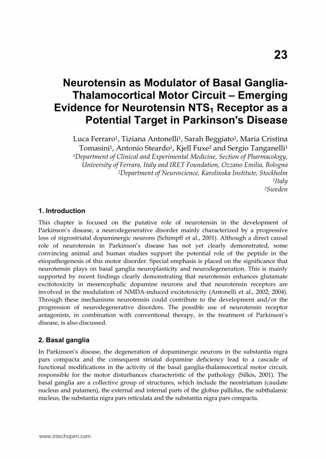
From a simplistic point of view, motor information coming from glutamatergic neurons
located in several areas of the cerebral cortex, reach the striatum, which represents the
primary input nucleus of the basal ganglia. These information, processed in the striatum, are
transmitted by the so called “direct” and “indirect” pathways, to the main output nuclei of
the basal ganglia (substantia nigra pars reticulata and the internal part of the globus
pallidus; Fig. 1). The “indirect pathway” encompasses a trisynaptic link including i)
GABAergic/enkephalinergic neurons, which connect the striatum to the external part of the
globus pallidus; ii) the external part of the globus pallidus GABAergic neurons projecting to
subthalamic nucleus and iii) glutamatergic subthalamic nucleus neurons, which project to
the basal ganglia output structures (internal part of the globus pallidus/substantia nigra
pars reticulata) and send collaterals to external part of the globus pallidus (Gerfen, 1992). On
the other hand, the “direct” monosynaptic pathway consists of GABAergic neurons which
directly connect the striatum to the main basal ganglia output structures (substantia nigra
pars reticulata and the internal part of the globus pallidus). Outputs from these nuclei
consist of inhibitory GABAergic neurons projecting to the ventral-anterior and ventrolateral
nuclei of the thalamus which, through excitatory glutamatergic fibers, project back to the
prefrontal and motor cortices. The differences in neuronal connectivity between the “direct”
and “indirect” pathways show dissimilar functional consequences: the stimulation of the
“direct pathway” inhibits substantia nigra pars reticulata and the internal part of the globus
pallidus activity, thus leading to a disinhibition of thalamocortical neurons and a
consequent facilitation of motor initiation. On the contrary, the stimulation of the “indirect
pathway” produces motor inhibition. In spite of the model of the “direct” and “indirect”
pathways is an oversimplification of the basal ganglia organization, it still represents the
cornerstone for modern research on the basal ganglia functions (for review, Smith and
Villalba, 2008).
Dopamine released by terminals of neurons located in substantia nigra pars compacta markedly affects the functional activity of the striatum. In the striatum dopamine D1 and D2 receptor subtypes are respectively expressed on the “direct” and “indirect” striatonigral pathways and modulate motor information (Gerfen, 2003). Although the degree of this anatomical separation of D1 and D2 receptors has been for a time a controversial topic, recent studies, using transgenic mice have confirmed that dopamine receptor subtypes have mainly a different expression on separate populations of GABAergic medium-spiny projection neurons (Wang et al., 2006; Galvan and Wichmann, 2008). Due to the different location of its receptor subtypes, striatal dopamine physiologically activates the “direct pathway” (D1 receptors) and inhibits the “indirect pathway” (D2), leading to an increase of thalamocortical motor drive (see above). In addition, striatal glutamate release from corticostriatal glutamatergic terminals is tonically inhibited by dopaminergic input coming from the substantia nigra pars compacta and activating dopamine D2 heteroreceptors (Bamford et al., 2004). In this synaptic arrangement, dopamine depletion within the striatum not only removes tonic dopamine inhibitory control over corticostriatal glutamatergic drive, but also induces an imbalance between the “direct” and the “indirect” pathways (Fig. 1). In particular, this deficit produces an overactivity of the GABAergic projections from the striatum to the external part of the globus pallidus, leading to an excessive inhibition of thalamocortical and brainstem motor systems. From a pathological point of view, the hyperactivity of striatopallidal GABAergic neurons is considered one of the anomaly responsible for generation of motor parkinsonian symptoms. Pharmacological interventions
www.intechopen.com
Neurotensin as Modulator of Basal Ganglia-Thalamocortical Motor Circuit – Emerging Evidence for Neurotensin NTS1 Receptor as a Potential Target in Parkinson's Disease
473
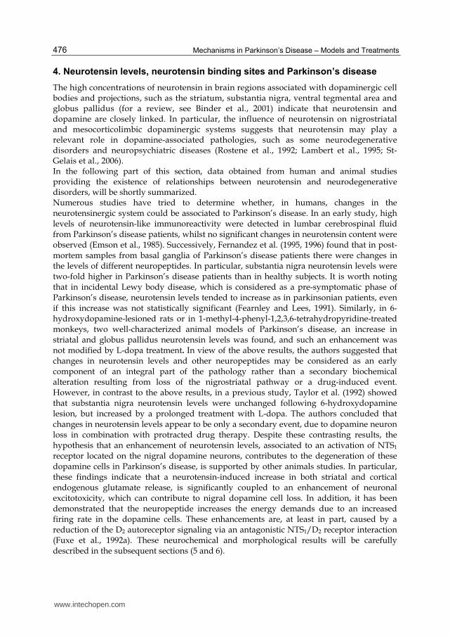
Fig. 1. The changes in the activity of basal ganglia circuits in normal state (’GO’) vs Parkinson’s Disease (’NO GO’) are indicated. Heavy arrows, high activity; thin arrows, low activity. Abbreviations used: GPl, globus pallidus, lateral; GPm, globus pallidus, medial; SNC, Substantia nigra, zona compacta; SNR,Substantia nigra, zona reticulata; SupCol, Superior colliculus; Form Ret, formatio retucularis; PPN, Pedunculo pontine nucleus (from Tanganelli et al., 2004).
www.intechopen.com
Mechanisms in Parkinson’s Disease – Models and Treatments
474
that can compensate for loss of dopamine and suppress the expression of motor symptoms in the pre-motor stages of Parkinson’s disease are represented by the reduction of: i) the excitatory corticostriatal inputs that excite striatal output neurons of the “indirect pathway” or ii) the overactivity of striato-pallidal GABAergic neurons. The use of selective D2 receptor agonist, A2A adenosine receptor antagonism, blockade of GABA receptors in the external part of the globus pallidus or reduction of the excitatory NMDA receptor-mediated corticostriatal inputs, impinging upon striatal output neurons of the “indirect pathway”, can be helpful for slowing progression of Parkinson’s disease symptoms.
3. Neurotensin and its receptors
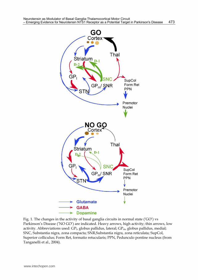
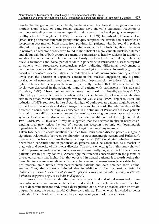
Neuropeptides represent undoubtedly one of the most common signaling molecules in the central nervous system. Accumulating evidence have implicated a vast number of neuropeptides and their receptors in the control of a wide range of physiological functions and pathological events, including neurodegenerative disorders. Like all neuropeptides, neurotensin, an endogenous 13 amino acid peptide (Figure 2), is
synthesized as part of a larger inactive precursor (Proneurotensin/neuromedin N).
Fig. 2. Sequence of neurotensin (modified from Ferraro et al., 2009).
The precursor molecule, a highly conserved polypeptide of 169-170 amino acid, contains one copy each of neurotensin and neuromedin N near the C-terminus and undergoes a differential tissue-specific cleavage at its four dibasic sites by proprotein convertases. Pro-neurotensin/neuromedin N may therefore be processed to generate different sets of peptides. Four biologically active products of Pro-neurotensin/neuromedin N processing have been described: neurotensin, neuromedin N, large neurotensin and large neuromedin N (Kitabgi, 2010). In the brain, Pro-neurotensin/neuromedin N processing mainly depends on proprotein convertase 2 activity and leads to high amounts of neurotensin and neuromedin N and small quantities of large neurotensin and large neuromedin N (Kitabgi, 2010). Using radioimmunoassay techniques, it has been demonstrated that the regional distribution of neurotensin and neuromedin N in brain tissues is, generally, the same. However, marked differences in the ratio of neurotensin over neuromedin N have been observed in different brain areas, being neurotensin generally more abundant in dopaminergic region such as substantia nigra pars compacta and ventral tegmental area. Once processed as an active peptide in neurons, neurotensin is stored in dense core vesicles.
The physiological inactivation of neurotensin is operated by endopeptidases belonging to
the family of metallopeptidases, which act on primary cleavage sites in the peptide
sequence: Arg8-Arg9, Pro10-Tyr11 and Tyr11-Ile12 bonds. Another mechanism that
produces an inactivation of neurotensin transmission is the process of neurotensin
internalization.
Neurotensin is widely expressed in nerve cells, fibers and terminals (Uhl, 1982; Emson et al.,
1985) and exhibits diverse biological actions in the regulation of central nervous system
www.intechopen.com
Neurotensin as Modulator of Basal Ganglia-Thalamocortical Motor Circuit – Emerging Evidence for Neurotensin NTS1 Receptor as a Potential Target in Parkinson's Disease
475
functions of mammals, including man. The peptide is also highly expressed in the
periphery, where it mainly acts as a modulator of the gastrointestinal and cardiovascular
systems (Wang and Evers, 1999). Neurotensin was originally isolated and sequenced from
bovine hypothalamus (Carraway and Leeman, 1973). Subsequent anatomical and functional
studies have provided evidence that, in the brain, neurotensin behaves as neurotransmitter
and/or neuromodulator (Nemeroff and Cain, 1985; Mendez et al., 1997). Neurotensin is
released by neurons through sodium and calcium-dependent mechanisms. Once released,
neurotensin produces its biological effects by interacting with three different receptor
subtypes (NTS1, NTS2 and NTS3/sortilin). The large distribution in the central nervous
system of this family of membrane receptors explains the wide range of physiologic and
pathologic effects mediated by the neuropeptide (Barroso et al., 2000). NTS1 and NTS2
receptors belong to the family of G-protein-coupled receptors with seven transmembrane
domains, which share 60% homology. The NTS1 receptor displays a high affinity for
neurotensin, while NTS2 receptor has a substantially lower affinity for the peptide and a
high affinity to levocabastine, a histamine H1 receptor antagonist (Chalon et al., 1996;
Vincent et al., 1999). NTS3/sortilin receptor is a single transmembrane protein located in
intracellular vesicles of neurons and glia and appears involved in cell sorting and in tropism
in cancer cells (Nouel et al., 1999; Mazella et al., 1998). NTS1 receptor is coupled to a variety
of signaling cascades, including production of inositol phosphates through activation of
phospholipase C, formation of cAMP and cGMP, and induction of mitogen-activated
protein kinase phosphorylation. Autoradiographic ligand binding, in situ hybridization, and
immunohistochemical studies have yielded abundant information on the distribution of
NTS1 receptors in mammalian brain. NTS1 receptors are markedly expressed in brain
regions rich in dopamine cell bodies, such as the substantia nigra pars reticulata and pars
compacta, the ventral tegmental area, and in projection areas of both nigrostriatal and
mesocorticolimbic dopaminergic pathways, such as striatum, nucleus accumbens and
frontal cortex (Palacios and Kuhar, 1981; Goulet et al., 1999; Boudin et al., 1998; Binder et al.,
2001). In the striatum and nucleus accumbens, NTS1 receptors are co-localized at post-
synaptic level with dopamine D2 receptors and, although in low density, at the pre-synaptic
levels too (Pickel et al., 2001; Delle Donne et al., 2004). This receptor co-distribution together
with the demonstration that neurotensin is localized within the nigrostriatal and mesolimbic
dopamine neurons explain the role that the neuropeptide plays in the modulation of
dopamine neurotransmission (Jennes et al., 1982). It is worth noting that in the striatum,
NTS1 receptors are significantly located on cortical glutamatergic terminals as well as on the
striatopallidal GABA neurons (Boudin et al., 1996; Alexander and Leeman, 1998; Tanji et al.,
1999). Finally, in the globus pallidus, neurotensin receptors (NTS1 and NTS2) exist in
different neurons and are located both pre-synaptically and post-synaptically (Fassio et al.,
2000; Sarret et al., 2003) thus regulating (mainly NTS1 receptors), both pallidal glutamatergic
and GABAergic transmission (Chen et al., 2004; 2006). Such distribution of NTS1 receptors
justifies the modulation that neurotensin exerts on the mesolimbic, mesocortical and
nigrostriatal dopamine neurons, as well as on glutamatergic and GABAergic neurones
(Deutch and Zahm, 1992; Fuxe et al., 1992 a,b; Rostene et al., 1992; Binder et al., 2001; Dobner
et al., 2003; Petrie et al., 2005). Most of the central and peripheral functions controlled by
NTS1 receptors have been elucidated by the use of the non-peptide neurotensin antagonist
SR48692, which preferentially binds NTS1 receptors (Gully et al., 1993; Rostene et al., 1997).
www.intechopen.com
Mechanisms in Parkinson’s Disease – Models and Treatments
476
4. Neurotensin levels, neurotensin binding sites and Parkinson’s disease
The high concentrations of neurotensin in brain regions associated with dopaminergic cell bodies and projections, such as the striatum, substantia nigra, ventral tegmental area and globus pallidus (for a review, see Binder et al., 2001) indicate that neurotensin and dopamine are closely linked. In particular, the influence of neurotensin on nigrostriatal and mesocorticolimbic dopaminergic systems suggests that neurotensin may play a relevant role in dopamine-associated pathologies, such as some neurodegenerative disorders and neuropsychiatric diseases (Rostene et al., 1992; Lambert et al., 1995; St-Gelais et al., 2006). In the following part of this section, data obtained from human and animal studies providing the existence of relationships between neurotensin and neurodegenerative disorders, will be shortly summarized. Numerous studies have tried to determine whether, in humans, changes in the neurotensinergic system could be associated to Parkinson’s disease. In an early study, high levels of neurotensin-like immunoreactivity were detected in lumbar cerebrospinal fluid from Parkinson’s disease patients, whilst no significant changes in neurotensin content were observed (Emson et al., 1985). Successively, Fernandez et al. (1995, 1996) found that in post-mortem samples from basal ganglia of Parkinson’s disease patients there were changes in the levels of different neuropeptides. In particular, substantia nigra neurotensin levels were two-fold higher in Parkinson’s disease patients than in healthy subjects. It is worth noting that in incidental Lewy body disease, which is considered as a pre-symptomatic phase of Parkinson’s disease, neurotensin levels tended to increase as in parkinsonian patients, even if this increase was not statistically significant (Fearnley and Lees, 1991). Similarly, in 6-hydroxydopamine-lesioned rats or in 1-methyl-4-phenyl-1,2,3,6-tetrahydropyridine-treated monkeys, two well-characterized animal models of Parkinson’s disease, an increase in striatal and globus pallidus neurotensin levels was found, and such an enhancement was not modified by L-dopa treatment. In view of the above results, the authors suggested that changes in neurotensin levels and other neuropeptides may be considered as an early component of an integral part of the pathology rather than a secondary biochemical alteration resulting from loss of the nigrostriatal pathway or a drug-induced event. However, in contrast to the above results, in a previous study, Taylor et al. (1992) showed that substantia nigra neurotensin levels were unchanged following 6-hydroxydopamine lesion, but increased by a prolonged treatment with L-dopa. The authors concluded that changes in neurotensin levels appear to be only a secondary event, due to dopamine neuron loss in combination with protracted drug therapy. Despite these contrasting results, the hypothesis that an enhancement of neurotensin levels, associated to an activation of NTS1 receptor located on the nigral dopamine neurons, contributes to the degeneration of these dopamine cells in Parkinson’s disease, is supported by other animals studies. In particular, these findings indicate that a neurotensin-induced increase in both striatal and cortical endogenous glutamate release, is significantly coupled to an enhancement of neuronal excitotoxicity, which can contribute to nigral dopamine cell loss. In addition, it has been demonstrated that the neuropeptide increases the energy demands due to an increased firing rate in the dopamine cells. These enhancements are, at least in part, caused by a reduction of the D2 autoreceptor signaling via an antagonistic NTS1/D2 receptor interaction (Fuxe et al., 1992a). These neurochemical and morphological results will be carefully described in the subsequent sections (5 and 6).
www.intechopen.com
Neurotensin as Modulator of Basal Ganglia-Thalamocortical Motor Circuit – Emerging Evidence for Neurotensin NTS1 Receptor as a Potential Target in Parkinson's Disease
477
Besides the changes in neurotensin levels, biochemical and histological investigations in post-mortem brain tissues of parkinsonian patients have shown a significant reduction of neurotensin-binding sites in several specific brain areas of the basal ganglia as respect to healthy subjects (Chinaglia et al. 1990; Fernandez et al., 1994). In particular, Chinaglia et al. (1990), using a receptor autoradiography technique, compared the distribution of neurotensin receptors in post-mortem brain tissues from parkinsonian patients, with that found in patients affected by progressive supranuclear palsy and-in age-matched controls. Significant decreases in neurotensin receptor density were found in the substantia nigra, caudate nucleus, putamen and globus pallidus of both groups of patients in comparison to healthy subjects. In addition, a significant decrement of neurotensin receptor density was found in the ventral tegmental area, nucleus accumbens and dorsal part of caudate in patients with Parkinson’s disease as regards to patients with progressive supranuclear palsy, indicating differential involvement of neurotensin receptor alterations in these two neurological disorders. Interestingly, in this cohort of Parkinson’s disease patients, the reduction of striatal neurotensin binding sites was lower than the decrease of dopamine content in this nucleus, suggesting only a partial localization of neurotensin receptors on nigrostriatal dopaminergic projections. Using in situ hybridization, it has been possible to more specifically illustrate that NTS1 receptor mRNA levels were decreased in the substantia nigra of patients with parkinsonism (Yamada and Richelson, 1995). These human results were confirmed in 1-methyl-4-phenyl-1,2,3,6-tetrahydropyridine-treated monkeys, where a decrease in the number of neurotensin-binding sites in the striatum and substantia nigra was found (Goulet et al., 1999; Tanji et al., 1999). The reduction of NTS1 receptors in the substantia nigra of parkinsonian patients might be related to the loss of the nigrostriatal dopaminergic neurons. In contrast, the interpretation of the decrease in neurotensin-binding sites observed in the striatum of Parkinson’s disease patients is certainly more difficult since, at present, the results concerning the pre-synaptic or the post-synaptic localization of striatal neurotensin receptors are still contradictory (Quirion et al., 1985; Cadet, 1991). However, it may be suggested that the decrease in striatal neurotensin-binding sites may reflect the loss of neurotensin receptors not only on dopaminergic nigrostriatal terminals but also on striatal GABAergic medium spiny neurons. Taken together, the above mentioned studies from Parkinson’s disease patients suggest a significant relationship between the alteration of neurotensinergic system and Parkinson’s disease. On the basis of these findings, Schimpff et al. (2001) evaluated whether plasma neurotensin concentrations in parkinsonian patients could be considered as a marker in diagnosis and severity of this motor disorder. The results emerging from this study showed that the plasma neurotensin concentrations were significantly higher in Parkinson’s disease patients than in healthy controls. Accordingly, neurotensin concentration in the plasma of untreated patients was higher than that observed in treated patients. It is worth noting that these findings were compatible with the enhancement of neurotensin levels detected in post-mortem brain tissues from parkinsonian patients and data obtained from animal studies. Thus, the authors concluded that in addition to the diagnostic criteria for Parkinson’s disease “measurement of extracted plasma neurotensin concentrations in patients with Parkinson may prove useful as an index in diagnosis”. In summary, it can be concluded that the increase in striatal and nigral neurotensin tissue concentrations, as well as in cerebrospinal fluid and plasma levels may be due either to a loss of dopamine neurons and/or to a dysregulation of neurotensin transmission on striatal output, favoring the striatopallidal GABAergic pathway. Further work is needed to better understand the role of neurotensin in the pathophysiology of Parkinson’s disease.
www.intechopen.com
Mechanisms in Parkinson’s Disease – Models and Treatments
478
5. Striatal neurotensin and Parkinson’s disease: neurochemical animal studies
5.1 Neurotensin modulation of pre- and post-synaptic D2 receptors. Relevance for the control of striatopallidal GABAergic projections
As stated above, the motor deficits that characterize Parkinson’s disease are associated to an imbalance on the functional activity of the “direct”–“indirect” circuits in favor of the “indirect pathway”, i.e. reduced activity in the “direct pathway” and/or increased activity in the “indirect pathway” (Obeso et al., 2000; 2008). Several lines of evidence indicate that neurotensin is co-localized and co-distributed with dopamine neurons of the basal ganglia, including the somatodendritic complex and axon terminals of various neuronal elements in the substantia nigra and striatum. This close anatomical relationship, reinforces functional findings demonstrating the existence of reciprocal modulations between neurotensinergic and dopaminergic systems in these brain areas (Hökfelt et al., 1984; Nemeroff and Cain, 1985; Blaha et al., 1990; Castel et al., 1994; Tanganelli et al., 1994; Rostène et al., 1997; Werkman et al., 2000). Intensive animal studies have well documented that neurotensin, in addition to its direct excitatory effects on dopamine neurons, significantly modulates D2 auto- and hetero-receptors functions through the activation of its high-affinity NTS1 receptor (Kalivas and Duffy, 1990; Werkman et al., 2000; Binder et al., 2001). The regulation of dopaminergic transmission, especially at the level of nigrostriatal and mesocorticolimbic dopamine pathways, by neurotensin (Kitabgy et al., 1989; Deutch and Zahm, 1992) is mainly due to an antagonistic action of the activated NTS1 receptor on D2 receptor recognition and signaling. In the striatum, neurotensin has been shown to reduce the affinity of D2 agonist binding sites and their transduction signals through a receptor-receptor interaction at both pre- and post-synaptic levels (Agnati et al., 1983; von Euler and Fuxe 1987; Shibata et al., 1987; Da-Silva et al., 1989; Fuxe et al., 1992 a,b). In particular, neurotensin by increasing the Kd value of D2 receptor agonist binding, significantly decreases the affinity of D2 receptors for endogenous dopamine and dopamine receptor agonists. The neurotensin-induced reduction of D2 receptor agonist affinity has been demonstrated both in sections and in membrane preparations. The presence at the cellular level of NTS1 and D2 receptors in the same axon terminals and dendrites (Delle Donne et al., 2004), together with the demonstrated antagonistic intramembrane NTS1/D2 receptor–receptor interactions using biochemical radioligand binding analysis in striatal membranes (Agnati et al., 1983; von Euler and Fuxe 1987; Tanganelli et al., 1989; Fuxe et al., 1992b; Li et al., 1995; Diaz-Cabiale et al., 2002; Antonelli et al., 2007), give indirect evidence for the existence of NTS1/D2 receptor heteromerization. By using intrastriatal monoprobe microdialysis and measuring dopamine release from striatal terminals, in vivo evidence has been obtained that the neurotensin/D2 antagonistic receptor–receptor interaction exists at the pre-junctional level in striatal dopamine transmission. This study demonstrate that, as expected, intrastriatal perfusion with the preferential dopamine D2 receptor agonist pergolide decreased local dopamine outflow, an effect which reflects a stimulation of terminal D2 auto-receptors causing the inhibition of striatal dopamine outflow. Interestingly, when neurotensin was co-perfused at a low nanomolar threshold concentration, together with pergolide, the inhibitory effect of the preferential dopamine D2 receptor agonist on dopamine release is fully abolished as measured in the striatum of awake unrestrained rats. This provides a functional in vivo correlate to the binding results indicating the existence of antagonistic neurotensin/D2 receptor–receptor interactions previously shown in neostriatal membranes and sections. A
www.intechopen.com
Neurotensin as Modulator of Basal Ganglia-Thalamocortical Motor Circuit – Emerging Evidence for Neurotensin NTS1 Receptor as a Potential Target in Parkinson's Disease
479
possible direct interaction between D2 and NTS1 receptors with the formation of heteromers has also been considered by Jomphe et al. (2006) as one of the possible, but not the exclusive, mechanisms underlying the functional control of striatal dopamine D2-mediated transmission by neurotensin. Biochemical and functional evidence suggests the existence of an antagonistic NTS1/D2 receptor-receptor interaction in rat neostriatum also at the post-synaptic level (Fuxe et al., 1992a; Ferraro et al., 1997). Post-synaptic D2 receptors in the neostriatum exist predominantly on medium sized GABAergic neurons of the “indirect pathway”, which project to the globus pallidus and exert an inhibitory influence on striopallidal GABA transmission (Reid et al., 1990; Ferre’ et al., 1993). Converging evidence suggests that the behavioural catalepsy associated with blockade of striatal D2 receptors is mediated by increased striopallidal GABA transmission, which leads to a decrease in thalamocortical motor drive (Drew et al., 1990; Osborne et al., 1994). Neurochemical findings, obtained employing in vivo dual-probe microdialysis technique, whereby one probe was implanted into the striatum and the other into the ipsilateral globus pallidus, demonstrated that intrastriatal perfusion with D2 agonists inhibits striopallidal GABA release (Reid et al., 1990; Ferre’ et al., 1993). On the contrary, D2 receptor antagonists enhance striopallidal GABA release (Drew et al., 1990). Interestingly, intrastriatal co-perfusion of neurotensin, at a concentration by itself ineffective on pallidal extracellular GABA levels, in combination with pergolide, fully antagonizes the inhibitory effects of the preferential D2 agonist on pallidal GABA release. The presence in the perfusate medium of the selective neurotensin receptor antagonist SR48692 removes the antagonistic effect of neurotensin, thus restoring the pergolide-induced inhibition of pallidal GABA levels. It is worth noting that higher concentrations of neurotensin, via a direct activation of NTS1 receptor subtypes, significantly increase pallidal GABA outflow. SR48692 fully counteracts the facilitatory effects of neurotensin, indicating the involvement of NTS1 receptors located on striopallidal GABAergic neurons in this effect. In view of the evidence showing that behavioural catalepsy in rodents and akinesia in humans are mediated by an increased striopallidal GABA transmission (Scheel-Kruger, 1986; Drew et al., 1990; Osborne et al., 1994), these findings suggest that the cataleptic profile of neurotensin (Shibata et al., 1987; Da-Silva et al., 1989) may be explained by its ability to influence neurotransmission in the “indirect pathway”. In particular, the cataleptic action of neurotensin may be in part related to an enhancement of endogenous neurotensin signalling and in part to a reduction of post-synaptic D2 receptors affinity. The existence of this intramembrane antagonistic neurotensin/D2 receptor interaction is also supported by the finding that haloperidol-induced catalepsy is associated with an increase in pallidal GABA release (Drew et al., 1990; Osborne et al., 1994). Briefly, neurotensin-induced increase of pallidal GABA release and the consequent activation of striopallidal GABA transmission, may represent the neurochemical substrate to explain the behavioural data indicating that the activation of striatal NTS1 receptors reduces motor activity (Poncelet et al., 1994).
5.2 Neurotensin modulation of striatal pre- and post-synaptic D2 receptors. Functional consequences on the activity of the “indirect pathway”
As previously reported, in the “indirect pathway” the striatopallidal GABAergic projection corresponds to the first neuron of the trisynaptic connection that projects to the substantia nigra pars reticulata. The GABAergic projection from the globus pallidus to the subthalamic nucleus represents the second neuron whereas the subthalamic nucleus glutamatergic cells
www.intechopen.com
Mechanisms in Parkinson’s Disease – Models and Treatments
480
projecting terminals to the substantia nigra pars reticulata and collaterals to the internal part of the globus pallidus, the third one. Thus, changes in the activation of striatopallidal GABA neurons lead to modifications of the activity of subthalamic nucleus glutamate neurons and consequent variations in substantia nigra pars reticulata and pallidal glutamate release. In view of the above data and to analyze the functional relevance of striatal NTS1 receptor activation on the activity of the “indirect pathway”, a dual-probe microdialysis analysis was planed. One probe was implanted into the striatum and the other one in the ipsilateral globus pallidus of the awake rat; the effects of neurotensin on striatal and pallidal glutamate levels were then measured. In this part of the present section the results coming from these microdialysis studies, will be summarized.
5.2.1 Effects of striatal NTS1 receptor activation on pallidal glutamate levels
Extracellular pallidal glutamate levels are mainly derived from the collaterals of the subthalamic nucleus-substantia nigra pars reticulata neurons (see above). Intrastriatal infusion with a high concentration of neurotensin increases striatal and pallidal glutamate as well as pallidal GABA levels (see also the above section). All these effects are counteracted by the local perfusion with the NTS1 receptor antagonist SR48692. Thus, the intrastriatal neurotensin-induced increase of pallidal glutamate levels may be related to a direct activation of somatodendritic NTS1 receptors located on the striatopallidal GABA neurons or to the antagonistic NTS1/D2 receptor-receptor interaction. The demonstration that the striatal neurotensin-induced increase in pallidal glutamate levels is counteracted by the intrapallidal perfusion of the GABAA receptor antagonist (-)-bicuculline, suggests that this effect is mediated via the activation of striatopallidal neurons. In fact, it seems likely that the stimulation of striatal NTS1 receptors, by increasing striatopallidal GABA release, reduces the activity of GABAergic neurons projecting from the globus pallidus to the subthalamic nucleus, thus increasing pallidal glutamatergic transmission. In other words, this sequence of GABA-mediated inhibitory modulations induces a disinhibition of the excitatory glutamatergic subthalamic nucleus-substantia nigra pars reticulata efferents which send axon collaterals to the globus pallidus (Alexander and Crutcher, 1990). The intrapallidal (-)-bicuculline perfusion was employed since previous studies demonstrated the role of GABAA receptor activation in regulating the pallidal output system toward the subthalamic nucleus (Kita, 1992; Amalric et al., 1994). Accordingly, an electrophysiological study (Soltis et al., 1994) demonstrated that the infusion of bicuculline into subthalamic nucleus increased the firing rate of pallidal neurons.
5.2.2 Effects of NTS1 receptor activation on striatal glutamate levels
Extracellular striatal glutamate levels are derived in part, from the terminals of cortical and
thalamic afferents (Sirinathsinghji and Heavens, 1989; Parent and Hazrati, 1995). As
previously stated, intrastriatal perfusion with neurotensin at a high concentration increases
striatal glutamate levels and this effect is fully counteracted by SR48692.
Immunohistochemical studies have shown that in the striatum and nucleus accumbens,
NTS1 receptors and dopamine D2 receptors are expressed on axon terminals, including the
glutamatergic ones (Delle Donne et al., 2004). In this context, it seems possible that at least
two mechanisms might underlay the neurotensin-mediated enhancement of striatal
glutamate release. The first one may be related to a direct activation of local NTS1 receptors
expressed on striatal glutamate terminals. The second mechanism implies that the formation
www.intechopen.com
Neurotensin as Modulator of Basal Ganglia-Thalamocortical Motor Circuit – Emerging Evidence for Neurotensin NTS1 Receptor as a Potential Target in Parkinson's Disease
481
of a NTS1/D2 heteromeric receptor complex mainly located on the plasma membrane of
striatal glutamate terminals, antagonizes the inhibitory D2 receptor mediated signaling
(see above) on the glutamate terminals, leading to an increase of glutamate release. The
presence of a functional antagonistic pre-synaptic NTS1/D2 interaction on glutamatergic
striatal terminals has been demonstrated since a threshold concentration of neurotensin
counteracts the D2 agonist quinpirole-induced inhibition of K+-evoked striatal glutamate
levels.
5.3 Neurochemical studies: concluding remarks
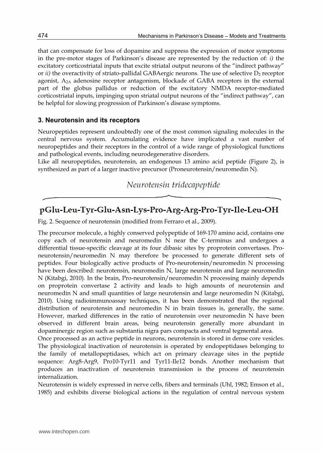
The analysis of all the above microdialysis results suggests that the over-activity of
striatopallidal GABA pathway is under the control of NTS1 receptors located both on
medium size spiny striatal GABAergic neurons and on striatal glutamatergic terminals. The
mechanisms involved in this control are mainly associated to a direct activation of NTS1
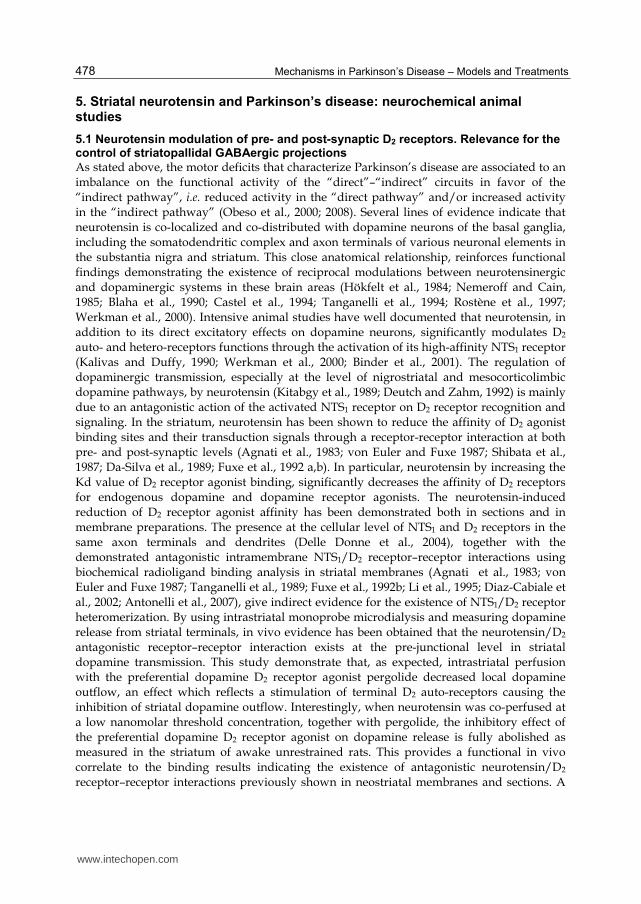
receptor homomer or to an antagonistic NTS1/D2 receptor-receptor interaction (Figure 3).
Based on these molecular mechanisms, it might be suggested that the activation of NTS1
receptors suppresses the inhibitory control mediated by nigrostriatal dopaminergic neurons
on striatopallidal GABAergic neurons and enhances the excitatory cortico-striatal
glutamatergic signaling. The final consequence of these combined opposite modulations is
the hyperactivity of the striatopallidal GABAergic neurons of the “indirect pathway” which
is considered one of the anomaly responsible for generation of motor parkinsonian
symptoms (see section 2). Thus, neurotensin receptor antagonists, by counteracting the
neurotensin-induced hyperactivity of striatopallidal GABAergic neurons, could be useful to
reduce motor symptoms in Parkinson’s disease patients.
6. Neurotensin and Parkinson’s disease: biochemical and morphological analyses in neuronal cell cultures
The substantial elevation in extracellular glutamate accompanied by an excessive activation
of excitatory amino acid receptors generates neuronal cell death (Sonsalla et al., 1998).
Glutamate has been one of the major focus of research into the excitotoxic basis of
neurodegenerative diseases (Choi, 1988; Meldrum and Garthwaite, 1990). In vivo and in
vitro studies have shown that neurotensin significantly increases endogenous glutamate
outflow in discrete rat brain regions, such as the striatum, globus pallidus, frontal cortex,
substantia nigra and parabrachial nucleus-ventrobasal thalamus (Sanz et al., 1993; Saleh,
1997; Ferraro et al., 1998, 2000, 2001). These findings suggest that neurotensin may play a
relevant role in reinforcing the effects exerted by glutamate on a variety of central nervous
system functions. In particular, the observations that neurotensin amplifies glutamate levels
and antagonizes the dopamine D2 receptor-mediated inhibition of dopamine transmission in
the basal ganglia (Fuxe et al., 1992 a,b), indicate that neurotensin may contribute to enhance
the firing rate and energy demands in the nerve cells. In this context, as illustrated in section
4, a putative role of neurotensin in the development of Parkinson’s disease, has been
suggested (Fernandez et al., 1995, 1996; Tanji et al., 1999; Schimpff et al., 2001).
In this paragraph, the effects of neurotensin in modulating the glutamate-induced
neurodegenerative effects in cultured rat mesencephalic dopaminergic (Antonelli et al.,
2002) and cortical (Antonelli et al., 2004) neurons will be shortly summarized.
www.intechopen.com
Mechanisms in Parkinson’s Disease – Models and Treatments
482
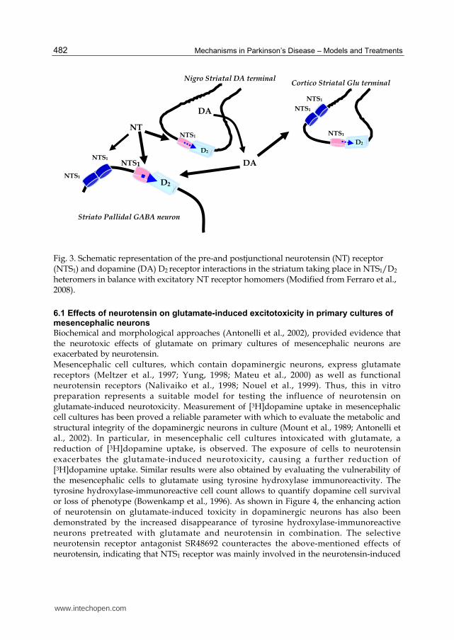
Fig. 3. Schematic representation of the pre-and postjunctional neurotensin (NT) receptor (NTS1) and dopamine (DA) D2 receptor interactions in the striatum taking place in NTS1/D2 heteromers in balance with excitatory NT receptor homomers (Modified from Ferraro et al., 2008).
6.1 Effects of neurotensin on glutamate-induced excitotoxicity in primary cultures of mesencephalic neurons
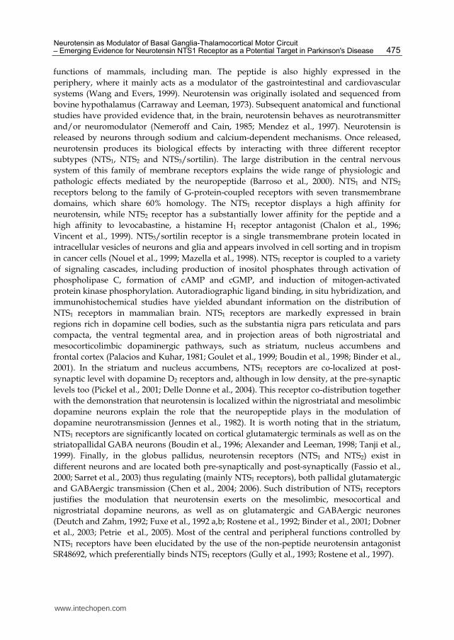
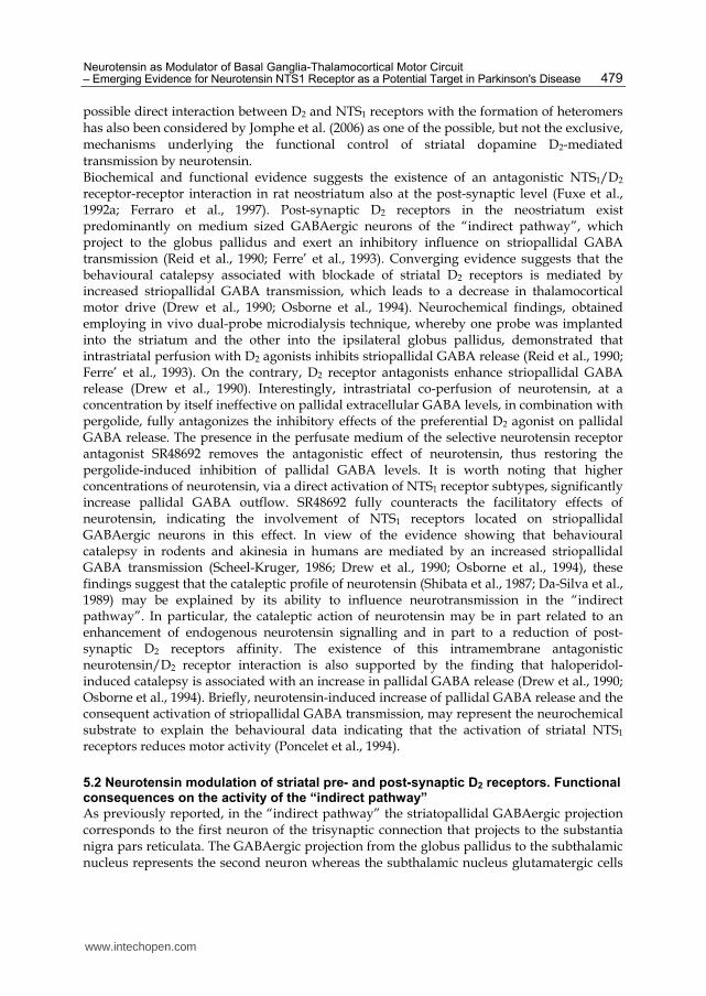
Biochemical and morphological approaches (Antonelli et al., 2002), provided evidence that the neurotoxic effects of glutamate on primary cultures of mesencephalic neurons are exacerbated by neurotensin. Mesencephalic cell cultures, which contain dopaminergic neurons, express glutamate receptors (Meltzer et al., 1997; Yung, 1998; Mateu et al., 2000) as well as functional neurotensin receptors (Nalivaiko et al., 1998; Nouel et al., 1999). Thus, this in vitro preparation represents a suitable model for testing the influence of neurotensin on glutamate-induced neurotoxicity. Measurement of [3H]dopamine uptake in mesencephalic cell cultures has been proved a reliable parameter with which to evaluate the metabolic and structural integrity of the dopaminergic neurons in culture (Mount et al., 1989; Antonelli et al., 2002). In particular, in mesencephalic cell cultures intoxicated with glutamate, a reduction of [3H]dopamine uptake, is observed. The exposure of cells to neurotensin exacerbates the glutamate-induced neurotoxicity, causing a further reduction of [3H]dopamine uptake. Similar results were also obtained by evaluating the vulnerability of the mesencephalic cells to glutamate using tyrosine hydroxylase immunoreactivity. The tyrosine hydroxylase-immunoreactive cell count allows to quantify dopamine cell survival or loss of phenotype (Bowenkamp et al., 1996). As shown in Figure 4, the enhancing action of neurotensin on glutamate-induced toxicity in dopaminergic neurons has also been demonstrated by the increased disappearance of tyrosine hydroxylase-immunoreactive neurons pretreated with glutamate and neurotensin in combination. The selective neurotensin receptor antagonist SR48692 counteractes the above-mentioned effects of neurotensin, indicating that NTS1 receptor was mainly involved in the neurotensin-induced
NT
DA
DA
Striato Pallidal GABA neuron
NTS1
NTS1
NTS1
NTS1
NTS1
D2
Nigro Striatal DA terminal
NTS1
D2
NTS1
D2
Cortico Striatal Glu terminal
www.intechopen.com
Neurotensin as Modulator of Basal Ganglia-Thalamocortical Motor Circuit – Emerging Evidence for Neurotensin NTS1 Receptor as a Potential Target in Parkinson's Disease
483
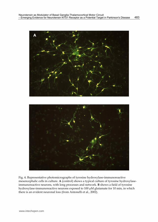
Fig. 4. Representative photomicrographs of tyrosine hydroxylase-immunoreactive mesencephalic cells in culture. A (control) shows a typical culture of tyrosine hydroxylase-immunoreactive neurons, with long processes and network. B shows a field of tyrosine hydroxylase-immunoreactive neurons exposed to 100 µM glutamate for 10 min, in which there is an evident neuronal loss (from Antonelli et al., 2002).
www.intechopen.com
Mechanisms in Parkinson’s Disease – Models and Treatments
484
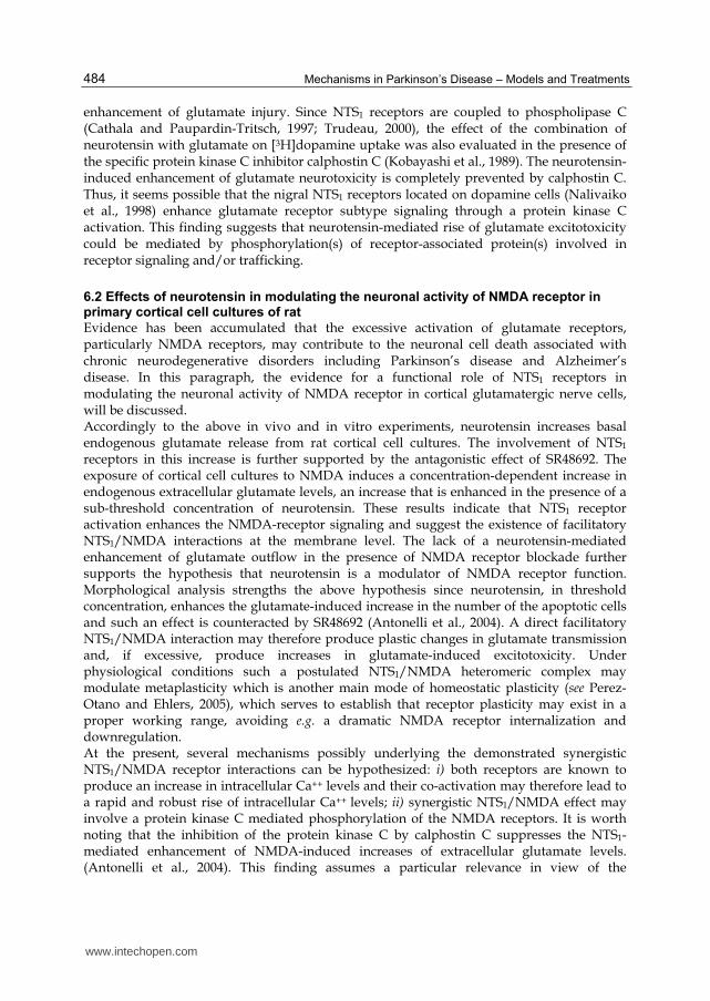
enhancement of glutamate injury. Since NTS1 receptors are coupled to phospholipase C (Cathala and Paupardin-Tritsch, 1997; Trudeau, 2000), the effect of the combination of neurotensin with glutamate on [3H]dopamine uptake was also evaluated in the presence of the specific protein kinase C inhibitor calphostin C (Kobayashi et al., 1989). The neurotensin-induced enhancement of glutamate neurotoxicity is completely prevented by calphostin C. Thus, it seems possible that the nigral NTS1 receptors located on dopamine cells (Nalivaiko et al., 1998) enhance glutamate receptor subtype signaling through a protein kinase C activation. This finding suggests that neurotensin-mediated rise of glutamate excitotoxicity could be mediated by phosphorylation(s) of receptor-associated protein(s) involved in receptor signaling and/or trafficking.
6.2 Effects of neurotensin in modulating the neuronal activity of NMDA receptor in primary cortical cell cultures of rat
Evidence has been accumulated that the excessive activation of glutamate receptors, particularly NMDA receptors, may contribute to the neuronal cell death associated with chronic neurodegenerative disorders including Parkinson’s disease and Alzheimer’s disease. In this paragraph, the evidence for a functional role of NTS1 receptors in modulating the neuronal activity of NMDA receptor in cortical glutamatergic nerve cells, will be discussed. Accordingly to the above in vivo and in vitro experiments, neurotensin increases basal endogenous glutamate release from rat cortical cell cultures. The involvement of NTS1 receptors in this increase is further supported by the antagonistic effect of SR48692. The exposure of cortical cell cultures to NMDA induces a concentration-dependent increase in endogenous extracellular glutamate levels, an increase that is enhanced in the presence of a sub-threshold concentration of neurotensin. These results indicate that NTS1 receptor activation enhances the NMDA-receptor signaling and suggest the existence of facilitatory NTS1/NMDA interactions at the membrane level. The lack of a neurotensin-mediated enhancement of glutamate outflow in the presence of NMDA receptor blockade further supports the hypothesis that neurotensin is a modulator of NMDA receptor function. Morphological analysis strengths the above hypothesis since neurotensin, in threshold concentration, enhances the glutamate-induced increase in the number of the apoptotic cells and such an effect is counteracted by SR48692 (Antonelli et al., 2004). A direct facilitatory NTS1/NMDA interaction may therefore produce plastic changes in glutamate transmission and, if excessive, produce increases in glutamate-induced excitotoxicity. Under physiological conditions such a postulated NTS1/NMDA heteromeric complex may modulate metaplasticity which is another main mode of homeostatic plasticity (see Perez-Otano and Ehlers, 2005), which serves to establish that receptor plasticity may exist in a proper working range, avoiding e.g. a dramatic NMDA receptor internalization and downregulation. At the present, several mechanisms possibly underlying the demonstrated synergistic NTS1/NMDA receptor interactions can be hypothesized: i) both receptors are known to produce an increase in intracellular Ca++ levels and their co-activation may therefore lead to a rapid and robust rise of intracellular Ca++ levels; ii) synergistic NTS1/NMDA effect may involve a protein kinase C mediated phosphorylation of the NMDA receptors. It is worth noting that the inhibition of the protein kinase C by calphostin C suppresses the NTS1-mediated enhancement of NMDA-induced increases of extracellular glutamate levels. (Antonelli et al., 2004). This finding assumes a particular relevance in view of the
www.intechopen.com
Neurotensin as Modulator of Basal Ganglia-Thalamocortical Motor Circuit – Emerging Evidence for Neurotensin NTS1 Receptor as a Potential Target in Parkinson's Disease
485
demonstration that protein kinase C is likely to be an important regulator of neuronal NMDA receptors in vivo. The activation of protein kinase C increases NMDA channel opening rate and a rapid delivery of functional NMDA receptors to the cell surface. Thus, regulation of neuronal NMDA receptors by protein kinases plays a critical role in synaptic transmission and synaptic plasticity of NMDA receptors. Since phospholipase C–protein kinase C–inositol triphosphate pathway is the major signal transduction of NTS1 receptors, it may be suggested that the existence of a neurotensin-mediated potentiation of NMDA receptors involves the activation of protein kinase C. Similarly to the above hypothesis, it has been demonstrated that mGluR1 activation potentiates NMDA receptors by an activation of protein kinase C (Skeberdis et al., 2001; Matsuyama et al., 2002); iii) it also seems possible that NTS1 receptor by forming a receptor heteromer with the NMDA receptor (NTS1/NMDA heteromer) may contribute to enhance NMDA receptor signalling and its surface expression; iv) a recent paper reported that neurotensin receptor agonists robustly increased extracellular concentrations of glycine in the rat prefrontal cortex (Li et al., 2010). It is well known that normal NMDA receptor function depends on not only the binding of glutamate to the receptor but also the binding of glycine to an allosteric site on this receptor. Thus, it could be suggested that neurotensin modulates NMDA receptor functions by modulating allosteric glycine activity. Taken together, the results obtained in mesencephalic and cortical cell cultures suggest that neurotensin receptor antagonists, by counteracting the neurotensin-induced amplification of glutamate excitotoxicity could display neuroprotective properties.
7. Neurotensin receptor blockade in an animal model of Parkinson’s disease
The postulated neuroprotective properties of neurotensin receptor antagonists are also supported by experiments (Ferraro et al., 2008) carried out in a rat model of Parkinson’s disease [unilateral nigral 6-hydroxydopamine induced lesion of the nigrostriatal DA pathway, hemiparkinson model]. In this study, behavioural and biochemical experiments have been performed in control animals and in 6-hydroxydopamine unilaterally lesioned rats chronically treated with saline or with the NTS1 receptor antagonist SR48692 from one-week before until one-week after the lesion. A conventional behavioural assessment using apomorphine-induced rotation was performed to quantify the unilateral nigrostriatal lesion-induced motor asymmetry after ipsilateral 6-hydroxydopamine injection. As expected, in 6-hydroxydopamine-lesioned rats, but not in control rats, the apomorphine injection produced a controlateral turning behaviour that significantly and progressively increased from week 1 to the 3rd week following the lesion. However, interestingly, in the SR48692-treated group, but not in the vehicle-treated group, the apomorphine-induced rotational behaviour is significantly reduced at each time of evaluation (day 7, 14 and 21 post lesion). Moreover, whereas the treatment stopped 2 weeks before, the effect of the compound remains significant. This finding suggests that systemic administration of NTS1 antagonist decreased the functional consequence of a partial dopaminergic lesion induced by intranigral application of the neurotoxin 6-hydroxydopamine in the rat. In view of the above behavioural findings, neurochemical experiments have been carried out in control animals and in rats chronically treated with SR48692 or its vehicle from one-week before until one week after the 6-hydroxydopamine injection. In particular, the responsivity to a challenge with NMDA has been assessed. The results obtained from this study demonstrate that in 6-hydroxydopamine-lesioned control and vehicle-treated rats,
www.intechopen.com
Mechanisms in Parkinson’s Disease – Models and Treatments
486
intrastriatal perfusion with NMDA induced a slight increase in glutamate extracellular levels that was significantly lower than that observed in sham-operated animals. Interestingly, in 6-hydroxydopamine-lesioned rats chronically treated with SR48692, the effect of intrastriatal perfusion of NMDA induced an increase in glutamate extracellular levels that was significantly higher with respect to that obtained in the group of 6-hydroxydopamine lesioned rats but still lower to that observed in control rats. These neurochemical results are in line with previous microdialysis data indicating that dopamine denervation is associated with a reduction of the enhancement of striatal glutamate transmission induced by a high micromolar NMDA concentration. Since it has been demonstrated that endogenous dopamine in the striatum facilitates strong excitatory inputs, the reduction of NMDA-stimulated glutamate levels in lesioned-animals could imply a loss of facilitatory dopamine receptor mediated signals. In this view, the observation that in rats chronically treated with SR48692 the excitatory response to a NMDA stimulus on the striatal glutamatergic transmission is partially restored may support a protective action of the NTS1 antagonist against 6-hydroxydopamine-induced dopamine neuron degeneration.
8. Conclusion
Parkinson's disease is associated to a progressive loss of nigrostriatal dopaminergic neurons. The decrease of dopamine levels in the striatum of parkinsonian patients is responsible for the main motor disturbances characteristic of the disease such as akinesia, muscular rigidity and tremor. The strict interactions between the tridecapeptide neurotensin and the dopaminergic systems lead to hypothesize that the peptide could be involved in some aspects of Parkinson’s disease. In this context, the present chapter discusses the putative role of neurotensin in the development and symptoms of Parkinson’s disease. Human studies provide evidence that in the basal ganglia of Parkinson’s disease patients there is an increase in neurotensin levels. These changes might be an integral part of the pathology rather than a consequence of the dopamine neuron degeneration. In addition, neurotensin receptor binding sites, especially in the nigrostriatal dopamine system, are reduced in brains of Parkinson’s disease patients and in the basal ganglia of hemiparkinsonian rats. This is probably a result of the ongoing degeneration of nigral dopamine cells in which the peptide actively participates. Based on neurochemical and morphological animals studies, the hypothesis is now introduced that the activation of NTS1 receptors by enhancing glutamate release and by amplifying the NMDA-mediated glutamate signalling contributes to the degeneration of dopaminergic neurons in Parkinson’s disease. In addition, the reduction of the D2 autoreceptor signaling due to the antagonistic NTS1/D2 receptor-receptor interaction, by enhancing the firing rate of dopamine neurons and energy demand may further contribute to this degeneration. The neurochemical studies have also clearly demonstrated that increased striatal neurotensin levels, by leading to an over-activity to the “indirect pathway” in the basal ganglia, could also play a role in motor Parkinson’s disease symptoms. In closing, in view of the presented results, it could be suggested that NTS1 antagonists, in combination with conventional drug treatments, may provide a possible novel therapeutic approach for the treatment of neurodegenerative pathologies, especially Parkinson’s disease. This hypothesis is supported by studies demonstrating the putative neuroprotective
www.intechopen.com

Neurotensin as Modulator of Basal Ganglia-Thalamocortical Motor Circuit – Emerging Evidence for Neurotensin NTS1 Receptor as a Potential Target in Parkinson's Disease
487
effects of the neurotensin receptor antagonist SR48692 (systemically administered) in an in vivo animal model of Parkinson’s disease. However, Mesnage et al. (2004) in an exploratory study reported that SR48692 could not improve parkinsonian motor disability. However, in this paper the authors reported that the lack of efficacy of NTS1 receptor antagonists could be attributed to the low dose used, as demonstrated by the absence of adverse events observed in any of the patients tested. In fact, it was concluded that further studies with higher doses of neurotensin receptor antagonists are needed. Taken together, the reported findings prompt to continue these preclinical studies in order to better understand the role of neurotensin in Parkinson’s disease development and symptoms.
9. Acknowledgement
This work has been supported by grants from Sanofi-Aventis and University of Ferrara (Fondo di Ateneo per la Ricerca Scientifica). The authors thank Dr. Jacqueline Fournier (Sanofi-Aventis) for her excellent scientific support during the research development.
10. References
Agnati, LF.; Fuxe, K.; Benfenati, F. & Battistini, N. (1983). Neurotensin in vitro markedly
reduces the affinity in subcortical limbic [3H]N-propylnorapomorphine binding
sites. Acta Physiol. Scan. 119, (December 1983), pp. 459-461, ISSN 0001-6772.
Alexander, GE. & Crutcher, M. (1990). Functional architecture of basal ganglia circuits:
neuronal substrates of parallel processing. Trends Neurosci. 13, (July 1990), pp. 266-
271, ISSN 0166-2236.
Alexander, MJ. & Leeman, SE. (1998). Widespread expression in adult rat forebrain of
mRNA encoding high-affinity neurotensin receptor. J. Comp. Neurol. 402,
(December 1998), pp. 475-500, ISSN 0021-9967.
Amalric, M.; Farin, D.; Dormont, JF. & Schmied, A. (1994). GABA-receptor activation in the
globus pallidus and entopeduncular nucleus: opposite effects on reaction time
performance in the cat. Exp. Brain Res. 102, pp.244-258, ISSN 0014-4819.
Antonelli, T.; Tomasini, MC.; Finetti, S.; Giardino, L.; Calzà, L.; Fuxe, K.; Soubriè, P.;
Tanganelli, S. & Ferraro, L. (2002). Neurotensin enhances Glutamate excitotoxicity
in mesencephalic neurons. J. Neurosci. Res. 70, (December 2002), pp. 766-773, ISSN
0360-4012.
Antonelli, T., Ferraro, L.; Fuxe, K.; Finetti, S.; Fournier, J.; Tanganelli, S.; De Mattei, M. &
Tomasini, M. C. (2004). Neurotensin enhances endogenous extracellular glutamate
levels in primariy cultures of rat cortical neurons. Involment of neurotensin
receptor in NMDA induced excitotoxicity. Cerebral Cortex 1, (April 2004), pp. 466-
473, ISSN 1047-3211.
Antonelli, T.; Tomasini, MC.; Fuxe, K.; Agnati, LF.; Tanganelli, S. & Ferraro, L. (2007)
Receptor-receptor interactions as studied with microdialysis. Focus on NTR/D2
interactions in the basal ganglia. J. Neural Transm. 114, (January 2007), pp. 105-13.
ISSN 0300-9564.
www.intechopen.com
Mechanisms in Parkinson’s Disease – Models and Treatments
488
Bamford, NS.; Robinson, S.; Palmiter, RD.; Joyce, JA.; Moore, C. & Meshul, CK. (2004)
Dopamine modulates release from corticostriatal terminals. J. Neurosci. 24, (October
2004), pp. 9541-9552, ISSN 0270-6474.
Barroso, S.; Richard, F.; Nicolas-Etheve, D.; Reversat, JL.; Bernassau, JM.; Kitabgi, P. &
Labbè-Julliè, C. (2000). Identification of residues involved in neurotensin binding
and modelling of the agonist binding site in neurotensin receptor 1. J. Biol. Chem.
275, (January 2000), pp. 328-336, ISSN 0021-9258.
Binder, EB.; Kinkead, B.; Owens, MJ. & Nemeroff, CB. (2001). Neurotensin and dopamine
interactions. Pharmacol. Rev. 53, (December 2001), pp. 453-486, ISSN 0031-6997.
Blaha, CD.; Coury, A.; Fibiger, HC. & Phillips, AG. (1990). Effects of neurotensin on DA
release and metabolism in the rat striatum and nucleus accumbens: cross-validation
using in vivo voltammetry and microdialysis. Neuroscience 34, pp. 699-705, ISSN
0306-4522.
Boudin, H.; Pélaprat, D.; Rostène, W. & Beaudet, A. (1996). Cellular distribution of
neurotensin receptors in rat brain: immunohistochemical study using an
antipeptide antibody against the cloned high affinity receptor, J. Comp. Neurol. 373,
(September 1996), pp. 76–89, ISSN 0021-9967.
Boudin, H.; Pelaprat, D.; Rostène, W.; Pickel, VM. & Beaudet, A. (1998). Correlative
ultrastructural distribution of neurotensin receptor proteins and binding sites in the
rat substantia nigra. J. Neurosci. 18, (October 1998), pp.8473-8484, ISSN 0270-6474.
Bowenkamp, KE.; David, D.; Lapchak, PL.; Henry, MA.; Granholm, AC.; Hoffer, BJ. &
Mahalik, TJ. (1996). 6-hydroxydopamine induces the loss of the dopaminergic
phenotype in substantia nigra neurons of the rat. A possible mechanism for
restoration of the nigrostriatal circuit mediated by glial cell line-derived
neurotrophic factor. Exp. Brain Res. 111, (September 1996), pp.1-7, ISSN 0014-4819.
Cadet, JL.; Kujirai, K. & Przedborski, S. (1991). Bilateral modulation of [3H]neurotensin
binding by unilateral intrastriatal 6-hydroxydopamine injections: evidence from a
receptor autoradiographic study. Brain Res. 564, (November 1991), pp. 37-44, ISSN
0006-8993.
Carraway, R. & Leeman, SE. (1973). The isolation of a new hypotensive peptide,
neurotensin, from bovine hypothalami. J. Biol. Chem. 248, (October 1973), pp.6854-
6861, ISSN 0021-9258.
Castel, MN.; Morino, P.; Nylander, I.; Terenius, L. & HöKfelt, T. (1994) Differential
dopaminergic regulation of the neurotensin striatonigral and striatopallidal
pathways in the rat. Eur. J. Pharmacol. 262, (September 1994), pp.1-10, ISSN 0014-
2999.
Cathala, L. & Paupardin-Tritsch, D. (1997). Neurotensin inhibition of the hyperpolarization-
activated cation current (Ih) in the rat substantia nigra pars compacta implicates the
protein kinase C pathway. J. Physiol. 503, (August 1997), pp.87-97, ISSN 0022-3751.
Chalon, P.; Vita, N.; Kaghad, M.; Guillemot, M.; Bonnin, J. & Delpech, B. (1996). Molecular
cloning of a levocabastine-sensitive neurotensin binding site. FEBS Lett. 386, (May
1996), pp. 91–94, ISSN 0014-5793.
www.intechopen.com

Neurotensin as Modulator of Basal Ganglia-Thalamocortical Motor Circuit – Emerging Evidence for Neurotensin NTS1 Receptor as a Potential Target in Parkinson's Disease
489
Chen, L.; Yung, KK. & Yung, WH. (2004). Neurotensin depolarizes globus pallidus neurons
in rats via neurotensin type-1 receptor. Neuroscience 125, pp.853-859, ISSN 0306-
4522.
Chen, L.; Yung, KK. & Yung, WH. (2006). Neurotensin selectively facilitates glutamatergic
transmission in globus pallidus. Neuroscience 141, (September 2006), pp.1871-1878,
ISSN 0306-4522 .
Chinaglia, G.; Probst, A. & Palacios, JM. (1990). Neurotensin receptors in Parkinson’s disease
and progressive supranuclear palsy: an autoradiographic study in basal ganglia.
Neuroscience 39, pp.351-360, 0306-4522.
Choi, DW. (1988). Glutamate neurotoxicity and diseases of the nervous system. Neuron 1,
(October 1988), pp.623-34. ISSN 0896-6273.
Da-Silva, SL.; Brandäo, ML. & Tomaz, C. (1989) Behavioral effects of neurotensin applied to
periventricular structures of rats. Braz. J. Med. Biol. Res. 22, pp.711-715, ISSN 0100-
879X.
Delle Donne, KT.; Chan , J.; Boudin, H.; Pélaprat, D.; Rostène, W. & Pickel, VM. (2004).
Electron microscopic dual labeling of high-affinity neurotensin and dopamine D2
receptors in the rat nucleus accumbens shell. Synapse. 52, (June 2004), pp.176-187,
ISSN 0887-4476.
Dobner, PR.; Deutch, AY. & Fadel, J. (2003). Neurotensin: dual roles in psychostimulant and
antipsychotic drug responses. Life Sci. 73,(June 2003), pp.801-811. Rev, ISSN 0024-
3205.
Deutch, AY. & Zahm, DS. (1992). The current status of neurotensin-dopamine interactions:
issues and speculations. Ann. N.Y. Acad. Sci. 668, pp.232-252, ISSN 0077-8923.
Diaz-Cabiale, Z., Fuxe, K.; Narvaez, JA.; Finetti, S.; Antonelli, T.; Tanganelli, S. & Ferraro, L.
(2002). Neurotensin-induced modulation of dopamine D2 receptors and their
function in rat striatum: Counteraction by NTR1-lik receptor antagonist.
Neuroreport 13, (May 2002), pp.763-766, ISSN 0959-4965.
Drew, KL.; O’Connor, WT.; Kehr, J. & Ungerstedt, U. (1990). Regional specific effects of
clozapine and haloperidol on GABA and dopamine release in rat basal ganglia.
Eur. J. Pharm. 187, (October 1990), pp. 385–397, ISSN 0014-2999
Emson, PC.; Horsfield, PM.; Goedert, M.; Rossor, MN. & Hawkes, CH. (1985). Neurotensin
in human brain: regional distribution and effects of neurological illness. Brain Res.
347, (November 1985), pp.239-244 ISSN 0006-8993.
Fassio, A.; Evans, G.; Grisshammer, R.; Bolam, JP.; Mimmack, M. & Emson, PC. (2000).
Distribution of the neurotensin receptor NTS1 in the rat CNS studied using an
amino-terminal directed antibody. Neuropharmacology 39, (June 2000), pp. 1430–
1442, ISSN 0028-3908.
Fearnley, JM. & Lees, AJ. (1991). Ageing and Parkinson's disease: substantia nigra regional
selectivity. Brain 114, (October 1991), pp. 2283-2301, ISSN 0006-8950.
Fernandez, A.; De Ceballos, ML. & Jenner, P. (1994). Neurotensin, substance P, delta and mu
opioid receptors are decreased in basal ganglia of Parkinson’s disease patients.
Neuroscience 61, (July 1994), pp.73-79, ISSN 0306-4522.
Fernandez, A.; Jenner, P.; Marsden, CD. & De Ceballos, ML. (1995). Characterization of
neurotensin-like immunoreactivity in human basal ganglia: increased neurotensin
www.intechopen.com
Mechanisms in Parkinson’s Disease – Models and Treatments
490
levels in substantia nigra in Parkinson's disease. Peptides. 16, pp.339-346, ISSN 0196-
9781.
Fernandez, A.; De Ceballos, ML.; Rose, S.; Jenner, P. & Marsden, CD. (1996) Alterations in
peptide levels in Parkinson's disease and incidental Lewy body disease. Brain 119
(Pt 3), (June 1996), pp.823-830, ISSN 0006-8950.
Ferraro. L.; O'Connor, WT.; Antonelli, T.; Fuxe, K. & Tanganelli, S. (1997). Differential effects
of intrastriatal neurotensin(1–13) and neurotensin(8–13) on striatal dopamine and
pallidal GABA release. A dual-probe microdialysis study in the awake rat. Eur. J.
Neurosci. 9, (September 1997), pp.1838-1846, ISSN 0953-816X.
Ferraro, L.; Tomasini, MC.; Siniscalchi, A.; Fuxe, K.; Tanganelli, S. & Antonelli, T. (2000).
Neurotensin increases endogenous glutamate release in rat cortical slices. Life Sci.
66, pp.927-936, ISSN 0024-3205.
Ferraro, L.; Tomasini, MC.; Fernandez, M.; Bebe, BW.; O'Connor, WT.; Fuxe, K.; Glennon,
JC.; Tanganelli, S & Antonelli, T. (2001). Nigral neurotensin receptor regulation of
nigral glutamate and nigroventral thalamic GABA transmission: a dual-probe
microdialysis study in intact conscious rat brain. Neuroscience 102, pp.113-120, ISSN
0306-4522.
Ferraro, L.; Tomasini, MC.; Mazza, R.; Fuxe, K.; Fournier, J.; Tanganelli, S. & Antonelli, T.
(2008). Neurotensin receptors as modulators of glutamatergic transmission. Brain
Res. Rev. 58 (August 2008) pp.365-373, ISSN 0165-0173.
Ferraro L, Tomasini MC, Beggiato S, Guerrini R, Salvadori S, Fuxe K, Calzà L, Tanganelli S,
Antonelli T. Emerging evidence for neurotensin receptor 1 antagonists as novel
pharmaceutics in neurodegenerative disorders. Mini Rev Med Chem. 9 (October
2009), pp. 1429-1438, ISSN: 1389-5575 (Print).
Ferrè, S.; O’Connor, WT.; Fuxe, K. & Ungerstedt, U. (1993). The striopallidal neuron: a main
locus for adenosine-dopamine interactions in the brain. J. Neurosci. 13, (December
1993) pp.5402–5406, ISSN 0270-6474.
Fuxe, K.; von Euler, G.; Agnati, LF.; Merlo Pich, E.; O’Connor, WT.; Tanganelli, S.; Li, XM.;
Tinner, B.; Cintra, A.; Carani, C. & Benfenati, F. (1992a). Intramembrane
interactions between neurotensin receptors and dopamine D2 receptors as a major
mechanism for the neuroleptic-like action of neurotensin. Ann. N.Y. Acad. Sci. 668,
pp.186-204, ISSN 0077-8923.
Fuxe, K.; O’Connor, WT.; Antonelli, T.; Osborne, PG.; Tanganelli, S.; Agnati, LF. &
Ungerstedt, U. (1992b). Evidence for a substrate of neuronal plasticity based on pre-
and postsynaptic neurotensin-dopamine receptor interactions in the neostriatum.
Proc. Natl. Acad. Sci. 89, (June 1992), pp.5591-5595, ISSN 0027-8424.
Galvan, A. & Wichmann T. (2008). Phatophisiology of Parkinsonism. Clinical Neurophysiol.
119, (July 2008), pp.1459-1474. Rew, ISSN 1388-2457.
Gerfen, CR. (1992). The neostriatal mosaic: multiple levels of compartmental organization in
the basal ganglia. Ann. Rev. Neurosci. 15, pp.285-320, ISSN 0147-006X.
Gerfen, CR. (2003). D1 dopamine receptor supersensitivity in the dopamine-depleted
striatum animal model of Parkinson's disease. Neuroscientist 9, (December 2003),
pp.455-62, ISSN 1073-8584.
www.intechopen.com
Neurotensin as Modulator of Basal Ganglia-Thalamocortical Motor Circuit – Emerging Evidence for Neurotensin NTS1 Receptor as a Potential Target in Parkinson's Disease
491
Goulet, M.; Morissette, M. & Grondin, R. (1999). Neurotensin receptors and dopamine
transporters: effects of MPTP lesioning and chronic dopaminergic treatments in
monkeys. Synapse 32, (June 1999), pp.153-64, ISSN 0887-4476.
Gully, D.; Canton, M.; Boidegrain, R.; Jeanjean, F.; Molimard, JC.; Poncelet, M.; Gueudet, C.;
Heaulme, M.; Leyris, A.; Brouard, A.; Pelaprat, D.; Labbe-Jullie, C.; Mazella, J.;
Soubrie, P.; Maffrand, JP.; Rostene, W.; Kitbagi, P. & Le Fur, G. (1993) Biochemical
and pharmacological profile of a potent and selective nonpeptide antagonist of the
neurotensin receptor. Proc. Natl. Acad. Sci. 90, (January 1993), pp.65–69, ISSN 0027-
8424.
Kalivas, PW. & Duffy, P. (1990). Effect of acute and daily neurotensin and enkephalin
treatments on extracellular dopamine in the nucleus accumbens. J. Neurosci. 10,
(September 1990), pp.2940-2949, ISSN 0270-6474.
Kita, H. (1992). Responses of globus pallidus neurons to cortical stimulation: intracellular
study in the rat. Brain Res. 589, (August 1992), pp.84-90, ISSN 0006-8993.
Kitabgi, P.; Herve, D.; Studler, JM.; Tramu, G.; Rostene, W. & Tassin, JP. (1989).
Neurotensin/dopamine interactions. Encephale 15, (January-February 1989), pp.91–
94, ISSN 0013-7006.
Kitabgi, P. (2010). Neurotensin and neuromedin N are differentially processed from a
common precursor by prohormone convertases in tissues and cell lines. Results
Probl. Cell Differ. 50, pp.50:85-96.
Kobayashi, E.; Nakano, H.; Morimoto, M. & Tamaoki, T. (1989). Calphostin C (UCN-1028C),
a novel microbial compound, is a highly potent and specific inhibitor of protein
kinase C. Biochem. Biophys. Res. Commun. 159, (March 1989), pp.548-553, ISSN 0006-
291X.
Hökfelt, T.; Everitt, BJ.; Theodorsson-Norheim, E. & Goldstein, M. (1984). Occurrence of
neurotensinlike immunoreactivity in subpopulations of hypothalamic,
mesencephalic, and medullary catecholamine neurons. J. Comp. Neurol. 222,
(February 1984), pp.543-59, ISSN 0021-9967.
Jennes, L.; Stumpf, WE. & Kalivas, PW. (1982). Neurotensin: Topographical distribution in
rat brain by immunohistochemistry. J. Comp. Neurol. 210, (September 1982), pp.211-
224, ISSN 0021-9967.
Jomphe, C.; Lemelin, PL.; Okano, H. & Trudeau, LE. (2006). Bidirectional regulation of
dopamine D2 and neurotensin NTS1 receptors in dopamine neurons. Eur. J.
Neurosci. 24, (November 2006), pp.2789-2800, ISSN 0953-816X.
Lambert, PD.; Gross, R.; Nemeroff, CB. & Kilts, CD., (1995). Anatomy and mechanisms of
neurotensin–dopamine interactions in the central nervous system. Ann. N. Y. Acad.
Sci. 757, (May 1995), pp. 377–389, ISSN 0077-8923.
Li, X M.; Ferraro, L.; Tanganelli, S.; O’Connor, WT.; Hasselrot, U.; Ungerstedt, U. & Fuxe, K.
(1995). Neurotensin peptides antagonistically regulate postsynaptic dopamine D2
receptor in rat nucleus accumbens: a receptor binding and microdialysis study. J.
Neural Transm. [Gen Sect], 102, pp.125-137, ISSN 0300-9564.
Li, Z.; Boules ,M.; Williams, K.; Peris, J. & Richelson, E. (2010) The novel neurotensin analog
NT69L blocks phencyclidine (PCP)-induced increases in locomotor activity and
www.intechopen.com
Mechanisms in Parkinson’s Disease – Models and Treatments
492
PCP-induced increases in monoamine and amino acids levels in the medial
prefrontal cortex. Brain Res., 1311, (January 2010), pp. 28-36, ISSN 0006-8993.
Mateu, G.; Privat, A.; Thibault. J. & Vignon, J. (2000). Modulation of glutamate neurotoxicity
on mesencephalic dopaminergic neurons in primary cultures by the presence of
striatal target cells. Int. J. Dev. Neurosci. 18, (October 2000), pp.607–613, ISSN 0736-
5748.
Matsuyama, S.; Higashi, H.; Maeda, H.; Greengard, P. & Nishi, A. (2002). Neurotensin
regulates DARPP-32 thr34 phosphorylation in neostriatal neurons by activation of
dopamine D1-type receptors. J. Neurochem. 81, (April 2002), pp.325-334, ISSN 0022-
3042.
Mazella, J.; Zsurger, N.; Navarro, V.; Chabry, J.; Kaghad, M. & Caput, D. (1998) The 100-kDa
neurotensin receptor is gp95/sortilin, a non-G-protein-coupled receptor, J. Biol.
Chem. 273, (October 1998), pp.26273–26276, ISSN 0021-9258.
Meldrum, B. & Garthwaite, J. (1990). Excitatory amino acid neurotoxicity and
neurodegenerative disease. Trends Pharmacol. Sci. 11, (September 1990) pp.379-387,
ISSN 0165-6147.
Meltzer, LT.; Christoffersen, CL. & Serpa, KA. (1997). Modulation of dopamine neuronal
activity by glutamate receptor subtypes. Neurosci. Biobehav. 21, (July 1997), pp.511–
518. ISSN 0149-7634.
Mendez, M.; Souaze, F.; Nagano, M.; Kelly, PA.; Rostene, W. & Forgez, P. (1997). High
affinity neurotensin receptor mRNA distribution in rat brain and peripheral tissues.
Analysis by quantitative RT-PCR. J. Mol. Neurosci. 9, (October 1997), pp.93–102,
ISSN 0895-8696.
Mesnage, V.; Houeto, JL.; Bonnet, AM.; Clavier, I.; Arnulf, I.; Cattelin, F.; Le Fur, G.; Damier,
P.; Welter, ML. & Agid Y. (2004). Neurokinin B, neurotensin and Cannabinoid
receptor antagonists and Parkinson disease. Clin. Neuropharmacol. 27, (May-June
2004), pp.108-110, ISSN 0362-5664.
Mount, H.; Welner, S.; Quirion, R. & Boksa, P. (1989). Glutamate stimulation
of3H]dopamine release from dissociated cell cultures of rat ventral mesencephalon.
J Neurochem 52, (april 1989) pp.1300–1310, ISSN 0022-3042.
Nalivaiko, E.; Michaud, JC.; Soubrié, P. & Le Fur, G. (1998). Electrophysiological evidence
for putative subtypes of neurotensin receptors in guinea-pig mesencephalic
dopaminergic neurons. Neuroscience 86, (October 1998), pp.799-811, ISSN 0306-
4522.
Nemeroff, CB. & Cain, ST. (1985). Neurotensin-dopamine interactions in the CNS. Trends
Pharmacol. Sci. 6, pp.201-205. ISSN 0165-6147.
Nouel, D.; Sarret, P.; Vincent, JP.; Mazella, J. & Beaudet, A. (1999). Pharmacological,
molecular and functional characterization of glial neurotensin receptors.
Neuroscience 94, pp.1189-1197, ISSN 0306-4522.
Obeso, JA.; Linazasoro, G.; Guridi, J.; Ramos, E. & Rodriguez-Oroz, MC. (2000). High
frequency stimulation of the subthalamic nucleus and levodopa induced
dyskinesias in Parkinson's disease. J. Neurol. Neurosurg. Psychiatry 68, (January
2000), pp.122-3, ISSN 0022-3050.
www.intechopen.com
Neurotensin as Modulator of Basal Ganglia-Thalamocortical Motor Circuit – Emerging Evidence for Neurotensin NTS1 Receptor as a Potential Target in Parkinson's Disease
493
Obeso, JA.; Marin, C.; Rodriguez-Oroz, C.; Blesa, J.; Benitez-Temiño, B.; Mena-Segovia. J.;
Rodríguez, M. & Olanow. CW. (2008). The basal ganglia in Parkinson's disease:
current concepts and unexplained observations. Ann Neurol. 64 Suppl2, (December
2008), S30-46, ISSN 0364-5134.
Osborne, PG.; O’Connor, WT.; Beck, O. & Ungerstedt, U. (1994). Acute vs chronic
haloperidol; Relationship between tolerance to catalepsy and caudate and
accumbens DA, GABA and Acetylcholine release. Brain Res. 643, (January 1994),
pp.20–30, ISSN 0006-8993.
Palacios, JM. & Kuhar, MJ. (1981).Neurotensin receptors are located on dopamine-
containing neurones in rat midbrain. Nature. 294, (December 1981), pp.587-589,
ISSN 0028-0836.
Parent, A. & Hazrati, LN. (1995). Functional anatomy of the basal ganglia. I. The cortico-
basal ganglia-thalamo-cortical loop. Brain Res Brain Res Rev 20, (January 1995),
pp.91–127, ISSN 0165-0173.
Perez-Otano, I. & Ehlers, M.D. (2005). Homeostatic plasticity and NMDA receptor
trafficking. Trends Neurosci. 28, (May 2005), pp. 229-238, ISSN 0165-6147.
Petrie, Ak.; Schmidt, D.; Busber, M.; Fadel, J.; Carraway, RE. & Deutch, A. (2005)
Neurotensin activates GABAergic interneurons in the prefrontal cortex. J. Neurosci.
25, (February 2005), pp.1629-1636, ISSN 0270-6474.
Poncelet, M.; Gueudet, C.; Gully, D.; Soubriè, P. & Le Fur, G. (1994) Turning behavior
induced by intrastriatal injection of neurotensin in mice: sensitivity to non-peptide
neurotensin antagonists. Naunyn Schmiedeberg's Arch. Pharmacol. 349, (January
1994), pp.57-60, ISSN 0028-1298.
Pickel, VM.; Chan, J.; Delle Donne, KT.; Boudin, H.; Pélaprat, D. & Rosténe, W. (2001). High-
affinity neurotensin receptors in the rat nucleus accumbens: subcellular targeting
and relation to endogenous ligand. J. Comp. Neurol. 435, (June 2001), pp.142-155,
ISSN 0021-9967.
Quirion, R.; Chiueh, CC.; Everist, HD. & Pert, A. (1985). Comparative localization of
neurotensin receptors on nigrostriatal and mesolimbic dopaminergic terminals,
Brain Res. 327, (February 1985), pp.385–389, ISSN 0006-8993.
Reid, MS.; O’Connor, WT.; Herrera-Marschitz, M. & Ungerstedt, U. (1990). The effect of
intranigral GABA and dynorphin A injections on striatal dopamine and GABA
release: evidence that dopamine provides inhibitory regulation of striatal GABA
neurons via D2 type receptors. Brain Res. 519, (June 1990), pp.255–260, ISSN 0006-
8993.
Rostene, W.; Brouard, A.; Dana, C.; Masuo, Y.; Agid, F.; Vial, M.; Lhiaubet, AM. Rostene, W.
& Pelaprat, D. (1992). Interaction between neurotensin and dopamine in the brain.
Morphofunctional and clinical evidence. Ann. N.Y. Acad. of Sci. 668, pp. 217–231,
ISSN 0077-8923.
Rostene, W.; Azzi, M.; Boudin, H.; Lepee, I.; Souaze, F.; Mendez-Ubach, M.; Betancur, C. &
Gully, D. (1997). Use of nonpeptide antagonists to explore the physiological roles of
neurotensin. Focus on brain neurotensin/dopamine interactions. Ann. N.Y. Acad.
Sci. 814,(April 1997), pp.125-141, ISSN 0077-8923.
www.intechopen.com
Mechanisms in Parkinson’s Disease – Models and Treatments
494
Saleh, TM.; Kombian, SB.; Zidichouski, JA. & Pittman, QJ. (1997). Cholecystokinin and
neurotensin inversely modulate excitatory synaptic transmission in the
parabrachial nucleus in vitro. Neuroscience 77, (March 1997), pp. 23-35, ISSN 0306-
4522.
Sanz, B.; Exposito, I. & Mora, F. (1993). Effects of neurotensin on the release of glutamic acid
in the prefrontal cortex and striatum of the rat. Neuroreport 4, (September 1993),
pp.1194-1196, ISSN 0959-4965.
Sarret, P.; Perron, A.; Stroh, T. & Beaudet, A. (2003). Immunohistochemical distribution of
NTS2 neurotensin receptors in the rat central nervous system. J. Comp. Neurol. 461,
(July 2003), pp.520–538, ISSN 0021-9967 .
Scheel-Krüger, J. (1986). Dopamine-GABA interactions: evidence that GABA transmits,
modulates and mediates dopaminergic functions in the basal ganglia and the
limbic system. Acta Neurol. Scand. Suppl. 107, pp.1-54, ISSN 0065-1427.
Shibata, K.; Yamada, K. & Furukawa, T. (1987). Possible neuronal mechanisms involved in
neurotensin-induced catalepsy in mice. Psychopharmacology (Berl). 91, pp.288-292,
ISSN 0033-3158.
Schimpff , RM.; Avard, C.; Fénelon, G.; Lhiaubet, AM.; Tennezé, L.; Vidailhet, M. & Rostène,
W. (2001). Increased plasma neurotensin concentrations in patients with
Parkinson’s disease .J Neurol Neurosurg. Psychiatry 70, (June 2001), pp.784–786, ISSN
0022-3050.
Silkis, I. (2001). The cortico-basal ganglia-thalamocortical circuit with synaptic plasticity. II.
Mechanism of synergistic modulation of thalamic activity via the direct and
indirect pathways through the basal ganglia. Biosystems. 59, (January 2001), pp.7-14,
ISSN 0303-2647.
Sirinathsinghji, DJS. & Heavens, RP. (1989). Stimulation of GABA release from the rat
neostriatum and globus pallidus in vivo by corticotropinreleasing factor. Neurosci.
Lett. 100, (May 1989), pp. 203–209, ISSN 0304-3940.
Skeberdis, VA.; Lan, J.; Opitz, T.; Zheng, X.; Bennett, MV. & Zukin, RS. (2001). mGluR1-
mediated potentiation of NMDA receptors involves a rise in intracellular calcium
and activation of protein kinase C. Neuropharmacology 40, (June 2001), pp.856-865,
ISSN 0028-3908.
Smith, Y. & Villalba, R. (2008). Striatal and extrastriatal dopamine in the basal ganglia: an
overview of its anatomical organization in normal and Parkinsonian brains. Mov
Disord. 23 Suppl (3), S534-547, ISSN 0885-3185.
Soltis, RP.; Anderson, LA.; Walters, JR. & Kelland, MD. (1994). A role for non-NMDA
excitatory amino acid receptors in regulating the basal activity of rat globus
pallidus neurons and their activation by the subthalamic nucleus. Brain Res. 666,
(December 1994), pp.21-30, ISSN 0006-8993.
Sonsalla, PK.; Albers, DS. & Zeevalk, GD. (1998). Role of glutamate in neurodegeneration of
dopamine neurons in several animal models of parkinsonism. Amino Acids. 14,
pp.69-74, ISSN 0939-4451.
St-Gelais, F.; Jomphe, C. & Trudeau, LE. (2006). The role of neurotensin in central nervous
system pathophysiology: what is the evidence? J. Psychiatry Neurosci. 31, (July
2006), pp. 229-245, ISSN 1180-4882.
www.intechopen.com
Neurotensin as Modulator of Basal Ganglia-Thalamocortical Motor Circuit – Emerging Evidence for Neurotensin NTS1 Receptor as a Potential Target in Parkinson's Disease
495
Tanganelli, S.; von Euler, G.; Fuxe, K.; Agnati, LF. & Ungerstedt, U. (1989). Neurotensin
counteracts apomorphine-induced inhibition of dopamine release as studied by
microdialysis in rat neostriatum. Brain Res. 502, (November 1989), pp.319-324, ISSN
0006-8993.
Tanganelli, S.; O'Connor, WT.; Ferraro, L.; Bianchi, C.; Beani, L.; Ungerstedt, U. & Fuxe, K.
(1994). Facilitation of GABA release by neurotensin is associated with a reduction
of dopamine release in rat nucleus accumbens. Neuroscience 60, (June 1994), pp.649-
657, ISSN 0306-4522.
Tanganelli, S.; Sandager Nielsen, K.; Ferraro, L.; Antonelli, T.; Kehr, J.; Franco, R.; Ferré, S.;
Agnati, LF.; Fuxe, K. & Scheel-Krüger, J. (2004). Striatal plasticity at the network
level. Focus on adenosine A2A and D2 interactions in models of Parkinson's
Disease. Parkinsonism Relat. Disord. 10, (July 2004), pp.273-80, ISSN 1353-8020.
Tanji, H.; Araki, T.; Fujihara, K. (1999). Alteration of neurotensin receptors in MPTP-treated
mice. Peptides 20, pp.803-807, ISSN 0196-9781.
Taylor, MD.; De Ceballos, ML.; Rose, S.; Jenner, P. & Marsden, CD. (1992). Effects of a
unilateral 6-hydroxydopamine lesion and prolonged L-3,4-
dihydroxyphenylalanine treatment on peptidergic systems in rat basal ganglia. Eur.
J. Pharmacol. 219, (August 1992), pp.183-192, ISSN 0014-2999 .
Trudeau, LE. (2000). Neurotensin regulates intracellular calcium in ventral tegmental area
astrocytes: evidence for the involvement of multiple receptors. Neuroscience 97,
pp.293-302, ISSN 0306-4522.
Uhl, GR. (1982). Distribution of neurotensin and its receptor in the central nervous system.
Ann. N. Y. Acad. Sci. 400, pp.132-49, ISSN 0077-8923.
Vincent, J.P.; Mazella, J. & Kitabgi, P. (1999). Neurotensin and neurotensin receptors. Trends
Pharmacol. Sci. 20, (July 1999) pp.302-309, ISSN 0165-6147.
Von Euler, G. & Fuxe, K. (1987). Neurotensin reduces the affinity of D2 receptors in rat
striatal membranes. Acta Physiol. Scand. 131, (December 1987), pp.625-626, ISSN
0001-6772.
Wang, Z.; Kai, L.; Day, M.; Ronesi, J.; Yin, HH. & Ding, J. (2006). Dopaminergic control of
corticostriatal long-term synaptic depression in medium spiny neurons is
mediated by cholinergic interneurons. Neuron 50, (May 2006), pp.443–452, ISSN
0896-6273.
Wang, XM. & Evers, BM. (1999). Characterization of early developmental pattern of
expression of neurotensin/neuromedin N gene in foregut and midgut. Dig. Dis. Sci.
44, (January 1999), pp.33-40, ISSN 0163-2116.
Werkman, TR.; Kruse, CG.; Nievelstein, H., Long, SK. & Wadman, WJ. (2000). Neurotensin
attenuates the quinpirole-induced inhibition of the firing rate of dopamine neurons
in the rat substantia nigra pars compacta and the ventral tegmental area.
Neuroscience 95, pp.417-423, ISSN 0306-4522.
Yamada, M. & Richelson, E. (1995). Heterogeneity of melanized neurons expressing
neurotensin receptor messenger RNA in the substantia nigra and the nucleus
paranigralis of control and Parkinson’s disease brain. Neuroscience 64, (January
1995), pp.405-417, ISSN 0306-4522.
www.intechopen.com
Mechanisms in Parkinson’s Disease – Models and Treatments
496
Yung, KK. (1998). Localization of ionotropic and metabotropic glutamate receptors in
distinct neuronal elements of the rat substantia nigra. Neurochem. Int. 33, (December
1998), pp.313–326, ISSN 0197-0186.
www.intechopen.com
Mechanisms in Parkinson's Disease - Models and TreatmentsEdited by Dr. Juliana Dushanova
ISBN 978-953-307-876-2Hard cover, 582 pagesPublisher InTechPublished online 08, February, 2012Published in print edition February, 2012
InTech EuropeUniversity Campus STeP Ri Slavka Krautzeka 83/A 51000 Rijeka, Croatia Phone: +385 (51) 770 447 Fax: +385 (51) 686 166www.intechopen.com
InTech ChinaUnit 405, Office Block, Hotel Equatorial Shanghai No.65, Yan An Road (West), Shanghai, 200040, China
Phone: +86-21-62489820 Fax: +86-21-62489821
Parkinson's disease (PD) results primarily from the death of dopaminergic neurons in the substantia nigra.Current PD medications treat symptoms; none halt or retard dopaminergic neuron degeneration. The mainobstacle to developing neuroprotective therapies is a limited understanding of the key molecular mechanismsthat provoke neurodegeneration. The discovery of PD genes has led to the hypothesis that misfolding ofproteins and dysfunction of the ubiquitin-proteasome pathway are pivotal to PD pathogenesis. Previouslyimplicated culprits in PD neurodegeneration, mitochondrial dysfunction, and oxidative stress may also act inpart by causing the accumulation of misfolded proteins, in addition to producing other deleterious events indopaminergic neurons. Neurotoxin-based models have been important in elucidating the molecular cascade ofcell death in dopaminergic neurons. PD models based on the manipulation of PD genes should prove valuablein elucidating important aspects of the disease, such as selective vulnerability of substantia nigra dopaminergicneurons to the degenerative process.
How to referenceIn order to correctly reference this scholarly work, feel free to copy and paste the following:
Luca Ferraro, Tiziana Antonelli, Sarah Beggiato, Maria Cristina Tomasini, Antonio Steardo, Kjell Fuxe andSergio Tanganelli (2012). Neurotensin as Modulator of Basal Ganglia- Thalamocortical Motor Circuit –Emerging Evidence for Neurotensin NTS1 Receptor as a Potential Target in Parkinson's Disease, Mechanismsin Parkinson's Disease - Models and Treatments, Dr. Juliana Dushanova (Ed.), ISBN: 978-953-307-876-2,InTech, Available from: http://www.intechopen.com/books/mechanisms-in-parkinson-s-disease-models-and-treatments/neurotensin-and-parkinson-s-disease

© 2012 The Author(s). Licensee IntechOpen. This is an open access articledistributed under the terms of the Creative Commons Attribution 3.0License, which permits unrestricted use, distribution, and reproduction inany medium, provided the original work is properly cited.


