

Nervous System
Subdivisions
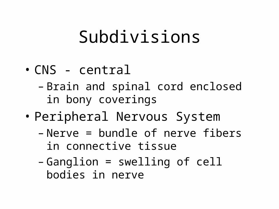
• CNS - central– Brain and spinal cord enclosed in
bony coverings
• Peripheral Nervous System– Nerve = bundle of nerve fibers in
connective tissue– Ganglion = swelling of cell bodies in
nerve

CNS
PNS

Brain
Cranial Nerves
Autonomic (involuntary) motor nerves
Somatic (voluntary) Sensory Nerves
Spinal Nerves
PNS
Spinal Cord

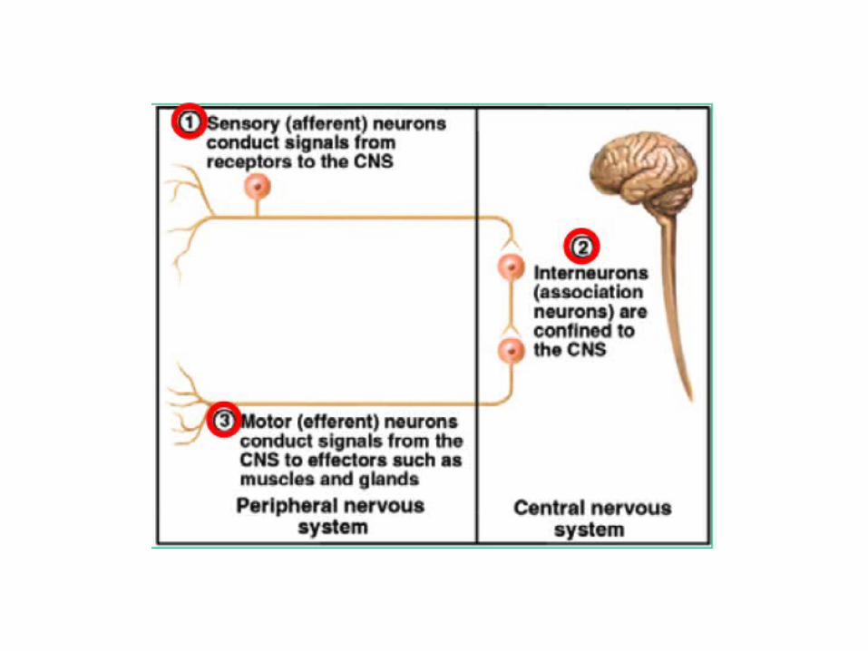
Fundamental Types of Neurons
• Sensory (afferent)– Receptors detect changes in body
and external environment– Information transmitted to brain or
spinal cord

• Interneurons (association neurons)– link sensory & motor pathways in CNS– 90% of our neurons are interneurons– Process, store & retrieve info
Fundamental Types of Neurons

• Motor (efferent) neurons– Send signals out to muscles and
gland cells– Organs that carry out responses
called effectors
Fundamental Types of Neurons

Fundamental properties of neurons
• Excitability (irritability) – Ability to respond to changes in the body
and external environment called stimuli
• Conductivity– Produce traveling electrical signals
• Secretion– When electrical signal reaches end of nerve
fiber, a chemical neurotransmitter is secreted
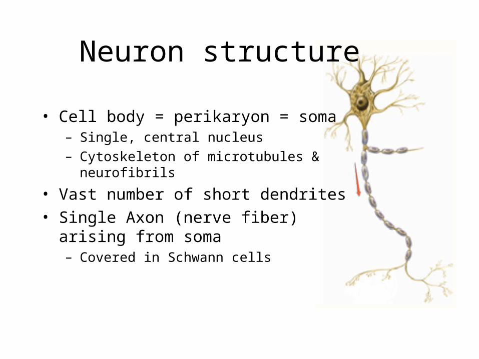
Neuron structure
• Cell body = perikaryon = soma– Single, central nucleus– Cytoskeleton of microtubules &
neurofibrils
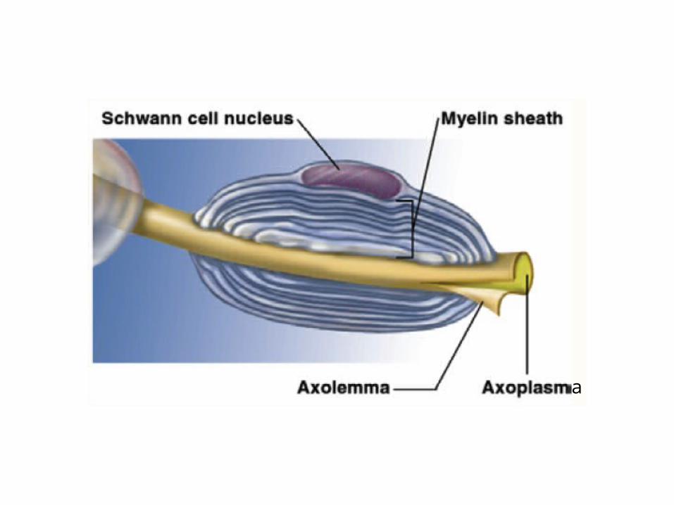
• Vast number of short dendrites• Single Axon (nerve fiber) arising
from soma– Covered in Schwann cells
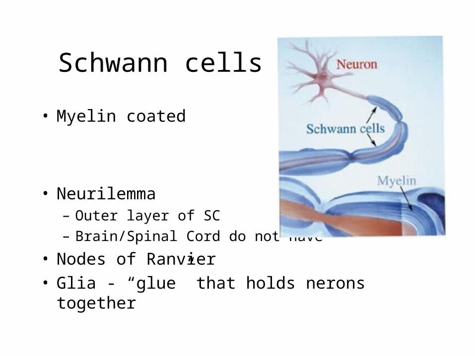
Schwann cells
• Myelin coated
• Neurilemma– Outer layer of SC– Brain/Spinal Cord do not have
• Nodes of Ranvier• Glia - “glue” that holds nerons together
a
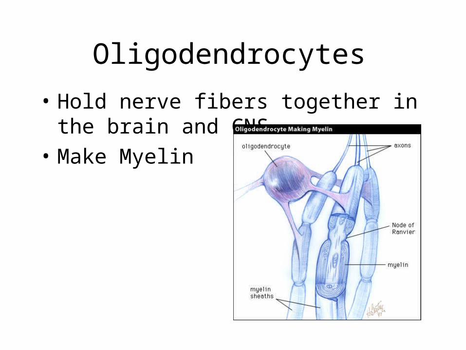
Oligodendrocytes
• Hold nerve fibers together in the brain and CNS
• Make Myelin

Multiple sclerosis
• Myelin disorder– Myelin loss/oligodendrocyte damage– = demyelinization of white matter in
CNS– Hard, plaque-like lesions -->
inflammation– Nerve conduction impaired– More common women 20-40yrs

Nerves• Groups of peripheral nerve fibers
(axons) - like cables– Covered in myelin = look white
• Axons in CNS (tracts) = white matter• Gray matter - unmyelinated dendrites
and axons

Endoneurium -tissue that wraps axons
Fascicles - groups of wrapped axons
Perineurium - tissue that wraps fascicles
Epineurium - tough, fibrous sheath that covers whole nerve
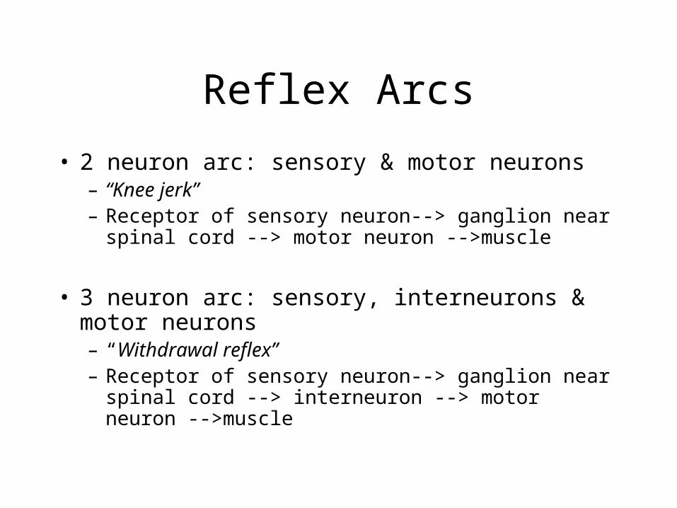
Reflex Arcs
• 2 neuron arc: sensory & motor neurons– “Knee jerk”– Receptor of sensory neuron--> ganglion near
spinal cord --> motor neuron -->muscle
• 3 neuron arc: sensory, interneurons & motor neurons– “Withdrawal reflex”– Receptor of sensory neuron--> ganglion near
spinal cord --> interneuron --> motor neuron -->muscle

GANGLIONINTERNEURON
MUSCLE
RECEPTOR
MOTORNEURON
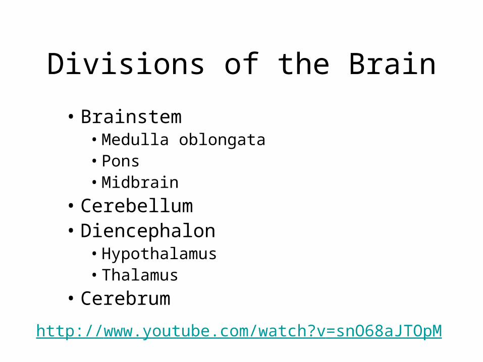
Divisions of the Brain
• Brainstem• Medulla oblongata• Pons• Midbrain
• Cerebellum• Diencephalon• Hypothalamus• Thalamus
• Cerebrum
http://www.youtube.com/watch?v=snO68aJTOpM
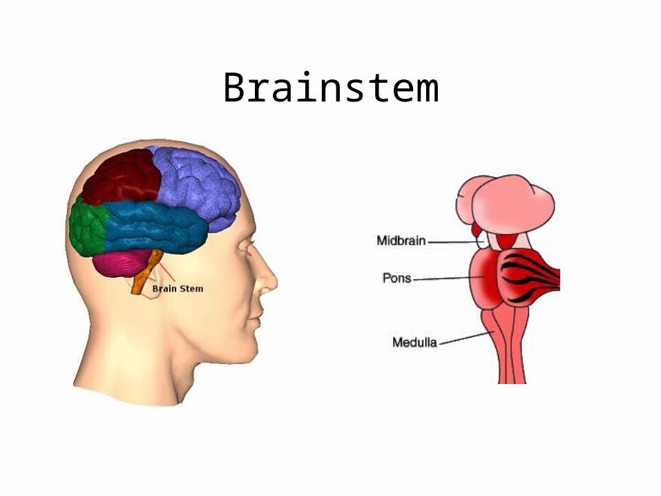
Brainstem
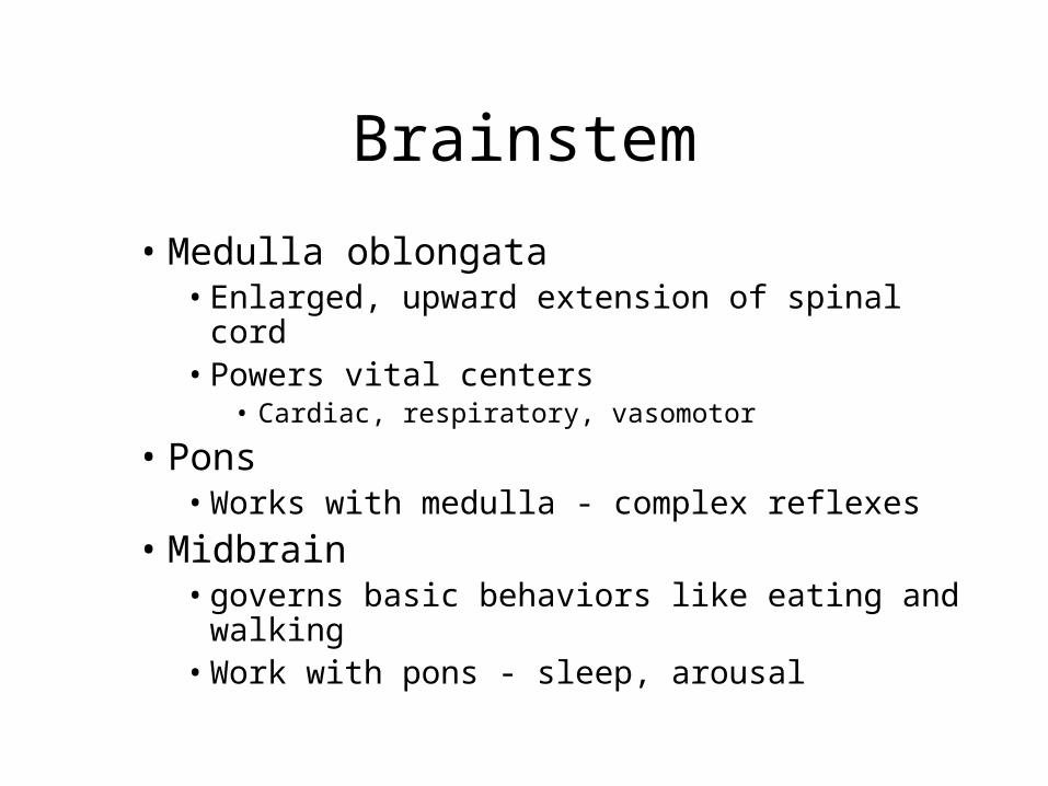
Brainstem
• Medulla oblongata• Enlarged, upward extension of spinal cord• Powers vital centers
• Cardiac, respiratory, vasomotor
• Pons• Works with medulla - complex reflexes
• Midbrain• governs basic behaviors like eating and
walking• Work with pons - sleep, arousal

Cerebellum
Cerebellum• Essential in normal movements
– Precise, smooth movements– Walking, drawing, hammering a nail
– processes input from other areas of the brain, spinal cord and sensory receptors to provide precise timing for movements

Diencephalon
• Hypothalamus– Impulses control glands, muscles, organs
• Heartbeat, intestines, vasomotory• ADH, RH
• Thalamus– Dumbbell-shaped– Produces sensations– Sensations with emotions– Arousal, alertness, activity

Cerebrum

Cerebrum
• Largest part of brain• Gyri - ridge• Sulci - grooves
– Fissure - deep sulci• Basal ganglia
– Gray matter in white matter– Automatic movements

Cerebrum• Corpus callosum -
connects left and right hemispheres
• Cerebral Cortex - layer of gray matter covering the cerebrum


Peripheral Nervous System (PNS)
• Made up of– Cranial nerves
• 12 pairs• conduct impulses from face/body to brain
– Table 9-2 p.921
– Spinal nerves• 21 pairs• Conduct impulses from body to spinal
cord
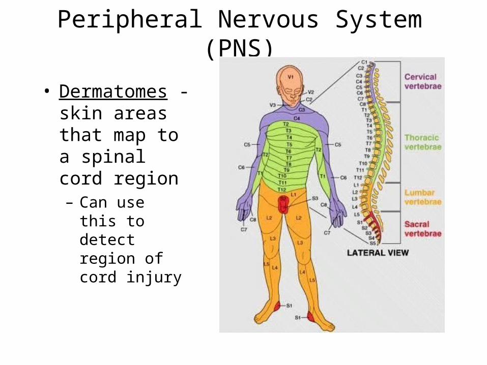
Peripheral Nervous System (PNS)
• Dermatomes - skin areas that map to a spinal cord region– Can use this
to detect region of cord injury
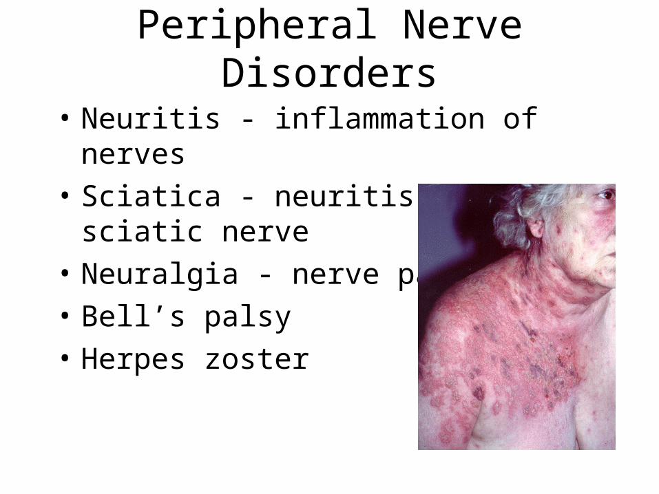
Peripheral Nerve Disorders
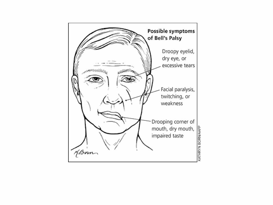
• Neuritis - inflammation of nerves• Sciatica - neuritis of sciatic nerve• Neuralgia - nerve pain• Bell’s palsy• Herpes zoster
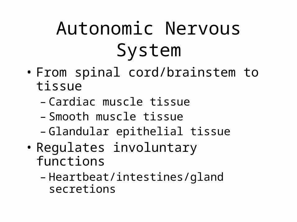
Autonomic Nervous System
• From spinal cord/brainstem to tissue– Cardiac muscle tissue– Smooth muscle tissue– Glandular epithelial tissue
• Regulates involuntary functions– Heartbeat/intestines/gland secretions
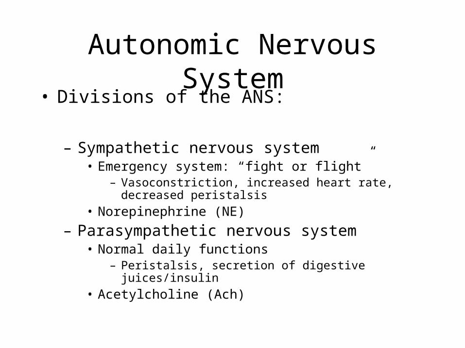
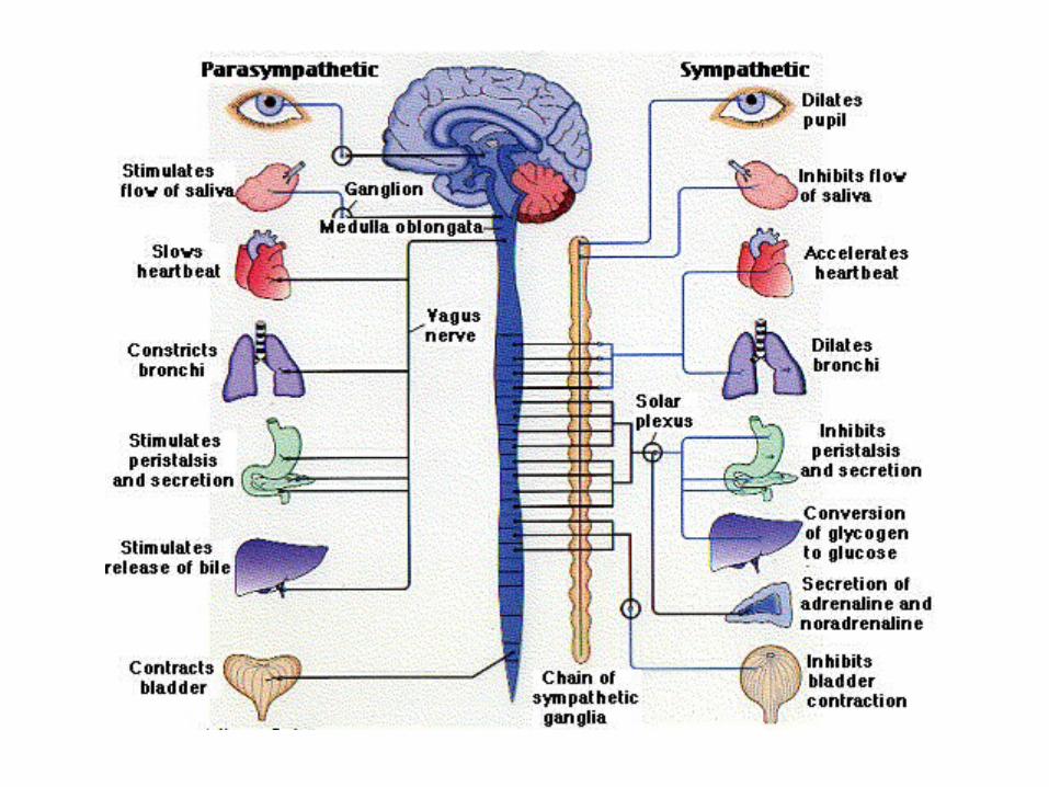
Autonomic Nervous System
• Divisions of the ANS:
– Sympathetic nervous system• Emergency system: “fight or flight”
– Vasoconstriction, increased heart rate, decreased peristalsis
• Norepinephrine (NE)
– Parasympathetic nervous system• Normal daily functions
– Peristalsis, secretion of digestive juices/insulin• Acetylcholine (Ach)
Other Neurotransmitters

1. Action Potential2. Vesicles Fuse3. NT released4. NT crosses synapse5. Binds with receptors6. New Action Potential Fires7. Reuptake of NT
Nerve Impulse
Other NTs (are many more):• Dopamine• Epinephrine• Serotonin• Histamine

The venom contains 5% acetylcholine
Asian giant hornet

Organophosphate Poisoning
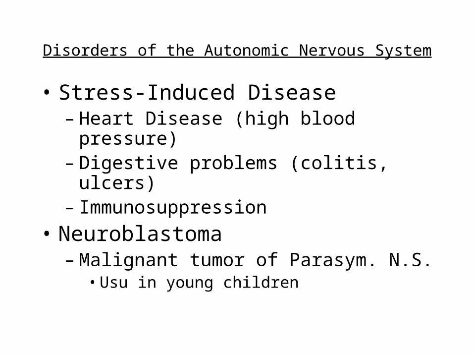
Disorders of the Autonomic Nervous System
• Stress-Induced Disease– Heart Disease (high blood pressure)– Digestive problems (colitis, ulcers)– Immunosuppression
• Neuroblastoma– Malignant tumor of Parasym. N.S.
• Usu in young children


![Introduction to the nervous system and nerve tissue[1]](https://cdn.vdocuments.mx/doc/165x107/55b5a338bb61eba3108b4796/introduction-to-the-nervous-system-and-nerve-tissue1.jpg)


