
Received: 08th Sept-2013 Revised: 28th Sept-2013 Accepted: 30th Sept-2013
Research Article
MORPHOLOGICAL STUDY ON LUMBOSACRAL TRANSITIONAL VERTEBRA IN ADULT INDIAN SACRA AND ITS CLINICAL IMPLICATIONS
Kosuri Kalyan Chakravarthi1*, Nelluri Venumadhav2, Siddaraju KS2, and Pandey S.N3
1, 3 Department of Anatomy, Mayo Institute of Medical Sciences, Faizabad Road, Gadia, Barabanki-225001 (UP) India.
2Department of Anatomy, Melaka Manipal Medical College (MMMC), Manipal University, Manipal, Karnataka, India
2Department of Anatomy, KMCT Medical College, Manassery, Calicut, Kerala, India. *Corresponding Author: Email Id: [email protected]
ABSTRACT: Lumbosacral transitional vertebra (Sacralization) is the fusion of 5Th lumber vertebra with the first segment of the sacrum it may be complete or incomplete. In complete sacralization body of the 5Th lumber vertebra completely fuses with the sacrum, where as in incomplete sacralisation shows a well defined joint line between the transverse process and the sacrum. Both forms may be either unilateral or bilateral. Such kind of abnormalities are importance while reporting the X ray, CT and MRI films, during surgical procedures at the Lumbosacral region and making a differential diagnosis for the low back ache patients. Accordingly the present study was designed to evaluate the incidence and morphological study of Sacralization (Lumbosacral transitional vertebra) in adult Indian sacra and its clinical significance. This study was carried out on 150 dry human sacra irrespective of age and sex at Mayo Institute of Medical Sciences- Barabanki,-UP, Melaka Manipal Medical College-Manipal University and Department of Anatomy, KMCT Medical College, Manassery-Calicut. It was observed that out of 150 sacra, 57 (38%) sacra showed sacralization. Out of 57 sacralized bones, 38 (25.33%) bones showed bilateral sacralization, whereas 19 (12.67%) bones showed unilateral sacralization. Such Lumbosacral transitional vertebra may increase the ricks of Disc bulge / herniation or pseudarthrosis (nonunions) with the ilium, degenerative sclerosis around the false joint, compression of lumber nerve roots, low back pain, and false administration of epidural or intradural anaesthetics in lumbosacral region. Its sound knowledge is not only enlightening for the orthopaedic surgeons, also vital for the clinical anatomists, forensic experts and morphologists. Key words: Lumber vertebra, lumbar nerve, pseudarthrosis, Sacralization, sacrum. INTRODUCTION Sacrum is a wedge shaped bone between two iliac bones normally it is formed by fusion of five sacral vertebrae. Lumbar vertebra is irregular, having large body, stout pedicles and thick lamina the main function of lumbar vertebra is to support the upper body, transfer weight from axial to appendicular skeleton, and provide mobility in the lower back. Lumbosacral transitional vertebrae are congenital anomalies of the lumbosacral region, which includes sacralisation of fifth lumbar vertebra, which occurs because of defect in the segmentation of the lumbosacral spine during development. Lumbosacral transitional vertebrae will affect the biomechanics of the lumbar spine. Such lumbosacral transitional vertebral anomalies may confuse or failure to recognize during spinal surgery which may leads to serious complications. Aihara T et al (2005) and Leboeuf C et al (1989) reported that, patients with lumbosacral transitional vertebrae are to have increased risk for advanced disc degeneration or disc herniation above the lumbosacral transitional vertebrae. Therefore, the aim of this study was to investigate the incidence and morphological study of sacralization (Lumbosacral transitional vertebra) in adult Indian sacra and discuss its clinical implications. METHODS AND MATERIALS In the present study 150 dry human sacra were examined in the Department of Anatomy Mayo Institute of Medical Sciences- Barabanki, Department of Anatomy, Melaka Manipal Medical College-Manipal University and Department of Anatomy, KMCT Medical College, Manassery-Calicut. All sacra were macroscopically inspected
International Journal of Applied Biology and Pharmaceutical Technology Page: 340 Available online at www.ijabpt.com

Kalyan et al
and it was recorded whether the sacralization (Lumbosacral transitional vertebra) of the 5Th lumber vertebra was unilateral or bilateral and whether it was complete or incomplete. Photographs of the Lumbosacral transitional vertebra were taken for proper documentation. RESULTS
• It was observed that out of 150 sacra, 57 (38%) sacra showed sacralization. • Out of 57 sacralized bones, 38 (25.33%) bones showed bilateral sacralization (Fig 1and 3), whereas 19
(12.67%) bones showed unilateral sacralisation (Fig 2and 4). • Incomplete with a narrow gap non fusion of body of 5th lumbar vertebra with sacrum were noted in 30 sacra
(20%) (Fig 1). • Unilateral fusion of body of 5th lumbar vertebra with sacrum was noted in 11 sacra (7.33%) (Fig 2and 4).
Fig 1: Showing bilateral sacralization. A- 5Th lumbar vertebra; Red Colour Arrows- Transverse process of 5Th lumbar vertebra both side is completely fused with sacrum; Black Arrow- A narrow gap showing incomplete non fusion of body of 5Th
lumbar vertebra with sacrum.
International Journal of Applied Biology and Pharmaceutical Technology Page: 341 Available online at www.ijabpt.com

Kalyan et al
Fig 2: Showing unilateral sacralization.
A- 5Th lumbar vertebra; Red Colour Arrow- Transverse process of 5Th lumbar vertebra on right side is completely fused with sacrum; Blue Arrow- Unilateral non fusion of body of 5Th lumbar vertebra with
sacrum.
International Journal of Applied Biology and Pharmaceutical Technology Page: 342 Available online at www.ijabpt.com
Kalyan et al
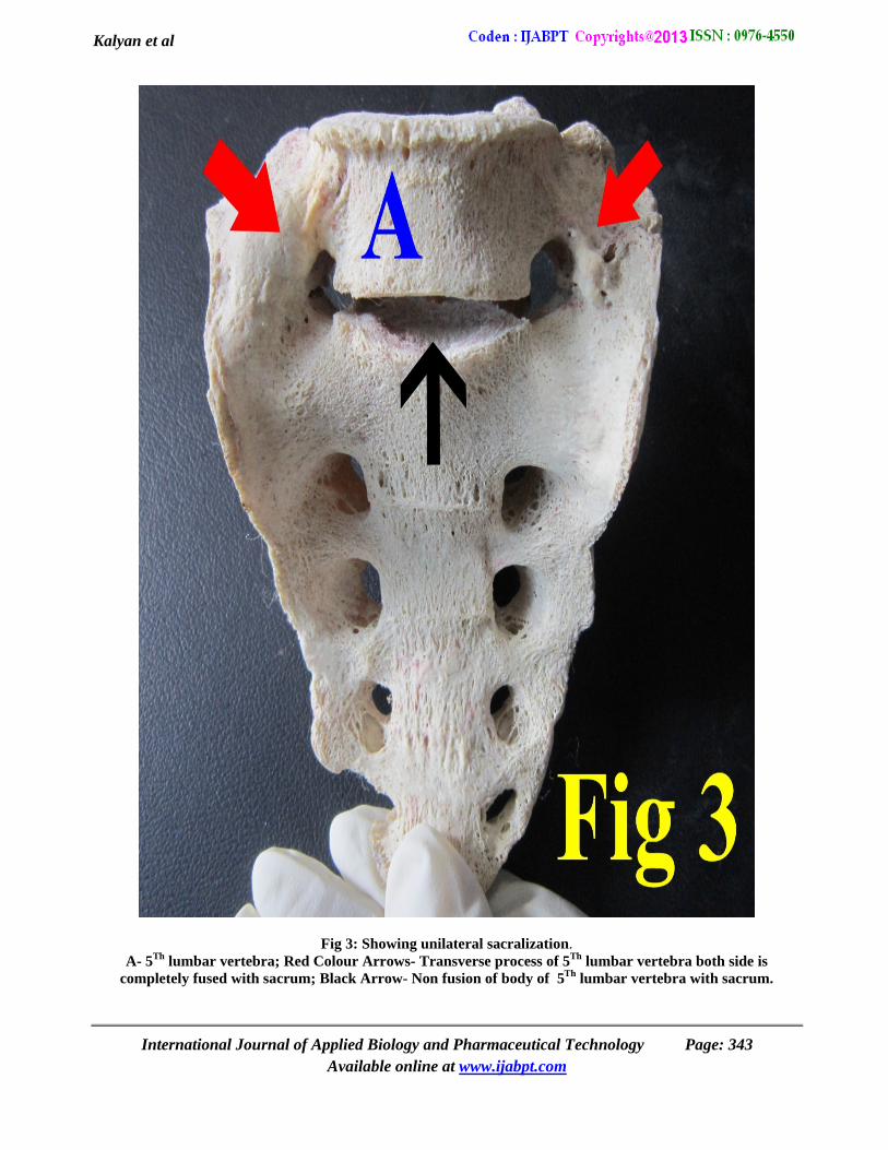
Fig 3: Showing unilateral sacralization. A- 5Th lumbar vertebra; Red Colour Arrows- Transverse process of 5Th lumbar vertebra both side is
completely fused with sacrum; Black Arrow- Non fusion of body of 5Th lumbar vertebra with sacrum.
International Journal of Applied Biology and Pharmaceutical Technology Page: 343 Available online at www.ijabpt.com

Kalyan et al
Fig 4: Showing unilateral sacralization.
A- 5Th lumbar vertebra; Red Colour Arrow- Transverse process of 5Th lumbar vertebra on right side is completely fused with sacrum; Black Arrow- Unilateral fusion of body of 5Th lumbar vertebra with sacrum;
Blue Arrow- Unilateral non fusion of body of 5Th lumbar vertebra with sacrum. DISCUSSION Disordered ossification or calcification of cartilages or ligamentous structures in various parts of the body is frequently observed which may seriously hamper clinical and diagnostic procedures such as compression to neighbouring structures or complications in the regional surgery (Kosuri Kalyan Chakravarthi 2013), (Kosuri Kalyan Chakravarthi 2013) (Kosuri Kalyan Chakravarthi 2012). The occurrence of lumbosacral transitional vertebra is linked to its embryological development and osteological defects. sacralization of 5th lumbar vertebra may causes pain are actual pressure on fifth lumber nerve, ligamentous strain around the sacralization or compression of soft tissues between bony joints resulting in pain along the sciatic nerve distribution. The present study shows that the incidence of sacralization of the fifth lumbar vertebra is 38 %. Based on the literature, sacralization varied by race and incidence the prevalence of Lumbosacral transitional vertebra in our study was much higher than the previous Indian study and the other reports reported in the literature (Table-1).
International Journal of Applied Biology and Pharmaceutical Technology Page: 344 Available online at www.ijabpt.com

Kalyan et al
Table 1: Incidence of Lumbosacral transitional vertebra (Sacralization) as reported by various Research Workers.
Research Workers Year Incidence of Sacralization (%)
Karan Bhagwan Khairnar 2013 6.6
Kubawat 2012 11.1 Hughes 2006 9.2
Steinberg 2003 14 Kim 2003 1.7
Chithriki 2002 5.0 Santiago 2001 11.6
Peh 1999 6.2 Hald 1995 7.8 Hahn 1992 7.5
Bustami 1989 10.0 Moore BH 1925 3.6
Present Study 2013 38
Such Sacralization (Lumbosacral transitional vertebra) of 5th lumber vertebra may misguide or confuse in recognizing correct numbering can theoretically lead to problems with the administration of epidural or intradural anaesthetics or wrong level surgery (Malanga GA 2004) in patients with Lumbosacral transitional vertebra. Lumbosacral transitional vertebra affects the position of the intercrestal line (Tuffier’s line) which corresponds to the level L4/L5 and is used as a landmark for needle insertion (Kim JT 1997). Sacralization of fifth lumbar vertebra may cause greater difficulty during labour because of less mobile pelvis and it may results in low back pain problem, Bertolotti (1917) reported the relationship between the low back pain and sacralisation of fifth lumbar vertebra. The sacralized transverse process of 5Th lumbar vertebra may form a pseudarthrosis (nonunions) with the ilium and may results in degenerative sclerosis around the false joint. In such cases lumber nerve roots may be altered and results in low back pain. Sacralization may also may leads to spondylolisthesis. Such occurrence of lumbosacral transitional vertebra is linked to its embryological development and osteological defects.
CONCLUSION
Lumbosacral transitional vertebra may increase the ricks of Disc bulge / herniation, pseudarthrosis (nonunions) with the ilium, degenerative sclerosis around the false joint, compression of lumber nerve roots, low back pain, and false administration of epidural or intradural anaesthetics in lumbosacral region. We believe that the present study has provided some important data which will contribute to the scientific literature, providing the anatomical data of Lumbosacral transitional vertebra in the Indian adult population. Sound knowledge of sacralization is not only enlightening for the orthopaedic surgeons, also vital for the clinical anatomist, Radiologists, Forensic experts Architectures and morphologists.
REFERENCES Aihara T, Takahashi K, Ogasawara A, Itadera E, Ono Y, Moriya H. (2005). Intervertebral disc degeneration
associated with lumbosacral transitional vertebrae: a clinical and anatomical study. J Bone Joint Surg 87: 687-91.
Leboeuf C, Kimber D, White K. (1989). Prevalence of spondylolisthesis, transitional anomalies and low intercrestal line in a chiropractic patient population. J Manipulative Physiol Ther 12: 200-204.
Kosuri Kalyan Chakravarthi, Nelluri Venumadhav, Huban Thomas. (2013). Ossified Cartilago Thyreoidea and Its Clinical Insight: A Cadaveric Study Int. J. Bioassays 2, 1044-47.
Kosuri Kalyan Chakravarthi, Nelluri Venumadhav, Ravindranath Gandrakota. (2013). Abnormal Bone Outgrowths and Osseous Structures around the Foramen Ovale May Leads to Mandibular Compression or Entrapment Neuropathy. Int. J. Bioassays 02, 922-25.
Kosuri Kalyan Chakravarthi, Sarath Babu K. (2012). An anatomical study of the pterygo-alar bar and porus crotaphitico buccinatorius. Int J Med Health Sci1: 3-9.
International Journal of Applied Biology and Pharmaceutical Technology Page: 345 Available online at www.ijabpt.com
Kalyan et al
Karan Bhagwan Khairnar, Manisha Bhausaheb Rajale. (2013). sacralization of lumbar vertebra. Indian journal of basic & applied medical research 2, 510-14.
Kubavat Dharati, Nagar SK, Malukar Ojaswini, Trivedi Dipali, Shrimankar Paras, Patil Sucheta. (2012). A study of sacralisation of fifth lumber vertebra in Gujarat. National Journal of Medical research. Vol 2 Issue 2, Apr-Jun
Hughes RJ, Saifuddin A. (2006). Numbering of lumbosacral transitional vertebrae on MRI: role of the iliolumbar ligaments. Am J Roentgenol 187, 59-65.
Steinberg EL, Luger E, Arbel R, Menachem A, Dekel S. (2003). A comparative roentgenographic analysis of the lumbar spine in male army recruits with and without lower back pain. Clin Radiol, 58: 985-9.
Kim JT, Bahk JH, Sung J. (2003). Influence of age and sex on the position of the conus medullaris and Tuffier’s line in adults. Anesthesiology 99, 1359-63.
Chithriki M, Jaibaji M, Steele RD. (2002). The anatomical relationship of the aortic bifurcation to the lumbar vertebrae: a MRI study. Surg Radiol Anat 24, 308-12.
Santiago FR, Milena GL, Herrera RO, Romero PA, Plazas PG.(2001). Morphometry of the lower lumbar vertebrae in patients with and without low back pain. Eur Spine J, 10: 228-33.
Peh WC, Siu TH, Chan JH. (1999). Determining the lumbar vertebral segments on magnetic resonance imaging. Spine, 24: 1852-5.
Hald HJ, Danz B, Schwab R, Burmeister K, Bähren W. (1995). Radiographically demonstrable spinal changes in asymptomatic young men. Rofo, 163: 4-8.
Hahn PY, Strobel JJ, Hahn FJ. (1992). Verification of lumbosacral segments on MR images: identification of transitional vertebrae. Radiology 182, 580-1.
Bustami F. (1989). The anatomical features and functional significance of lumbar transitional vertebra. Jordan Med J 23,49-59
Moore BH, Illinois C. (1925). Sacralization of the fifth lumbar vertebra. J Bone Joint Surg 7: 271-78. Malanga GA, Cooke PM. 2004. Segmental anomaly leading to wrong level disc surgery in cauda equina syndrome.
Pain Physician 7, 107-110. Kim NH, Suk KS. (1997). The role of transitional vertebrae in spondylolysis and spondylolytic spondylolisthesis.
Bull. Hosp Jt Dis 56; 161-6. Bertolotti M. (1917). Contributto alla conoscenza dei vici di differenzarione regionale del rachide con speciale
riguards all asimilazzione sacrale della v. lombare. Radiologique Medica 4:113-44.
International Journal of Applied Biology and Pharmaceutical Technology Page: 346 Available online at www.ijabpt.com


