MODULE 5 & 6
ELBOW, WRIST, & HAND
ELBOW
Constitute 6% of all fractures
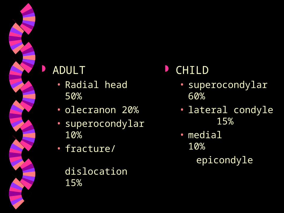
ADULT• Radial head 50%
• olecranon 20%
• superocondylar10%
• fracture/ dislocation
15%
CHILD• superocondylar
60%
• lateral condyle15%
• medial 10%
epicondyle

CHILDREN
Comparison views often needed
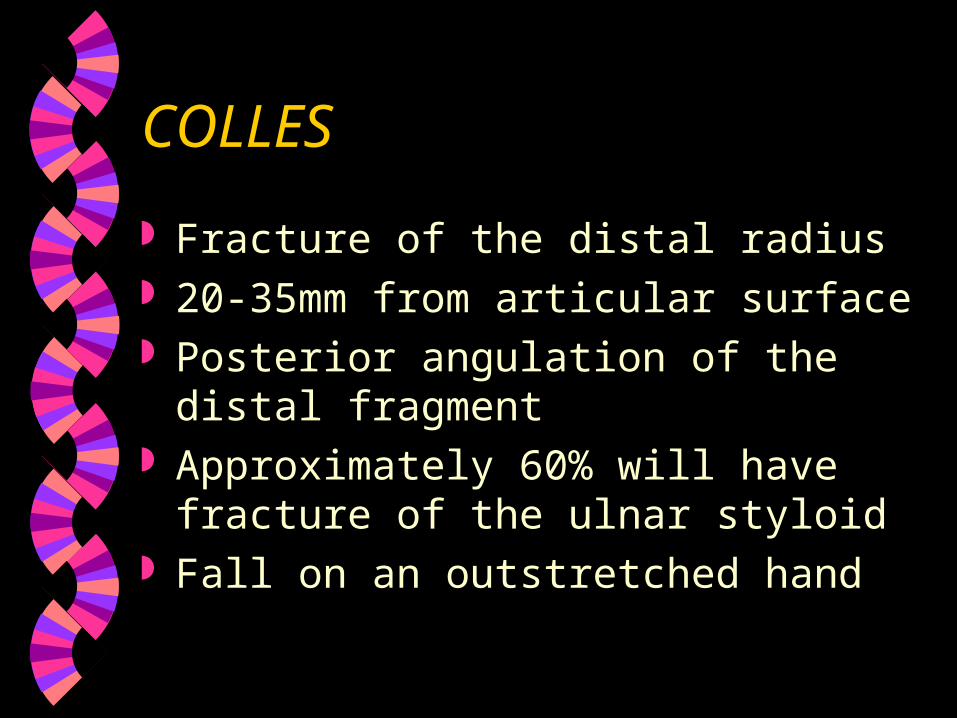
COLLES
Fracture of the distal radius 20-35mm from articular surface Posterior angulation of the distal fragment Approximately 60% will have fracture of
the ulnar styloid Fall on an outstretched hand
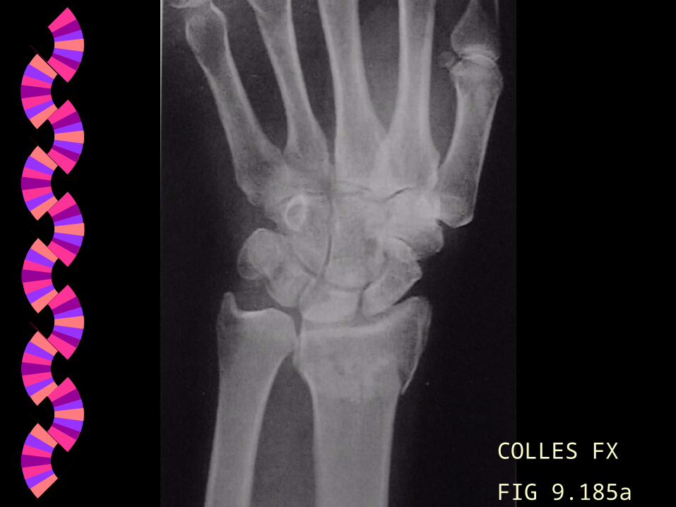
COLLES FX
FIG 9.185a
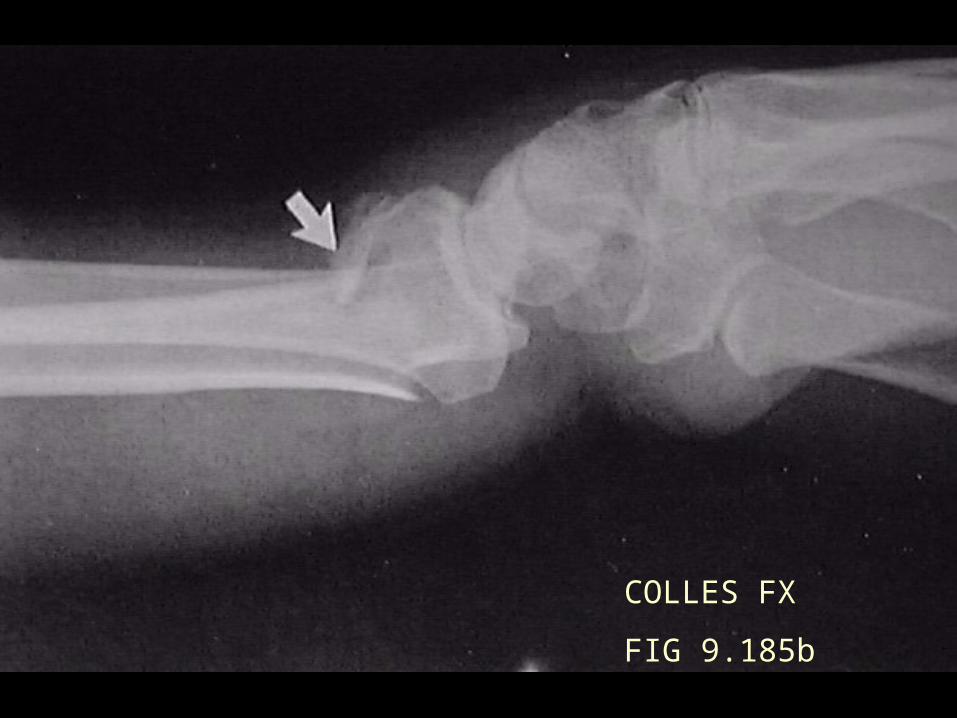
COLLES FX
FIG 9.185b
SMITH’S
Fracture of the distal radius Anterior angulation of the distal fragment
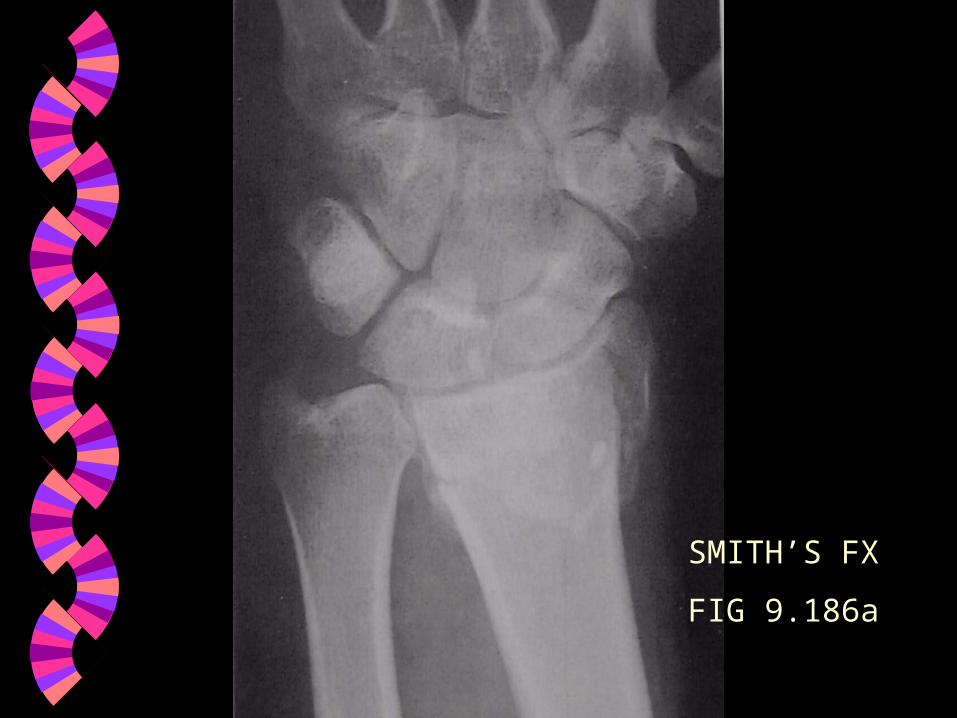
SMITH’S FX
FIG 9.186a
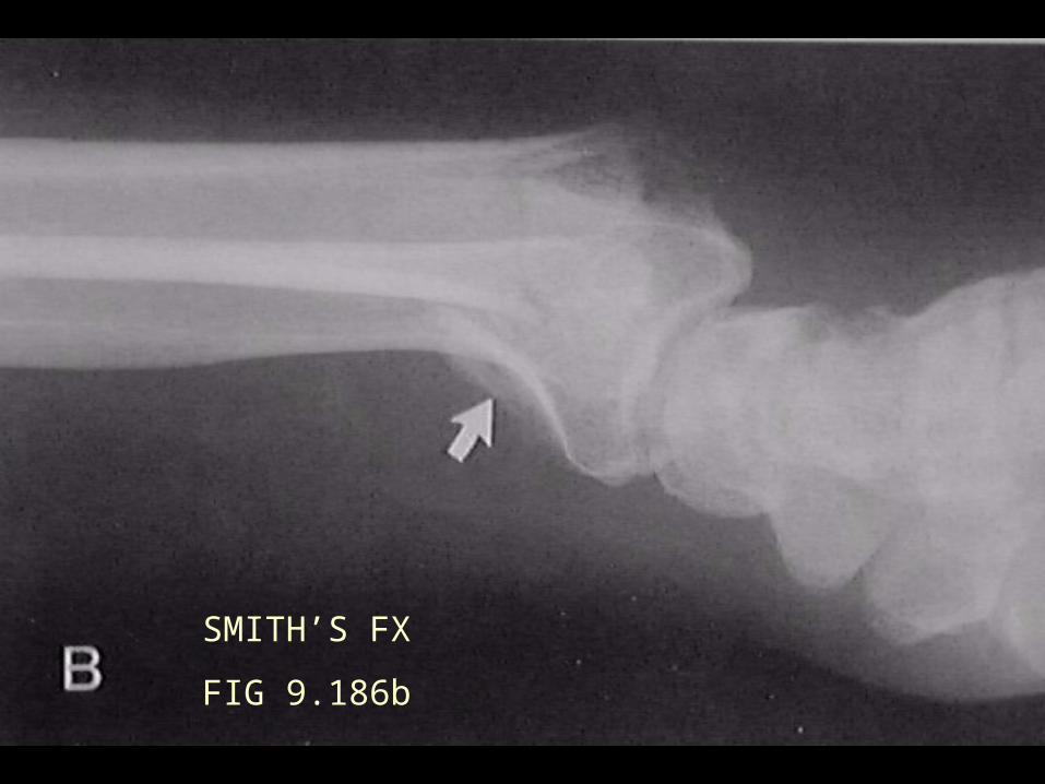
SMITH’S FX
FIG 9.186b
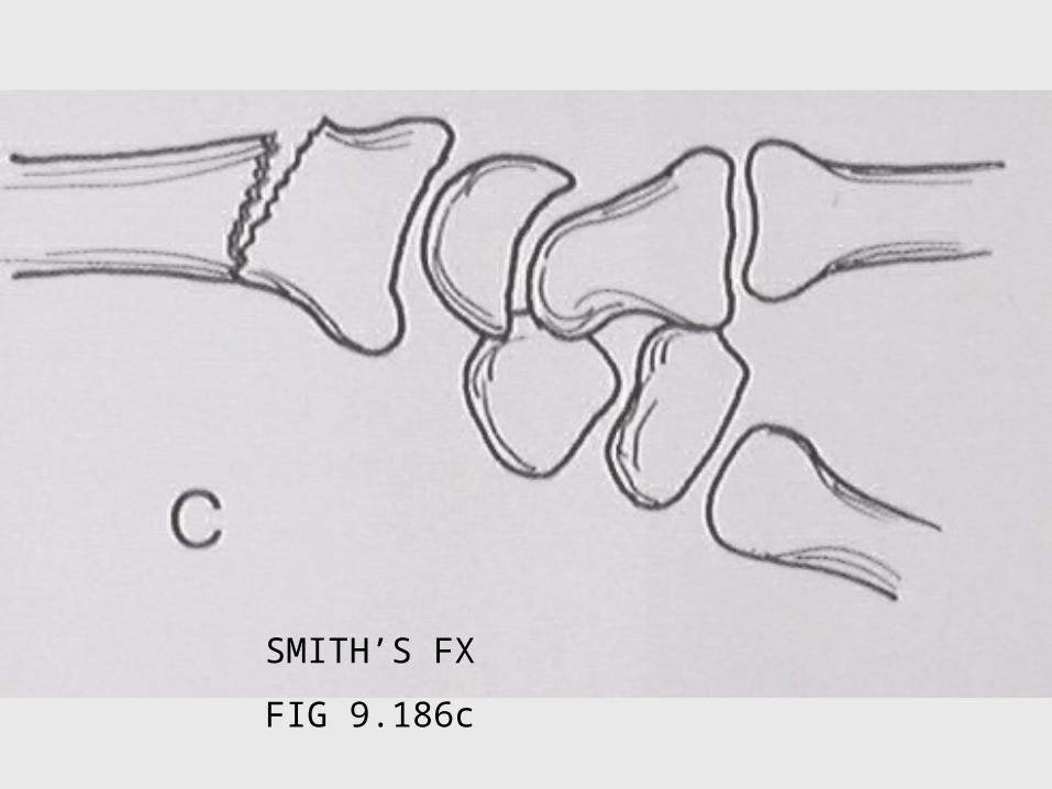
SMITH’S FX
FIG 9.186c
GALEAZZI
Distal radius fracture with ulnar dislocation
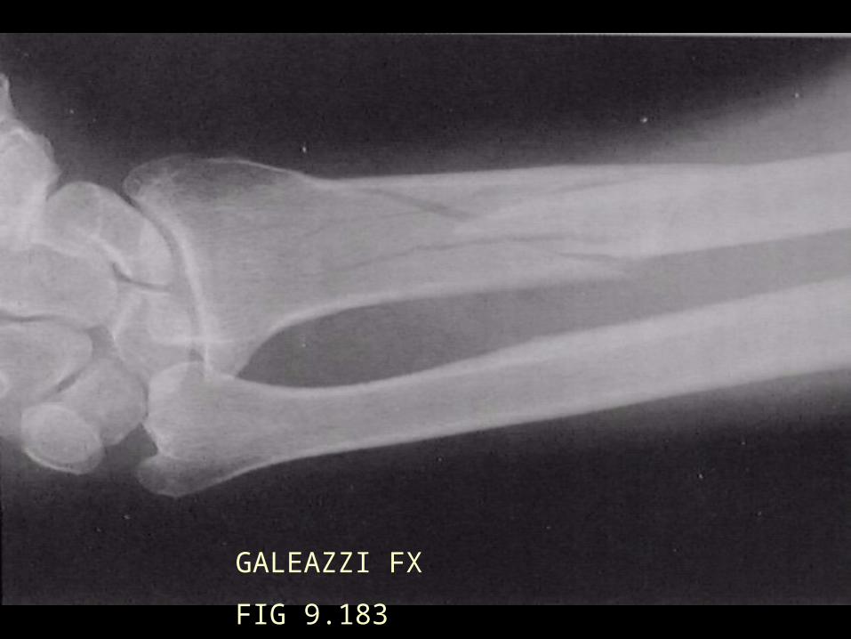
GALEAZZI FX
FIG 9.183

MONTEGGIA
Proximal ulnar fracture with radial dislocation
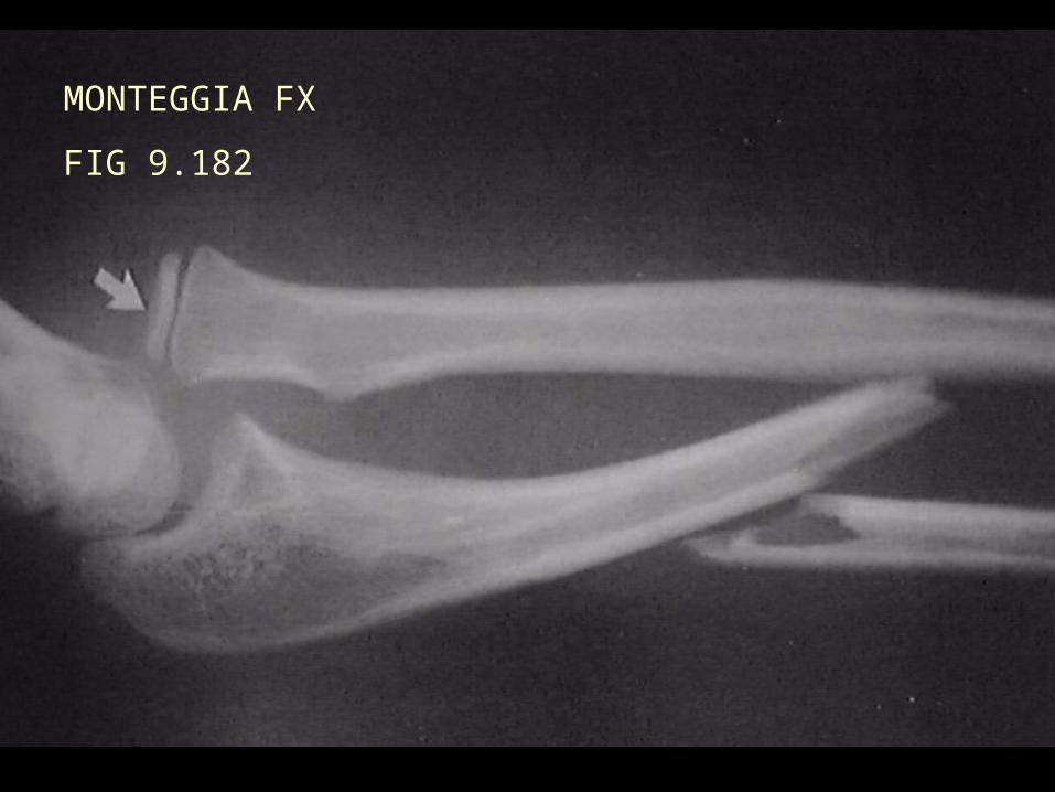
MONTEGGIA FX
FIG 9.182
HEAD
DIE
DIE
NORMAL X-RAY ANATOMY
Elbow Wrist Hand
ELBOW POSITIONING SLIDES
AP Lateral Jones view Oblique view - has been replaced by radial
head - capitellum view
WRIST POSITIONING SLIDES
PA Ulnar Deviation Lateral Oblique
HAND POSITIONING SLIDES
PA Oblique Lateral
MEASUREMENTS
Axial relationships Carpal alignment
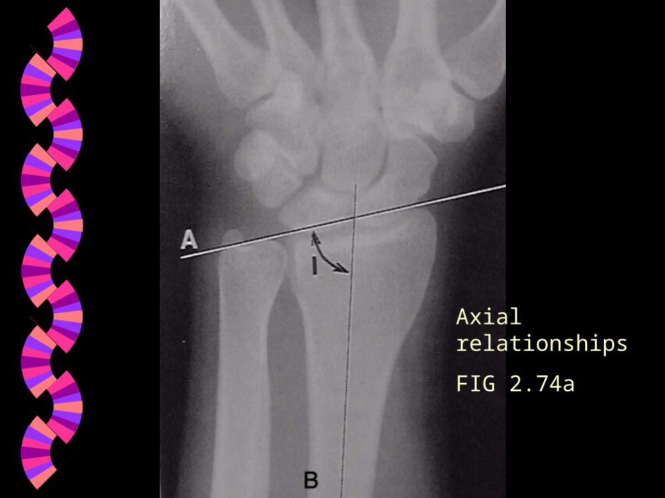
Axial relationships
FIG 2.74a
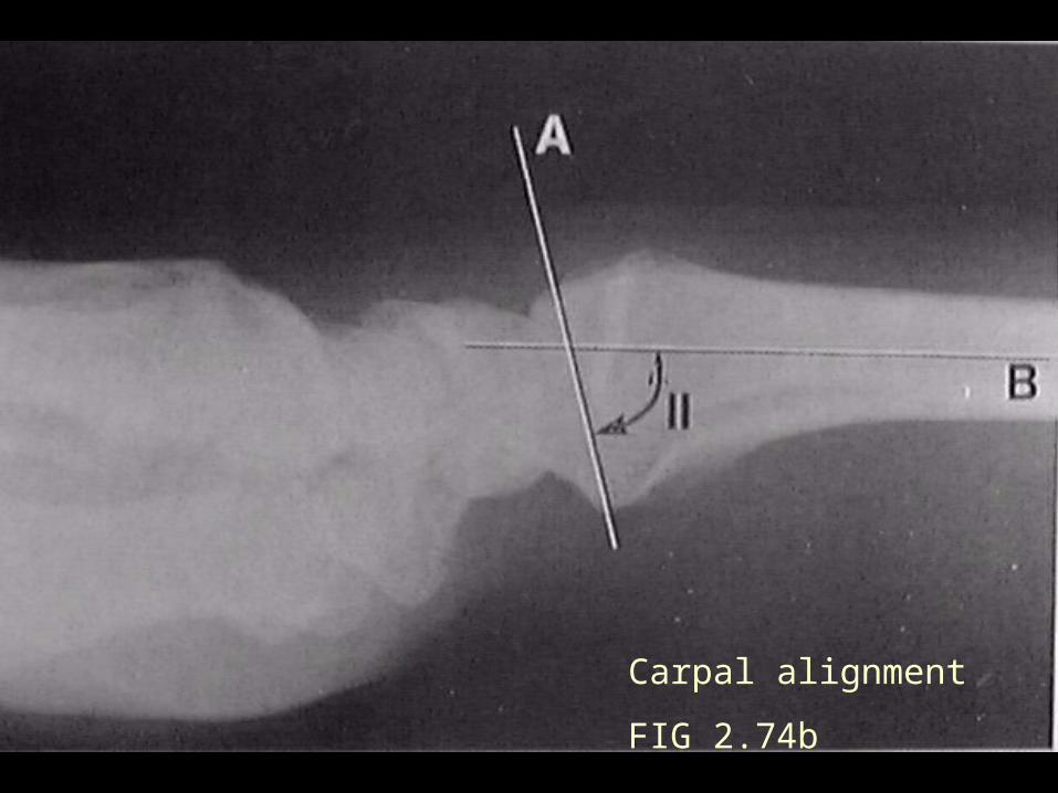
Carpal alignment
FIG 2.74b


