
miR-200c attenuates P-gp mediated MDR and metastasis by targeting JNK2/c-Jun signaling pathway in colorectal cancer
Hua Sui1#, Guo-Xiang Cai2#, Shu-Fang Pan3, Wan-Li Deng4, Yu-Wei Wang2, Zhe-Sheng Chen5,
San-Jun Cai2, Hui-Rong Zhu1, Qi Li1*
1 Department of Medical Oncology, Shuguang Hospital, Shanghai University of Traditional Chinese Medicine, Shanghai 201203, China,
2 Department of Colorectal Surgery, Fudan University Shanghai Cancer Center, Shanghai 200032, China,
3 Shanghai University of Traditional Chinese Medicine, Shanghai 201210, China,
4 Oncology Department I of Traditional Chinese Medical Hospital affiliated Xinjiang Medical University, Xinjiang 830000, China,
5 Department of Pharmaceutical Sciences, College of Pharmacy and Health Sciences, St. John’s University, Queens, NY 11439, USA #These authors contributed equally to this work. Correspondence to: Prof./Dr. Qi Li, Department of Medical Oncology, Shuguang Hospital, Shanghai University of Traditional Chinese Medicine. 528 Zhangheng Rd, Shanghai 201203, P.R.China Tel:+8621 20256517; Fax:+8621 20256533, E-mail of Qi Li: [email protected] Running title: miR-200c induce MDR and metastasis Keywords: Multidrug resistance; ABCB1/P-glycoprotein; miR-200c; JNK signaling pathway; metastasis
Abbreviations list: CRC: colorectal cancers; MDR: multidrug resistance; ABCB1: ATP-binding cassette (ABC) transporter-subfamily B member 1; miRNAs: microRNAs; VCR: vincristine; L-OHP: Oxaliplatin; FOLFOX or CapeOX: the standard chemotherapy regimen for treatment of colorectal cancer
Grant support: National Natural Science Foundation of China (81202812, H Sui; 81373862, HR Zhu; 81001055, GX Cai), Shanghai Pujiang Program (13PJD008, GX Cai), Program of Shanghai Committee of Science and Technology (13140902500, Q Li), Shanghai Municipal Education Commission (2011JW57, H Sui; 12ZZ118 Q Li), Shanghai Municipal Health Bureau (20114Y013, H Sui; 2010019, Q Li) and National High Technology Research and Development Program (2012AA02A506, SJ Cai)
Conflict of Interest Statement: Authors have no conflicts of interest.
Total number of figures and tables: Six figures, four supplementary figures, seven supplementary tables
on January 6, 2020. © 2014 American Association for Cancer Research. mct.aacrjournals.org Downloaded from
Author manuscripts have been peer reviewed and accepted for publication but have not yet been edited. Author Manuscript Published OnlineFirst on September 9, 2014; DOI: 10.1158/1535-7163.MCT-14-0167

Abstract
MicroRNA-200c (miR-200c) recently emerged as an important regulator of tumorigenicity and
cancer metastasis, however, its role in regulating multidrug resistance (MDR) remains unknown. In
the current study, we found that the expression levels of miR-200c in recurred and metastatic
colorectal cancers (CRC) were significantly lower, while the JNK2 expression was higher
compared to primary tumors. We showed that in MDR CRC cells, miR-200c targeted the 3’UTR of
JNK2 gene. Over-expression of miR-200c attenuated the levels of p-JNK, p-c-Jun, P-gp and
MMP-2/-9, the downstream factors of JNK signaling pathway, resulting in increased sensitivity to
chemotherapeutic drugs, which was accompanied by heightened apoptosis and decreased cell
invasion and migration. Moreover, in an orthotopic MDR CRC mouse model, we demonstrated that
over-expression of miR-200c effectively inhibited the tumor growth and metastasis. At last, in the
tumor samples from locally advanced CRC patients with routine post-surgical chemotherapy, we
observed an inverse correlation between the levels of mRNA expression of miR-200c and JNK2,
ABCB1, MMP-9 thus predicting patient therapeutic outcomes. In summary, we found that
miR-200c negatively regulated the expression of JNK2 gene and increased the sensitivity of MDR
CRC cells to chemotherapeutic drugs, via inhibiting the JNK2/p-JNK/p-c-Jun/ABCB1 signaling.
Restoration of miR-200c expression in MDR CRC may serve as a promising therapeutic approach
in MDR induced metastasis.
Keywords: Multidrug resistance; ABCB1/P-glycoprotein; miR-200c; JNK signaling pathway;
metastasis
on January 6, 2020. © 2014 American Association for Cancer Research. mct.aacrjournals.org Downloaded from
Author manuscripts have been peer reviewed and accepted for publication but have not yet been edited. Author Manuscript Published OnlineFirst on September 9, 2014; DOI: 10.1158/1535-7163.MCT-14-0167

1. Introduction
Invasion/metastasis and multidrug resistance (MDR) are two of the unfavorable factors causing
failures in cancer treatment. Several studies have demonstrated that drug-resistant cancer cells
derive easily from the more invasive or metastatic sub-populations within the tumor, although the
detailed mechanism remains unclear (1,2). MDR is usually mediated by a series of integral
membrane proteins, including ATP-binding cassette (ABC) transporter-subfamily B member 1
(ABCB1/ P-glycoprotein, P-gp), subfamily C member-1/2 (ABCC-1/2, MRP-1/2), and subfamily G
member 2 (ABCG2, BCRP, MXR, ABCP) (3). P-gp, a 170-kDa membrane phosphoglycoprotein
encoded by the ABCB1 gene and the most characterized member of energy-dependent drug efflux
pumps involved in MDR, recently has been suggested to facilitate invasion/metastasis (4,5,6). Thus
a better understanding of the molecular mechanisms underlying MDR mediated metastasis is in dire
need.
JNK is a set of enzymes activated (dual phosphorylated) by MAPK (mitogen-activated protein
kinase) kinases MKK4 and MKK7, in response to a variety of stress signals such as UV irradiation,
chemotherapy damage, osmotic stress, hypoxia/anoxia and hyperthermia (7). The JNK family
consists of JNK1, JNK2, and JNK3, each with multiple isoforms generated through alternative
splicing. JNK3 is expressed predominantly in the brain whereas both JNK1 and JNK2 are expressed
ubiquitously (8). The JNKs were shown to bind to the serine/threonine domain of downstream
kinases (c-Jun, ATF2, and Elk-1), to phosphorylate/activate them (9). Previously, we have shown
that, the activation of the JNK signal transduction cascade by phosphorylating c-Jun on serines
63/73 leads to enhanced, P-gp mediated MDR in CRC cells (10). Consistent with our results, more
evidence has been published to confirm that activation of JNK signal signaling plays a role in
chemoresistance via the upregulation of ABCB1 expression (11). However, how JNK signaling
modulates drug resistance in CRC has not been fully addressed yet.
Recently, microRNAs (miRNAs), a class of non-coding RNA, were found to play important roles
in various fundamental biological processes, such as cell proliferation, apoptosis and differentiation
(12,13,14). Functioning as regulatory molecules, miRNAs are able to modulate gene expression by
inhibiting the protein translation process and/or degrading the respective target messenger RNA
(15). It is then plausible to consider miRNAs as therapeutic targets in cancer. Increasing number of
studies have shown that miRNAs may regulate chemoresistance and be involved in the modulation
on January 6, 2020. © 2014 American Association for Cancer Research. mct.aacrjournals.org Downloaded from
Author manuscripts have been peer reviewed and accepted for publication but have not yet been edited. Author Manuscript Published OnlineFirst on September 9, 2014; DOI: 10.1158/1535-7163.MCT-14-0167

of drug resistance–related pathways in cancer cells. Xia et al. showed that miR-15b and miR-16
regulated MDR by targeting BCL2 in human gastric cancer cells (16). More interestingly,
heightened expression levels of miR-200c were found in clinical recurrent and metastatic CRC
samples (17,18), although the underlining mechanism remains unclear.
Here we report for the first time: (1) miR-200c was significantly down-regulated in human CRC
MDR cells and in clinical recurrent tumor samples; (2) miR-200c targeted JNK2 gene 3’-UTR
directly to affect phosphorylated JNK mediated signaling; (3) over-expression miR-200c
down-regulated the levels of ABCB1/P-gp via JNK signaling pathway, resulting in increased
sensitivity to chemotherapeutic drugs and decreased metastasis in vitro and in vivo.
2. Materials and methods 2.1. Cell culture and reagents
The human colorectal cancer HCT8, HCT116 parental cell lines, gastric cancer SGC7901 and
hepatic carcinoma Bel7402 parental cell lines were purchased from the Shanghai Cell Collection
(Shanghai, China). All the sensitive cell line authentication was assessed using short tandem repeat
(STR) DNA profiling method every year in our lab and the latest verification was done in June,
2013. HCT116/L-OHP MDR cell line was established and maintained in our laboratory by stepwise
drug selection from the parental cells as reported previously in August, 2011(19). HCT8/V,
SGC7901/DDP MDR and Bel/Fu MDR cell lines were obtained from Keygen Biotech Co., Ltd.
(Nanjing, China) in June, 2013. The MDR was measured by MMT assay as reported previously
(20-23), and the results presented in Supplementary Table S1-4. Cells were grown in RPMI 1640
medium supplemented with 10% (v/v) heat-inactivated fetal calf serum, 2 mM glutamine, 100
units/ml penicillin, and 100 �g/ml streptomycin (Invitrogen, Carlsbad, CA) at 37°C in a 5% CO2
humidified atmosphere. HCT8/V cells were routinely maintained in a medium containing 2,000
ng/L vincristine (VCR), HCT116/MDR cells in a medium containing 5,000 ng/mL Oxaliplatin
(L-OHP), SGC7901/DDP cells in a medium containing 1000 ng/mL cis-Diamminedichloro
platinum (cDDP), and Bel/Fu cells in a medium containing 2000 ng/L VCR, MMC and L-OHP
were purchased from Shenzhen Main Luck Pharmaceuticals Co., Ltd. (Shenzhen, China), cDDP
from Qilu Pharmaceutical Co., Ltd. (Shandong, China), and 5-Fu from Shanghai Xudong Haipu
Pharmaceutical Co., Ltd. (Shanghai, China). Monoclonal antibodies against ABCB1, ABCC-1/2,
BCRP, JNK1, JNK2, p-JNK, c-Jun, p-c-Jun, AP-1, ATF-2, p-ATF-2 and dehydrogenase (GAPDH)
on January 6, 2020. © 2014 American Association for Cancer Research. mct.aacrjournals.org Downloaded from
Author manuscripts have been peer reviewed and accepted for publication but have not yet been edited. Author Manuscript Published OnlineFirst on September 9, 2014; DOI: 10.1158/1535-7163.MCT-14-0167

antibodies were products of Cell Signaling Technology (Beverly, MA, USA).
2.2. miRNA microarray analysis Raw data were retrieved from the Array Express public gene expression database (E-BUGS-134,
E-GEOD-30009, E-GEOD-29702, E-GEOD-30034, E-GEOD-24460 and Chin dataset). Total
RNAs were extracted from culture cells with Trizol (Invitrogen, Carlsbad, CA), and analyzed using
an miRNA microarray (Shanghai Genechem Co. Ltd., Shanghai, China). The data adjustments
included data filtering, log2 transformation, gene centering and normalization. MDR and parental
cell samples were analyzed using t-tests, P values < 0.05 were selected as significant for cluster
analysis.
2.3. Patients’ samples To determine the associations among miR-200c, JNK2 and ABCB1 on the mRNA levels in CRC,
total RNAs of tumor and adjacent non-tumor colorectal tissues of 30 primary CRC tumors and
matched liver/lung metastasis samples from the Fudan University Shanghai Cancer Center and
Shuguang Hospital Shanghai University of Traditional Chinese Medicine were obtained for analysis.
The basic clinical characteristics of the 30 patients are presented in Supplementary Table S5.
Whole blood samples were obtained from healthy donors or patients with CRC s at Shuguang
Hospital Shanghai University of Traditional Chinese Medicine (Shanghai, China). Details of sample
collection, processing and relevant corresponding clinical data are provided in Supplementary
Table S6.
All of the donors or their guardians provided written consent and ethics permission was obtained
for the use of all samples. This study was approved by the Medical Ethics and Human Clinical Trial
Committee of the affiliated hospitals, Shanghai University of Traditional Chinese Medicine. Plasma
was separated from blood samples as previously described (24). 2.4. RNA and miRNA extraction, Quantitative RT-PCR
Total RNA was isolated and purified from cultured cells, plasma and tissue samples by the
RNeasy Mini Kit (Qiagen, Inc.). miRNA was prepared by miRcute miRNA isolation kit (Tiangen,
Co,. Ltd.). For cDNA synthesis, 1 �g of total RNA and 0.2�g of small RNA was reverse-transcribed
using oligo-dT primers and the Superscript Amplification System (Life Technologies, Carlsbad, CA,
USA). Sequences of all the primers are shown in Supplementary Table S7. Quantitative RT-PCR
was carried out using SYBR Green PCR Master Mix (Life Technologies). The PCR conditions were
set up as previously described (10). Amplification of GAPDH RNA, a relatively invariant internal
on January 6, 2020. © 2014 American Association for Cancer Research. mct.aacrjournals.org Downloaded from
Author manuscripts have been peer reviewed and accepted for publication but have not yet been edited. Author Manuscript Published OnlineFirst on September 9, 2014; DOI: 10.1158/1535-7163.MCT-14-0167

reference was performed in parallel. The relative level of miRNA expression was calculated by the
change in cycle threshold method. RNA U6 levels were used as internal reference. 2.5. Transfection of plasmids, siRNAs, miRNA mimics, miRNA inhibitors, and lentivirus production
On-target siRNAs were used to knock down JNK2 expression (Sequences of all the primers are
shown in Supplementary Table S8). miRNA mimics and inhibitors used in this study were
previously described (25). Transfection procedures were performed according to manufacturers'
instructions, with Lipofectamin 2000 as transfection reagent (Invitrogen). Briefly, 2×10⁴ cells were
plated in each well of a 6-well plate and incubated overnight. A mixture of Lipofectamine 2000
with siRNA (50 nM), MiRNA mimic or inhibitor (50 nM) was added onto the cells, followed by a
48-h incubation in regular medium. miR-200c and miR-control lentiviral particles used to transfect
HCT8/V cells were generated by using viral packaging 293T cells. The GFP positive cells,
transfected with miR-200c-GFP-Lentivirus, were sorted and the stable clones were cultured as
previously described (25). 2.6. 3’UTR luciferase reporter assay
Full-length JNK2 (2787 nt) and ABCB1 (379 nt) 3’UTR were synthesized and cloned into the
pmiR-GLO vector (Applied Biosystems, Foster City, CA) containing a luciferase reporter gene
(JNK2 or ABCB1 3’UTR wt). The putative miR-200c recognition sites in JNK2 or ABCB1 3’UTR
were subjected to site-directed mutagenesis (JNK2 or ABCB1 3’UTR mut), and the mutated
sequences were validated by DNA sequencing. To determine the effects of miR-200c on the activity
of ABCB1 3’UTR, pmiR- ABCB1-3’UTR, pmiR- ABCB1-3’UTR-mut or negative control vector,
along with a normalized construct TK-Renilla, were co-transfected into HCT8/V cells using
TurboFectTM. Fourty-eight hours later, the transfected cells were lysed for detection of luciferase
activity. HCT8/V cells co-transfected with pmiR-JNK2-3’UTR, pmiR-JNK2-3’UTR-mut or
negative control vector were similarly examined. 2.7. Western blot analysis
Whole cell lysates for Western blot analysis of ABCB1, ABCC-1/2, BCRP, JNK1, JNK2, p-JNK,
c-Jun, p-c-Jun, ATF-2, p-ATF-2, Elk-1 and �-actin expression were prepared as previously reported
(10). Briefly, the cells were lysed on ice in immunoprecipitation assay buffer for 2 h before being
homogenized using a mortar and pestle. The homogenized sample was centrifuged, and the
supernatant was collected and stored at -80°C. Densitometric analysis was done using the Scion
Imaging software (Scion Corporation), with �-actin as internal reference.
on January 6, 2020. © 2014 American Association for Cancer Research. mct.aacrjournals.org Downloaded from
Author manuscripts have been peer reviewed and accepted for publication but have not yet been edited. Author Manuscript Published OnlineFirst on September 9, 2014; DOI: 10.1158/1535-7163.MCT-14-0167

2.8. Cell Viability Assays
Cell proliferation was determined using the CCK-8 cell count kit according to manufacturers'
instructions. Briefly, cells were seeded in 96-well plates at 1×10⁴ cells/ well. When the cells
reached 60% confluence, the medium was removed and replaced with fresh medium containing
varying concentrations of anti-tumor drug and incubated for 48 h. The CCK-8 assay was then
performed: after 4 h of incubation with culture medium containing the CCK-8 reagent, the
absorbance was read at 450 nm using a microplate assay reader (Labsystems Dragon, Wellscan).
Relatively inhibitory rate of cell growth was calculated according to the formula listed below. R =
(A2-A1)/A2×100% and P = A1/A2×100% in which R was relative inhibitory rate and P was relative
proliferation ratio of cell growth; A1 was mean absorbance value of transfected cells, and A2 mean
absorbance value of untransfected control cells without any drug treatment. All experiments were
done with 5 replicates per experiment and repeated at least 3 times.
2.9. Apoptosis assay in vitro
Flow cytometry was used to detect apoptosis by determining the relative amount of
AnnexinV-FITC-positive-PI-negative cells, as previously described (26). Unstained cells, cells
stained with Annexin V-FITC alone, and cells stained with propidium iodide alone were used as
controls. Singly stained cells were used to adjust electronic compensation on FL1 and FL2
channels.
2.10. HPLC Analysis
HPLC (Supelco Co., Ltd. USA) analysis was performed on a 1200 system using a diamond C18
reversed-phase column (4.6 mm × 250 mm, 5 μm). The mobile phase consisted of methanol and
water (55:45, v/v), potassium dihydrogen phosphate 0.06 mol/L and adjusted to pH 5.0 at a flow
rate of 0.7 mL/min. The sample volume injected was 20 �L. The detection wavelength was set at
297 nm.
2.11. Cell invasion, migration and wound healing assays
The Matrigel invasion assay was done using the BD Biocoat Matrigel Invasion Chamber (pore
size: 8 mm, 24-well; BD Biosciences, USA) following the manufacturer's protocol (27). From five
randomly selected fields, the invading cells were counted under a light microscope. For
wound-healing assays, cell monolayers were scratched with a clean pipette tip and cell migration
on January 6, 2020. © 2014 American Association for Cancer Research. mct.aacrjournals.org Downloaded from
Author manuscripts have been peer reviewed and accepted for publication but have not yet been edited. Author Manuscript Published OnlineFirst on September 9, 2014; DOI: 10.1158/1535-7163.MCT-14-0167

was observed for up to 24 h.
2.12. Immunohistology analysis
The hydrated paraffin section were incubated in a blocking solution (10% donkey serum +5%
nonfat dry milk +4% BSA +0.1% Triton X-100) for 10 min, and then incubated at 4°C overnight
with anti-P-gp antibody. After washing with PBS, the sections were incubated with diluted (1:200)
biotinylated secondary antibody for 30 min. Subsequently, the sections were washed again in PBS
and incubated for 30 min with the preformed avidin-horseradish peroxidase macromolecular
complex. Development of peroxidase reaction was achieved by incubation in 0.01%
3,3-diaminobenzidine tetrahydrochloride (DAB) in PBS containing 0.01% hydrogen peroxide for
approximately 5 min at room temperature. Sections were then washed thoroughly in tap water,
counterstained in haematoxylin, dehydrated in absolute alcohol, cleared in xylene and mounted in
synthetic resin for microscopic examination.
2.13. Animals and Xenograft Model Male athymic nude mice (NCr-nu), 8-12 weeks old, were purchased from Sino-British SIPPR/BK
lab Animal Co., Ltd (Shanghai, China, license No. SCXK 2008-0016), and maintained under
specific-pathogen-free conditions. All animal protocols were approved by the Institutional Animal
Use and Care Committee. All the experiments and animal care were approved by Shanghai Medical
Experimental Animal Care Commission and in accordance with the Provision and General
Recommendation of Chinese Experimental Animals Administration Legislation.
The athymic nude mice were injected by HCT116/MDR cells stably infected with control or
miR-200c lentivirus (1.0x10⁶/mouse), randomized into 6 groups (n = 12 per group) as the followed:
mouse groups 1 to 3, subcutaneous injection; groups 4 to 6, colon orthotopical transplantation.
Groups 1 and 4: control cells, groups 2 and 5: cells with lentivirus control, group 3 and 6: cells with
lentivirus miR-200c. When the xenograft tumors reached an average size of 100 mm3, all the
animals were given an intraperitoneal injection oxaliplatin every other day and the injection dosage
(5mg/kg) was half of the maximum tolerated dose (MTD) as previously described (28).
The body weight of the animals and the two perpendicular diameters (A and B) were recorded
every 3 days and tumor volume (V) was estimated according to the following formula (16) :V=
/6×[(A+B)/2]3. Six mice were sacrificed in each group on the 28th day after treatment, the other 6
mice in the same group were observed longer for survival time. Tumor samples were excised from
on January 6, 2020. © 2014 American Association for Cancer Research. mct.aacrjournals.org Downloaded from
Author manuscripts have been peer reviewed and accepted for publication but have not yet been edited. Author Manuscript Published OnlineFirst on September 9, 2014; DOI: 10.1158/1535-7163.MCT-14-0167

the sacrificed mice and weighed. The survival time for each group and overall significance was
plotted on a Kaplan-Meier survival curve also using GraphPad Prism.
2.14. Bioluminescence imaging
Bioluminescence imaging and data acquisition were performed using D-luciferin potassium salt
and the IVIS 100 imaging system coupled to the Living Image software (Xenogen) as previously
reported (25).
2.15. H and E Staining
The colon tissues containing primary tumors, liver and lungs harvested from CRC xenograft
tumor group 5 and 6 mice were explanted, imaged, and immediately fixed in 10% neutral buffered
formalin for 24 h. The tissues were then processed, embedded in paraffin, and sectioned for
hematoxylin and eosin (H and E) staining.
2.16. Immunofluorescence analysis.
Immunofluorescence analyses of P-gp in MDR cells were done as previously described (26).
Briefly, cells were incubated with the following primary antibodies: P-gp, JNK2, p-JNK and
p-c-Jun, washed by PBS, and incubated with desmin antibody (1:400), followed by incubation with
fluorescence-conjugated antibody. Cells were counterstained with Hoeschst for 5 min and mounted.
Pericyte coverage was determined by the percentage of vessels with 50% or more coverage by the
fluorescence associated, desmin-positive cells in 5 random fields at 400x magnification for each
vision.
3. Results 3.1. miR-200c expression is lower in MDR CRC cells, recurrent and metastatic CRC tumors
First, we compared levels of miRNAs in the CRC MDR cells with their parental cells using
miRNA microarray analysis. In total, 13 miRNAs were found to be significantly up-regulated and 4
were down-regulated (Supplementary Fig. 1). Quantitative PCR was used to confirm the identified
differential levels of miRNAs in these CRC cell lines. Remarkably, all MDR cancer cells exhibited
lower miR-200c expression levels than that in parental cells (Fig. 1A). Next, using qRT-PCR, we
examined the mRNA expression levels of miR-200c and ABCB1 in 30 sets of primary CRCs (PC)
and their matched liver and lung metastases. We found that ABCB1/P-gp levels were higher in liver
and lung metastases compared to the primary sites (Fig. 1B left & C), while miR-200c levels were
significantly lower in recurrent or metastatic CRC tissues (liver and lung) than that in primary
on January 6, 2020. © 2014 American Association for Cancer Research. mct.aacrjournals.org Downloaded from
Author manuscripts have been peer reviewed and accepted for publication but have not yet been edited. Author Manuscript Published OnlineFirst on September 9, 2014; DOI: 10.1158/1535-7163.MCT-14-0167

tissues (Fig. 1B right & D). These results suggest that the low expression of miR-200c is associated
with CRC tumor recurrence or metastasis. Therefore, we hypothesized that miR-200c and
ABCB1/P-gp were probably involved in the development of CRC MDR phenotype. 3.2. miR-200c targets JNK2 3’ UTR
With this knowledge of a correlation between miR-200c and ABCB1/P-gp expression, we next
looked at whether ABCB1 gene was the direct target of miR-200c. To test this hypothesis, we
generated reporter constructs containing the full-length 3’UTR of ABCB1 gene upstream of the
luciferase open reading frame, and surprisingly, we did not see that the ABCB-1 directed luciferase
activity was affected by miR-200c mimics (miR-200cover), which was supposed to increase the
miR-200c levels (Fig. 2A & B). Neither the luciferase activity was altered when the seed sequences
of predicted miR-200c binding sties in ABCB1 3’UTR was mutated (Fig. 2B). These results
indicate that 3’UTR of ABCB1 is not the direct target of miR-200c.
We then sought to identify other factors, the expression of which may be the direct targets of
miR-200c. Our previous result showed that JNK signal transduction was associated with MDR in
CRC (10), and JNK2 was one of the target genes of miR-200c. Another report illustrated that
miR-200c regulates JNK2 in breast cancer (29). Knowing these, we next decided to focus on JNK2
to explore whether it is the direct target of miR-200c in MDR CRC. We examined whether JNK2
3’UTR activity was affected by the miR-200c mimics with a luciferase reporter construct (Fig. 2C).
As shown in Fig. 2D, a significant decrease in JNK2 3’UTR luciferase activity was observed in
HCT8/V cells in the presence of miR-200c mimics, compared to the miR-control and the mimics or
inhibitor of miR-153 (miR-153over/inhibitor ), which is an unrelated miRNA. This effect of miR-200c
was specific, as the site-directed mutation of the seed region of JNK2 3’UTR where miR-200c has
been shown to bind, reversed the inhibition of luciferase activity (Fig. 2D, Supplementary Table
3). We next determined if the protein product of JNK2 gene was affected by the negative regulation
of miR-200c on JNK2 3’UTR. By Western blot analyses, we demonstrated the protein levels of
JNK2 were up-regulated by an inhibitor of miR-200c both in HCT8/V and its parental cells (Fig.
2E). Meanwhile, we validated that miR-200c mimics indeed increased, while miR-200c inhibitor
decreased, the miR-200c mRNA level (Supplementary Fig. 2). Taken together, these data
supported our hypothesis that the expression of JNK2 was, at least in part, regulated by miR-200c
in MDR CRC cells.
3.3. miR-200c regulates JNK signaling as well as levels of p-c-Jun, P-gp and MMP-2/9
on January 6, 2020. © 2014 American Association for Cancer Research. mct.aacrjournals.org Downloaded from
Author manuscripts have been peer reviewed and accepted for publication but have not yet been edited. Author Manuscript Published OnlineFirst on September 9, 2014; DOI: 10.1158/1535-7163.MCT-14-0167

Previous evidence indicated that JNK2 is a miR-200c target, and its expression is directly
regulated by miR-200c (29). To test the effect of miR-200c on JNK2 mediated signaling, we
determined the levels of the major components of the JNK signaling pathway, including JNK1,
JNK2, p-JNK, c-Jun, p-c-Jun, ATF-2, p-ATF-2 and Elk-1 in the CRC cell overexpressing miR200c.
As control, the JNK2 targeting small hairpin RNA (shRNA) constructs (sh-JNK2-1, sh-JNK2-2 and
sh-JNK2-3) were used to reduce JNK2 expression (Supplementary Fig. 3A, B, C). We first
assessed the expression levels of miR-200c and JNK2 in MDR CRC cell lines HCT8/V,
HCT116/MDR and their parental cell lines. Our data confirmed that the expression of miR-200c
was significantly lower while JNK2 levels were higher in MDR cells relative to their parental lines
(Fig. 3 A&B). Secondly, we found that the expression of JNK2 and p-JNK were increased by a
miR-200c inhibitor, but decreased by miR-200c overexpression in CRC cells, suggesting an
important role of miR-200c in the activating process of JNK signaling cascade (Fig. 3C). To
determine if miR-200c can modulate downstream factors of JNK signaling, we measured the
protein levels of c-Jun, ATF-2 and Elk-1 in HCT8/V cells treated with miR-200c mimics or
miR-200c inhibitor, and found that the level of phosphorylated form of c-Jun was enhanced, but not
that of ATF-2 and Elk-1 (Fig. 3D). This implied that ATF-2 and Elk-1 may not belong to the JNK
signaling pathway upon which miR-200c has an effect. By similar experimental approach, we found
the level of ABCB1 encoded P-gp was attenuated by miR-200c overexpression while increased by
miR-200c inhibitor. In contrast, the expression of other components of cell membrane-bound ATP
binding cassette (ABC) transporters such as BCRP and MRP1/2, were not significantly altered by
miR-200c (Fig. 3E).
To gain a mechanistic understanding of how miR-200c modulated JNK signaling mediated MDR
phenotype, we transfected the MDR CRC HCT8 cells with JNK shRNA constructs (sh-JNK2),
miR-200c (miR-200cover) and/or JNK2 expressing plasmid (JNK2over). As the JNK2 knocking down
or miR-200c overexpression decreased the levels of JNK2, p-JNK, c-Jun, p-c-Jun and P-gp, the
inhibitory effects of miR-200c in these MDR cells were reversed markedly by the JNK2over (Fig.
3F). Immunofluorescence analyses showed that miR-200c overexpression or sh-JNK2 decreased
P-gp expression, but the admixture of miR-200cover and JNK2over offset the inhibitory effect of
miR-200cover , suggesting that miR-200c regulates the expression of P-gp specifically via JNK2
medicated-JNK signaling pathway (Fig. 3G). Similar results were obtained in HCT116/MDR cells
(Supplementary Fig. 3D). Together, these data propose an important regulatory role for miR-200c
on January 6, 2020. © 2014 American Association for Cancer Research. mct.aacrjournals.org Downloaded from
Author manuscripts have been peer reviewed and accepted for publication but have not yet been edited. Author Manuscript Published OnlineFirst on September 9, 2014; DOI: 10.1158/1535-7163.MCT-14-0167

in CRC MDR, probably through modulating JNK signaling dependent P-gp expression.
Next, we assessed the expression levels of Bcl-2, Bcl-xl, Survivin, MMP2/9 and TIMP-1/2, the
important proteins regulating invasion and migration, in HCT8/V cells treated by miR-200c
inhibition. Western blot analysis determined that the protein levels of Bcl-2, Bcl-xl, and Survivin
did not significantly change, but the protein levels of MMP2/9 were increased, while the levels of
TIMP-1/2 (suppressors of MMPs) were lowered upon miR-200c inhibition, in HCT8/V cells (Fig.
3H&I). 3.4. miR-200c modulates MDR and cancer cell invasion/migration in vitro
Next we determined if miR-200c reversed the P-gp mediated MDR. We first established that
either increasing the expression of miR-200c (miR-200cover) or decreasing JNK2 protein levels
(sh-JNK2) reduced cell growth in CRC cell lines HCT8/V and HCT116/MDR treated with
chemotherapy agents such as cDDP, 5-FU, MMC and THP (Fig. 4 A&B and Supplementary Fig.
4). It is important to note, the overexpression of JNK2 (JNK2over) rescued the cells from
miR-200cover induced growth inhibition. Flow cytometry analyses then demonstrated an increased
apoptosis accompanying miR-200cover or sh-JNK2 expression in CRC cells (Fig. 4C). Employing
HPLC assay, we next discovered that the intracellular accumulation of chemotherapeutic agents
such as VCR in HCT8/V cells was increased by miR-200cover in a dose-dependent manner (Fig. 4D).
These data suggested that miR-200c significantly decreased the efflux of chemotherapeutic drugs in
a P-gp-overexpressing MDR cancer cell, resulting in higher intracellular drug concentration and
longer retaining time, enhancing the tumoritoxic effect of the drug.
Previous studies have shown that transfection with miR-200c inhibits cell invasion and migration
in human cancer cell lines (30). Herein, we investigated the effect of miR-200c reconstitution on the
invasive and migratory capabilities of MDR CRC cell lines. As shown in Fig. 4E, miR-200cover
significantly reduced HCT8/V cells’ capability of invading through the Matrigel-coated transwell.
Similar results were seen in wound-healing assays, in which miR-200cover in MDR cell lines
markedly reduced cell migration (Fig. 4F). In conclusion, these data support a role for
miR-200c/JNK2 axis in regulating the MDR and invasion/migration.
3.5. miR-200c reverses P-gp mediated MDR and metastasis in vivo To further test our hypothesis of miR-200c’s role in MDR in vivo, we first established a
xenograft tumor model of fluorescence-labeled MDR CRC cell line, control vs miR-200cover. When
we treated these tumor bearing mice with oxaliplatin, a clinically used chemotherapy agent, the
on January 6, 2020. © 2014 American Association for Cancer Research. mct.aacrjournals.org Downloaded from
Author manuscripts have been peer reviewed and accepted for publication but have not yet been edited. Author Manuscript Published OnlineFirst on September 9, 2014; DOI: 10.1158/1535-7163.MCT-14-0167

miR-200cover tumors are smaller than the controls (Fig. 5A). We next tested the in vivo metastatic
potential of miR-200cover CRC cells HCT116/L in a colonic orthotopic xenograft tumor model, and
found fewer colonic metastatic sites in livers and lungs of the miR-200cover group (Fig. 5B and
Supplementary Table S9). It is important to note that our procedures to set up xenograftic tumors
using lentivirus infected cells had minimal detrimental effect to the animals. Immunohistochemistry
analyses of these tumor confirmed that the levels of JNK2, p-JNK, p-c-Jun, P-gp and MMP-2/9
were decreased in miR-200cover tumors, compared controls (Fig. 5C). In summary, our data
purported that in vivo tumor growth and metastasis of MDR CRC were inhibited by miR-200c
reconstitution via repressing JNK2/c-Jun/P-gp signaling.
3.6. Relationship of miR-200c and JNK2, MMPs after chemotherapy As blood corpuscle has been reported to contain high levels of RNA activity (24), we screened
miR-200c expression in plasma samples of the patients with poor response to chemotherapy. We
found that the patients with poor response to chemotherapy had lower amount of miR-200c, but
higher levels of ABCB1, JNK2 and MMP-2/-9 in their plasma samples. And more interestingly, the
levels of miR-200c progressively decreased, while the expression levels of ABCB1, JNK2 and
MMP-9 increased, as more rounds of chemotherapy were administrated (Fig. 6A&B&C&D).
However, from Fig. 6E we could observe that level of MMP-2 was not changed significantly in the
colon cancer patients during the chemotherapy process.
Lastly, we examined the levels of JNK2 and MMP-2/-9 mRNA in 30 pairs of primary CRCs and
their matched liver and lung metastases. The results indicated that the expression of JNK2 and
MMP-2/-9 were significantly higher, inversely correlated with the low levels of miR-200c in
metastasized CRC sites compared to the primary tumors (Fig. 7A&B&C&D). To summarize, these
data suggest that the attenuated expression of miR-200c correlated with up-regulated JNK2
expression, promoting P-gp function, MDR phenotype and metastasis in CRC.
4. Discussion
Resistance to anticancer drug therapies and tumor metastases are the main causes of morbidity
and mortality of cancer patients. MDR is a complicated multifaceted phenomenon, which is
mediated by a spectrum of integral membrane proteins, including ABCB1/P-gp, ABCC-1/2 and
ABCG2/BCRP. To date, the mechanisms of regulating the expression levels of these proteins
remain largely unexplored. Recently, several lines of evidence have purported to study the drug
on January 6, 2020. © 2014 American Association for Cancer Research. mct.aacrjournals.org Downloaded from
Author manuscripts have been peer reviewed and accepted for publication but have not yet been edited. Author Manuscript Published OnlineFirst on September 9, 2014; DOI: 10.1158/1535-7163.MCT-14-0167

resistance and invasion/metastasis as a single entity. It was reported that miRNAs can regulate the
expression of certain proteins and genes, which function as tumor suppressors or oncogenes in the
occurrence and development of tumor MDR and metastases (31-33). Our current study elucidated
that miR-200c was down-regulated in MDR cells compared with the parent cells and a similar
phenomenon also occurred in clinical recurred and metastatic tumors.
Some previous studies reported drug resistant tumor cells are more invasive/metastatic than
non-resistant parental cells, as these drug-resistant cells have acquired enhanced invasive ability in
addition to their known MDR phenotype (34). Our previous studies have shown that miR-200c is
associated with MDR phenotype in several resistant cells and clinical recurrence/metastasis CRC
samples. However, the molecular mechanism underlying miRNAs-mediated MDR remains unclear.
Zhu et al reported that miR-200c might play an important role in the development of MDR in
human cancer cell lines by targeting the anti-apoptotic genes BCL2 and XIAP (35). However, as
previously observed, miR-200c did not directly bind to the 3’-UTR of the MDR-1, MRP-1/2, or
BCRP gene which have been considered as critical MDR regulators. Then, what is the target of
miR-200c? How does it regulate MDR in CRC? We propose that miR-200c might regulate drug
resistance factors differently depending on the particular type of malignancies.
Although bioinformatic analyses have shown that ABCB13’UTR may be targeted by miR-200c,
our study demonstrated that miR-200c could not alter the activity of ABCB1 3’UTR. Our
previously data also showed that the activation of JNK signaling induced ABCB1/P-gp expression
as well as phosphorylation of c-Jun (10). Hence we wondered whether JNK signaling cascade was
the target of miR-200c in P-gp mediated MDR. Our data supported this hypothesis by
demonstrating that JNK2 is a direct target gene of miR-200c. Similar results were observed for
miR-200c, as it is involved in activating the expression of JNK2 in CRC, resulting in altered
expression of a repertoire of cancer-related genes (36).
JNK2 binds the phosphorylated form of c-Jun at the NH2-terminal activation domain (37).
Several studies have found the presence of a highly activated JNK protein in the P-gp-associated
MDR variants of drug resistant cancers (38, 39). Previous work from our group and others have
illustrated that JNK activation is an important part of the cellular response to variable anticancer
drugs and may also play a role in the MDR phenotype (11, 40). However, so far, no evidence has
been found to give any clue of an involvement of microRNAs in JNK signaling-mediated MDR. In
our current study, for the first time, we have confirmed that overexpression of miR-200c decreased
on January 6, 2020. © 2014 American Association for Cancer Research. mct.aacrjournals.org Downloaded from
Author manuscripts have been peer reviewed and accepted for publication but have not yet been edited. Author Manuscript Published OnlineFirst on September 9, 2014; DOI: 10.1158/1535-7163.MCT-14-0167

the levels of JNK2, p-JNK p-c-Jun, ABCB1/P-gp and MMP-2/-9 in MDR CRC cells. In addition,
our analyses showed that miR-200c did not affect other critical transporters in MDR such as MRP-1,
MRP-2 and BCRP, consistent with the reported function of JNK (41-43). We also found that ATF-2
and Elk-1, the other two major players downstream of activated JNK in MDR CRC cells, were not
affected by miR-200c on the expression levels, suggesting miR-200c might selectively target the
JNK2/p-JNK/p-c-Jun signaling pathway. Lastly, we found that miR-200c induced a significant
decrease in the expression of ABCB1-mediated P-gp and MMP-2/-9, which was offset by JNK2
overexpression. In summary, our study demonstrated that miR-200c repressed JNK2, which in turn
inhibited P-gp mediated MDR in vitro, rendering MDR cancer cells sensitive to chemotherapeutic
agents, by inducing cell apoptosis and enhancing the intracellular drug accumulation in a dose
dependent manner. Moreover, overexpression of miR-200c inhibited MDR cell invasion and
migration. Further tests in in vivo xenograft and orthotopic transplantation models of CRC cells
overexpressing miR-200c recapitulated the drug re-sensitizing and invasion/migration inhibitory
effects of miR-200c in human MDR CRC cells in vitro.
Lastly, we found that the patients with poor response to chemotherapy had significantly lower
levels of miR-200c in their plasma compared with patients before chemotherapy, and the levels
progressively decreased along with the rounds of chemotherapy. We have also observed an inverse
correlation between the expression levels of miR-200c and that of JNK2, ABCB1 and MMP-2/-9,
which predicts the treatment outcome in locally advanced CRC patients administrated with standard
post-surgery chemotherapy regime (FOLFOX or CapeOX). We further demonstrated that the
decreased levels of miR-200c correlated with the clinical data of patients responding poorly to
chemotherapy (44). It indicates that decreased miR-200c expression may be the main mechanism
that positively regulates P-gp and MMP-2/-9 expression in MDR CRC.
In conclusion, our findings in this report indicate that miR-200c expression inversely correlated
with ABCB1/P-gp expression in CRC tumors. Further analysis has shown that over-expression of
miR200c inhibited MDR and metastasis through down-regulation of ABCB1/P-gp in vitro and in
vivo, at least in part through the inhibition of JNK pathway. This is the first piece of evidence
linking the effects of miR-200c on JNK2/p-JNK/p-c-Jun signaling pathway mediated MDR
phenotype and metastasis. Our study provides a new mechanism for cancer cell MDR and may pave
a novel way of treating these chemo-resistant cancers by targeting miR-200c.
Acknowledgement
on January 6, 2020. © 2014 American Association for Cancer Research. mct.aacrjournals.org Downloaded from
Author manuscripts have been peer reviewed and accepted for publication but have not yet been edited. Author Manuscript Published OnlineFirst on September 9, 2014; DOI: 10.1158/1535-7163.MCT-14-0167

We thank Drs. Qing Ji and Jian Sun for technical assistance. We thank Dr. S. Paul Gao for
manuscript copyedited assistance.
References 1. Lu L, Zhou D, Jiang X, Song K, Li K, Ding W. Loss of E-cadherin in multidrug resistant breast cancer cell
line MCF-7/Adr: possible implication in the enhanced invasive ability. Eur Rev Med Pharmacol Sci 2012;16:1271-9.
2. Qiang F, Guangguo R, Yongtao H, Dandan D, Hong Y. Multidrug resistance in primary tumors and metastases in patients with esophageal squamous cell carcinoma. Pathol Oncol Res 2013;19:641-8.
3. Liang Z, Wu H, Xia J, Li Y, Zhang Y, Huang K, et al. Involvement of miR-326 in chemotherapy resistance of breast cancer through modulating expression of multidrug resistance-associated protein 1. Biochem Pharmacol 2010;79:817-24.
4. Ward AB, Szewczyk P, Grimard V, Lee CW, Martinez L, Doshi R, et al. Structures of P-glycoprotein reveal its conformational flexibility and an epitope on the nucleotide-binding domain. Proc Natl Acad Sci U S A 2013;110:13386-91.
5. Sui H, Fan ZZ, Li Q. Signal transduction pathways and transcriptional mechanisms of ABCB1/Pgp-mediated multiple drug resistance in human cancer cells. The Journal of International Medical Research 2012;40:426-35.
6. Bankovi� J, Andrä J, Todorovi� N, Podolski-Reni� A, Miloševi� Z, Miljkovi� D, et al. The elimination of P-glycoprotein over-expressing cancer cells by antimicrobial cationic peptide NK-2: the unique way of multi-drug resistance modulation. Exp Cell Res 2013;319:1013-27.
7. Raciti M, Lotti LV, Valia S, Pulcinelli FM, Di Renzo L. JNK2 is activated during ER stress and promotes cell survival. Cell Death Dis 2012;3:e 429.
8. Sabapathy K, Hochedlinger K, Nam SY, Bauer A, Karin M, Wagner EF. Distinct Roles for JNK1 and JNK2 in Regulating JNK Activity and c-Jun-Dependent Cell Proliferation. Mol Cell 2004;15:713-25.
9. Zhan X, Feng X, Kong Y, Chen Y, Tan W. JNK signaling maintains the mesenchymal properties of multi-drug resistant human epidermoid carcinoma KB cells through snail and twist1. BMC Cancer 2013;13:180.
10. Sui H, Zhou S, Wang Y, Liu X, Zhou L, Yin P, et al. COX-2 contributes to P-glycoprotein-mediated multidrug resistance via phosphorylation of c-Jun at Ser63/73 in colorectal cancer. Carcinogenesis 2011;32:667-75.
11. Wang N, Li Z, Tian F, Feng Y, Huang J, Li C, et al. PKC� inhibited apoptosis by decreasing the activity of JNK in MCF-7/ADR cells. Exp Toxicol Pathol 2012;64:459-64.
12. Takeyama H, Yamamoto H, Yamashita S, Wu X, Takahashi H, Nishimura J, et al. Decreased miR-340 expression in bone marrow is associated with liver metastasis of colorectal cancer. Mol Cancer Ther 2014;13:976-85.
13. Jia W, Eneh JO, Ratnaparkhe S, Altman MK, Murph MM. MicroRNA-30c-2* expressed in ovarian cancer cells suppresses growth factor-induced cellular proliferation and downregulates the oncogene BCL9. Mol Cancer Res 2011;9:1732-45.
14. Wei J, Wang F, Kong LY, Xu S, Doucette T, Ferguson SD, et al. miR-124 inhibits STAT3 signaling to enhance T cell-mediated immune clearance of glioma. Cancer Res. 2013;73:3913-26.
15. Bartel DP. MicroRNAs: target recognition and regulatory functions. Cell 2009;136:215-33. 16. Xia L, Zhang D, Du R, Pan Y, Zhao L, Sun S, et al. miR-15b and miR-16 modulate multidrug resistance by
targeting BCL2 in human gastric cancer cells. Int J Cancer 2008;123:372-9. 17. Wang M, Zhang P, Li Y, Liu G, Zhou B, Zhan L, et al. The quantitative analysis by stem-loop real-time PCR
on January 6, 2020. © 2014 American Association for Cancer Research. mct.aacrjournals.org Downloaded from
Author manuscripts have been peer reviewed and accepted for publication but have not yet been edited. Author Manuscript Published OnlineFirst on September 9, 2014; DOI: 10.1158/1535-7163.MCT-14-0167

revealed the microRNA-34a, microRNA-155 and microRNA-200c overexpression in human colorectal cancer. Med Oncol 2012;29:3113-8.
18. Chen J, Wang W, Zhang Y, Chen Y, Hu T. Predicting distant metastasis and chemoresistance using plasma miRNAs. Med Oncol 2014;31:799.
19. Lu H, Sun J, Xu JH, Fan ZZ. Establishment of an oxaliplatin-resistant human colon carcinoma HCT116/L-OHP cell and preliminary exploration for the mechanism of resistance. Tumor 2011;31:675-81.
20. Sui H, Zhu HR, Wu J, Nikitin AY, Cai JF, Fan ZZ, Li Q. Effets of Jianpi Jiedu Recipe on Reversion of P-glycoprotein–mediated Multidrug Resistance through COX-2 pathway in Colorectal Cancer. Chin J Integr Med 2013;doi: 1111117/s11655-112-1257-x.
21. Li CL, Liu JW, Sa XY, Deng WL, Xu JH, Fan ZZ. Preliminary study on establishment of a VCR resistant colon carcinoma cell line and its underlyingmechan ism of drug resistance. Tumor 2009;29:31-4.
22. Gu W, Zhang YN, Li B, Han J, Cheng BB, Ling CQ. Establishment of a multidrug- resistant cell line BEL-7402/5-FU of human hepatocellular carcinoma and its biological characteristics. Journ al of Chinese Integrative Medicine 2006;4:265-70.
23. Pan HM, Fei HX, Du JP, Chen ZH, Zhang T. Construction of a cisplatin-induced human gastric cancer drug resistant cell line World Chinese Journal of Digestology 2007;15:2009-13.
24. Huang Z, Huang D, Ni S, Peng Z, Sheng W, Du X. Plasma microRNAs are promising novel biomarkers for early detection of colorectal cancer. Int J Cancer 2010;127:118-26.
25. Lin PC, Chiu YL, Banerjee S, Park K, Mosquera JM, Giannopoulou E, et al. Epigenetic repression of miR-31 disrupts androgen receptor homeostasis and contributes to prostate cancer progression. Cancer Res 2013;73:1232-44.
26. Sui H, Liu X, Jin BH, Pan SF, Zhou LH, Yu NA, et al. Zuo Jin Wan, a Traditional Chinese Herbal Formula, reverses P-gp mediate MDR in vitro and in vivo. ECAM 2013;doi.org/10.1155/2013/957078.
27. Chen D, Zhang Y, Wang J, Chen J, Yang C, Cai K, et al. MicroRNA-200c overexpression inhibits tumorigenicity and metastasis of CD117+CD44+ovarian cancer stem cells by regulating epithelial-mesenchymal transition. J Ovarian Res 2013;6:50.
28. Kolinsky K, Shen BQ, Zhang YE, Kohles J, Dugan U, Zioncheck TF, et al. In vivo activity of novel capecitabine regimens alone and with bevacizumab and oxaliplatin in colorectal cancer xenograft models. Mol Cancer Ther 2009;8:75–82.
29. Rokavec M, Wu W, Luo JL. IL6-mediated suppression of miR-200c directs constitutive activation of inflammatory signaling circuit driving transformation and tumorigenesis. Mol Cell 2012;45:777-89.
30. Hur K, Toiyama Y, Takahashi M, Balaguer F, Nagasaka T, Koike J, et al. MicroRNA-200c modulates epithelial-tomesenchymal transition (EMT) in human colorectal cancer metastasis. Gut 2013;62:1315-26.
31. Feng DD, Zhang H, Zhang P, Zheng YS, Zhang XJ, Han BW, et al. Down-regulated miR-331-5p and miR-27a are associated with chemotherapy resistance and relapse in leukaemia. J Cell Mol Med 2011;15:2164-75.
32. Zhang P, Bill K, Liu J, Young E, Peng T, Bolshakov S, et al. MiR-155 is a liposarcoma oncogene that targets casein kinase-1� and enhances �-catenin signaling. Cancer Res 2012;72:1751-62.
33. Yu CC, Tsai LL, Wang ML, Yu CH, Lo WL, Chang YC, et al. miR145 targets the SOX9/ADAM17 axis to inhibit tumor-initiating cells and IL-6-mediated paracrine effects in head and neck cancer. Cancer Res 2013;73:3425-40.
34. Lu L, Zhou D, Jiang X, Song K, Li K, Ding W. Loss of E-cadherin in multidrug resistant breast cancer cell line MCF-7/Adr: possible implication in the enhanced invasive ability. Eur Rev Med Pharmacol Sci 2012;16:1271-9.
35. Zhu W, Xu H, Zhu D, Zhi H, Wang T, Wang J, et al. miR-200bc/429 cluster modulates multidrug resistance of human cancer cell lines by targeting BCL2 and XIAP. Cancer Chemother Pharmacol 2012;69:723-31.
on January 6, 2020. © 2014 American Association for Cancer Research. mct.aacrjournals.org Downloaded from
Author manuscripts have been peer reviewed and accepted for publication but have not yet been edited. Author Manuscript Published OnlineFirst on September 9, 2014; DOI: 10.1158/1535-7163.MCT-14-0167

36. Rokavec M, Wu W, Luo JL. IL6-Mediated Suppression of miR-200c Directs Constitutive Activation of Inflammatory Signaling Circuit Driving Transformation and Tumorigenesis. Mol Cell 2012;45:777-89.
37. Davies C, Tournier C. Exploring the function of the JNK (c-Jun N-terminal kinase) signalling pathway in physiological and pathological processes to design novel therapeutic strategies. Biochem Soc Trans 2012;40:85-9
38. Zhang W, Chen BA, Jin JF, He YJ, Niu YQ. Involvement of c-Jun N-terminal kinase in reversal of multidrug resistance of human leukemia cells in hypoxia by 5-bromotetrandrine. Leuk Lymphoma 2013;54:2506-16.
39. Li Z, Min W, Gou J. Knockdown of cyclophilin A reverses paclitaxel resistance in human endometrial cancer cells via suppression of MAPK kinase pathways. Cancer Chemother Pharmacol 2013;72:1001-11.
40. Tang PM, Zhang DM, Xuan NH, Tsui SK, Waye MM, Kong SK, et al. Photodynamic therapy inhibits P-glycoprotein mediated multidrug resistance via JNK activation in human hepatocellular carcinoma using the photosensitizer pheophorbide a. Mol Cancer 2009;8:56.
41. Yan F, Wang XM, Liu ZC, Pan C, Yuan SB, Ma QM. JNK1, JNK2, and JNK3 are involved in P-glycoprotein-mediated multidrug resistance of hepatocellular carcinoma cells. Hepatobiliary Pancreat Dis Int 2010;9:287-95.
42. Bark H, Xu HD, Kim SH, Yun J, Choi CH. P-glycoprotein down-regulates expression of breast cancer resistance protein in a drug-free state. FEBS Lett 2008;582:2595-600.
43. Tajitsu Y, Ikeda R, Nishizawa Y, Mataki H, Che XF, Sumizawa T, et al. Molecular basis for the expression of major vault protein induced by hyperosmotic stress in SW620 human colon cancer cells. Int J Mol Med 2013;32:703-8.
44. Tang H, Deng M, Tang Y, Xie X, Guo J, Kong Y, et al. miR-200b and miR-200c as prognostic factors and mediators of gastric cancer cell progression. Clin Cancer Res 2013;19:5602-12.
on January 6, 2020. © 2014 American Association for Cancer Research. mct.aacrjournals.org Downloaded from
Author manuscripts have been peer reviewed and accepted for publication but have not yet been edited. Author Manuscript Published OnlineFirst on September 9, 2014; DOI: 10.1158/1535-7163.MCT-14-0167

Figure Legends Figure 1. miRNA expression in MDR cancer cells and primary colorectal metastasis cancer
samples
(A) The different levels of miRNA screened by miRNA microarray analysis were validated with qRT-PCR in
four pairs MDR cell HCT8/V, HCT116/L-OHP, SGC7901/DDP, Bel7402/Fu vs. parental cells. (B) mRNA levels of
ABCB1 and miR-200c in primary tumor samples (N = 30), hepatic metastasis (N = 29), and lung metastasis (N = 7)
were quantified by qRT-PCR Expression levels of ABCB1 mRNA (C) and miR-200c (D) in matched primary CRC
(PC) and the matched liver and lung metastasis. The bold horizontal bar represents mean expression levels; *p<0.05,
**p<0.01, t test.
Figure 2. miR-200c targets JNK2 3’UTR
(A) The box shows a miR-200c predicted binding site, 312-334 in ABCB1 3’-UTR. The sequences of ABCB1
3’-UTR and its mutants used in this study are indicated. (B) Luciferase reporter assay results showing the effect
of miR-200c on ABCB1 3’-UTR. Luciferase constructs ABCB1-WT, ABCB1-MUT, or pmiR-GLO vector alone were
co-transfected with TK-Renilla plasmid into HCT8/V cells with miR-200c mimics/inhibitor (miR-200cover/inhibitor) or
miR-153 mimics/inhibitor (miR-153over/inhibitor). Luciferase activity was measured and normalized with Renilla
luciferase values. The mean and standard errors with triplicate experiments are shown. (C) The box shows a
miR-200c predicted binding site, 353-381 in JNK2 3’-UTR. The sequences of JNK2 3’-UTR and its mutants
used in this study are indicated. (D) Luciferase reporter assay showing the effect of miR-200c on JNK2.
Luciferase constructs JNK2-WT, JNK2-MUT, or pmiR-GLO vector alone were co-transfected with TK-Renilla
plasmid into HCT8/V cells. Luciferase activity was measured and normalized with Renilla luciferase values. The
mean and standard errors from triplicate experiments are indicated. The effect of miR-153, an unrelated miRNA
which doesn’t bind 3’-UTR of JNK2 is indicated. (E) The effect of miR-200c manipulation on protein levels of
JNK2 in MDR cell HCT8/V and its parental cells. Left: lysates from the cells treated with miR-200c mimics and
inhibitor were probed for JNK2. Right: quantitative analysis of the Western blot data from the left.
on January 6, 2020. © 2014 American Association for Cancer Research. mct.aacrjournals.org Downloaded from
Author manuscripts have been peer reviewed and accepted for publication but have not yet been edited. Author Manuscript Published OnlineFirst on September 9, 2014; DOI: 10.1158/1535-7163.MCT-14-0167

Figure 3. miR-200c regulates JNK2/p-JNK/p-c-Jun/ABCB1 signaling pathway and the levels of
MMP2/9 and TIMP-1/2
(A) Relative expression levels of miR-200c were measured by quantitative PCR in colon carcinoma MDR cells
and its parental cells. (B) Relative expression levels of JNK2 were detected by Western blots in colon carcinoma
MDR cells and its parental cells. (C, D, E, H, I) Western blots showing expression levels of JNK1, JNK2, c-Jun,
ATF-2, Elk-1, phosphorylated JNK, c-Jun, Bcl-2, Bcl-xl, Survivin, MMP2/9 and TIMP-1/2 in cells treated with
miR-200c mimics (miR-200cover), miR-200c inhibitor (miR-200cinhibitor) or miR-control (Vector) for as described in
Materials. (F) Western blots showing expression levels of JNK2, P-gp, phosphorylated JNK and c-Jun in cells
transfected with miR-200c mimics (miR-200cover), JNK2 siRNA (sh-JNK2), PEGF-JNK2 (JNK2over), miR-200c
mimics (miR-200cover), miR-control (Vector) or co-transfection with PEGF-JNK2 and miR-200c mimics (JNK2over
/miR-200cover) as described in Materials. (G) HCT8/V cells examined for the presence of P-gp by
immunofluorescence analysis as described in Materials. The experiment was performed for thrice with similar results.
*P < 0.05, **P < 0.01.
Figure 4. miR-200c modulates MDR phenotype and cancer cell invasion/migration
(A) miR-200c modulates the chemo-sensitivity of HCT8/V cells. The cell proliferation of VCR treated HCT8/V
cells transfected with miR-200c mimics, JNK2 siRNA or PEGF-JNK2. Data are presented as mean SD of
triplicate experiments. *P<0.05 vs. Vector group; P<0.01 sh-JNK2 vs. JNK2over; P<0.01 JNK2over/miR-200c
over vs. miR-200c over. (B) The cell proliferation of HCT8/V cells treated with miR-200c mimics (50nM) in
combination with cDDP, 5-Fu and HTP. (C) miR-200c sensitizes HCT8/V cells to VCR-induced apoptosis via
inhibiting JNK2. Flow cytometry analysis of apoptosis (annexin V+) for HCT8/V cells transfected with miRNA
or siRNA control, miR-200cover, sh-JNK2, JNK2over, or PEGF-JNK2, and treated with VCR (100 μg/mL, half of the
IC50 based on the data shown in Supplementary Table. S6, 7) for 24 h. (D) Upper panel: Fold changes of
intracellular VCR accumulation in HCT8/V cells was measured by a validated HPLC method as described in
the Method and Materials. The data are presented as the mean ± SD from at least three experiments. *P<0.05
vs. Vector group; P<0.01 sh-JNK2 vs. JNK2over; P<0.01 JNK2over/miR-200c over vs. miR-200c over. Lower panel:
Intracellular concentration of VCR in miR-200c mimics treated HCT8 cells was measured by HPLC. The data
are presented as the mean ± SD from at least three experiments. . *P<0.05 vs. miR-200c mimics 0 nM group. (E)
Cell invasion assay result using Matrigel-coated transwell (upper panel, representative pictures of invasion chambers;
bottom panel, average counts from five random microscopic fields). (F) Wound-healing assay. Images were taken 0
and 24 h after wound formation.
on January 6, 2020. © 2014 American Association for Cancer Research. mct.aacrjournals.org Downloaded from
Author manuscripts have been peer reviewed and accepted for publication but have not yet been edited. Author Manuscript Published OnlineFirst on September 9, 2014; DOI: 10.1158/1535-7163.MCT-14-0167
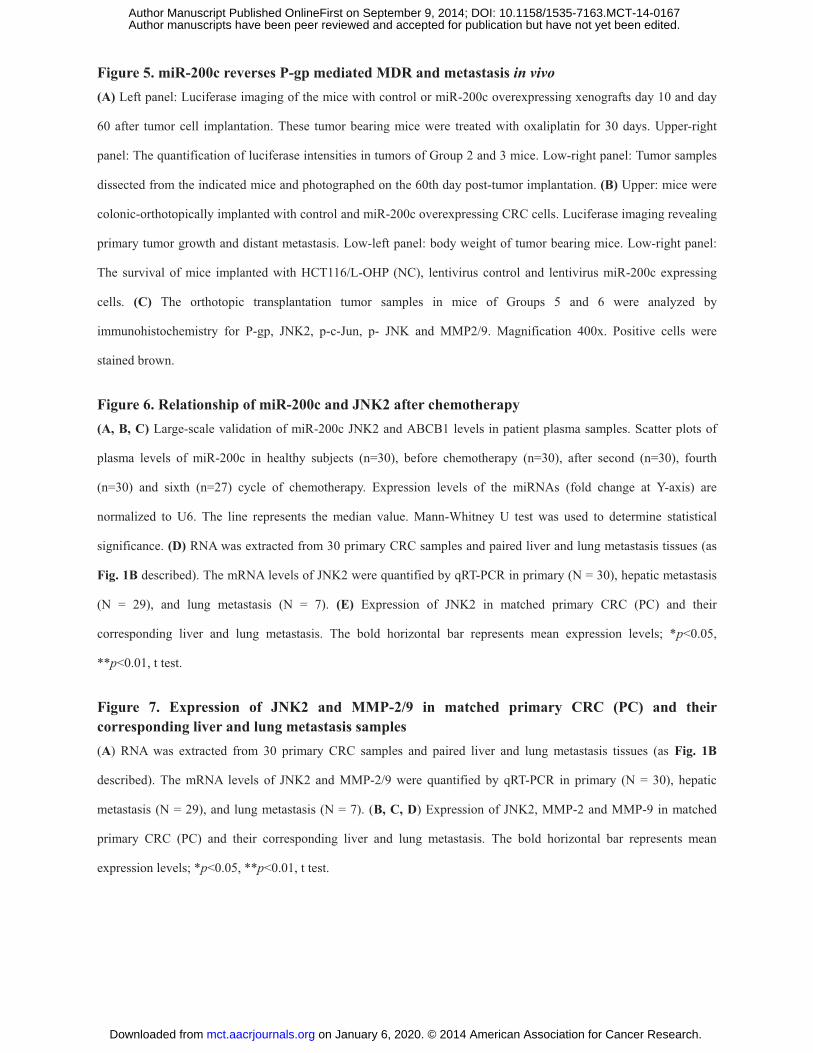
Figure 5. miR-200c reverses P-gp mediated MDR and metastasis in vivo (A) Left panel: Luciferase imaging of the mice with control or miR-200c overexpressing xenografts day 10 and day
60 after tumor cell implantation. These tumor bearing mice were treated with oxaliplatin for 30 days. Upper-right
panel: The quantification of luciferase intensities in tumors of Group 2 and 3 mice. Low-right panel: Tumor samples
dissected from the indicated mice and photographed on the 60th day post-tumor implantation. (B) Upper: mice were
colonic-orthotopically implanted with control and miR-200c overexpressing CRC cells. Luciferase imaging revealing
primary tumor growth and distant metastasis. Low-left panel: body weight of tumor bearing mice. Low-right panel:
The survival of mice implanted with HCT116/L-OHP (NC), lentivirus control and lentivirus miR-200c expressing
cells. (C) The orthotopic transplantation tumor samples in mice of Groups 5 and 6 were analyzed by
immunohistochemistry for P-gp, JNK2, p-c-Jun, p- JNK and MMP2/9. Magnification 400x. Positive cells were
stained brown.
Figure 6. Relationship of miR-200c and JNK2 after chemotherapy (A, B, C) Large-scale validation of miR-200c JNK2 and ABCB1 levels in patient plasma samples. Scatter plots of
plasma levels of miR-200c in healthy subjects (n=30), before chemotherapy (n=30), after second (n=30), fourth
(n=30) and sixth (n=27) cycle of chemotherapy. Expression levels of the miRNAs (fold change at Y-axis) are
normalized to U6. The line represents the median value. Mann-Whitney U test was used to determine statistical
significance. (D) RNA was extracted from 30 primary CRC samples and paired liver and lung metastasis tissues (as
Fig. 1B described). The mRNA levels of JNK2 were quantified by qRT-PCR in primary (N = 30), hepatic metastasis
(N = 29), and lung metastasis (N = 7). (E) Expression of JNK2 in matched primary CRC (PC) and their
corresponding liver and lung metastasis. The bold horizontal bar represents mean expression levels; *p<0.05,
**p<0.01, t test.
Figure 7. Expression of JNK2 and MMP-2/9 in matched primary CRC (PC) and their corresponding liver and lung metastasis samples (A) RNA was extracted from 30 primary CRC samples and paired liver and lung metastasis tissues (as Fig. 1B
described). The mRNA levels of JNK2 and MMP-2/9 were quantified by qRT-PCR in primary (N = 30), hepatic
metastasis (N = 29), and lung metastasis (N = 7). (B, C, D) Expression of JNK2, MMP-2 and MMP-9 in matched
primary CRC (PC) and their corresponding liver and lung metastasis. The bold horizontal bar represents mean
expression levels; *p<0.05, **p<0.01, t test.
on January 6, 2020. © 2014 American Association for Cancer Research. mct.aacrjournals.org Downloaded from
Author manuscripts have been peer reviewed and accepted for publication but have not yet been edited. Author Manuscript Published OnlineFirst on September 9, 2014; DOI: 10.1158/1535-7163.MCT-14-0167

Published OnlineFirst September 9, 2014.Mol Cancer Ther Hua Sui, Guo-Xiang Cai, Shu-Fang Pan, et al. targeting JNK2/c-Jun signaling pathway in colorectal cancermiR-200c attenuates P-gp mediated MDR and metastasis by
Updated version
10.1158/1535-7163.MCT-14-0167doi:
Access the most recent version of this article at:
Material
Supplementary
http://mct.aacrjournals.org/content/suppl/2014/09/09/1535-7163.MCT-14-0167.DC1
Access the most recent supplemental material at:
Manuscript
Authoredited. Author manuscripts have been peer reviewed and accepted for publication but have not yet been
E-mail alerts related to this article or journal.Sign up to receive free email-alerts
Subscriptions
Reprints and
To order reprints of this article or to subscribe to the journal, contact the AACR Publications
Permissions
Rightslink site. Click on "Request Permissions" which will take you to the Copyright Clearance Center's (CCC)
.http://mct.aacrjournals.org/content/early/2014/09/09/1535-7163.MCT-14-0167To request permission to re-use all or part of this article, use this link
on January 6, 2020. © 2014 American Association for Cancer Research. mct.aacrjournals.org Downloaded from
Author manuscripts have been peer reviewed and accepted for publication but have not yet been edited. Author Manuscript Published OnlineFirst on September 9, 2014; DOI: 10.1158/1535-7163.MCT-14-0167


