
1
MicroRNA-7 directly targets Reg1 in pancreatic cells 1
2
Shawna Downing1¶, Fan Zhang1¶, Zijing Chen1, Emmanuel S. Tzanakakis1,2* 3
4
1Department of Chemical and Biological Engineering, Tufts University, Medford, MA 02155 5
2Clinical and Translational Science Institute, Tufts Medical Center, Boston, MA 02111 6
¶Equal contribution 7
Running title: MicroRNA-7 regulates Reg1 expression 8
9
Shawna Downing, Graduate Student, Chemical and Biological Engineering, Tufts University, Medford, MA, 10
02155, [email protected] 11
Fan Zhang, Graduate Student, Chemical and Biological Engineering, Tufts University, Medford, MA, 02155, 12
Zijing Chen, Graduate Student, Chemical and Biological Engineering, Tufts University, Medford, MA, 02155, 14
Emmanuel S. Tzanakakis, Professor, Chemical and Biological Engineering, Tufts University, Medford, MA, 16
02155, [email protected] 17
18
*Corresponding author: 19
Emmanuel S. Tzanakakis 20
Professor 21
Department of Chemical and Biological Engineering 22
Tufts University 23
Science and Technology Center, Room 276A 24
Medford, MA 02155 USA 25
Phone: +1-617-627-0831 26
Fax: +1-617-627-3991 27
E-mail address: [email protected] 28
Downloaded from www.physiology.org/journal/ajpcell at Tufts Univ Hirsh Health Sciences Library (130.064.085.171) on July 1, 2019.
2
29
List of abbreviations 30
3’UTR: 3’ untranslated region 31
AD: Alzheimer disease 32
EGFR: Epidermal growth factor receptor 33
hfPCR: high fidelity polymerase chain reaction 34
PanIN: pancreatic intraepithelial neoplasia 35
PDAC: pancreatic ductal adenocarcinoma 36
PMSF: phenylmethanesulfonyl fluoride 37
PVDF: polyvinylidene difluoride 38
39
40
41
Downloaded from www.physiology.org/journal/ajpcell at Tufts Univ Hirsh Health Sciences Library (130.064.085.171) on July 1, 2019.
3
Abstract 42
Regenerating islet-derived (Reg) proteins, which were first discovered in the pancreas, are associated with 43
increased proliferation, prevention of apoptosis and enhanced differentiation in normal and disease states but 44
very little is known about the regulation of their expression. We hypothesized that Reg expression is influenced 45
by microRNAs. Bioinformatic analysis predicted Reg1 to be a target of microRNA-7 (miR-7), which influences 46
pancreatic beta-cell function. To this end, we investigated the effects of miR-7 on Reg1 expression in pancreatic 47
acinar and islet beta-cells. High levels of Reg1 were noted by immunostaining and western blotting in acinar 48
cells in contrast to islet cells. A reciprocal expression pattern was observed for miR-7. Overexpression of miR-7 49
resulted in Reg1 mRNA suppression and reduction of secreted Reg1 protein. Conversely, miR-7 knockdown led 50
to increases in Reg1. Targeting of Reg1 by miR-7 was confirmed via luciferase activity assays. In contrast, 51
miR-7 did not directly repress the human ortholog of Reg1, REG1A, as well as REG1B indicating species 52
differences in the regulation of Reg expression. This is the first account of microRNA modulation of any Reg 53
member warranting studies to fill gaps in our knowledge of Reg protein biology, particularly in disease 54
contexts. 55
56
Keywords: Reg proteins; microRNA-7; pancreas; islets; acinar cells. 57
58
Downloaded from www.physiology.org/journal/ajpcell at Tufts Univ Hirsh Health Sciences Library (130.064.085.171) on July 1, 2019.

4
59
Introduction 60
Regenerating islet-derived (Reg) proteins are C-type lectin-like proteins originally discovered in patients 61
with chronic calcific pancreatitis (10) while subsequent studies linked Regs further to pancreatitis (5, 22, 32) but 62
also to pancreatic cancer and diabetes. In particular, murine Reg1 and its human ortholog REG1A (also known 63
as lithostathine) are expressed in the exocrine pancreas and hyperplastic islets but not in normal islets. 64
Transgenic mice expressing Reg1 from a glucagon promoter in their islets exhibit apoptosis of β-cells with 65
ensuing diabetes and develop malignant tumors (48). Yet, Reg1 helps restore β-cell mass after pancreatectomy 66
in rats (43) and prevents or delays the development of diabetes in non-obese diabetic mice (44). Elevated 67
plasma Reg1 has been observed in mice with pancreatic intraepithelial neoplasia (PanIN) lesions (16), while 68
REG1A and REG1B are detected in the serum and urine of pancreatic ductal adenocarcinoma (PDAC) patients 69
(30, 39). Reg1 has also been implicated in extrapancreatic pathologies including colon cancer (49), seminoma 70
(33), hepatocellular carcinoma (8), cholangiocarcinoma (21) and Alzheimer disease (AD) (11, 12). 71
Despite the protein’s significant presence in various pathophysiological contexts, especially as they relate to 72
pancreas, very little is known about the regulation of the Reg1 gene expression. Miscellaneous factors including 73
IL-6 and dexamethasone (13), PDGF and growth hormone (19), and gastrin (3) reportedly influence Reg1 levels 74
in different cell types, partly through pertinent response elements on the gene promoter. Beyond autocrine or 75
paracrine signals, regulation of protein production or RNA stability can be mediated by microRNAs (miRNA), 76
which are short (18-25 nucleotides) single-stranded RNA molecules that cleave or translationally repress their 77
specific target gene mRNAs (7). MicroRNA-modulated gene expression underlies diverse cellular processes 78
including proliferation, apoptosis, differentiation and tumorigenesis. To this end, several microRNAs regulate 79
pancreatic cell function, metabolism and differentiation. However, no miRNA has been identified to date that 80
targets Reg genes in any cell type including pancreatic cells. 81
Here, we performed bioinformatic analysis of the 3’ untranslated region (3’UTR) of the Reg1 mRNA 82
seeking miRNAs with matching seed regions. The mmu-miR-7a-5p and mmu-miR-7b-5p featuring identical 83
Downloaded from www.physiology.org/journal/ajpcell at Tufts Univ Hirsh Health Sciences Library (130.064.085.171) on July 1, 2019.

5
seed regions were among the miRNAs identified as potentially targeting the murine Reg1 mRNA. The 84
aforementioned miRNAs belong to a group comprising Mir7a-1, Mir7a-2 and Mir7b (9) all of which have the 85
same seed sequence. The miR-7 family is evolutionary conserved across primates, rodents, and zebrafish 86
exhibiting a neuroendocrine pattern of expression (4, 6, 9, 28, 47). In the adult rat and human pancreases, the 87
islet/acinar tissue ratio of miR-7 expression is greater than 200 (6, 9, 26). Transgenic mice overexpressing miR-88
7a in β-cells develop diabetes due to impaired insulin secretion and β-cell dedifferentiation without significant 89
changes in proliferation and apoptosis (29). In humans, miR-7 is also downregulated in PDAC and ampullary 90
adenocarcinoma compared to normal pancreas (41). 91
Given the apparent inverse localization of miR-7 and Reg1 in the pancreatic acinar and islet compartments, 92
we considered the possibility of miR-7 targeting Reg1 thereby influencing its expression in line with our in 93
silico analysis. Here, we show that miR-7 is a negative regulator of the murine Reg1. The repression of Reg1 by 94
miR-7 was observed in acinar cells and β-cells. Further, the human REG1A (homolog of the murine Reg1) and 95
REG1B are not direct targets of miR-7, pointing to differences among species in the determinants of Reg 96
expression. This is the first account of miRNA modulation of the expression of a member of the Reg protein 97
family. 98
99
Materials and Methods 100
101
Housing and care of mice and collection of pancreatic tissues were approved by the Institutional Animal 102
Care and Use Committee at Tufts University. 103
104
Murine pancreatic islet and exocrine tissue isolation 105
Isolation of pancreatic tissue was performed as we reported (50). Eight-week old C57/BL6 mice were 106
injected intraperitoneally with 270 mg/kg ketamine/15 mg/kg xylazine (anesthesia overdose). A cannula was 107
prepared by filling with collagenase solution (CIzyme, Vitacyte, Indianapolis, IN) a syringe fitted with a 27 G 108
Downloaded from www.physiology.org/journal/ajpcell at Tufts Univ Hirsh Health Sciences Library (130.064.085.171) on July 1, 2019.
6
needle. After an incision made in the lower abdomen (V cut), the pancreas was exposed, the pancreatic duct was 109
clamped off at its duodenal insertion with a small bulldog clamp and the cannula was inserted into the duct 110
proximal to the liver. Collagenase solution (3 ml/mouse) was injected to fully inflate the pancreas, which was 111
subsequently removed and placed in a 50-ml conical tube for 20-30 min in a 37oC water bath. At the end of the 112
incubation, RPMI 1640 medium with 10% FBS was added (~20 ml) to each tube. The tubes were hand shaken 113
vigorously for 5-10 seconds to break up the tissue and were kept in ice. Samples were washed three times to 114
remove the collagenase by centrifugation at 180xg for 1.5 min. The supernatant was poured off, medium was 115
added (~25 ml) and the samples were vortexed gently. The suspension was filtered through a 400-µm wire mesh 116
(VWR, Randor, PA). The filtrate containing islets and exocrine tissue was spun at 180xg for 1.5 min and the 117
supernatant was removed. The pellet was resuspended in 10-15 ml Histopaque 1077 (Sigma-Aldrich, St. Louis, 118
MO) and vortexed until the suspension was homogeneous. After overlaying with 10 ml of RPMI 1640 medium 119
the sample was spun for 20 min at 1750xg with very slow acceleration and no braking at 10oC. Islets were 120
collected from the interface with a 10-ml serological pipette and placed in a 50-ml conical tube. Exocrine tissue 121
was collected from the bottom of the gradient and harvested to 50 ml-conical tubes. After three washes with 122
RPMI 1640 with 10% FBS and centrifugation at 180xg for 1.5 min the islet fraction was transferred to 6-cm 123
sterile culture dishes for islet picking under a microscope (Leica Microsystems Inc., Buffalo Grove, IL). Islet 124
and the exocrine tissue were immediately processed for total RNA extraction or immunohistochemistry (see 125
pertinent section below). 126
127
Cloning and Vector Preparation 128
The murine miR-7 (mmu-miR-7a-2) and 3’UTR of Reg1 were cloned by high fidelity PCR (hfPCR) from 129
genomic DNA isolated from MIN6 cells. The following primers were utilized: mmu-miR-7a-2; forward (F): 5’-130
ATATAGATCTGAAGGTGGCTAGCGTGA-3’, reverse (R): 5’-CCCGTCGACTGAAATGACCAGCAC-3’ 131
(underlined: BglII and SalI sites). The miR-7 inhibitor (miR-7 sponge) sequence was designed (14) with two 132
repeats of the miR-7 binding site: 5’-ACAACAAAATCACTAGTCTTCCA-3’ and generated by hfPCR using 133
Downloaded from www.physiology.org/journal/ajpcell at Tufts Univ Hirsh Health Sciences Library (130.064.085.171) on July 1, 2019.
7
overlapping primers. The microRNA-67 from C. elegans (cel-miR67; accession MI0000038) was also included 134
in the study as an unrelated microRNA control. The corresponding synthesized DNA (Eurofins Genomics, 135
Louisville, KY) was used as a template for hfPCR amplification with the primers: (F) 5’-136
GGAGATCTATTCCAACTCGATCA-3’, R: 5’-CCGTCGACAATAAACGAAATT-3’ (underlined: BglII and 137
SalI sites). The amplification product of miR-7, miR-7 inhibitor, or cel-miR-67 was ligated into the 138
pSuper.GFP/neo vector (Oligoengine, Seattle, WA) between the BglII and SalI sites (pSuper.GFP/neo-miR-7). 139
The murine Reg1 3’UTR was cloned from MIN6 cell genomic DNA using the primers: F: 5’-140
GGGAATTCAGTCACCTGAAAAAAAATAGTCA-3’, R: 5’-CCACTAGTGCAACATTGTAAAGGTGT-3’ 141
(underlined: EcoRI and SpeI sites). Genomic DNA was also extracted from human embryonic kidney 293 142
(HEK293) cells for cloning the 3’UTRs of REG1A and REG1B with the primers: REG1A 3’UTR; F: 5’-143
GGGAATTCAGGCAACTGGAAAATACATG-3’, R: 5’-CCACTAGTGACAGCACAATAGTGGAAAC-3’, 144
and REG1B 3’UTR; F: 5’-GGGAATTCAGGAAGCTGAAAAATGGATGT-3, R: 5’-145
CCACTAGTGAGCAAATGCAGAAGACAGAA-3’ (underlined: EcoRI and SpeI sites). The pGL3 luciferase 146
vector (Promega, Madison, WI) was modified by (i) replacing the CMV promoter with the EF1α promoter and 147
(ii) inserting a multiple cloning site (MCS) segment in the XbaI site flanking the 3’ end of the luciferase gene. 148
The MCS contained restriction sites for EcoRI and SpeI for insertion of the 3’UTR of Reg1 (Luc-3’UTR-Reg1), 149
REG1A or REG1B. A mutated Reg1 3’UTR sequence (97-GTCTTCC-103 97-GTCCCTT-103; +1 denoting the 150
nucleotide immediately after the Reg1 stop codon) was generated by high-fidelity PCR using a corresponding 151
synthesized template (Integrated DNA Technologies, Coralville, IA) and was inserted in the above luciferase 152
vector (Luc-Δ3’UTR-Reg1) as described for the wild-type Reg1 3’UTR. All constructs were verified by 153
sequencing. 154
155
Cell culture and transfection 156
MIN6 and βTC -cells, 266-6 acinar cells (ATCC, Manassas, VA) and HEK293 cells were cultured in 157
DMEM medium (Thermo Fisher Scientific) with 10% fetal bovine serum (FBS) and penicillin-streptomycin 158
Downloaded from www.physiology.org/journal/ajpcell at Tufts Univ Hirsh Health Sciences Library (130.064.085.171) on July 1, 2019.

8
(100 U/ml – 50 g/ml). Cells were manually passaged every 5-7 days with TrypLE (Life Technologies, 159
Carlsbad, CA) at a 1:4-1:6 ratio, and media were replaced every 3-4 days. The cultures were maintained in 5% 160
CO2/95% air at 37 oC. Cells were stained with Trypan Blue dye (Life Technologies) and counted in a 161
hemocytometer or with the TC20 automated cell counter (Bio-Rad, Hercules, CA). 162
For transfection, cells were grown to 70-90% confluence and plasmids were delivered using Lipofectamine 163
2000 (Invitrogen). Transfection of miR7 (C-310592-07) or cel-miR67 (CN-001000-01) mimic (Dharmacon 164
Inc., Lafayette, CO) was carried out with RNAiMax (Invitrogen, Carlsbad, CA) following the manufacturer’s 165
instructions. 166
167
RNA extraction, RT-PCR and quantitative PCR analysis 168
Total cellular RNA was extracted with Trizol (Life Technologies) according to manufacturer’s instructions. 169
Reverse transcription was performed at 42 ºC for 60 min with 1 µg total RNA using ImProm-II reverse 170
transcriptase (Promega, Madison, WI) and 250 ng oligo(dT)12-18 primers (Thermo Fisher Scientific). The 171
reverse transcriptase was heat-inactivated at 70 °C for 15 min. The resulting complimentary DNA (cDNA) was 172
analyzed on a StepOne Plus thermocycler (Applied Biosystems, Foster City, CA) by quantitative PCR (qPCR) 173
for 40 cycles and 58-60 ºC annealing temperature depending on each primer set. The sequences for primers 174
used in this study are listed in Table 1. 175
For miRNA expression analysis, total RNA was converted to cDNA with the qScript microRNA cDNA 176
synthesis kit (Quantabio, Beverly, MA) using an oligo-dT adapter primer (Table 1). The resulting cDNA was 177
amplified by qPCR (PerfeCTa SYBR Green SuperMix, Quantabio) with a universal PCR primer combined with 178
a primer targeting the mmu-mir7a-5p or the RNU6-2. 179
Analysis was performed based on the ΔΔCT method (17) with Actb and RNU6-2 used as endogenous 180
controls for the expression of Reg1 and mmu-mir7a-5p, respectively. 181
182
Immunocytochemistry 183
Downloaded from www.physiology.org/journal/ajpcell at Tufts Univ Hirsh Health Sciences Library (130.064.085.171) on July 1, 2019.

9
Cells were fixed in 4% paraformaldehyde (Sigma-Aldrich) for 20 min and permeabilized with 0.1% Triton 184
X-100 in PBS for 20 min at room temperature (nuclear marker staining). After three 5-min washes with PBS 185
after each step, samples were blocked with 3% normal donkey serum (NDS; Jackson ImmunoResearch 186
Laboratories Inc., West Grove, PA) in PBS for 30 min. Incubation was carried out at 4 ºC with a sheep anti-187
murine Reg1 antibody (AF1657; R&D Systems, Minneapolis, MN) followed by treatment with a donkey anti-188
sheep Rhodamine Red-X-conjugated antibody (713-295-147; Jackson ImmunoResearch) at room temperature 189
for 1 h. Nuclear DNA was stained with DAPI (Sigma-Aldrich). Controls were stained with IgG instead of 190
primary antibody. Immunostaining was visualized with a Leica TCS SPE confocal microscope (Leica 191
Microsystems Inc., Buffalo Grove, IL). 192
193
Immunohistochemistry 194
Paraffin-embedded sections were dehydrated with increasing concentrations of ethanol (80–100%). After 195
three 15-min washings with PBS, samples were blocked for 20 min in 3% NDS at 37°C and then incubated 196
overnight at 4°C with guinea pig anti-murine insulin (A0564; Dako/Agilent Technologies, Carpinteria, CA) and 197
sheep anti-murine Reg1 primary antibodies. Following three more washes with PBS, the sections were 198
incubated with AffiniPure donkey anti-guinea pig Alexa Fluor 488 and anti-sheep Rhodamine Red-X (706-545-199
148 and 713-295-147, respectively; Jackson ImmunoResearch) secondary antibodies for 30 min at 37°C. The 200
samples were then washed three times with PBS, mounted with SlowFade Diamond Antifade medium 201
containing DAPI (Invitrogen) and visualized by confocal microscopy. 202
203
Western blot analysis 204
Total protein was isolated using lysis buffer containing Tris-HCl (50 mM, pH 8), NaCl (150 mM), NP40 205
(1%), sodium dodecyl sulfate (SDS) (0.1%), sodium deoxycholate (1%), protease inhibitor cocktail including 206
PMSF, and phosphatase inhibitors (1 mM sodium fluoride, 5 mM sodium pyrophosphate, 5 mM sodium 207
orthovanadate; all from Sigma-Aldrich). Protein concentration was determined via the Bradford method (Pierce 208
Downloaded from www.physiology.org/journal/ajpcell at Tufts Univ Hirsh Health Sciences Library (130.064.085.171) on July 1, 2019.

10
Biotechnology, Rockford, IL). Samples were separated in 10% SDS-PAGE and transferred to polyvinylidene 209
difluoride (PVDF) membranes as described before (25). The membranes were blocked with 5% skim milk and 210
incubated overnight at 4 oC with diluted primary antibodies against murine Reg1 (1:1,000) and -actin (1:1,000; 211
#4970, Cell Signaling Technology, Beverly, MA). Then, membranes were washed four times (10-min) with 212
TBST and incubated for 1 h at room temperature with corresponding horseradish peroxidase (HRP)-linked 213
secondary antibodies (1:10,000; 713-035-147, 711-035-152, Jackson ImmunoResearch). After further washing 214
with TBST, enhanced chemiluminescence reagent (Pierce Biotechnology) was added and the membranes were 215
visualized with a C-DiGit blot scanner (LI-COR Biotechnology, Lincoln, NE). Densitometry was performed 216
using the Image Studio software (LI-COR) (42). 217
218
Dual Luciferase Assay 219
For determination of luciferase activity, cells in 24-well plates were transfected with a total of 500 ng of 220
plasmids per well as stated. Luciferase activity was assessed using the Dual Luciferase Assay Kit (Promega, 221
Madison, WI) 24-72 h post-transfection. Luminescence was detected in a microplate reader (Spectramax i3x, 222
Molecular Devices, San Jose, CA) with firefly luciferase activity normalized to the respective Renilla luciferase 223
signal for each sample. 224
225
Enzyme immunosorbent assay (ELISA) 226
ELISA was performed to detect protein secreted in the culture medium. Cells were grown to 70-90% 227
confluence and the medium was replaced by low-serum medium (DMEM with 1% FBS). After 30 h, the 228
medium was collected and cells were harvested with TrypLE (Life Technologies, Carlsbad, CA) and 229
resuspended in fresh medium. Cells were counted using a hemocytometer while the collected medium was 230
added to 96-well EIA/RIA plates (Corning Inc, Corning, NY). Fresh low-serum medium served as control. 231
Recombinant murine Reg1 (R&D Systems) was plated at different dilutions to obtain a standard curve as we 232
reported (25). After overnight incubation at 4°C wells were coated with a blocking buffer of 3% BSA and 0.1% 233
Downloaded from www.physiology.org/journal/ajpcell at Tufts Univ Hirsh Health Sciences Library (130.064.085.171) on July 1, 2019.

11
azide in 0.1% TBST for 90 min at 37°C. Following four washes with 0.1% TBST, samples were incubated for 2 234
h at room temperature with biotinylated sheep anti-mouse Reg1 (1:2000; BAF1657, R&D Systems) diluted in 235
buffer of 0.1% TBST with 1% BSA and 0.1% azide. Wells were again washed four times and a secondary 236
rabbit anti-biotin antibody (#5597, Cell Signaling Technology) in the same dilution buffer was added for 1 h at 237
room temperature. The wells were washed 4 times with 0.1% TBST and incubated with 1-StepTM Ultra TMB-238
ELISA substrate solution (Thermo Fisher Scientific) per manufacturer’s instructions. Total Reg concentration 239
was normalized by the number of cells in each sample. 240
241
Statistical analysis 242
Data are expressed as mean ± standard deviation (SD) unless stated otherwise from at least three 243
independent experiments analyzed in triplicates. ANOVA and the posthoc Tukey test were performed using 244
Prism (v. 8, GraphPad Software, La Jolla, CA). Values of p<0.05 were considered as significant. 245
246
Results 247
Murine Reg1 is a predicted target of miR-7 248
We set out to search in silico for miRNAs with binding sites on the 3’UTR of the murine Reg1. 249
Interrogation of the TargetScan database (1) revealed several putative miRNAs targeting Reg1 (Table 2). Of 250
those miRNAs, the seed region of miR-7a-5p and miR-7b-5p was an exact match for the 97-103 nucleotide 251
segment of the 3’UTR of Reg1 (Fig. 1A). Given the involvement of the miR-7 family in pancreatic cell function 252
and pathologies (15, 29), we decided to further investigate its potential role on the expression of murine Reg1. 253
Of note, we found no putative binding site(s) for miR-7 in the 3’UTR sequences of the genes encoding the 254
human REG1A (Reg1 ortholog) and REG1B proteins despite the miR-7 family being conserved in mice and 255
humans and the high homology between the murine and human Reg proteins. 256
257
Reciprocal expression of Reg1 and miR-7 in murine pancreatic exocrine and islet cells 258
Downloaded from www.physiology.org/journal/ajpcell at Tufts Univ Hirsh Health Sciences Library (130.064.085.171) on July 1, 2019.

12
We proceeded to investigate experimentally whether Reg1 is a target of miR-7. First, the expression profiles 259
of Reg1 and miR-7 were established by examining both exocrine cells and islet β-cells. Higher levels of miR-7 260
were detected in MIN6 (7.08-fold, p=0.0164) and βTC β-cells (44.46-fold, p=0.009) than in 266-6 acinar cells 261
(Fig. 1B). This miR-7 expression pattern was mirrored in murine primary islets (30.08-fold, p=5x10⁻⁴) and 262
exocrine tissue. 263
Conversely, Reg1 expression was lower in β-cells than in 266-6 cells (MIN6: 26.9-fold lower, βTC: 12.4-264
fold lower, both with p<10-4). Similarly, primary islets exhibited lower Reg1 levels (4.97-fold, p=7x10⁻⁴) 265
compared to exocrine explants (Fig. 1C). Moreover, 266-6 cells were positive for Reg1 unlike MIN6 cells (Fig. 266
2A) and Reg1 was detected in exocrine regions but not in islets of pancreatic tissue sections (Fig. 2B). These 267
findings were corroborated by Western blotting (Fig. 2C). Secreted Reg1 protein was present in supernatants 268
from 266-6 cell cultures but was not detectable in those from MIN6 and βTC cells (Fig. 2D). 269
Taken together, these findings demonstrate that increased miR-7 expression coincides with lower levels of 270
Reg1 in β-cells and primary islets while these profiles are inverted in exocrine cells. 271
272
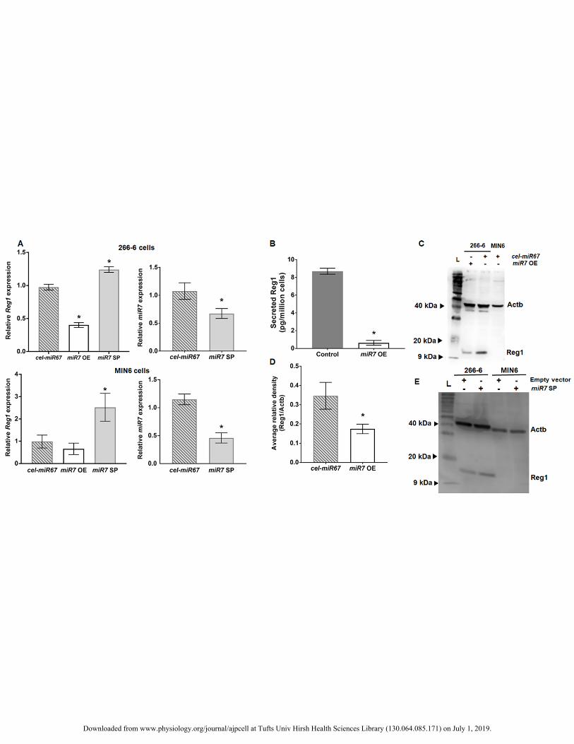
Direct targeting of murine Reg1 by miR-7 273
The reciprocal expression of Reg1 and miR-7 in exocrine and endocrine cells along with the discovery in 274
silico of a region of the Reg1 3’UTR matching the miR-7 seed sequence prompted us to investigate whether 275
miR-7 represses Reg1 directly. To this end, miR-7 was overexpressed or suppressed so that Reg1 levels could 276
be analyzed. Overexpression of miR-7 (transfection with miR7 OE plasmid) led to a 60% reduction of Reg1 277
expression (Fig. 3A; p=5.5x10⁻³, n=3) in 266-6 cells. Accordingly, the amounts of Reg1 released in the medium 278
were greater in control cells than in those overexpressing miR-7 (Fig. 3B). The content of Reg1 in the latter 279
cells (miR7 OE) was almost half (Figs. 3C-D) of that of cells transfected with cel-miR67. The cel-miR-67 was 280
chosen because (i) it is natively expressed in C. elegans reducing the risk for confounding interactions with 281
murine cell miRNA moieties, and (ii) there are no apparent sites on the 3’UTR of Reg1 matching the cel-miR-282
67 seed region. The expression of cel-miR-67 did not affect Reg1 or miR-7 levels. Upon inhibition of miR-7 in 283
Downloaded from www.physiology.org/journal/ajpcell at Tufts Univ Hirsh Health Sciences Library (130.064.085.171) on July 1, 2019.
13
MIN6 cells, which natively exhibit pronounced miR-7 levels, Reg1 expression was increased (Fig. 3A) but there 284
was no detectable change in Reg1 protein (Fig. 3E). Similarly, overexpression of miR-7 resulted in reduction of 285
Reg1 mRNA. These results show that the constructs used in this study were effective for miR-7 modulation. 286
More importantly, augmenting or suppressing miR-7 levels causes significant changes in Reg1. 287
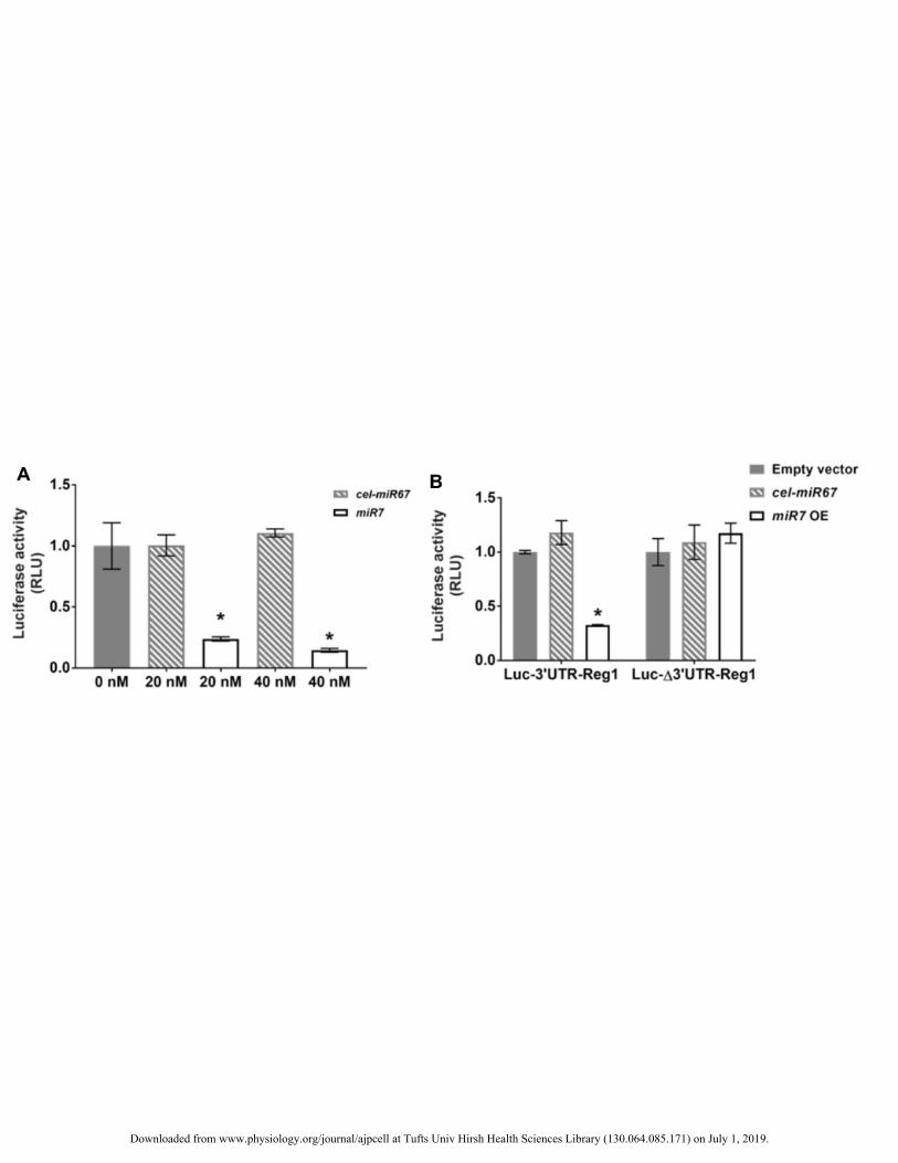
Direct repression of the expression of a gene by a particular microRNA involves binding of the latter to the 288
3’UTR of the target mRNA. Therefore, the 3’UTR of Reg1 was placed downstream of the luciferase gene (Luc-289
3’UTR-Reg1). When Luc-3’UTR-Reg1 was delivered with a miR-7 mimic to 266-6 cells, luciferase activity 290
was lower supporting the notion that miR-7 directly interacts with the 3’UTR of Reg1 (Fig. 4A). No differences 291
were noted in luciferase activity between cells treated with cel-miR67 mimic or vehicle. Furthermore, 266-6 292
cells were co-transfected with the miR-7 overexpression plasmid and either the Luc-3’UTR-Reg1 vector or a 293
luciferase construct with the Reg1 3’UTR but with its miR-7 binding site mutated (Luc-Δ3’UTR-Reg1). Despite 294
overexpression of miR-7, luciferase signal was significantly reduced only in Luc-3’UTR-Reg1-transfected cells 295
(Fig. 4B). Again, co-expression of cel-miR67 with either of the aforementioned luciferase constructs did not 296
result in differences in luciferase activity in comparison to cells receiving the empty expression vector. 297
These results prove that miR-7 directly targets the 3’UTR of murine Reg1 in line with the bioinformatic 298
analysis-based identification of Reg1 as a miR-7 target. 299
300
Human REG1A and REG1B are not targets of miR-7 301
We also assessed whether the human REG1A (ortholog of murine Reg1) and REG1B, which together 302
comprise the human REG1 subgroup, are also targeted by miR-7. Cells were transfected with a construct 303
containing the luciferase gene upstream of the 3’UTR of the human REG1A (Luc-3’UTR-REG1A) or REG1B 304
(Luc-3’UTR-REG1B). Additionally, these cells were co-transfected either with the miR-7 OE or empty 305
(control) vector. There was no difference in luciferase activity in 266-6 cells receiving each luciferase vector 306
regardless of the expression of miR-7 (Fig. 5A). 307
Downloaded from www.physiology.org/journal/ajpcell at Tufts Univ Hirsh Health Sciences Library (130.064.085.171) on July 1, 2019.

14
The findings support our in silico analysis indicating the absence of regions matching the miR-7 seed on the 308
REG1A and REG1B 3’UTRs. 309
310
Discussion 311
Regulation of the expression of Reg family members remains puzzling despite their discovery almost four 312
decades ago. This is surprising considering that Reg proteins are implicated in major pathologies of the 313
pancreas and other tissues including the gastrointestinal tract and central nervous system. Here, we showed that 314
miR-7 directly targets the murine Reg1 modulating its expression. This is aligned with the reciprocal expression 315
of miR-7 and Reg1 in the acinar and islet cells although the physiological significance of this pattern is unclear. 316
Both miR-7 and Reg1 influence the differentiation and proliferation of β-cells. Overexpression of Mir-7a in 317
murine islets causes a decline in β-cell differentiation markers such as Pdx1, Nkx6-1, Mafa and Neurod1 318
whereas pancreatic islets of Rip-Cre Mir7a2fl/fl mice exhibit higher steady-state levels of these transcripts 319
compared to controls (29). MiR-7 expression drops during islet compensation at the onset of diabetes in BKS 320
db/db mice (29). MiR-7 also regulates adult β-cell proliferation negatively as it targets five components of the 321
mTOR signaling pathway (46). In contrast, Reg1 is considered a β-cell mitogen with its expression increasing in 322
regenerating or hyperplastic islets although the exact mechanism is unknown (24, 45). Delayed development of 323
diabetes and larger islet volume are observed in the offspring of NOD mice crossed with transgenic mice 324
overexpressing Reg1 from an insulin promoter (Ins-Reg) (44). After damage due to encephalomyocarditis virus 325
infection, the islets of Reg1-/- mice exhibit lower BrdU labeling compared to those of wild-type mice (2). 326
Moreover, diabetic NOD mice exposed to human REG1A display a greater β-cell mass than control animals 327
(20). When combined with our findings of miR-7 targeting Reg1, a picture emerges of potentially coordinated 328
Reg1/miR-7 activities. Further investigation will be necessary to determine if these activities have significant 329
implications in the physiology for pancreatic cells. It will be particularly interesting to examine if control of 330
murine β-cell proliferation by miR-7 may partly be realized through adjustment of Reg1 expression. 331
Downloaded from www.physiology.org/journal/ajpcell at Tufts Univ Hirsh Health Sciences Library (130.064.085.171) on July 1, 2019.
15
Expression of Reg1 has been detected in E9-E12 mouse embryos but not in E8.5 and Reg2, which exhibits a 332
76% amino acid homology with Reg1, appears on E12 (37). Conversely, miR-7 expression is low between 333
E10.5-11.5 and increases at E13.5-E14.5 with strong localization in pancreatic endocrine cells and the neural 334
tube (34). Of note, the Reg2 3’UTR exhibits no matching regions for the miR-7 seed based on bioinformatic 335
analysis. This is in line with the restricted expression of Reg2 mRNA and protein in the exocrine tissue (40). 336
Hence, the upregulation in Reg2 may compensate the repression of Reg1 due to rising miR-7 levels but the 337
functional equivalence of Reg2 and Reg1 (despite their high amino acid homology) is not established. 338
It should be noted that the profile of Reg1 has not been studied in miR-7 mutant mice. As discussed already, 339
deletion of Mir7a2 in the islets of Rip-Cre Mir7a2fl/fl mice led to improved glucose tolerance via increased 340
insulin secretion (29). However, the available gene expression data (GSE48195) are not sufficient for 341
comprehensive statistical analysis to determine specific changes in Reg1 levels. Moreover, mice with 342
conditional excision of miR-7a-2 exhibit a reduction – but not complete ablation – of miR-7 expression most 343
likely due to compensatory effects by other miR-7 family members (36). Hence, information about the 344
relationship between Reg1 and miR-7 in vivo has been challenging to obtain from published studies involving 345
miR-7 transgenic mice. This is further exacerbated by the plurality of miR-7 target genes. 346
Inhibition of miR-7 in β-cells resulted in increased Reg1 expression but no changes were detected in the 347
corresponding protein. A longer period of miR-7 suppression may be necessary for a substantial change in the 348
protein level while additional mechanisms influencing the production of Reg protein cannot be ruled out. 349
The interaction between miR-7 and Reg1 may also be appreciated in other contexts beyond pancreas 350
pathophysiology. MiR-7 is expressed in various regions of the brain of humans and mice (18, 28) and is linked 351
to neuronal differentiation and function while its dysregulation may contribute to neurological disorders, 352
including AD (38). To this end, aged animals have lower Reg1 expression than healthy animals in a murine 353
model of AD (31). Hence, addressing whether miR-7 affects the expression of Reg1 in this context similar to 354
what we have demonstrated in pancreatic cells is of significant interest. Similarly, Reg1 and miR-7 have been 355
Downloaded from www.physiology.org/journal/ajpcell at Tufts Univ Hirsh Health Sciences Library (130.064.085.171) on July 1, 2019.

16
implicated in different types of cancer in studies involving rodent models (23, 27, 35) opening prospects for 356
further investigation of the Reg1/miR-7 relationship as a contributing factor. 357
We found that miR-7 directly targets the murine reg1 but not the human REG1A and REG1B. Other genes 358
also have miR-7 target sites in their 3’UTR that are poorly conserved among species. The human epidermal 359
growth factor receptor (EGFR) gene features two segments targeted by miR-7 in its 3’UTR whereas the murine 360
Egfr has a single 3’UTR region albeit with low similarity to the miR-7 seed (23). In fact, overexpression of 361
miR-7 does not alter Egfr expression in MIN6 cells (46). The difference we report here between the murine 362
Reg1 and the human REG1A and REG1B may underlie yet unknown disparities among species in the 363
regulation of Reg and therefore in its function in normal and disease states. It also cautions about extrapolating 364
findings on Reg proteins from studies involving rodents to humans. Nonetheless, the discovery of the 365
modulation of a Reg family gene in murine cells by a miRNA motivates the exploration of a similar mechanism 366
applicable to human Reg genes. To this end, ongoing work focuses on the identification of miRNAs targeting 367
the human Reg members that may potentially lead to novel insights into Reg protein biology. 368
369
Acknowledgements 370
Funding support has been provided by the National Science Foundation (NSF, CBET1743367) to EST. 371
372
Author Contributions 373
F. Zhang, S. Downing and Z. Chen performed research; F. Zhang, S. Downing, Z. Chen and E. Tzanakakis 374
analyzed data; F. Zhang, S. Downing and E. Tzanakakis wrote the paper; E. Tzanakakis designed research. 375
376
Figure Legends 377
Figure 1. (A) Alignment of a putative miR-7 binding sequence in the murine Reg1 3’-UTR (nucleotides 97–378
103) is shown with the seed regions (capital letters) of mmu-miR-7a-5p and mmu-miR-7b-5p. Expression of (B) 379
mmu-miR-7 and (C) Reg1 in MIN6 and βTC β-cells as well as primary islets and exocrine tissue. *p<0.05, 380
Downloaded from www.physiology.org/journal/ajpcell at Tufts Univ Hirsh Health Sciences Library (130.064.085.171) on July 1, 2019.
17
**p<0.01 vs. the expression of 266-6 cells (for MIN6 and βTC β-cells) or exocrine tissue (for islets) from n=4 381
separate experiments analyzed in triplicates. Normalization of mmu-miR-7 and Reg1 data was performed 382
relative to the expression of RNU6-2 and Actb, respectively. 383
384
Figure 2. (A) Immunocytochemistry of 266-6 and MIN6 cells for Reg1. Nuclear DNA staining (DAPI) is also 385
shown along with merged images. Control samples (middle row) were stained only with the secondary 386
antibody. Bars: 25 µm. (B) Reg1 protein expression in pancreatic tissue. Insulin expression demarcates islet β-387
cells. Bars: 50 µm. (C) Western blot for the expression of Reg1 in 266-6, MIN6, and βTC cells. The loading 388
control beta-actin (Actb) is also shown. L: ladder. (D) The concentration of Reg1 secreted in the culture 389
medium was determined by ELISA (n=3 experiments in triplicates), ND: not detectable. 390
391
Figure 3. (A) Relative Reg1 expression in 266-6 and MIN6 cells transfected with either a miR-7 overexpression 392
(OE) or inhibition (sponge; SP) plasmid. Results from cells transfected with a plasmid for an unrelated control 393
microRNA (cel-miR67) are included. *p<0.05, n=3 experiments vs. Reg1 levels for cells transfected with empty 394
vector. The miR-7 levels after transfection with miR7 SP or cel-miR67 plasmid are shown. * p<0.05, n=3 395
experiments vs. miR7 expression for cells transfected with empty vector. (B) ELISA results showing the 396
amounts of Reg1 released by 266-6 cells transfected with either an empty vector (Control) or the miR-7 OE 397
plasmid. *p<0.05, n=3 experiments in triplicates. (C) A representative blot is shown of Reg1 content in 266-6 398
cells transfected with an cel-miR7 or miR-7 OE plasmid. A MIN6 cell sample is also shown. Loading control: 399
Actb. (D) Results of densitometric analysis of western blots such as the one shown in (C). The results are the 400
average (n=3) values of the ratio of intensities of the Reg1 and corresponding Actb bands for each 266-6 cell 401
sample shown in (C). *p<0.05 vs. cells transfected with the cel-miR67 plasmid. (E) Western blot for Reg1 from 402
266-6 and MIN6 cells transfected with either the empty miRNA expression vector (pSuper) or miR-7 sponge 403
(miR7 SP). The loading control, Actb, is also shown. 404
405
Downloaded from www.physiology.org/journal/ajpcell at Tufts Univ Hirsh Health Sciences Library (130.064.085.171) on July 1, 2019.
18
Figure 4. (A) Acinar 266-6 cells were transfected with a luciferase vector featuring the 3’UTR of Reg1 (Luc-406
3’UTR-Reg1) and incubated with various concentrations of miR-7 or cel-miR-67 mimic before measuring 407
luciferase activity. Results are shown as relative activity compared to cells treated with vehicle only (0 nM). 408
*p<0.0001 vs. cells transfected with cel-miR67 mimic at the same concentration, n=4-7 independent 409
experiments in triplicates. (B) Luciferase activity was reduced with overexpression of miR-7 (miR7 OE, light 410
bars) in cells co-transfected with Luc-3’UTR-Reg1 but not in those with the luciferase vector carrying the 411
3’UTR of Reg1 with a scrambled seed region (Luc-Δ3’UTR-Reg1). Dark bars depict results of cells transfected 412
with an empty vector. *p<0.0001, n=3 experiments. No differences were noted in cells transfected with each of 413
the aforementioned luciferase vectors and the cel-miR67 plasmid (hatched bars). 414
415
Figure 5. miR7 does not target the 3’UTR of human REG1A and REG1B. Luciferase activity results are shown 416
for 266-6 cells from n=3 independent experiments with triplicate measurements. 417
418
References 419
1. Agarwal V, Bell GW, Nam JW, and Bartel DP. Predicting effective microRNA target sites in 420
mammalian mRNAs. Elife 4: 2015. 421
2. Aida K, Kobayashi T, Takeshita A, Jimbo E, Nishida Y, Yagihashi S, Hosoi M, Fukui T, 422
Sugawara A, and Takasawa S. Crucial role of Reg I from acinar-like cell cluster touching with islets 423
(ATLANTIS) on mitogenesis of beta cells in EMC virus-induced diabetic mice. Biochem Biophys Res Commun 424
503: 963-969, 2018. 425
3. Ashcroft FJ, Varro A, Dimaline R, and Dockray GJ. Control of expression of the lectin-like protein 426
Reg-1 by gastrin: role of the Rho family GTPase RhoA and a C-rich promoter element. Biochem J 381: 397-427
403, 2004. 428
Downloaded from www.physiology.org/journal/ajpcell at Tufts Univ Hirsh Health Sciences Library (130.064.085.171) on July 1, 2019.

19
4. Ason B, Darnell DK, Wittbrodt B, Berezikov E, Kloosterman WP, Wittbrodt J, Antin PB, and 429
Plasterk RH. Differences in vertebrate microRNA expression. Proceedings of the National Academy of 430
Sciences of the United States of America 103: 14385-14389, 2006. 431
5. Bimmler D, Schiesser M, Perren A, Scheele G, Angst E, Meili S, Ammann R, and Graf R. 432
Coordinate regulation of PSP/reg and PAP isoforms as a family of secretory stress proteins in an animal model 433
of chronic pancreatitis. J Surg Res 118: 122-135, 2004. 434
6. Bravo-Egana V, Rosero S, Molano RD, Pileggi A, Ricordi C, Dominguez-Bendala J, and Pastori 435
RL. Quantitative differential expression analysis reveals miR-7 as major islet microRNA. Biochem Biophys Res 436
Commun 366: 922-926, 2008. 437
7. Bushati N, and Cohen SM. microRNA functions. Annu Rev Cell Dev Biol 23: 175-205, 2007. 438
8. Cavard C, Terris B, Grimber G, Christa L, Audard V, Radenen-Bussiere B, Simon MT, Renard 439
CA, Buendia MA, and Perret C. Overexpression of regenerating islet-derived 1 alpha and 3 alpha genes in 440
human primary liver tumors with beta-catenin mutations. Oncogene 25: 599-608, 2006. 441
9. Correa-Medina M, Bravo-Egana V, Rosero S, Ricordi C, Edlund H, Diez J, and Pastori RL. 442
MicroRNA miR-7 is preferentially expressed in endocrine cells of the developing and adult human pancreas. 443
Gene Expr Patterns 9: 193-199, 2009. 444
10. De Caro A, Lohse J, and Sarles H. Characterization of a protein isolated from pancreatic calculi of 445
men suffering from chronic calcifying pancreatitis. Biochem Biophys Res Commun 87: 1176-1182, 1979. 446
11. de la Monte SM, Ozturk M, and Wands JR. Enhanced expression of an exocrine pancreatic protein in 447
Alzheimer's disease and the developing human brain. The Journal of clinical investigation 86: 1004-1013, 1990. 448
12. Duplan L, Michel B, Boucraut J, Barthellemy S, Desplat-Jego S, Marin V, Gambarelli D, Bernard 449
D, Berthezene P, Alescio-Lautier B, and Verdier JM. Lithostathine and pancreatitis-associated protein are 450
involved in the very early stages of Alzheimer's disease. Neurobiol Aging 22: 79-88, 2001. 451
Downloaded from www.physiology.org/journal/ajpcell at Tufts Univ Hirsh Health Sciences Library (130.064.085.171) on July 1, 2019.

20
13. Dusetti NJ, Mallo GV, Ortiz EM, Keim V, Dagorn JC, and Iovanna JL. Induction of 452
lithostathine/reg mRNA expression by serum from rats with acute pancreatitis and cytokines in pancreatic 453
acinar AR-42J cells. Arch Biochem Biophys 330: 129-132, 1996. 454
14. Ebert MS, Neilson JR, and Sharp PA. MicroRNA sponges: competitive inhibitors of small RNAs in 455
mammalian cells. Nat Methods 4: 721-726, 2007. 456
15. Esguerra JL, Bolmeson C, Cilio CM, and Eliasson L. Differential glucose-regulation of microRNAs 457
in pancreatic islets of non-obese type 2 diabetes model Goto-Kakizaki rat. PloS one 6: e18613, 2011. 458
16. Faca VM, Song KS, Wang H, Zhang Q, Krasnoselsky AL, Newcomb LF, Plentz RR, Gurumurthy 459
S, Redston MS, Pitteri SJ, Pereira-Faca SR, Ireton RC, Katayama H, Glukhova V, Phanstiel D, Brenner 460
DE, Anderson MA, Misek D, Scholler N, Urban ND, Barnett MJ, Edelstein C, Goodman GE, Thornquist 461
MD, McIntosh MW, DePinho RA, Bardeesy N, and Hanash SM. A mouse to human search for plasma 462
proteome changes associated with pancreatic tumor development. PLoS Med 5: e123, 2008. 463
17. Fan Y, Hsiung M, Cheng C, and Tzanakakis ES. Facile engineering of xeno-free microcarriers for the 464
scalable cultivation of human pluripotent stem cells in stirred suspension. Tissue engineering Part A 20: 588-465
599, 2014. 466
18. Farh KK, Grimson A, Jan C, Lewis BP, Johnston WK, Lim LP, Burge CB, and Bartel DP. The 467
widespread impact of mammalian MicroRNAs on mRNA repression and evolution. Science 310: 1817-1821, 468
2005. 469
19. Francis PJ, Southgate JL, Wilkin TJ, and Bone AJ. Expression of an islet regenerating (reg) gene in 470
isolated rat islets: effects of nutrient and non-nutrient growth factors. Diabetologia 35: 238-242, 1992. 471
20. Gross DJ, Weiss L, Reibstein I, van den Brand J, Okamoto H, Clark A, and Slavin S. Amelioration 472
of diabetes in nonobese diabetic mice with advanced disease by linomide-induced immunoregulation combined 473
with Reg protein treatment. Endocrinology 139: 2369-2374, 1998. 474
Downloaded from www.physiology.org/journal/ajpcell at Tufts Univ Hirsh Health Sciences Library (130.064.085.171) on July 1, 2019.

21
21. Harada K, Zen Y, Kanemori Y, Chen TC, Chen MF, Yeh TS, Jan YY, Masuda S, Nimura Y, 475
Takasawa S, Okamoto H, and Nakanuma Y. Human REG I gene is up-regulated in intrahepatic 476
cholangiocarcinoma and its precursor lesions. Hepatology 33: 1036-1042, 2001. 477
22. Hayakawa T, Naruse S, Kitagawa M, Nakae Y, Harada H, Ochi K, Kuno N, Kurimoto K, and 478
Hayakawa S. Pancreatic stone protein and lactoferrin in human pancreatic juice in chronic pancreatitis. 479
Pancreas 10: 137-142, 1995. 480
23. Horsham JL, Ganda C, Kalinowski FC, Brown RA, Epis MR, and Leedman PJ. MicroRNA-7: A 481
miRNA with expanding roles in development and disease. Int J Biochem Cell Biol 69: 215-224, 2015. 482
24. Ishii C, Kawazu S, Tomono S, Ohno T, Shimizu M, Kato N, Fukuda M, Ito Y, Kurihara S, Murata 483
K, and Komeda K. Appearance of a regenerating (reg) gene protein in pancreatic islets of remission 484
BB/Wor//Tky rats. Endocr J 40: 269-273, 1993. 485
25. Jing D, Kehoe DE, and Tzanakakis ES. Expression of Reg family proteins in embryonic stem cells 486
and its modulation by Wnt/beta-catenin signaling. Stem Cells Dev 19: 1307-1319, 2010. 487
26. Joglekar MV, Joglekar VM, and Hardikar AA. Expression of islet-specific microRNAs during 488
human pancreatic development. Gene Expr Patterns 9: 109-113, 2009. 489
27. Judd LM, Menheniott TR, Ling H, Jackson CB, Howlett M, Kalantzis A, Priebe W, and Giraud 490
AS. Inhibition of the JAK2/STAT3 pathway reduces gastric cancer growth in vitro and in vivo. Plos One 9: 491
e95993, 2014. 492
28. Landgraf P, Rusu M, Sheridan R, Sewer A, Iovino N, Aravin A, Pfeffer S, Rice A, Kamphorst AO, 493
Landthaler M, Lin C, Socci ND, Hermida L, Fulci V, Chiaretti S, Foa R, Schliwka J, Fuchs U, Novosel A, 494
Muller RU, Schermer B, Bissels U, Inman J, Phan Q, Chien M, Weir DB, Choksi R, De Vita G, Frezzetti 495
D, Trompeter HI, Hornung V, Teng G, Hartmann G, Palkovits M, Di Lauro R, Wernet P, Macino G, 496
Rogler CE, Nagle JW, Ju J, Papavasiliou FN, Benzing T, Lichter P, Tam W, Brownstein MJ, Bosio A, 497
Borkhardt A, Russo JJ, Sander C, Zavolan M, and Tuschl T. A mammalian microRNA expression atlas 498
based on small RNA library sequencing. Cell 129: 1401-1414, 2007. 499
Downloaded from www.physiology.org/journal/ajpcell at Tufts Univ Hirsh Health Sciences Library (130.064.085.171) on July 1, 2019.

22
29. Latreille M, Hausser J, Stutzer I, Zhang Q, Hastoy B, Gargani S, Kerr-Conte J, Pattou F, Zavolan 500
M, Esguerra JL, Eliasson L, Rulicke T, Rorsman P, and Stoffel M. MicroRNA-7a regulates pancreatic beta 501
cell function. The Journal of clinical investigation 124: 2722-2735, 2014. 502
30. Makawita S, Dimitromanolakis A, Soosaipillai A, Soleas I, Chan A, Gallinger S, Haun RS, 503
Blasutig IM, and Diamandis EP. Validation of four candidate pancreatic cancer serological biomarkers that 504
improve the performance of CA19.9. BMC Cancer 13: 404, 2013. 505
31. Marchal S, Givalois L, Verdier JM, and Mestre-Frances N. Distribution of lithostathine in the mouse 506
lemur brain with aging and Alzheimer's-like pathology. Neurobiol Aging 33: 431 e415-425, 2012. 507
32. Matozaki T, Sakamoto C, Suzuki T, Chujo S, Matsuda K, Wada K, Nakano O, Konda Y, 508
Nishizaki H, Nagao M, and et al. Idiopathic chronic calcifying pancreatitis with diabetes mellitus. Analysis of 509
pancreatic stone protein gene. Digestive diseases and sciences 38: 963-967, 1993. 510
33. Mauro V, Carette D, Chevallier D, Michiels JF, Segretain D, Pointis G, and Senegas-Balas F. Reg I 511
protein in healthy and seminoma human testis. Histol Histopathol 23: 1195-1203, 2008. 512
34. Nieto M, Hevia P, Garcia E, Klein D, Alvarez-Cubela S, Bravo-Egana V, Rosero S, Damaris 513
Molano R, Vargas N, Ricordi C, Pileggi A, Diez J, Dominguez-Bendala J, and Pastori RL. Antisense miR-514
7 impairs insulin expression in developing pancreas and in cultured pancreatic buds. Cell transplantation 21: 515
1761-1774, 2012. 516
35. Parikh A, Stephan AF, and Tzanakakis ES. Regenerating proteins and their expression, regulation 517
and signaling. Biomol Concepts 3: 57-70, 2012. 518
36. Park CY, Jeker LT, Carver-Moore K, Oh A, Liu HJ, Cameron R, Richards H, Li Z, Adler D, 519
Yoshinaga Y, Martinez M, Nefadov M, Abbas AK, Weiss A, Lanier LL, de Jong PJ, Bluestone JA, 520
Srivastava D, and McManus MT. A resource for the conditional ablation of microRNAs in the mouse. Cell 521
Rep 1: 385-391, 2012. 522
Downloaded from www.physiology.org/journal/ajpcell at Tufts Univ Hirsh Health Sciences Library (130.064.085.171) on July 1, 2019.

23
37. Perfetti R, Raygada M, Wang Y, Zenilman ME, Egan JM, Denno KM, Sadler TW, and Shuldiner 523
AR. Regenerating (reg) and insulin genes are expressed in prepancreatic mouse embryos. J Mol Endocrinol 17: 524
79-88, 1996. 525
38. Pichler S, Gu W, Hartl D, Gasparoni G, Leidinger P, Keller A, Meese E, Mayhaus M, Hampel H, 526
and Riemenschneider M. The miRNome of Alzheimer's disease: consistent downregulation of the miR-527
132/212 cluster. Neurobiol Aging 50: 167 e161-167 e110, 2017. 528
39. Radon TP, Massat NJ, Jones R, Alrawashdeh W, Dumartin L, Ennis D, Duffy SW, Kocher HM, 529
Pereira SP, Guarner posthumous L, Murta-Nascimento C, Real FX, Malats N, Neoptolemos J, Costello 530
E, Greenhalf W, Lemoine NR, and Crnogorac-Jurcevic T. Identification of a Three-Biomarker Panel in 531
Urine for Early Detection of Pancreatic Adenocarcinoma. Clin Cancer Res 21: 3512-3521, 2015. 532
40. Sanchez D, Baeza N, Blouin R, Devaux C, Grondin G, Mabrouk K, Guy-Crotte O, and Figarella 533
C. Overexpression of the reg gene in non-obese diabetic mouse pancreas during active diabetogenesis is 534
restricted to exocrine tissue. J Histochem Cytochem 48: 1401-1410, 2000. 535
41. Schultz NA, Werner J, Willenbrock H, Roslind A, Giese N, Horn T, Wojdemann M, and Johansen 536
JS. MicroRNA expression profiles associated with pancreatic adenocarcinoma and ampullary adenocarcinoma. 537
Mod Pathol 25: 1609-1622, 2012. 538
42. Taylor SC, Berkelman T, Yadav G, and Hammond M. A defined methodology for reliable 539
quantification of Western blot data. Mol Biotechnol 55: 217-226, 2013. 540
43. Terazono K, Yamamoto H, Takasawa S, Shiga K, Yonemura Y, Tochino Y, and Okamoto H. A 541
novel gene activated in regenerating islets. The Journal of biological chemistry 263: 2111-2114, 1988. 542
44. Unno M, Nata K, Noguchi N, Narushima Y, Akiyama T, Ikeda T, Nakagawa K, Takasawa S, and 543
Okamoto H. Production and characterization of Reg knockout mice: reduced proliferation of pancreatic beta-544
cells in Reg knockout mice. Diabetes 51 Suppl 3: S478-483, 2002. 545
45. Unno M, Yonekura H, Nakagawara K, Watanabe T, Miyashita H, Moriizumi S, Okamoto H, Itoh 546
T, and Teraoka H. Structure, chromosomal localization, and expression of mouse reg genes, reg I and reg II. A 547
Downloaded from www.physiology.org/journal/ajpcell at Tufts Univ Hirsh Health Sciences Library (130.064.085.171) on July 1, 2019.

24
novel type of reg gene, reg II, exists in the mouse genome. The Journal of biological chemistry 268: 15974-548
15982, 1993. 549
46. Wang Y, Liu J, Liu C, Naji A, and Stoffers DA. MicroRNA-7 regulates the mTOR pathway and 550
proliferation in adult pancreatic beta-cells. Diabetes 62: 887-895, 2013. 551
47. Wienholds E, Kloosterman WP, Miska E, Alvarez-Saavedra E, Berezikov E, de Bruijn E, Horvitz 552
HR, Kauppinen S, and Plasterk RH. MicroRNA expression in zebrafish embryonic development. Science 553
309: 310-311, 2005. 554
48. Yamaoka T, Yoshino K, Yamada T, Idehara C, Hoque MO, Moritani M, Yoshimoto K, Hata J, 555
and Itakura M. Diabetes and tumor formation in transgenic mice expressing Reg I. Biochem Biophys Res 556
Commun 278: 368-376, 2000. 557
49. Zenilman ME, Kim S, Levine BA, Lee C, and Steinberg JJ. Ectopic expression of reg protein: A 558
marker of colorectal mucosa at risk for neoplasia. J Gastrointest Surg 1: 194-201; discussion 201-192, 1997. 559
50. Zhang F, and Tzanakakis ES. Optogenetic regulation of insulin secretion in pancreatic beta-cells. Sci 560
Rep 7: 9357, 2017. 561
562
Downloaded from www.physiology.org/journal/ajpcell at Tufts Univ Hirsh Health Sciences Library (130.064.085.171) on July 1, 2019.

Table 1. Primers used in this study (shown in a 5’ to 3’ orientation).
Gene Forward primer Reverse primer Amplicon size (bp)
Reg1 CTCATGCCTGATCGTCCTGTC AGCCCAAGTTAAACGGTCTTC 142
Actb GTGACGTTGACATCCGTAAAGA GCCGGACTCATCGTACTCC 245
Mir-7* CGTGGAAGACTAGTGATTTTGTTG miRNA reverse primer 86
Rnu6 GCAAATTCGTGAAGCGTTCC
miRNA reverse primer 104
miRNA reverse primer
GCATAGACCTGAATGGCGGTAAGGGTGTGGTAGGCGAGACATTTTTTTTTTTT
TTTTTTTT
* The primer is homologous to both Mir7-1 and Mir7-2.
Downloaded from www.physiology.org/journal/ajpcell at Tufts Univ Hirsh Health Sciences Library (130.064.085.171) on July 1, 2019.
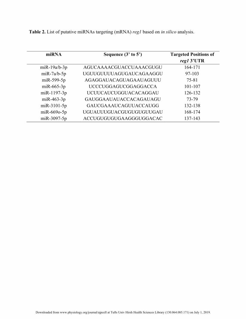
Table 2. List of putative miRNAs targeting (mRNA) reg1 based on in silico analysis.
miRNA Sequence (3’ to 5’) Targeted Positions of reg1 3’UTR
miR-19a/b-3p AGUCAAAACGUACCUAAACGUGU 164-171 miR-7a/b-5p UGUUGUUUUAGUGAUCAGAAGGU 97-103 miR-599-5p AGAGGAUACAGUAGAAUAGUUU 75-81 miR-665-3p UCCCUGGAGUCGGAGGACCA 101-107 miR-1197-3p UCUUCAUCUGGUACACAGGAU 126-132 miR-463-3p GAUGGAAUAUACCACAGAUAGU 73-79 miR-3101-5p GAUCGAAAUCAGUUACCAUGG 132-138 miR-669o-5p UGUAUUUGUACGUGUGUGUUGAU 168-174 miR-3097-5p ACCUGUGUGUGAAGGGUGGACAC 137-143
Downloaded from www.physiology.org/journal/ajpcell at Tufts Univ Hirsh Health Sciences Library (130.064.085.171) on July 1, 2019.
Downloaded from www.physiology.org/journal/ajpcell at Tufts Univ Hirsh Health Sciences Library (130.064.085.171) on July 1, 2019.

Downloaded from www.physiology.org/journal/ajpcell at Tufts Univ Hirsh Health Sciences Library (130.064.085.171) on July 1, 2019.
Downloaded from www.physiology.org/journal/ajpcell at Tufts Univ Hirsh Health Sciences Library (130.064.085.171) on July 1, 2019.
Downloaded from www.physiology.org/journal/ajpcell at Tufts Univ Hirsh Health Sciences Library (130.064.085.171) on July 1, 2019.

Downloaded from www.physiology.org/journal/ajpcell at Tufts Univ Hirsh Health Sciences Library (130.064.085.171) on July 1, 2019.


