
201
Medical Diagnostic ImagingMedical Diagnostic Imaging Laboratories
Lab Name Location Person in Charge Programs Served Courses Served
Patient Care and Management (2) W12-040 • Mawadah Mubarak • Medical Diagnostic
Imaging Dept.• Patient Care and
Management (2)
Introduction to MDI
W12-040W12-139M12-033
• Zarmeena Noorajan • Medical Diagnostic Imaging Dept. • Introduction to MDI
Radiographic Technique & Skeletal Radiography
W12-040M12-033
• Sura Majid• Asma Abdi
• Medical Diagnostic Imaging Dept.
• Skeletal Radiography• Skeletal Radiography CP• Radiographic Technique• Radiographic Technique
Clinical Practice
Medical Imaging Equipment (1)
W12-040M12-033 • Sura Majid • Medical Diagnostic
Imaging Dept.• Medical Imaging Equipment
(1)
Medical Imaging Equipment (2)
W12-040M12-033 • Dr. Entesar • Medical Diagnostic
Imaging Dept.• Medical Imaging Equipment
(2)
Medical Imaging & Processing & Diagnostic Imaging
W12-040M12-033 • Zarmeena Noorajan • Medical Diagnostic
Imaging Dept.
• Medical Imaging & Processing
• Diagnostic Imaging
Radiologic Anatomy
W12-040W12-139 • Asma Abdi • Medical Diagnostic
Imaging Dept. • Radiologic Anatomy
Radiologic Pathology& Image Interpretation
W12-040W12-139 • Mawadah Mubarak • Medical Diagnostic
Imaging Dept.• Radiologic Pathology &
Image Interpretation
Picture Archiving & Communication System
W12-139 • Mawadah Mubarak • Medical Diagnostic Imaging Dept.
• Picture Archiving Communication System
• Radiologic Pathology & Image Interpretation
• Radiologic Anatomy• Diagnostic Imaging
Ultrasonography M12-033 • Sura Majid • Medical Diagnostic Imaging Dept.
• Ultrasonography• Medical Imaging Clinical
Practice (2)
Computed Tomography W12-139 • Zarmeena Noorajan • Medical Diagnostic
Imaging Dept.
• Computed Tomography Medical Imaging Clinical Practice (1)
Magnetic Resonance Imaging
W12-139 • Asma Abdi • Medical Diagnostic Imaging Dept.
• Magnetic Resonance Imaging Medical Imaging Clinical Practice (2)

Cent
ral L
abor
ator
ies C
atal
og PATIENT CARE AND MANAGEMENT (2) LABORATORY
INTRODUCTIONThis Laboratory deals with taking care of the patient in the Medical Diagnostic Imaging Department including intravenous techniques, contrast agents and basic patient care procedures common at the Radiology Department. Departmental organization and issues of practice in an imaging department are examined together with medical-legal issues and local rules on regulation governing practices.
EQUIPMENT AND INSTRUMENTS • General X-Ray Machine• Portable X-Ray Machine• X-Ray Films and Cassettes• Whole Body Phantoms With Internal
Anatomy• Hand Washing Sink
• Stretcher• Transfer Board • Hydraulic Lift Machine• Wheel Chair• Stethoscope• Sphygmomanometer
EXPERIMENTS • Infection Control- Hand Washing Technique• Infection Control- Contact Precaution Technique• Aseptic Techniques• Pharmacology – Interpreting Medication Leaflet • Patient Transfer Techniques• Drug Administration• Vital Signs
TESTS AND SERVICES• Contact Precaution Awareness Lab for Infectious Disease in a Medical Imaging Setting

203
INTRODUCTION TO MEDICAL DIAGNOSTIC IMAGING LABORATORY
INTRODUCTION This course provides the foundation for all following MDI courses. It provides an insight into the field of Medical Imaging Technology, including plain radiography, ultrasound, computerized tomography, magnetic resonance imaging and radionuclide imaging. It provides the student with an understanding of the role of diagnostic imaging in health care, and an understanding of the place of modern medical diagnostic technology within the hospital health care team.
EQUIPMENT AND INSTRUMENTS • General X-Ray Machine• Portable X-Ray Machine• X-Ray Films and Cassettes• Whole Body Phantoms with Internal Anatomy• CR System• Dosimeter• Lead Shield
EXPERIMENTS • Orientation to MDI Labs• X-Ray Machine and Operating Steps • Imaging Accessories• Darkroom • MDI Department Workflow and Radiation Protection • Imaging Modalities • Basic Radiation Protection (Distance) • Basic Radiation Protection (Shielding) • Basic Radiation Protection (Time)

Cent
ral L
abor
ator
ies C
atal
og RADIOGRAPHIC TECHNIQUE & SKELETAL RADIOGRAPHY LABORATORY
INTRODUCTIONSkeletal Radiography Laboratory begins the practical experience in the fundamentals of practical radiography. It develops the skills necessary for the examination of patients and for producing actual radiographs. This course provides practical experience of the theory of Skeletal Radiography. Radiographic Technique completes the examination of the fundamentals of practical radiography. It continues development of the foundations of the skills necessary for the examination of patients, and for producing actual radiographs. This Laboratory provides practical experience for the theory of the axial skeleton and skull imaging technique. Students build technical skills as well as confidence in practicing radiography within the laboratory setting before engaging with real patients at clinical sites.
EQUIPMENT AND INSTRUMENTS • PACS (Picture Archiving and Communication System) with Post Processing and Viewing
System• General X-Ray Machine• General X-Ray Machine with Tomography • Portable X-Ray Machine• X-Ray Films and Cassettes• Automatic Film Processor• CR (Computed Radiography) System• Film Digitizer• Dry Film Processor• Whole Body Phantoms with Internal Anatomy• Viewing Boxes

205
EXPERIMENTS • Chest Radiography• Bony Thorax Radiography• Upper Extremities Radiography• Lower Extremities Radiography• Skull Radiography• Abdomen Radiography • Spine Radiography • Special Procedures Radiography
TESTS AND SERVICES• General X-Ray
o Chest o Bony Thorax
• Upper Extremities • Lower Extremities• Skull• Abdomen• Spine
Cent
ral L
abor
ator
ies C
atal
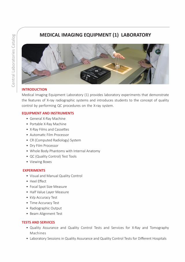
og MEDICAL IMAGING EQUIPMENT (1) LABORATORY
INTRODUCTIONMedical Imaging Equipment Laboratory (1) provides laboratory experiments that demonstrate the features of X-ray radiographic systems and introduces students to the concept of quality control by performing QC procedures on the X-ray system.
EQUIPMENT AND INSTRUMENTS • General X-Ray Machine• Portable X-Ray Machine• X-Ray Films and Cassettes• Automatic Film Processor• CR (Computed Radiology) System• Dry Film Processor• Whole Body Phantoms with Internal Anatomy• QC (Quality Control) Test Tools• Viewing Boxes
EXPERIMENTS • Visual and Manual Quality Control• Heel Effect • Focal Spot Size Measure• Half Value Layer Measure • kVp Accuracy Test• Time Accuracy Test • Radiographic Output • Beam Alignment Test
TESTS AND SERVICES• Quality Assurance and Quality Control Tests and Services for X-Ray and Tomography
Machines• Laboratory Sessions in Quality Assurance and Quality Control Tests for Different Hospitals

207
MEDICAL IMAGING EQUIPMENT (2) LABORATORY
INTRODUCTIONMedical Imaging Equipment Laboratory (2) introduces the student to the concept of quality control tests (QCs) in special medical imaging modalities including: CT, MRI, and PET. QC tests which are routinely carried out in clinics and medical centers to ensure that images produced are with high quality and within standard tolerance limits. In this lab, CT, MRI, and PET DICOM, images taken during monthly clinical QC with several accredited phantoms will be analyzed.
EQUIPMENT AND INSTRUMENTS:• DICOM Images of CT QC, MRI QC, and PET QC• DICOM Reader Software, OsiriX MD (Personal License)• Video Sessions for Setup Demonstration, Since the Lack of Accessibility to a CT, MRI or PET
Scanner• DICOM Images of the Following Accredited Phantoms:
a. NEMA Body Phantom SetTMb. Flanged Jaszczak ECT Phantomc. Respiratory Gated Body Phantom
EXPERIMENTS • CT QC:
a. Geometry Check b. Uniformity Checkc. Contrast to Noise Ratio (CNR) d. CT Number Calibratione. Head CT (Without a Contrast) Simulation Session with Ms. Asma Abdi.
• MRI QC:a. Geometry Check b. Ghost Artifactc. Contrast Noise Ratio d. Water vs. Copper Sulfate Contrast
• PET QC:a. Uniformity Checkb. Geometry Checkc. Sphere to Background Ration (SBR)

Cent
ral L
abor
ator
ies C
atal
og MEDICAL IMAGING AND PROCESSING & DIAGNOSTIC IMAGING LABORATORY
INTRODUCTIONThe Medical Imaging and Processing Laboratory introduces students to the photographic processes involved in the production of radiographs. Quality control issues are also examined. It involves practical work using the film processor and accessory equipment while applying the principles of film storage, safety and mixing in darkrooms.The Diagnostic Imaging Laboratory is a complete medical imaging and processing Laboratory. The contents are designed to impart an understanding of the components, principles and operation of digital imaging systems found in diagnostic radiology. Computed Radiography, Digital Radiography and Digital Dynamic Imaging, are introduced. Guidelines for selecting exposure factors and evaluating images within a digital system assist students in bridging between film-based and digital imaging systems. Principles of quality management including quality assurance and quality control are studied and carried out in the lab
EQUIPMENT AND INSTRUMENTS • PACS (Picture Archiving and Communication System) with Post Processing and Viewing
System• General X-Ray Machine• X-Ray Films and Cassettes• Automatic Film Processor• CR (Computed Radiology) System• DR (Digital Radiology) System• Dry Film Processor• Whole Body Phantoms with Internal Anatomy• QC (Quality Control) Test Tools• Viewing Boxes

209
EXPERIMENTS • Automatic Film Processor: Component and Function• Characteristic Curve• Darkroom Safelight Test• Image Artifact• The Air Gap Technique • The Effect of Heel Effect on Image Quality • Field Size: Effects on Density & Contrast • The Effect of kVp on Contrast• Introduction to Computed Radiography• Introduction to Digital Radiography• The Effect of Alignment on Shape Distortion• The Effect of Distance on Shape Distortion• Post Processing Images
TESTS AND SERVICES• Quality Assurance and Quality Control Tests and Services for X-Ray and Tomography
Machines• Laboratory Sessions in Quality Assurance and Quality Control Tests for Different Hospitals

Cent
ral L
abor
ator
ies C
atal
og RADIOLOGIC ANATOMY LABORATORY
INTRODUCTIONThis Laboratory relates images on radiographs, and other imaging modalities, to basic anatomical knowledge. It develops a fundamental understanding of medical imaging information relative to radiographic positioning. The Laboratory also provides an awareness of common anomalies that may be encountered in general radiographic practice.
EQUIPMENT AND INSTRUMENTS • PACS (Picture Archiving and Communication System) with Post Processing and Viewing
System• Viewing Boxes• Radiographic Images of the Whole Body in Different Modalities
EXPERIMENTS • Head Radiograph Reading• Neck Radiograph Reading• Thoracic Region Radiograph Reading• Abdominal Region Radiograph Reading• Pelvis Region Radiograph Reading
TESTS AND SERVICES• Second Opinion Consultation

211
RADIOLOGIC PATHOLOGY AND IMAGE INTERPRETATION LABORATORY
INTRODUCTIONThis Laboratory surveys medical and surgical diseases with emphasis placed on radiographic manifestations of disease processing and the alteration of radiographic techniques to compensate for the presence of disease.
EQUIPMENT AND INSTRUMENTS • PACS (Picture Archiving and Communication System) with Post processing and Viewing
System• Viewing Boxes• Radiographic Images of the Whole Body in Different Modalities
EXPERIMENTS • Respiratory System Pathology Image Reading• Respiratory System Pathology Image Reading• Alimentary Tract Pathology Image Reading• The Hepatobiliary System Pathology Image Reading• Genitourinary System Pathology Image Reading• Osseous System and Joints Pathology Image Reading• Neoplasia Pathology Image reading• Central Nervous System Pathology Image Reading• Osseous System and Joints Pathology Image Reading
TESTS AND SERVICES• Second Opinion Consultation

Cent
ral L
abor
ator
ies C
atal
og PICTURE ARCHIVING & COMMUNICATION SYSTEM LABORATORY
INTRODUCTIONThis Laboratory provides an introduction to Picture Archiving and Communication System (PACS) and its role in all of the medical imaging modalities. It also covers the fundamentals of computing, networking, DICOM, image acquisition, workflow, RIS, HIS, Image Compression, Digital Image visualization and Voice recognition. Images of different diagnostic modalities are archived and retrieved by viewing stations within the Laboratory or by any personal computers linked to the UOS LAN. In the practical sessions, students practice on PACS to fully grasp theory. Small projects for developing PACS and Tele-radiology are also carried out.
EQUIPMENT AND INSTRUMENTS • PACS (Picture Archiving and Communication System) with Post Processing and Viewing
System• CR (Computed Radiology) System• Film Digitizer• Dry Film Processor• On Line PACS (www.uospacs.com)
EXPERIMENTS • Binary Code – Hexadecimal • Digital Acquisition: CR • The Image File - Bandwidth • Image Workflow • Workstation Components • Workflow Profile • Storage Media • Image Processing• Visual Display Equipment • Image Compression• Acquisition Technologies • Digital Image Visualization
TESTS AND SERVICES• Digitizing Analog Films on CD • Copying Films • Copying Digital Images on CD/DVD

213
ULTRASONOGRAPHY LABORATORY
INTRODUCTIONThis Laboratory provides students with basic concepts and terminology as well as scanning protocols and techniques for the ultrasound examination of different body parts, together with sufficient practice to enable them to play a useful role in the health system. The Laboratory prepares students so that with further study they will be ready to acquire international recognition as registered ultrasound technologists. Students practice on life-size models as well as on each other to gain the skills needed to practice in hospitals.
EQUIPMENT AND INSTRUMENTS • Ultrasound Machine with a Doppler• Ultrasound Phantoms• Ultrasound Simulator
EXPERIMENTS • Breast Ultrasound• Abdomen Ultrasound• Thyroid Ultrasound• Doppler Ultrasound • Gynecology Ultrasound• Obstetric Ultrasound
TESTS AND SERVICES• General Ultrasound Scanning• BMD by Ultrasound (this service will be available in the near future)
Cent
ral L
abor
ator
ies C
atal
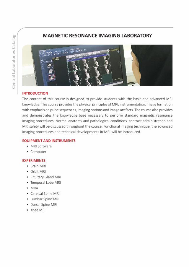
og MAGNETIC RESONANCE IMAGING LABORATORY
INTRODUCTION The content of this course is designed to provide students with the basic and advanced MRI knowledge. This course provides the physical principles of MRI, instrumentation, image formation with emphasis on pulse sequences, imaging options and image artifacts. The course also provides and demonstrates the knowledge base necessary to perform standard magnetic resonance imaging procedures. Normal anatomy and pathological conditions, contrast administration and MRI safety will be discussed throughout the course. Functional imaging technique, the advanced imaging procedures and technical developments in MRI will be introduced.
EQUIPMENT AND INSTRUMENTS • MRI Software • Computer
EXPERIMENTS • Brain MRI • Orbit MRI • Pituitary Gland MRI• Temporal Lobe MRI • MRA • Cervical Spine MRI • Lumbar Spine MRI • Dorsal Spine MRI • Knee MRI

215
COMPUTED TOMOGRAPHY LABORATORY
INTRODUCTIONWith the help of CT simulator software, the students will have clinical hands- on training for basic Computed Tomography clinical examinations within the classroom. This lab helps the students to understand the basic concepts related to the acquisition and scanning protocols. It will also enable the students to be introduced into a clinical scenario. Further, it helps them to understand how to screen a patient before undergoing a CT examination. With further practice and knowledge in the health care settings equip them to practice as a CT technologist.
EQUIPMENT • The Simulator Software
SIMULATED TRAINING ON:• CT Head• CT Chest• CT Neck• CT Abdomen & Pelvis• CT Lumbar Spine • CT Wrist


