Japanese MedakaJapanese Medaka
Mitotic ImagesMitotic Images
Embryonic DevelopmentEmbryonic Development
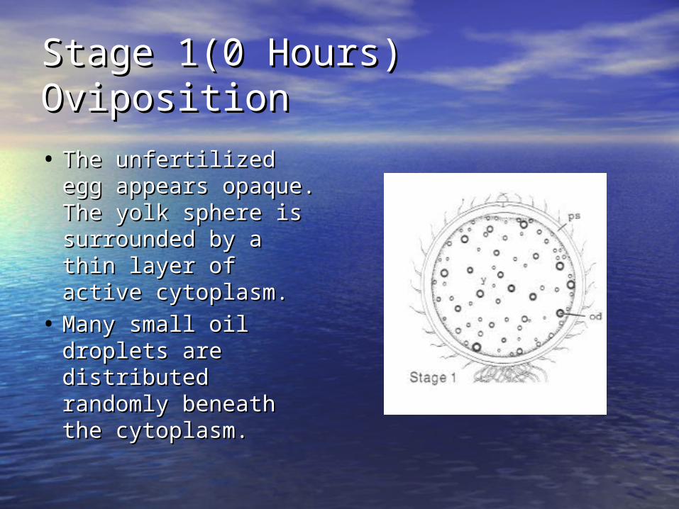
Stage 1(0 Hours)Stage 1(0 Hours)Oviposition Oviposition
• The unfertilized egg The unfertilized egg appears opaque. The appears opaque. The yolk sphere is yolk sphere is surrounded by a thin surrounded by a thin layer of active layer of active cytoplasm. cytoplasm.
• Many small oil droplets Many small oil droplets are distributed are distributed randomly beneath the randomly beneath the cytoplasm. cytoplasm.
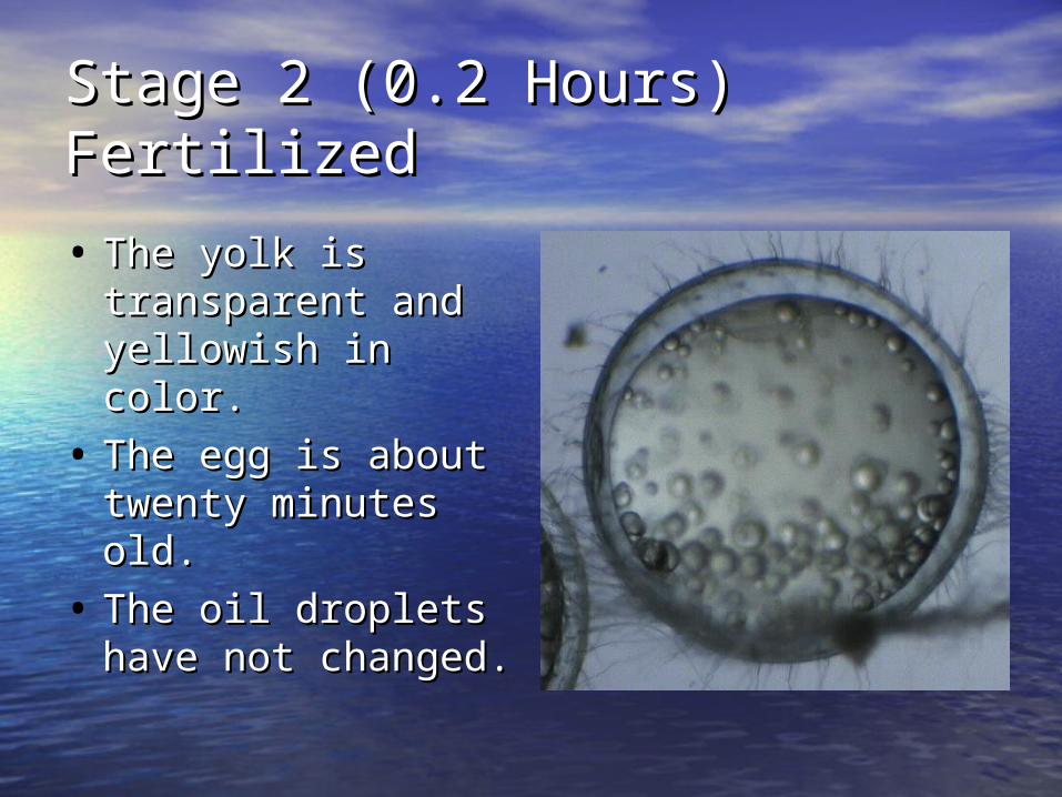
Stage 2 (0.2 Hours)Stage 2 (0.2 Hours)FertilizedFertilized
• The yolk is The yolk is transparent and transparent and yellowish in color.yellowish in color.
• The egg is about The egg is about twenty minutes twenty minutes old.old.
• The oil droplets The oil droplets have not changed.have not changed.
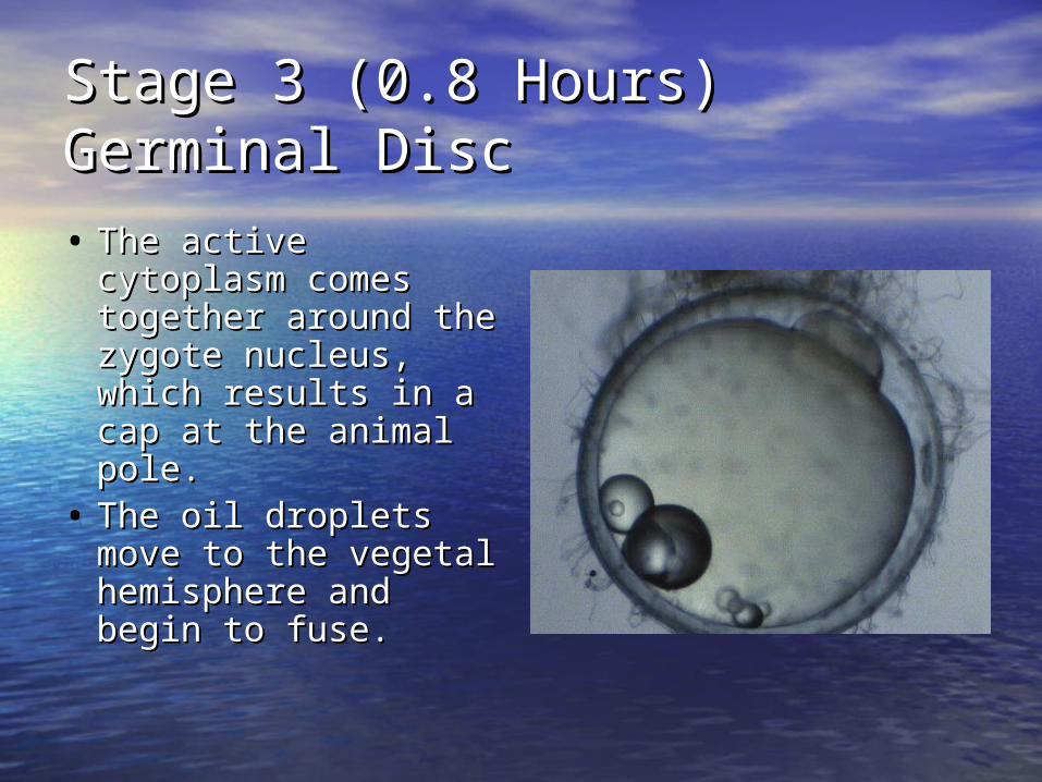
Stage 3 (0.8 Hours)Stage 3 (0.8 Hours)Germinal DiscGerminal Disc
• The active The active cytoplasm comes cytoplasm comes together around the together around the zygote nucleus, zygote nucleus, which results in a which results in a cap at the animal cap at the animal pole. pole.
• The oil droplets The oil droplets move to the vegetal move to the vegetal hemisphere and hemisphere and begin to fuse.begin to fuse.
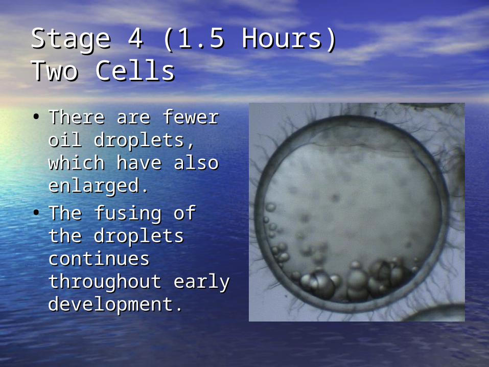
Stage 4 (1.5 Hours)Stage 4 (1.5 Hours)Two CellsTwo Cells
• There are fewer oil There are fewer oil droplets, which droplets, which have also enlarged.have also enlarged.
• The fusing of the The fusing of the droplets continues droplets continues throughout early throughout early development.development.
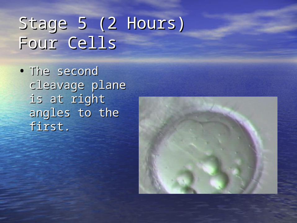
Stage 5 (2 Hours)Stage 5 (2 Hours)Four CellsFour Cells
• The second The second cleavage plane is cleavage plane is at right angles to at right angles to the first.the first.
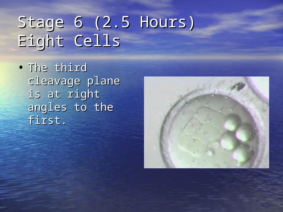
Stage 6 (2.5 Hours)Stage 6 (2.5 Hours)Eight CellsEight Cells
• The third cleavage The third cleavage plane is at right plane is at right angles to the first.angles to the first.
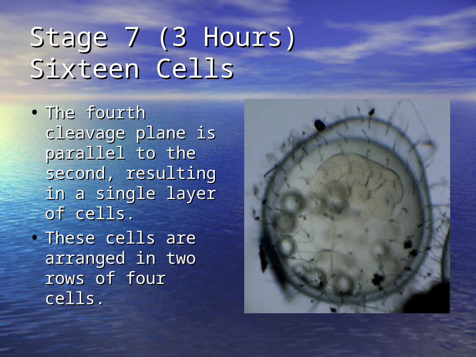
Stage 7 (3 Hours)Stage 7 (3 Hours)Sixteen CellsSixteen Cells
• The fourth The fourth cleavage plane is cleavage plane is parallel to the parallel to the second, resulting in second, resulting in a single layer of a single layer of cells.cells.
• These cells are These cells are arranged in two arranged in two rows of four cells.rows of four cells.
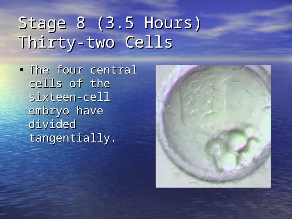
Stage 8 (3.5 Hours)Stage 8 (3.5 Hours)Thirty-two CellsThirty-two Cells
• The four central The four central cells of the sixteen-cells of the sixteen-cell embryo have cell embryo have divided divided tangentially.tangentially.
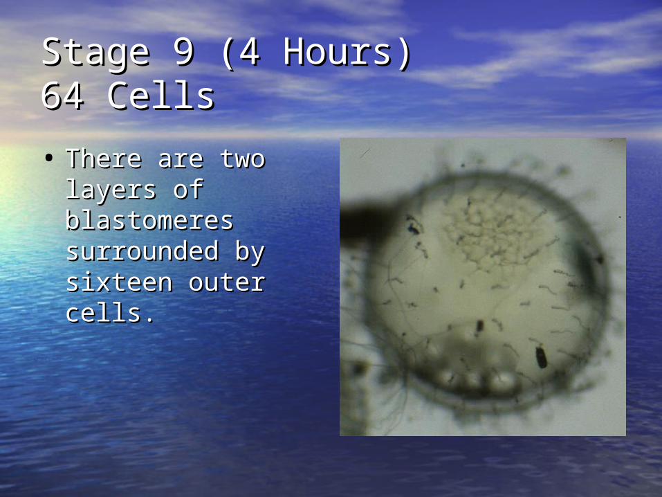
Stage 9 (4 Hours)Stage 9 (4 Hours)64 Cells64 Cells
• There are two There are two layers of layers of blastomeres blastomeres surrounded by surrounded by sixteen outer cells.sixteen outer cells.
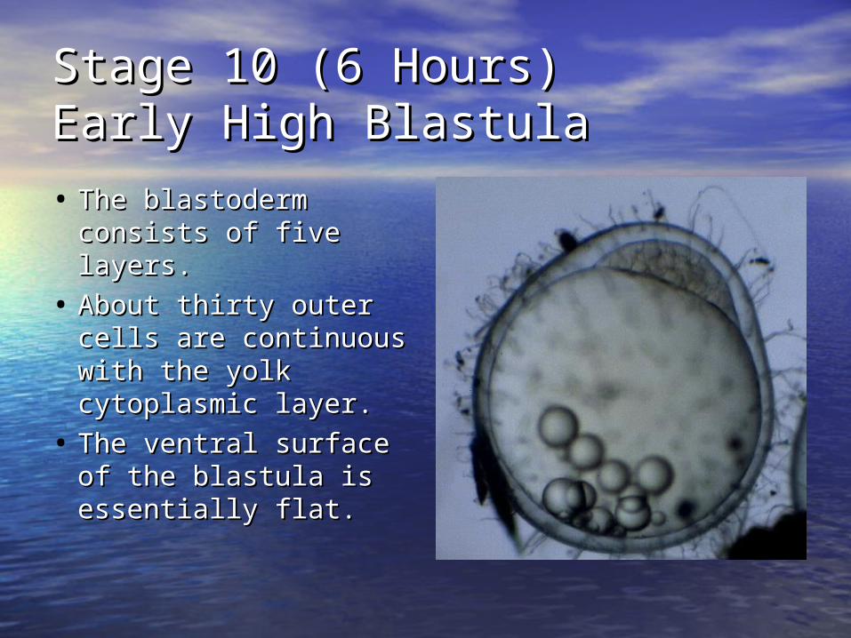
Stage 10 (6 Hours)Stage 10 (6 Hours)Early High BlastulaEarly High Blastula
• The blastoderm The blastoderm consists of five consists of five layers. layers.
• About thirty outer About thirty outer cells are continuous cells are continuous with the yolk with the yolk cytoplasmic layer. cytoplasmic layer.
• The ventral surface The ventral surface of the blastula is of the blastula is essentially flat.essentially flat.
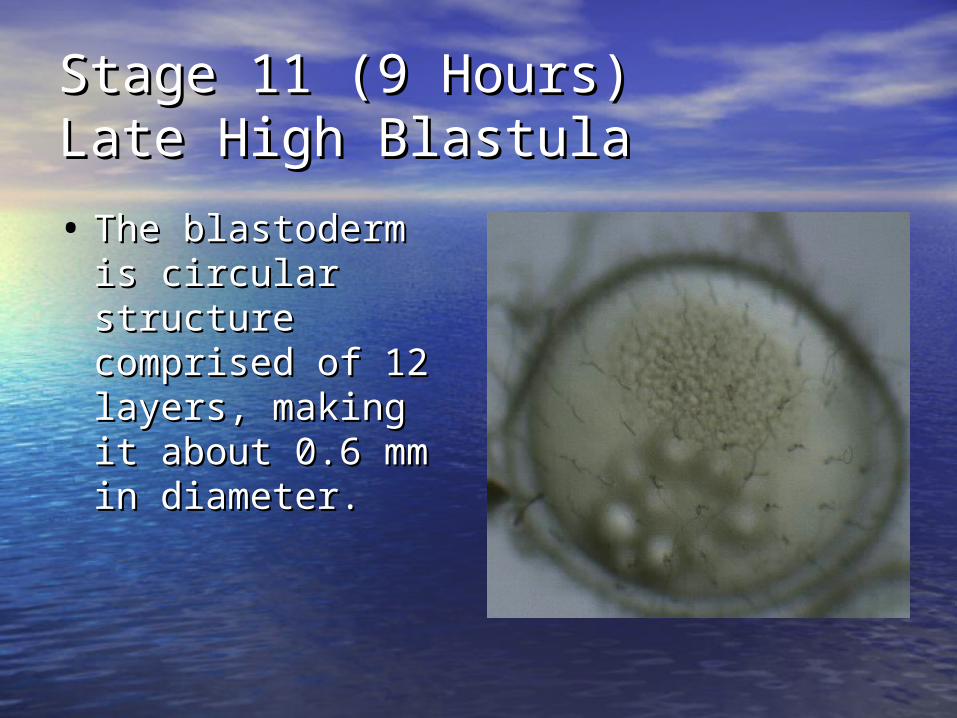
Stage 11 (9 Hours)Stage 11 (9 Hours)Late High BlastulaLate High Blastula
• The blastoderm is The blastoderm is circular structure circular structure comprised of 12 comprised of 12 layers, making it layers, making it about 0.6 mm in about 0.6 mm in diameter.diameter.
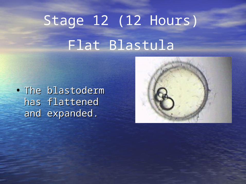
• The blastoderm The blastoderm has flattened and has flattened and expanded.expanded.
Stage 12 (12 Hours)
Flat Blastula
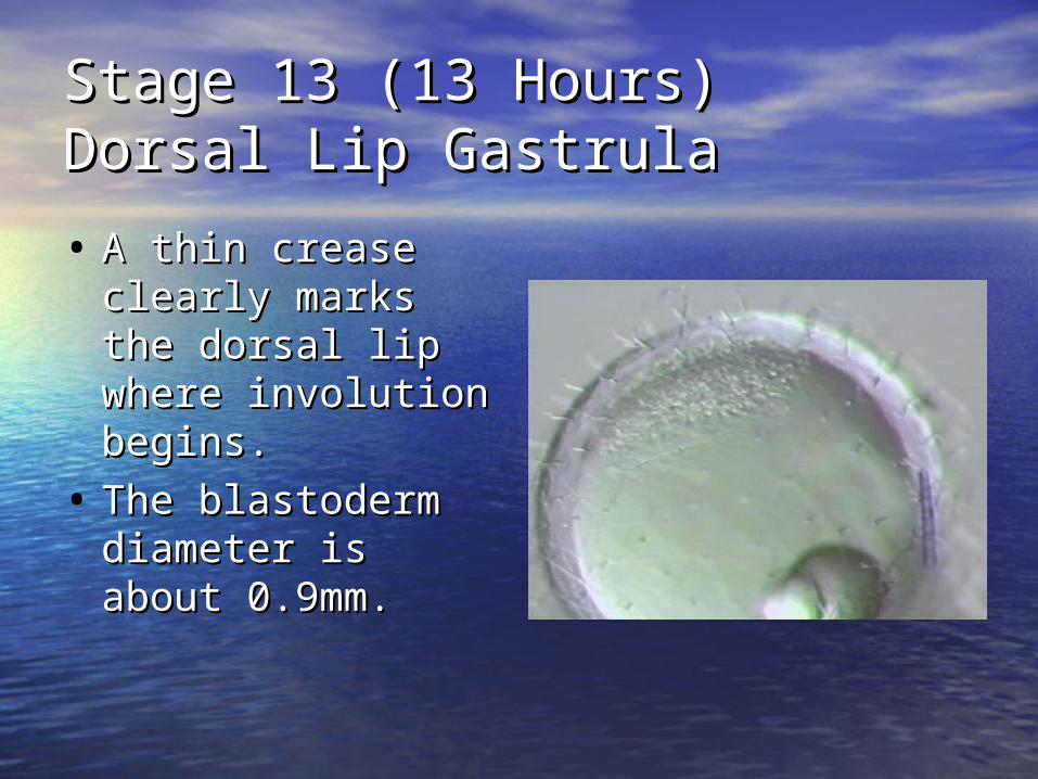
Stage 13 (13 Hours)Stage 13 (13 Hours)Dorsal Lip GastrulaDorsal Lip Gastrula
• A thin crease A thin crease clearly marks the clearly marks the dorsal lip where dorsal lip where involution begins. involution begins.
• The blastoderm The blastoderm diameter is about diameter is about 0.9mm.0.9mm.
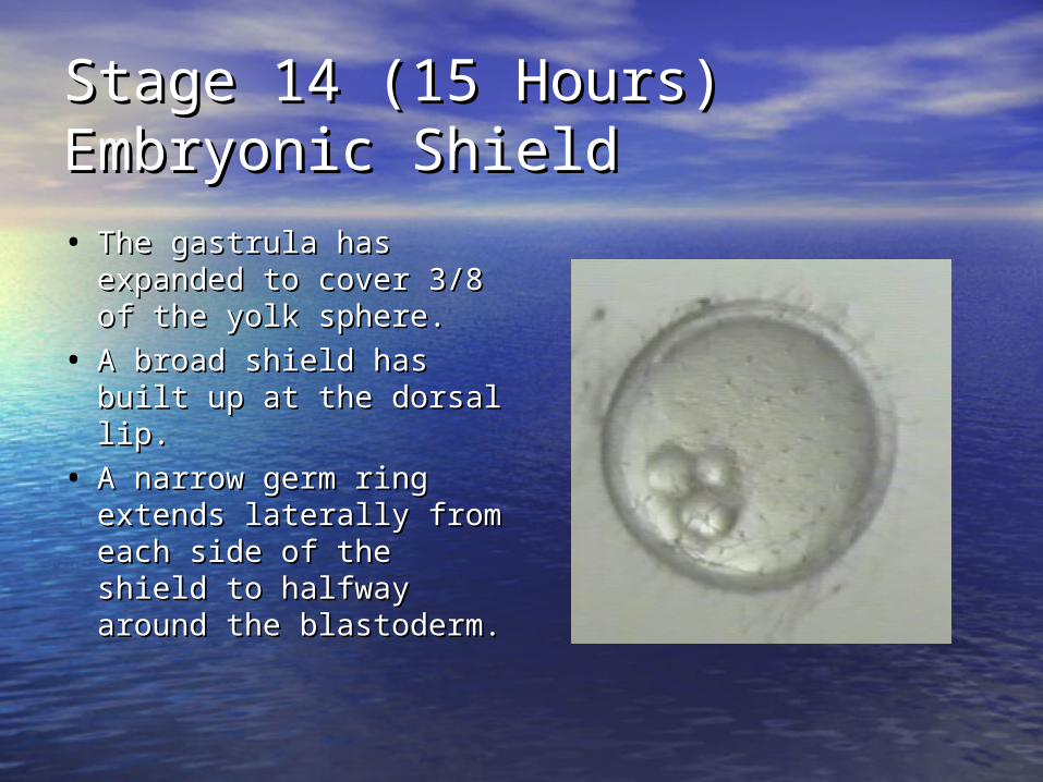
Stage 14 (15 Hours)Stage 14 (15 Hours)Embryonic ShieldEmbryonic Shield
• The gastrula has The gastrula has expanded to cover 3/8 expanded to cover 3/8 of the yolk sphere.of the yolk sphere.
• A broad shield has A broad shield has built up at the dorsal built up at the dorsal lip.lip.
• A narrow germ ring A narrow germ ring extends laterally from extends laterally from each side of the shield each side of the shield to halfway around the to halfway around the blastoderm.blastoderm.
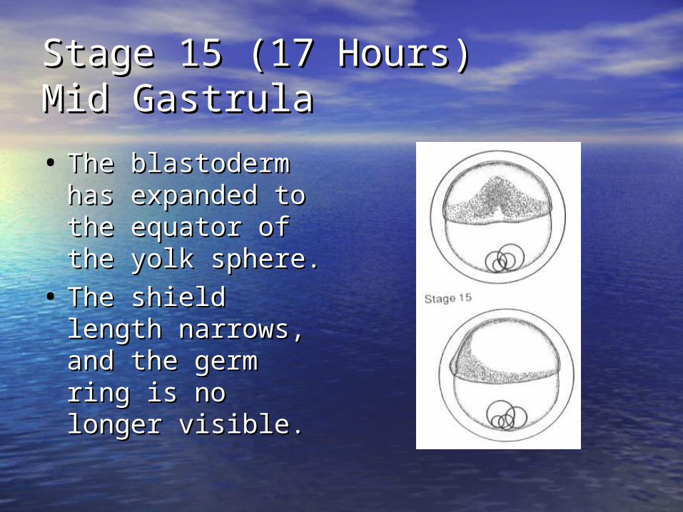
Stage 15 (17 Hours)Stage 15 (17 Hours)Mid GastrulaMid Gastrula
• The blastoderm The blastoderm has expanded to has expanded to the equator of the the equator of the yolk sphere.yolk sphere.
• The shield length The shield length narrows, and the narrows, and the germ ring is no germ ring is no longer visible.longer visible.
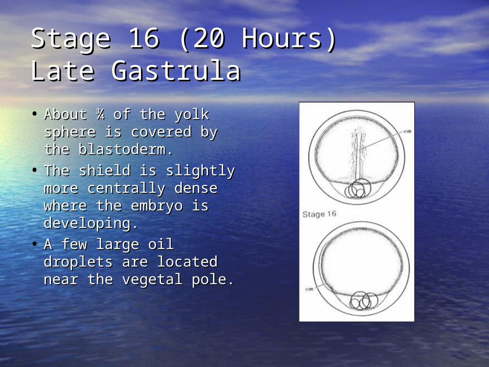
Stage 16 (20 Hours)Stage 16 (20 Hours)Late GastrulaLate Gastrula
• About ¾ of the yolk About ¾ of the yolk sphere is covered by sphere is covered by the blastoderm.the blastoderm.
• The shield is slightly The shield is slightly more centrally dense more centrally dense where the embryo is where the embryo is developing.developing.
• A few large oil A few large oil droplets are located droplets are located near the vegetal pole.near the vegetal pole.
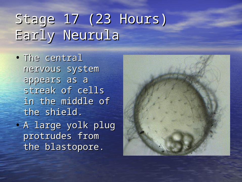
Stage 17 (23 Hours)Stage 17 (23 Hours)Early Neurula Early Neurula
• The central The central nervous system nervous system appears as a streak appears as a streak of cells in the of cells in the middle of the middle of the shield.shield.
• A large yolk plug A large yolk plug protrudes from the protrudes from the blastopore.blastopore.
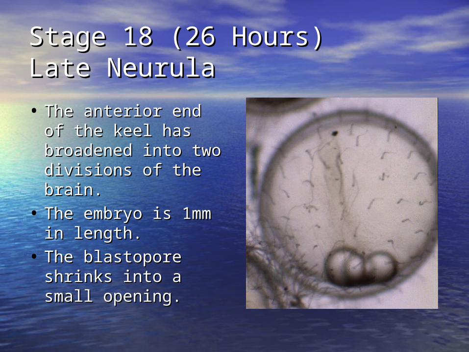
Stage 18 (26 Hours)Stage 18 (26 Hours)Late Neurula Late Neurula
• The anterior end of The anterior end of the keel has the keel has broadened into two broadened into two divisions of the divisions of the brain.brain.
• The embryo is 1mm The embryo is 1mm in length.in length.
• The blastopore The blastopore shrinks into a small shrinks into a small opening.opening.
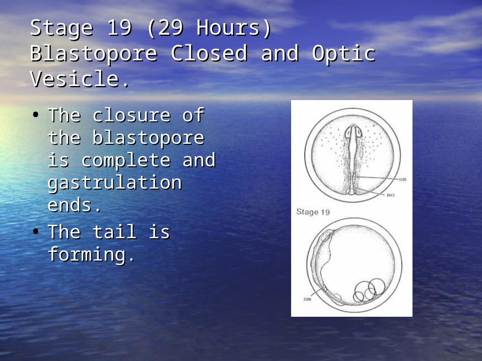
Stage 19 (29 Hours)Stage 19 (29 Hours)Blastopore Closed and Optic Vesicle.Blastopore Closed and Optic Vesicle.
• The closure of the The closure of the blastopore is blastopore is complete and complete and gastrulation ends.gastrulation ends.
• The tail is forming.The tail is forming.
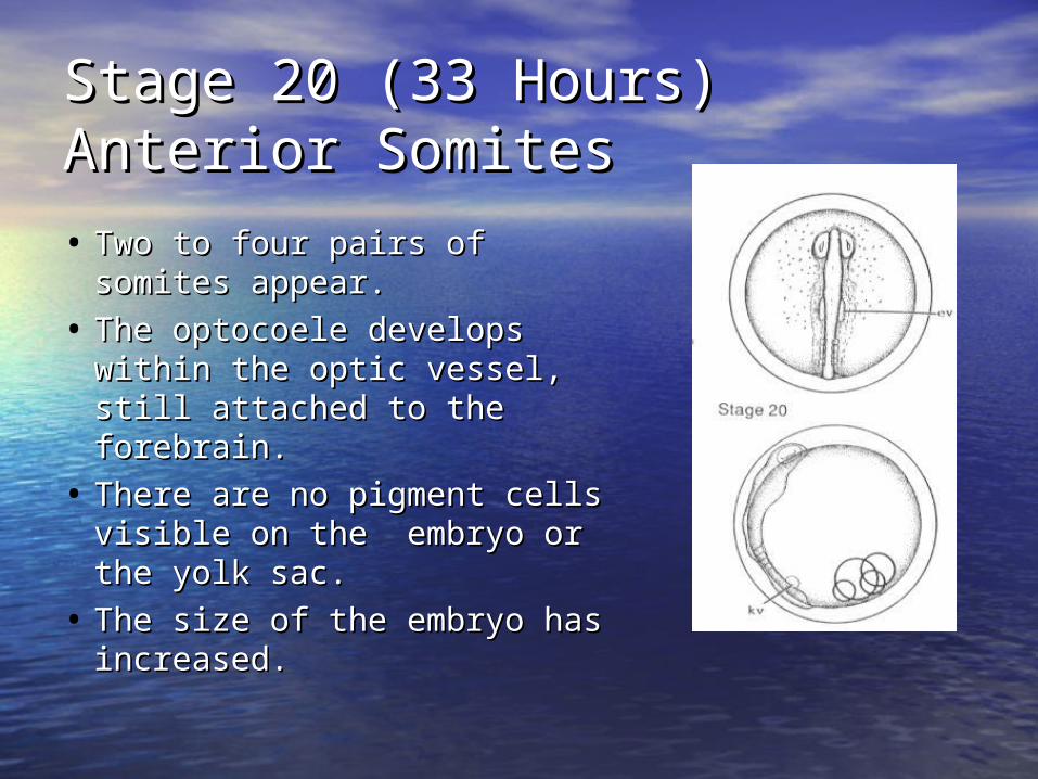
Stage 20 (33 Hours)Stage 20 (33 Hours)Anterior SomitesAnterior Somites
• Two to four pairs of somites Two to four pairs of somites appear. appear.
• The optocoele develops The optocoele develops within the optic vessel, still within the optic vessel, still attached to the forebrain.attached to the forebrain.
• There are no pigment cells There are no pigment cells visible on the embryo or the visible on the embryo or the yolk sac.yolk sac.
• The size of the embryo has The size of the embryo has increased.increased.
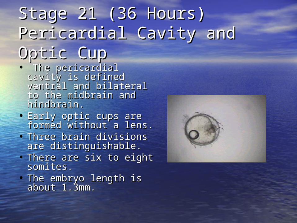
Stage 21 (36 Hours)Stage 21 (36 Hours)Pericardial Cavity and Optic Pericardial Cavity and Optic CupCup• The pericardial cavity is The pericardial cavity is
defined ventral and defined ventral and bilateral to the midbrain bilateral to the midbrain and hindbrain.and hindbrain.
• Early optic cups are Early optic cups are formed without a lens.formed without a lens.
• Three brain divisions are Three brain divisions are distinguishable.distinguishable.
• There are six to eight There are six to eight somites.somites.
• The embryo length is The embryo length is about 1.3mm.about 1.3mm.
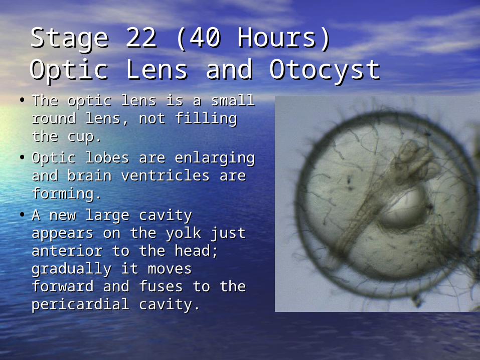
Stage 22 (40 Hours)Stage 22 (40 Hours)Optic Lens and OtocystOptic Lens and Otocyst
• The optic lens is a small The optic lens is a small round lens, not filling the round lens, not filling the cup.cup.
• Optic lobes are enlarging Optic lobes are enlarging and brain ventricles are and brain ventricles are forming.forming.
• A new large cavity appears A new large cavity appears on the yolk just anterior to on the yolk just anterior to the head; gradually it the head; gradually it moves forward and fuses to moves forward and fuses to the pericardial cavity.the pericardial cavity.
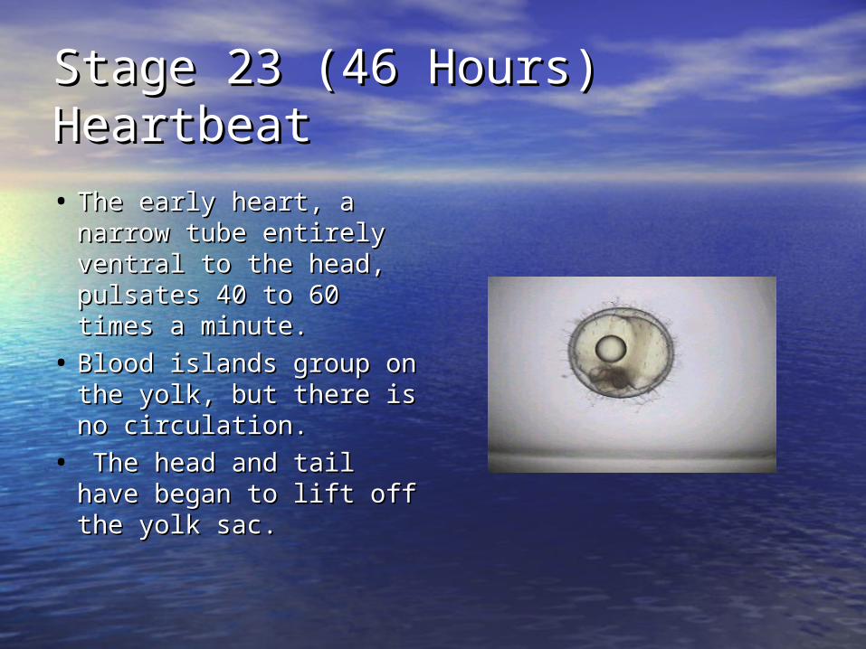
Stage 23 (46 Hours)Stage 23 (46 Hours)HeartbeatHeartbeat
• The early heart, a The early heart, a narrow tube entirely narrow tube entirely ventral to the head, ventral to the head, pulsates 40 to 60 times pulsates 40 to 60 times a minute. a minute.
• Blood islands group on Blood islands group on the yolk, but there is no the yolk, but there is no circulation.circulation.
• The head and tail have The head and tail have began to lift off the yolk began to lift off the yolk sac.sac.
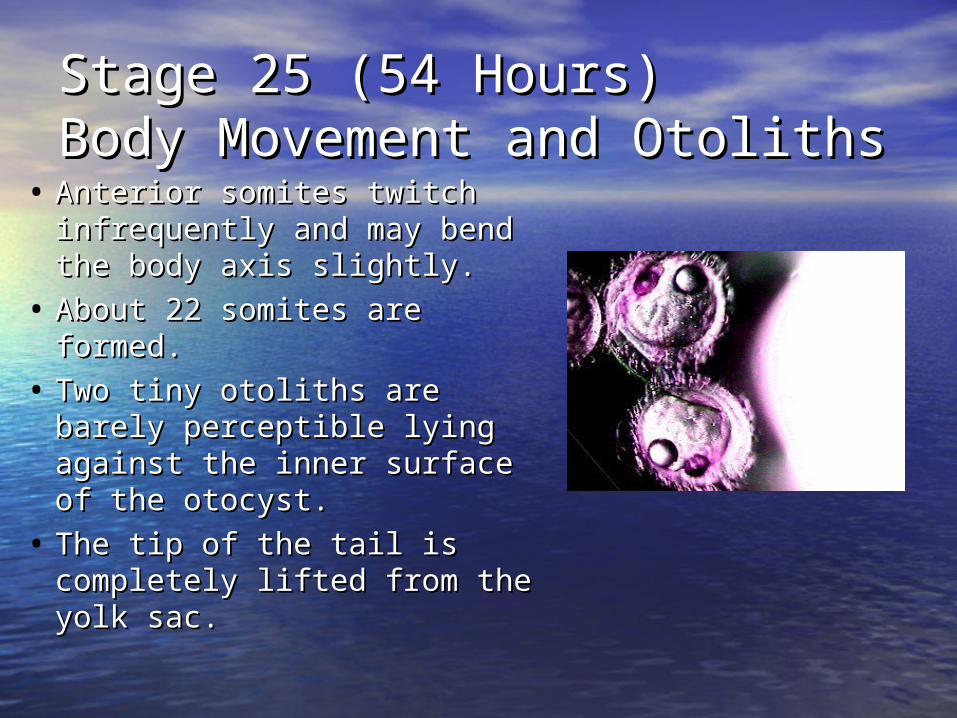
Stage 25 (54 Hours)Stage 25 (54 Hours)Body Movement and OtolithsBody Movement and Otoliths
• Anterior somites twitch Anterior somites twitch infrequently and may bend infrequently and may bend the body axis slightly.the body axis slightly.
• About 22 somites are formed.About 22 somites are formed.
• Two tiny otoliths are barely Two tiny otoliths are barely perceptible lying against the perceptible lying against the inner surface of the otocyst.inner surface of the otocyst.
• The tip of the tail is The tip of the tail is completely lifted from the completely lifted from the yolk sac.yolk sac.
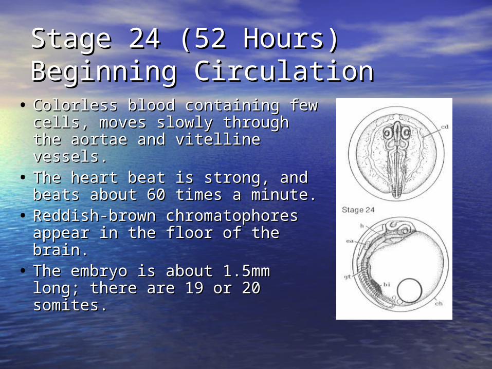
Stage 24 (52 Hours)Stage 24 (52 Hours)Beginning CirculationBeginning Circulation
• Colorless blood containing few Colorless blood containing few cells, moves slowly through the cells, moves slowly through the aortae and vitelline vessels.aortae and vitelline vessels.
• The heart beat is strong, and The heart beat is strong, and beats about 60 times a minute.beats about 60 times a minute.
• Reddish-brown chromatophores Reddish-brown chromatophores appear in the floor of the brain. appear in the floor of the brain.
• The embryo is about 1.5mm The embryo is about 1.5mm long; there are 19 or 20 somites.long; there are 19 or 20 somites.
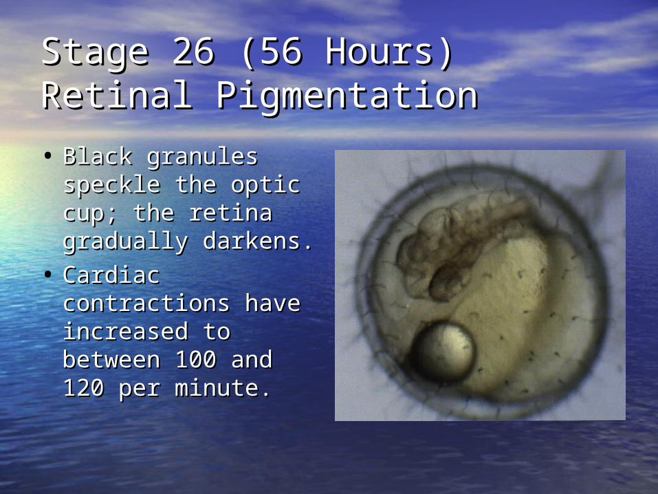
Stage 26 (56 Hours)Stage 26 (56 Hours)Retinal PigmentationRetinal Pigmentation
• Black granules Black granules speckle the optic speckle the optic cup; the retina cup; the retina gradually darkens.gradually darkens.
• Cardiac Cardiac contractions have contractions have increased to increased to between 100 and between 100 and 120 per minute.120 per minute.
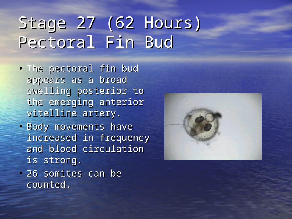
Stage 27 (62 Hours)Stage 27 (62 Hours)Pectoral Fin BudPectoral Fin Bud
• The pectoral fin bud The pectoral fin bud appears as a broad appears as a broad swelling posterior to the swelling posterior to the emerging anterior vitelline emerging anterior vitelline artery. artery.
• Body movements have Body movements have increased in frequency increased in frequency and blood circulation is and blood circulation is strong.strong.
• 26 somites can be 26 somites can be counted.counted.
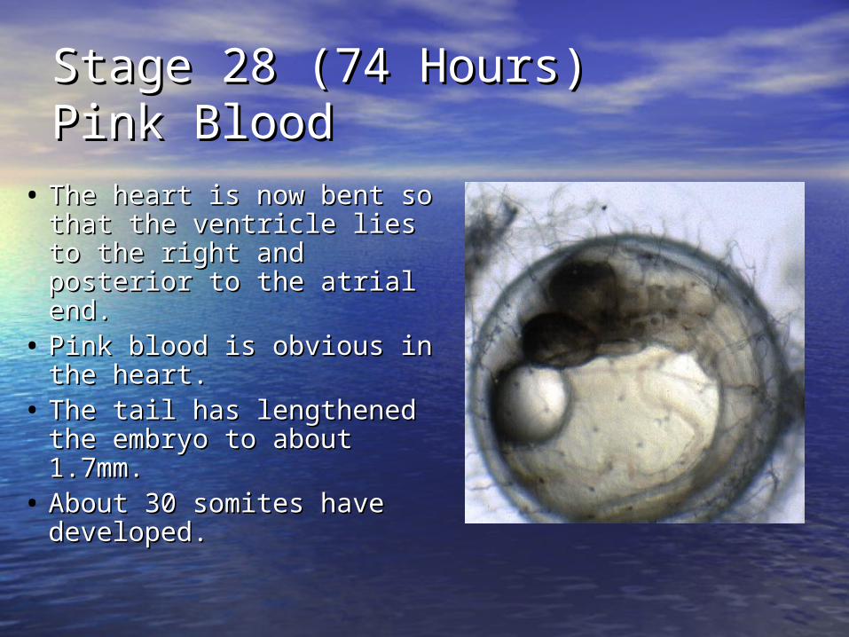
Stage 28 (74 Hours)Stage 28 (74 Hours)Pink BloodPink Blood
• The heart is now bent so The heart is now bent so that the ventricle lies to that the ventricle lies to the right and posterior to the right and posterior to the atrial end.the atrial end.
• Pink blood is obvious in Pink blood is obvious in the heart. the heart.
• The tail has lengthened The tail has lengthened the embryo to about the embryo to about 1.7mm.1.7mm.
• About 30 somites have About 30 somites have developed.developed.
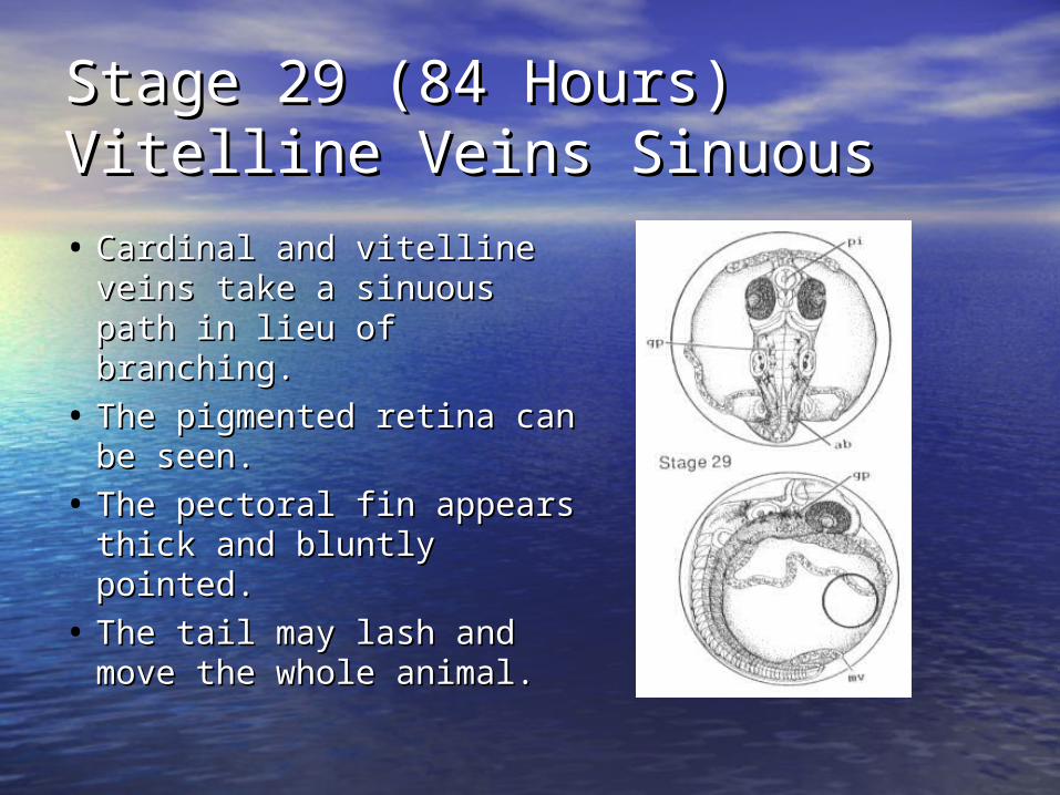
Stage 29 (84 Hours)Stage 29 (84 Hours)Vitelline Veins SinuousVitelline Veins Sinuous
• Cardinal and vitelline Cardinal and vitelline veins take a sinuous veins take a sinuous path in lieu of branching.path in lieu of branching.
• The pigmented retina The pigmented retina can be seen.can be seen.
• The pectoral fin appears The pectoral fin appears thick and bluntly thick and bluntly pointed. pointed.
• The tail may lash and The tail may lash and move the whole animal.move the whole animal.
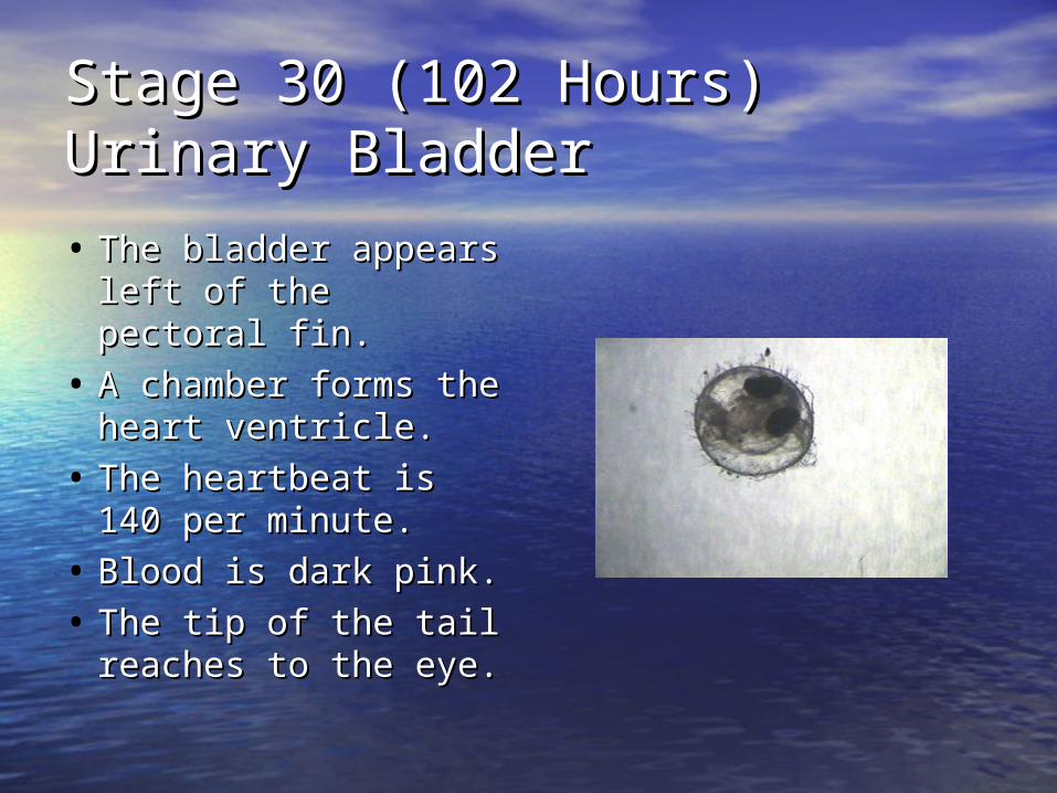
Stage 30 (102 Hours)Stage 30 (102 Hours)Urinary BladderUrinary Bladder
• The bladder appears The bladder appears left of the pectoral left of the pectoral fin.fin.
• A chamber forms the A chamber forms the heart ventricle.heart ventricle.
• The heartbeat is 140 The heartbeat is 140 per minute.per minute.
• Blood is dark pink.Blood is dark pink.
• The tip of the tail The tip of the tail reaches to the eye.reaches to the eye.
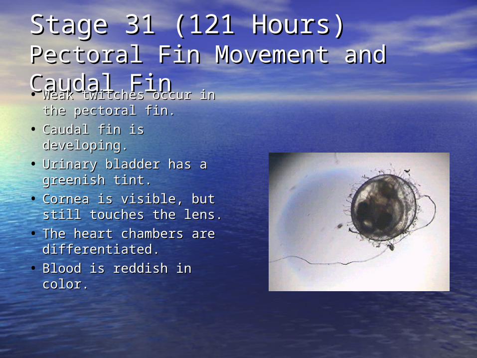
Stage 31 (121 Hours)Stage 31 (121 Hours)Pectoral Fin Movement and Caudal Pectoral Fin Movement and Caudal FinFin• Weak twitches occur in Weak twitches occur in
the pectoral fin.the pectoral fin.
• Caudal fin is Caudal fin is developing.developing.
• Urinary bladder has a Urinary bladder has a greenish tint.greenish tint.
• Cornea is visible, but Cornea is visible, but still touches the lens.still touches the lens.
• The heart chambers are The heart chambers are differentiated.differentiated.
• Blood is reddish in color.Blood is reddish in color.
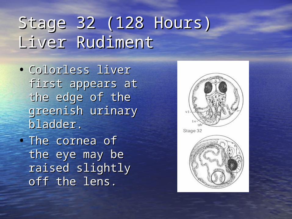
Stage 32 (128 Hours)Stage 32 (128 Hours)Liver RudimentLiver Rudiment
• Colorless liver first Colorless liver first appears at the appears at the edge of the edge of the greenish urinary greenish urinary bladder. bladder.
• The cornea of the The cornea of the eye may be raised eye may be raised slightly off the lens.slightly off the lens.
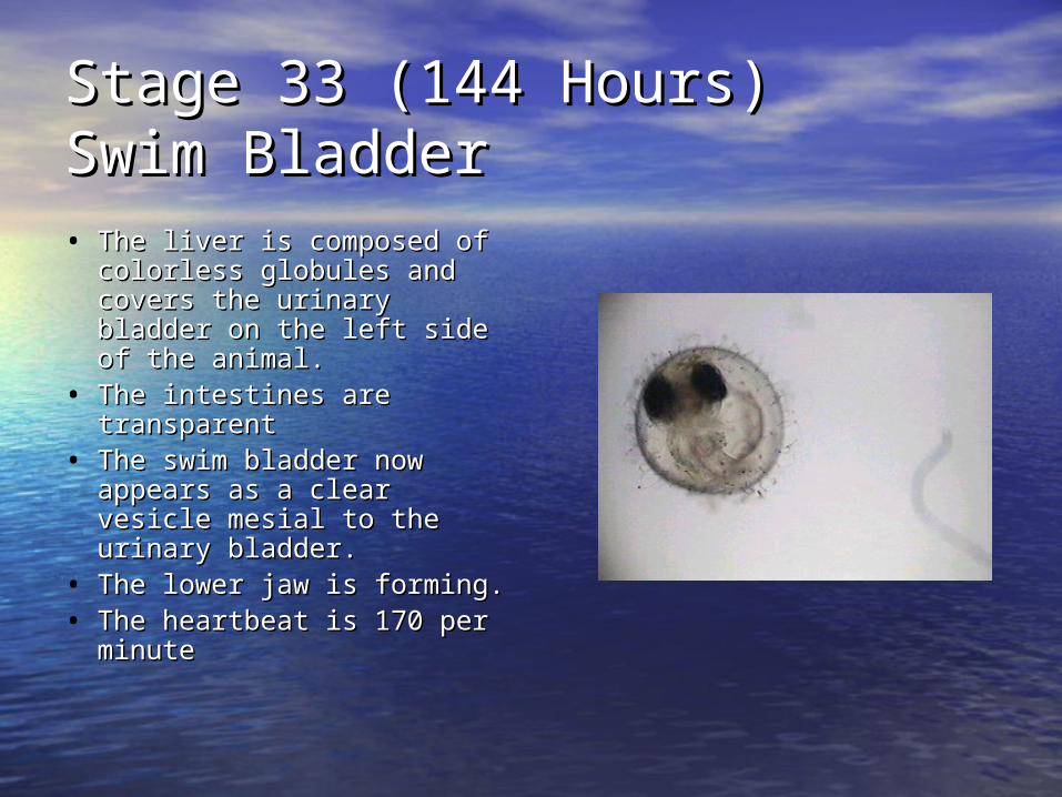
Stage 33 (144 Hours)Stage 33 (144 Hours)Swim BladderSwim Bladder• The liver is composed of The liver is composed of
colorless globules and colorless globules and covers the urinary bladder covers the urinary bladder on the left side of the on the left side of the animal.animal.
• The intestines are The intestines are transparenttransparent
• The swim bladder now The swim bladder now appears as a clear vesicle appears as a clear vesicle mesial to the urinary mesial to the urinary bladder. bladder.
• The lower jaw is forming. The lower jaw is forming. • The heartbeat is 170 per The heartbeat is 170 per
minuteminute
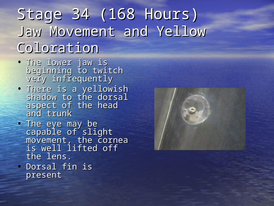
Stage 34 (168 Hours)Stage 34 (168 Hours)Jaw Movement and Yellow Jaw Movement and Yellow ColorationColoration• The lower jaw is The lower jaw is
beginning to twitch beginning to twitch very infrequentlyvery infrequently
• There is a yellowish There is a yellowish shadow to the dorsal shadow to the dorsal aspect of the head aspect of the head and trunkand trunk
• The eye may be The eye may be capable of slight capable of slight movement, the cornea movement, the cornea is well lifted off the is well lifted off the lens.lens.
• Dorsal fin is presentDorsal fin is present
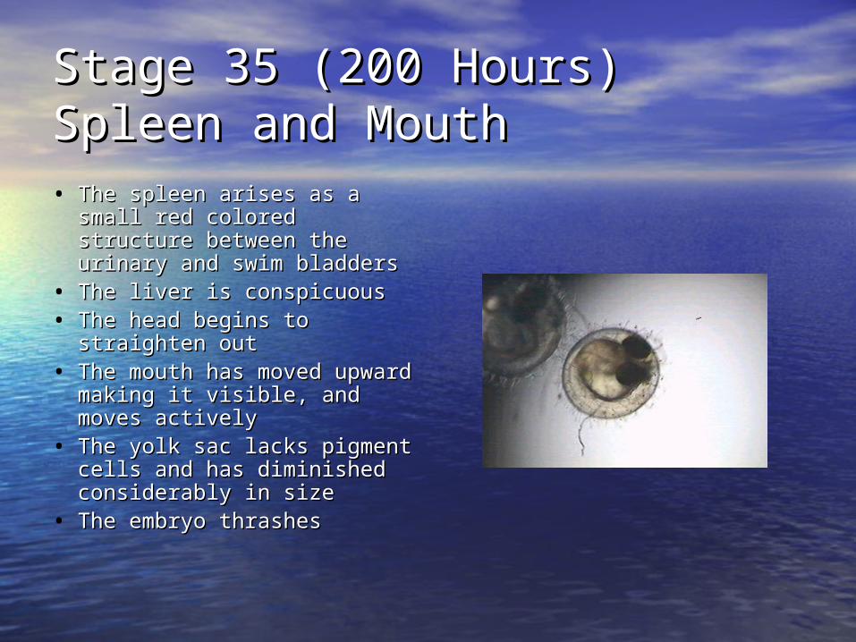
Stage 35 (200 Hours)Stage 35 (200 Hours)Spleen and MouthSpleen and Mouth• The spleen arises as a small The spleen arises as a small
red colored structure red colored structure between the urinary and between the urinary and swim bladdersswim bladders
• The liver is conspicuous The liver is conspicuous • The head begins to The head begins to
straighten outstraighten out• The mouth has moved The mouth has moved
upward making it visible, upward making it visible, and moves activelyand moves actively
• The yolk sac lacks pigment The yolk sac lacks pigment cells and has diminished cells and has diminished considerably in sizeconsiderably in size
• The embryo thrashes The embryo thrashes
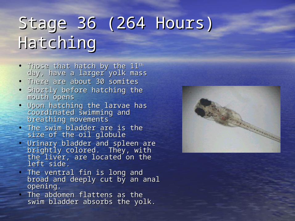
Stage 36 (264 Hours)Stage 36 (264 Hours)HatchingHatching• Those that hatch by the 11Those that hatch by the 11thth day, day,
have a larger yolk masshave a larger yolk mass• There are about 30 somitesThere are about 30 somites• Shortly before hatching the Shortly before hatching the
mouth opensmouth opens• Upon hatching the larvae has Upon hatching the larvae has
coordinated swimming and coordinated swimming and breathing movements breathing movements
• The swim bladder are is the size The swim bladder are is the size of the oil globuleof the oil globule
• Urinary bladder and spleen are Urinary bladder and spleen are brightly colored. They, with the brightly colored. They, with the liver, are located on the left side. liver, are located on the left side.
• The ventral fin is long and broad The ventral fin is long and broad and deeply cut by an anal and deeply cut by an anal opening. opening.
• The abdomen flattens as the The abdomen flattens as the swim bladder absorbs the yolk. swim bladder absorbs the yolk.
Caring for the Young Medaka larvae begin to feed the following
day of hatching The aquarium must be “baby proofed”, the
filters must be shut off to prevent injury and the plants at the bottom of the tank need to be good oxygenators.
The water temperature should be 16-28°C The fry must be fed protozoa for at least the
first 7 days and newly hatched brine shrimp for the next 2 weeks.
Finely chopped white worms and tropical fish food may be fed sparingly to the 3 week old Medaka fry.

DO NOT OVERFEED!!! When fed wisely and not overcrowded,
Medaka young grow 15mm in length within 4-6 weeks
They may now be cared for like adults Newly hatched fry are always in danger of
being eaten by older fry Medakas mature in 2 to 6 months They have a life span of four or more years
Helpful Hints

What We Thought About the Lab!!
Jody Manners, Tracy Rhodes, and Alex Smith surveyed 25 people to see what they thought about the Medaka lab.
76% thoroughly enjoyed the lab 16% mostly enjoyed the lab 8% enjoyed only certain parts of
the lab
Our Opinion on the Lab
Throughout this lab we have learned many things about developing embryos. This experience helped to complete our understanding of this critical topic. Using the laptops and microscopes was very rewarding and helped us learn first hand.

Credits Alex Smith, Tracy Rhodes, and Jody Manners Animations from Medakafish Homepage at
http://biol1.bio.nagoya-u.ac.jp:8000.html Clarion University Karen Anderson, Clarion University Mrs. Maine Mrs. Maine’s and Mrs. Wolfgang’s Academic
Biology Classes Pictures from BioG 101-104,
http://biog-101-104.bio.cornell.edu/


