Download - ISOLATION, PURIFICATION, SEROLOGY AND NATURE

ISOLATION, PURIFICATION, SEROLOGY AND NATUREOF ROSE MOSAIC VIRUS
by
ROBERT S. HALLIWELL
A THESIS
submitted to
OREGON STATE UNIVERSITY
in partial fulfillment ofthe requirements for the
degree of
DOCTOR OF PHILOSOPHY
June 1962

APPROVED;
Professor of Botany and Plant Pathology
In Charge of Major
lairmaiy of Department of Botrai0 (7 <7
Chairman of School Graduate Committee
i of Graduate SchoolODean
Date thesis is presented May 16, 1962
Typed by Claudia Annis
Redacted for privacy
Redacted for privacy
Redacted for privacy
Redacted for privacy

ACKNOWLEDGEMENT
The author wishes to express his gratitude to Dr. J. A. Milbrath
for his encouragement and guidance throughout the course of this
investigation and to Dr. R. E. Ford for his advice and assistance in
the serological studies.
Thanks are also due to Dr. F. H. Smith, Dr. R. A. Young,
Dr. I. W. Deep, and Dr. C. H. Wang for their helpful criticism and
advice in preparing this manuscript. He is grateful to H. H. Millsap
for taking the pictures, and J. D. Newstead for the electron micro
graphs used in this thesis.
The writer expresses his appreciation to Dr. R. W. Fulton of
the Plant Pathology Department of the University of Wisconsin for
supplying his isolate of rose mosaic virus for this study.
This project was made possible by support from the Oregon Bulb,
Florist and Nursery Council.
TABLE OF CONTENTS
Page
Introduction 1
Review of Literature 3
Materials and Methods 10
I. Plant inoculation technique 10II. Plant culture 10
Results 11
I. Isolation of rose mosaic virus of rose, 11
A. Attempted mechanical transmission of rosemosaic virus 11
B. Virus inhibitors in rose tissue extracts 11
C. Isolation of rose mosaic by graft transmissionto V. rosa and peach 12
D. Comparison of rose mosaic virus isolates andtomato ring spot type culture on herbaceoushosts 13
i. Chenopodium amaranticolor 14ii. Vigna sinensis, black cowpea. 14
iii. Phaseolus vulgaris, varieties Bountiful
and Sutter' s Pink 17
iv. Cucumis sativus, variety ChicagoPickling 19
II. Prunus ring spot virus in rose 19
A. Prevalence of Prunus ring spot virus in rose 19B, Transmission of Prunus ring spot virus from
rose to herbaceous hosts 20
III. Survey for rose virus indicator hosts 20IV. Purification of rose mosaic virus 21
A. Selection of host and assay plants 21B. Purification procedure. 21
V. Ultraviolet absorption of rose mosaic virus 25VI. Electron microscopy of rose mosaic virus 27
VII. Serological study of rose mosaic virus 30
A. Preparation of antigen 30B. Intmunization procedure 30C. Serological tests 30
TABLE OF CONTENTS
(continued)
Page
D. Serological relationship of rose mosaic virus.... 32
i. Preparation of the antigen.... 32ii. Immunization procedure 32
iii. Serological tests 34
E. Cross-precipitation tests 34
Discussion 36
Summary 41
Bibliography 43
LIST OF FIGURES
Figure Page
1 Typical rose mosaic in some hybrid tea roses... 15
2 Comparison of local lesions caused by rose mosaicisolates and tomato ring spot virus type culture onChenopodium amaranticolor 16
3 Comparison of systemic symptoms of some rose mosaicvirus isolates and tomato ring spot virus ontrifoliate leaves of black cowpea 18
4 Outline of the procedure used in purification of rosemosaic virus isolate F-l 24
5 Twenty-five to fifty percent sucrose densitygradient column of preparations from healthy cowpeasand from cowpeas infected with rose mosaic isolateF-l 26
6 Ultraviolet absorption spectra of componentsseparated from partially purified rose mosaic virusby density gradient centrifugation 28
7 Electron micrograph of particles occurring in thetwo zones of a density gradient column of a rosemosaic virus preparation which were not observedin healthy plant preparations , 29
LIST OF TABLES
Table Page
I Immunization schedule used in the preparation ofrose mosaic virus antiserum 31
II Immunization schedule used in the preparation oftomato ring spot antiserum 33
III Titer of tomato ring spot antiserum reaction withhomologue and rose mosaic virus isolate F-l inmicro-precipitin tests „ 35
ISOLATION, PURIFICATION, SEROLOGY AND NATUREOF ROSE MOSAIC VIRUSES
INTRODUCTION
The incidence of virus diseases of roses appears to be increasing
in the United States, probably because of the methods used to propagate
the great number of rose varieties affected. Numerous complaints
regarding diseased stock, both foreign and domestic, attest to the
seriousness of the problem. Growers in many areas are faced with the
choice of investing in a certification program that would guarantee
disease-free stock or having their plants quarantined.
Four different rose viruses have been reported in the United
States. Common rose mosaic, rose yellow mosaic and rose streak are the
most common, and rosette of rose or witches broom, which is more de
structive than the mosaics, has been reported occasionally.
Surveys for rose mosaic in Oregon rose nurseries, and in test
gardens containing roses from all parts of the United States, showed
that most plants had symptoms suggestive of virus infection. Most
infected varieties displayed symptoms on the late spring and early
summer foliage, and some symptomless varieties were assayed and found to
harbor a virus in a latent condition. Many rootstocks have become
infected by the practice of cutting rootstock materials from plants in
nursery rows which were previously budded with infected varieties, and
many new varieties have been propagated on such stock.
The effect of rose mosaic and other viruses on the general vigor,
longevity, hardiness, flower production, and color stability has not
been reported. The high percentage of virus infection in roses, the

frequent latent characteristic of the virus in some varieties, and a
lack of a reliable assay method, makes the selection of disease-free
stock extremely difficult. Thermal inactivation of several viruses in
living plants has now been demonstrated and may provide a method for
the cure of virus infected rose plants. However, the results of heat
inactivation studies would be difficult to evaluate if no virus assay
methods are available. Before disease-free roses can be selected
reliable assay methods must be developed and a certification or
cleanup program initiated.
Viruses, suspected of being rose mosaic, have been transmitted
from rose to herbaceous hosts, but the methods of transmission used
were unreliable and time consuming. Identification of rose viruses
has been difficult because of the variability obtained in results of
transmission studies, and erroneous conclusions sometimes drawn. A
reliable technique for the transmission of the rose viruses to her
baceous hosts would also facilitate a study of their physical and
biological properties. A technique for the purification of rose
mosaic virus has not been reported, consequently this virus has not
been characterized by electron microscopy, by spectrophotometry or by
serological methods.
This study was initiated to develop methods to recognize, isolate
and study the viruses which infect rose and determine their effect on
rose plants.

REVIEW OF LITERATURE
The first published records of a virus-like disease of rose in
the United States was by Taubenhaus in 1923 (37) and 1925 (38). He
described a mosaic symptom of rose in Texas, but no experiments were
initiated to determine the true cause of the disease. Rose mosaic
was first described as a virus disease by White in 1928 (45), and
named "Infectious Chlorosis." Surveys for rose mosaic in commercial
rose plantings throughout the United States, revealed a high percent
age of affected plants (28, 46, 47, 48). When evidence of the wide
spread nature of rose mosaic was presented to the rose growers
(33, 48, 49), resolutions were adopted to investigate the possibility
of quarantine action. Furthermore, believing European rootstocks
virus free, the growers resolved to lift import embargos (53). White
(50, p. 50a) rejected these resolutions as an answer to the problem,
and pointed out that "the source of the disease does not rest solely
with the stock upon which forcing varieties are budded. The budwood
itself is just as serious a carrier as the stock." He advocated
inspection and certification of budwood as well as rootstocks.
Milbrath (32, 33) believed that many of the survey reports on
the prevalence of rose mosaic could have been cases of mistaken
identity. He maintained that insect damage, nutritional disorders
and genetic disturbances were often mistaken for rose mosaic. He
also questioned the authenticity of the disease, since infected buds
grafted to healthy roses did not always produce symptoms on the host,
or at least not the same type symptom. He postulated that if rose
mosaic were transmitted by grafting, it should become systemic, and
all the leaves should subsequently show symptoms. He also noted that
diseased field-grown roses did not show symptoms after growing for a
period in a greenhouse.
In response to Milbrath's skepticism, White (51) in 1932,
effectively segregated the rose mosaic syndrome from non-pathological
disorders with similar symptoms. Chlorotic conditions of rose caused
by common rose insects, genetic disturbances and nutritional dis
orders, were compared to those caused by rose mosaic virus. Rose
mosaic was proven to be an authentic virus disease.
The occurrence of rose mosaic in American grown rootstocks was
determined by Brierley and Smith (3, 4). Between the years 1929 and
1935, they indexed rootstocks on the rose variety Mme. Butterfly. No
virus was detected in 103 plants of Texas Wax, or 27 of Multiflora,
and only in one plant of 21 Odorata and 3 plants of 208 Ragged Robin
rootstocks. Approximately 15 percent of 804 Manetti rootstocks were
found to be infected. These figures were considerably lower than
those reported from field surveys (28, 52).
Investigations of rose viruses by various American workers
indicated that there were three basic rose viruses, 1) rose mosaic,
2) rose yellow aosaic and 3) rose streak. This compilation, recorded
in the Review of Applied Mycology (7, p. 45-46), favors the classi
fication proposed by Brierley et al. (3). Some of the numerous
diseases originally described were concluded to be synonymous.

Infectious chlorosis, veinal chlorosis (51) and rose virus I (41)
appeared to be synorayswas with Brierley's rose mosaic. Thomas' (41)
rose viruses 2 and 3 were considered to be rose yellow mosaic.
The rose viras disease, rose rosette, was reported in the
United States by Thomas (42) in 1953. The description of this dis
figuring disease resembles that of rose witches broom reported by
Conners (8, 9) 12 years earlier in Canada.
Rose mosaic has been reported from Europe (22, 26) and
New Zealand (14). Rose wilt, a lethal virus disease of rose, was
also reported from Europe (18) and Australia (12, 20). This disease,
which was characterized by a cellular necrosis in the cortex,
medullary rays, and phloem, was shown by Grieve (20, 21) to be trans
mitted both mechanically and by insects. Neither Grieve nor Mushin
(27) were able to produce an antiserum for the rose wilt virus.
The rose cowl-forming virus, reported from Europe, was demon
strated by Klastersky (24) to be sap transmissible. Lime and elm
trees displaying similar cowl-like symptoms were suspected of harbor
ing the same virus (25).
Fry and Hunter (14) reported that a rose line-pattern virus and
rose vein-banding virus infected roses in New Zealand. The vein-
banding and line-pattern virus manifestations were transmitted
independently and retained their identity on different rose varieties,
This phenomenon prevailed on the authors to report the diseases as
separate entities.
Symptom manifestations of rose viruses have not been
dependable criteria of disease. Rose viruses can be latent in many
varieties and show seasonal symptoms in others (3, p. 640-641; 41,
p. 658). Baker et al. (1) demonstrated the effect of temperature on
symptom expression of a rose mosaic virus. They showed that diseased
plants, symptomless when grown out doors, developed symptoms when
transplanted under glass which is contrary to the report by Milbrath
(32, p. 542). They also noted that symptoms were somewhat depressed
when the temperature in the greenhouse was lowered at night. A
constant temperature of 17° C. appeared optimum for the expression of
symptoms in the varieties Rome Glory and Peerless.
The rose viruses are not readily transmitted mechanically, so
grafting methods are used predominantly. Fulton (15) reported an
instance of mechanical transmission of rose mosaic to cucumber
(Cucumis sativus L.) and to cowpea (Vigna sinensis (Torner) Savt.).
Mechanical transmission of the rose virus from rose to herbaceous
hosts was not accomplished after raid-summer. The rose virus isolate
was transmitted from infected cowpea to 25 plant species in 7 families,
including rose.
Numerous attempts to transmit the rose viruses with insects have
failed. White (47, 48) and Nelson (34) were the first to attempt
insect transfer, but reported negative results. Brierley et al.
(3, p,. 646-648) reported insect transmission trials with rose mosaic
and rose streak. They tested 42 species in the families Cicadellidae,
Cercopidae, Membracidae,, Araepidae, Fulgoridae, Aphiidae and Coccidae
in the order Homoptera and one species of thrips. None of these

insects transmitted the viruses. In addition, they demonstrated that
viruliferous vectors of strawberry crinkle virus (Philaenum
leucophthalmas L.), aster yellows virus (Macrosteles divisus Unler.)
and red raspberry mosaic (Amphorophora rubi Kalt.) were unable to
transmit any of these viruses to rose.
Viruses that commonly infect rosaceous hosts other than
Rosa spp., have been graft-transmitted to rose. Thomas (39, p. 583;
40, p. 641; 41, p. 655-657) transmitted apple mosaic and peach mosaic
to rose, and conversely, rose mosaic to apple but not to peach. Rose
mosaic and apple mosaic were regarded as separate entities on the
basis of symptom expression of the rose viruses in apple, and the
extremely slow systemic movement of apple mosaic in rose (20 to 27
months to produce symptoms 6 inches from the graft). Peach mosaic was
likewise separated from the rose viruses on the basis of results from
cross-inoculation tests.
Plum line-pattern virus has been transmitted to roses (10) where
it caused pronounced mottling, distortion and stunting of the leaves.
A general reduction in vigor was also observed. Valleau (43, p. 101),
in 1932, recognized the similarities of the symptoms of apple mosaic,
plum mosaic (plum line-pattern) and rose mosaic on their respective
hosts.
Cochran (6) placed buds from 11 standard rose varieties on peach
and observed that virus symptoms developed on some of these trees.
These symptoms were similar to those caused by the peach ring spot
virus which he was studying from Prunus hosts. Gilmer (19) recovered

8
two viruses when he inoculated peach trees with buds from infected
Multiflora rose. These viruses were isolated on cucumber and compared
with 123 isolates from Prunus spp. The rose virus isolates were
similar to Prunug ring spot virus with regard to thermal inactiva
tion, aging in vitro, effects of ions on infectivity, and response to
changes in pH. Electron micrographs prepared by Willison et al. (54,
p. 100-101) of the rose viruses isolated by Gilmer, suggested that
the two isolated were separate entities.
In Italy, Gualaccini (22) grafted rose buds infected with a
yellows type of rose mosaic to Kwanzan flowering cherry; severe
necrosis and death of Kwanzan resulted. He concluded that a severe
strain of the ring spot of Prunus was the virus involved. However,
Milbrath (30) showed that when this virus was present in pure culture
it caused only mild chlorotic patterns on Kwanzan. Apparently the
rose virus used by Gualaccini was not the ring spot virus of Prunus.
Fulton (16, 17) obtained clear infectious extracts containing
rose mosaic virus by adsorbing the macerated tissue on hydrated
calcium phosphate. Infected tissue macerates extracted in buffers of
pH 6.5 to 8.7 produced infectious supernatants. Ionic strength of
the extracting buffer was critical and infectious supernatants were
only obtained within certain molar limits.
Willison (54) reported the partial purification of two rose
viruses from cucumber by differential centrifugation. Electron micro
graphs showed that particles of one isolate were spheres about 40 mu
in diameter while those of the other isolate were spheres with a
diameter of approximately 28 mu. The particle sizes were determined
on the basis of the differences in the distribution of the particles
occurring in preparations from diseased tissue to those from healthy
tissue.
Brierley et al. (2, 3, 4) submitted evidence that rose mosaic,
yellow mosaic and streak were separate entities. Host reaction
studies and cross-protection investigations showed that, in some cases,
roses of the Talisman variety infected with both rose mosaic and
yellows mosaic displayed characteristic symptoms of each virus. The
rose mosaics, likewise, did not interfere with the development of the
rose streak virus syndrome.
Exposure of virus-infected rose cuttings to high temperatures as
a physio-therapeutic treatment was first investigated by Newton (35).
He reported that immersion of infected rose cuttings for one hour in
water at 45° C. or 15 minutes immersion in a one percent KMn04 solu
tion was not effective. According to Thomas (41, p. 659-660), rose
mosaic can survive in cuttings completely covered by sand held at
30° C. for 26 days, and in cuttings covered with moist sphagnum held
at 36° C. for 14 days. Immersion of diseased cuttings in water at
45° C. for 30 minutes was also ineffective. He also found that rose
yellow mosaic virus (rose virus 2 and 3 Thorn.) survived similar treat
ments. In 1960 Holmes (23) presented evidence of successful heat
inactivation of rose virus in vivo. Rose plants in which the soil
temperature was held at 33.5 to 34° C. for one week and then for
three weeks at 36° C. or longer, did not display a reoccurrence of
of mosaic symptoms during a 40 week period of observation.

10
METHODS AND MATERIALS
I. Plant inoculation technique.
Woody plant seedlings were top-worked with the test plant using
a whip-and-tongue graft, and inoculum was introduced by placing a
chip bud below the graft. The inoculated seedlings were planted in
number 10 cans and placed in the greenhouse.
Leaves of herbaceous hosts were inoculated mechanically. In
fected leaves of herbaceous hosts were macerated in a mortar in
0.02 M Na2HP04 at the approximate ratio of 1:5 (weight:volume).
Carborundum powder (600 mesh) was dusted on the leaves and the
extracted sap was rubbed on the healthy leaves with the forefinger,
after which excess inoculum and carborundum were removed by washing
with tap water.
II. Plant culture.
Herbaceous plants used in this study were grown from seeds sown
on a loam-peat moss mixture in number 10 cans and covered with one
half inch of sand. The plants were maintained in a greenhouse at an
approximate temperature of 70° F. during the daylight hours and 65°
F. at night. Sixteen hours of artificial light were provided during
the winter months. All inoculations were made on young, vigorously
growing seedlings.

11
RESULTS
I. Isolation of rose mosaic virus of rose.
A. Attempted mechanical transmission of rose mosaic virus from
rose. Numerous techniques employed in attempts to mechanically
transmit rose mosaic virus (RMV) from infected rose tissue were
unsuccessful. Reagents were added to the extracting buffer in an
attempt to inactivate the virus inhibitors present in rose tissue
extracts. Chelating agents such as EDTA, cysteine HCl, and ascorbic
acid were used to remove metallic ions; reducing agents, sodium
sulfite and cysteine were added to protect the virus from oxidizing
agents. KCN was used primarily to poison the proteinase enzyme
systems, and a gelatin solution and nicotine sulfate were used to
precipitate the tannins.
The reagents, separately or in combination, were added to the
buffer in which the infected rose tissue was macerated. The solu
tions were also vacuum-infiltrated into the intercellular spaces of
infected leaves prior to maceration. Mechanical transmission of RMV
was not accomplished by any of these treatments.
B. Virus inhibitors in rose tissue extracts. Experiments were
conducted to determine the effects of rose tissue extracts on the
infectivity of viruses. One tenth of a gram of leaf tissue from
black cowpea infected with alfalfa mosaic virus (AMV) was macerated
in 25 ml of phosphate buffer and strained through four layers of
cheesecloth. One ml of an extract from rose leaves of different

12
ages, or from roots, obtained in an identical manner, was added to
one ml of the cowpea extract to give a dilution of each component
of 1:500. Extract to be used on control plants was prepared by add
ing one ml of buffer to the virus preparation from cowpea in place
of the rose tissue extract. Primary leaves of 20 black cowpeas were
inoculated and observed for local lesion development. Similar tests
were conducted, using only rose root extracts, on tobacco ring spot
virus (TRS) and cucumber mosaic virus (CMV).
Extracts from mature and senescent leaves of rose had little
inhibitory effect on AMV, but extracts from young succulent terminal
leaves of rose reduced local lesion formation by 58 percent of that
of the controls. Contrarily, rose root extracts stimulated AMV
local lesion development on cowpea from 35 to 97 percent above that
of the controls. The stimulatory effects of root extracts were less
pronounced on the TRS and CMV isolates. Increases in local lesion
count of 15 and 11 percent respectively were observed.
C. Isolation of rose mosaic virus by graft transmission to
Vinca and Peach. Rose mosaic was transmitted to both peach and
Vinea rosa L. when infected buds were chip- or T-budded on these
hosts. Although no true organic union could be expected with these
plants, the virus was able to move from the rose tissue and infect
the host plants. Approximately 90 percent of the V. rosa plants
became infected if at least 3 diseased buds were used on each plant.
When peaches were graft-inoculated with infected rose buds, RMV

13
could be recovered from the terminal leaves 6-8 weeks later by
mechanical inoculation, to cucumbers black cowpea, or Bountiful bean
(Phaseolas vulgaris L.). Although the inoculated peaches were
observed for 5 months, the only symptom that developed was a mild
mosaic. Rose mosaic virus was mechanically transmitted from graft-
inoculated V. rosa 3-4 weeks after inoculation to cowpea or cucumber,
and since V. rosa did not require a dormant period, isolations could
be conducted throughout the year. As symptoms of RMV infection in
V. rosa were not always apparent, inoculated plants were assayed for
infectivity on cucumber or cowpea. Mild mosaic symptoms on V. rosa
were only observed on the terminal leaves during the spring and
early summer months.
Rose mosaic virus was mechanically transmitted from graft-
inoculated V. rosa to cowpea with greater ease and frequency than to
cucumber. Symptoms on cowpea, indicative of infection, were general
ly not observed on the inoculated primary leaf or on the systemically
infected first trifoliate leaves, but they did develop on subsequent
trifoliate leaves. However, strong symptoms of infection developed
on the inoculated primary and first trifoliate leaves of cowpea when
the primary leaves were inoculated with a virus solution of high
titer.
D. Comparison of rose mosaic virus isolates and tomato ring spot
type culture on herbaceous hosts. Rose showing typical mosaic virus
symptoms (Figure 1) were selected for isolation. Eight RMV isolates

14
were studied on herbaceous hosts and on the basis of host reactions,
four different isolates were distinguished and selected for further
study. Fulton's (15) isolate of RMV, designated F-l in this sutdy,
was treated as the type culture and Oregon RMV isolates, designated
P-4, V-1B and P-2 were compared with this culture in herbaceous
hosts. The symptoms and host range of the RMV isolates were similar
to several isolates of tomato ring spot (TomRS) isolated from
diseased gladiolus. These observations led to a comparative study
of RMV and TomRS.
i. Chenopodium amaranticplor Coste and Reyn. Local lesions
formed by the RMV isolates on the inoculated leaves of C. amaranti
cplor varied from small necrotic flecks, produced by isolates V-1B
and P-2, to small chlorotic ring spots of isolate P-4. However,
isolate P-4 would infect C. amaranticolor only during the Spring
months. American type culture 13 of tomato ring spot virus produced
necrotic ring spots similar to those produced by RMV isolate F-l
(Figure 2). Systemic symptoms induced by the RMV isolates and TomRS
virus were similar and the virus isolates could not be distinguished
by the symptoms.
ii. Vigna sinensis, black cowpea. Local reactions on the
inoculated primary leaves of black cowpea varied from chlorotic
spots to large necrotic lesions. However, reaction on the primary
leaves were unreliable because the occurrence of symptoms fluctuated
more between individual plants, seasons of the year, and inoculum
titers than between virus isolates.

Figure 1. Typical rose mosaic in some hybrid tearoses. 1 and 2, rose yellow mosaic,3 and 4, common rose mosaic.
15
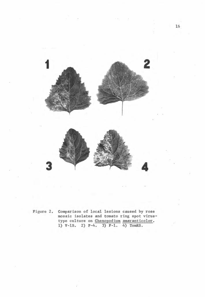
Figure 2. Comparison of local lesions caused by rosemosaic isolates and tomato ring spot virus-type culture on Chenopodium amaranticolor.1) V-1B. 2) P-4. 3) F-l. 4) TomRS.
16
17
Systemic symptoms on the trifoliate leaves were consistently
different among virus isolates and usually were not influenced by
the factors which affected the occurrence of symptoms on the primary
leaves. Isolate F-1 usually produced bright yellow chlorotic patterns
in the form of vein banding, ring spots or line patterns, but
necrotic lesions were seldom observed. Isolate P-4 generally re
mained latent except in the spring months or when inoculum of high
titer was used, when vein clearing and necrotic flecking were
observed. Isolate P-2 and V-1B both caused necrotic and chlorotic
lesions on the trifoliate leaves. Tomato ring spot virus reaction
was similar to that produced by P-4 in its symptom expression
(Figure 3) and in its failure to produce symptoms except in the
spring or with high titer. The severity and frequency of occurrence
of systemic symptoms on cowpea, in the case of all five viruses
tested, appeared to be influenced by the titer of the inoculum.
iii. Phaseolus vulgaris, varieties Bountiful and Sutter's
Pink. Rose mosaic virus isolate F-1 and TomRS virus were tested
on the following bean varieties! Bountiful, Sutter's Pink, Red
Kidney, pole bean - Bluelake, Golden Cluster Wax, Dwarf Horticulture,
Pinto and Great Northern. Bountiful bean and Sutter's Pink were the
only two varieties in which symptoms occurred consistently. All
four RMV isolates and TomRS virus caused chlorotic lesions on the
inoculated primary leaves and necrotic lesions on the systemically
infected trifoliate leaves. Differences among the five isolates
were not distinguishable on the basis of host symptoms produced on
either bean variety.

P-4V- IB
Figure 3. Comparison of systemic symptoms of somerose mosaic virus isolates and tomato
ring spot virus on trifoliate leaves ofblack cowpea.
18

19
iv. Cucumis sativus, variety Chicago Pickling. The rose
virus isolates and ToraRS culture produced chlorotic lesions on the
inoculated cotyledons, and necrotic lesions on the first true leaf
of cucumber. Death of the infected plants generally followed within
1% to 2 weeks after inoculation. Differences between the RMV and
TomRS isolates were not readily distinguished by symptoms on
cucumber.
II. Prunus ring spot virus in rose.
A. Prevalence of Prunus ring spot virus in rose. A survey for
Prunus ring spot virus (PRSV) in rose rootstocks in commercial
nurseries was conducted during the summers of 1959, 1960 and 1961.
Budwood was selected from 71 Manetti, 181 Multiflora and 38 miscel
laneous rootstocks. These were tested for PRSV on field grown Shiro-
fugen (Prunus serrulata) (29). Fifty-five hybrid tea roses, grown on
the Oregon State University plant pathology experimental farm, were
also tested. Buds from a prune tree showing prune dwarf symptoms
were used as a source of PRSV for a control.
None of the buds from the Manetti and Multiflora roses were
infected. An Odorata rose tested positive for PRSV and 25 of the
55 hybrid tea roses proved to be infected with PRSV. Forty-four of
the 55 tea roses had displayed symptoms of RMV, but there was no
correlation between the PRSV infection and the plants showing RMV
symptoms.
Evidence for the natural spread of PRSV among roses was

20
observed. Eleven of the 55 hybrid tea roses which had indexed
negative for PRSV were re-indexed the following year. Seven of the
eleven roses revested assayed positive for PRSV on Shiro-fugen.
B. Transmission of Prunus ring spot virus from rose to
herbaceous hosts. A Prunus ring spot virus, of the non-yellows
type (31), was mechanically and graft-transmitted from rose to
cucumber. PRSV was recovered on cucumber from the terminal leaves
of peach seedlings grafted with rose buds infected with PRSV.
Mechanical transmission of PRSV directly from infected rose tissue
to cucumber was accomplished on two different occasions. Rose
leaves were macerated in phosphate buffer, and the extract was
applied to the cotyledons of the cucumber. PRSV could not be trans
mitted to black cowpea or Bountiful bean, but V. rosa buds infected
with PRSV caused a typical necrotic reaction when budded to Shiro-
fugen. Symptoms caused by PRSV isolates from rose compared favor
ably with those caused by known isolated of PRSV of Cucurbita maxima
Dene. var. Buttercup and C. pepo L. var. White Scallop.
III. Survey for rose virus indicator hosts.
Five different host plants were tested during a search for an
index host for the rose viruses. Each plant selected had either
been observed or reported to be an indicator host for a specific
virus. The indicator hosts included Jonathan apple, Hopa crab,
Shiro plum, Prunus tomentosa, and Shiro-fugen, and the respective
viruses included apple mosaic virus, stem pitting virus, plum

21
line-pattern virus and Prunus ring spot virus. These viruses were
used as inoculum for the control plants.
No discreet virus symptoms were observed on the Hopa crab or
Shiro plum seedlings inoculated with RMV, but Shiro-fugen and
P. tomentosa reacted characteristically to the rose buds infected
with PRSV (13, 29). Virus symptoms were not observed on Jonathan
apple, but an increase in mildew (Podosphaera leucotricha (E & E)
Salm.) susceptibility was observed on seedlings inoculated with the
rose viruses.
The rose mosaic viruses were readily transmitted mechanically
from the terminal leaves of infected Jonathan apple, but mechanical
recovery of the virus was not accomplished from the other test
plants.
IV. Purification of rose mosaic virus.
A. Selection of host and assay plants. Black cowpea, cucumber
and Bountiful bean were infected with RMV and assayed for infectivity
on C. amaranticolor and compared for virus titer. Juice extracted
from cucumber cotyledons and trifoliate leaves of cowpea was more
infectious than extracts from the primary and trifoliate leaves of
Bountiful bean. Black cowpea was selected as a host over cucumber
because of its ease of culture and maintenance.
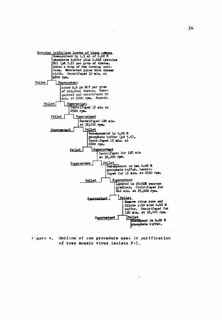
B. Purification procedure. A modification of the technique,
reported by Fulton (16, 17), for the clarification of tissue mac
erates infected with virus was adapted to the purification of RMV.

22
Trifoliate leaves of black cowpea infected with the F-l isolate were
briefly sprayed with Dow Corning anti-foam A and macerated in a
morter in 1% volumes (weight:volume) of 0.02 M Na2HP0,. The buffer
was titrated to pH of 7.5 with phosphoric acid and 0.01 percent
cysteine HCL added just prior to using. Buffer of the same formu
lation, less the cysteine complement, was used throughout the
remainder of the procedure. The macerated tissue was expressed
through four layers of cheesecloth with a hand press, and the extract
centrifuged for 10 minutes at 4,200 rpm in a Servall angle centri
fuge. Six-tenths gram of freshly prepared hydrated calcium phosphate
(HCP) was added to the extract for each gram of original tissue. The
mixture was homogenized in a beaker with a nagnetic stirrir. Mixing
was augmented by drawing and expelling the homogenate through a 30 ml
syringeo The homogenate was centrifuged for 10 minutes at 2,500 rpm.
The supernatant was again adsorbed with HCP as described above. The
clear supernatant was then centrifuged for 15 minutes at 6,500 rpm
to remove remaining traces of HCP. The extraction and clarification
procedures were conducted in a cold room at 5° C., and the extract
was at all times refrigerated or packed in ice.
Ultracentrifugation (Spinco No. 40 head) for 120 minutes at
38,000 rpm (127,640 x G), yielded an opalescent pellet and a free
flowing non-opalescent, gelatinous pellet. Both pellets were
resuspended in buffer and centrifuged for 15 minutes at 6,500 rpm.
The virus was again pelleted by centrifugation for 120 minutes at
38,000 rpm, resuspended in buffer, and centrifuged for 15 minutes at
63500 rpm.

23
Extraneous host materials were removed from the partially puri
fied virus suspension by density gradient centrifugation. The virus-
containing solution was layered on a 25 to 50 percent sucrose
gradient and centrifuged for 240 minutes at 25,000 rpm (90,000 x G)
in a Spinco SW 25 swinging bucket rotor. The virus zone was removed,
diluted 1:5 with buffer and centrifuged at 38,000 for 120 minutes.
The pellet was re-suspended in buffer and centrifuged for 15 minutes
at 6,500 rpnu An outline of the purification procedure is
illustrated in Figure 4.
The molarity of the phosphate buffer used in the extraction
process was found to be very critical. Infectivity of the clarified
preparation decreased as the ionic strength of the buffer was either
increased or decreased. Hydrogen ion concentration of the buffer
was less critical. Virus activity was detected in clarified prepara
tions extracted in buffers between the pH range of 7.0 to 8.0.
Clarified virus preparations extracted in buffer without an oxidative
protectant were low in infectivity. Agents with reducing and
chelating potentials, such as cysteine HCl, added to the extracting
buffer prevented loss of activity during clarification. Chelating
agents incorporated in the buffer, with and without the cysteine
complement, were not as effective as cysteine alone.
Longevity of the partially purified virus was greater when
suspended in buffer than when suspended in distilled water. Virus
suspensions in buffer were still infectious after 5 days storage at
5° C, but suspensions in distilled water were no longer infectious,
after similar storage.
Infected trifoliate leares of black coifpeaHomogenised In 1.5 ml of 0.02 Mphosphate buffer plus 0.0l£ cysteineHCl (pH 7.5) per gram of tissue.Added a drop of Dow Corning anti-foam. Kxtracted juice thru cheesecloth. Centrifuged 10 min. atU200 rpm.
Pellet | |SupernatantAdded 0.6 gm HCP per gramof original tissue. Homogenized and centrifuged 10min. at 2500 rpm. Repeat.
ISupernatantCentrifuged 15 min at
Pellet f
Pellet f
6500 rpm.
Supernatant
ICentrifuged 120 min.|at 38,000 rpm.
Supernatant II PelletResuspeneded in 0.02 Mphosphate buffer (pH 7.5).Centrifuged 15 min. at6500 rpm.
Pellet 1 ISupernatantCentrifuged for 120 minat 38,000 rpm.
~| PelletIResuspended
Supernatantr in 1ml 0.02 M
phosphate buffer. Centrifuged for 15 min. at 6500 rpm.
Pellet 11 SupernatantLayered on 25-50$ sucrosegradient. Centrifuged for2U0 min. at 25*000 rpm.
Izellei-Remove virus zone anddilute It 10 with 0.02 Mbuffer. Centrifuged for120 min. at 38,000 rpm.
"TPelletsuspend in 0.02 M
sphate buffer.
tant ISimerna
Supernatant T
figure 4. Outline of cne procedure used in purificationof rose mosaic virus isolate F-l.
24
25
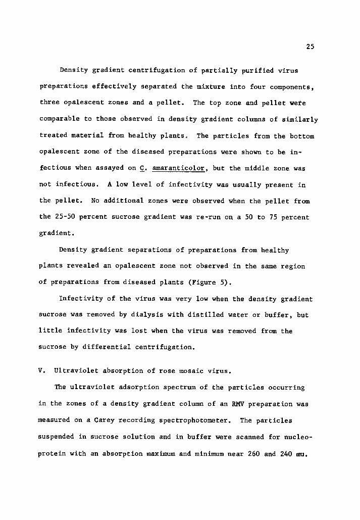
Density gradient centrifugation of partially purified virus
preparations effectively separated the mixture into four components,
three opalescent zones and a pellet. The top zone and pellet wete
comparable to those observed in density gradient columns of similarly
treated material from healthy plants. The particles from the bottom
opalescent zone of the diseased preparations were shown to be in
fectious when assayed on C. amaranticolor, but the middle zone was
not infectious. A low level of infectivity was usually present in
the pellet. No additional zones were observed when the pellet from
the 25-50 percent sucrose gradient was re-run on a 50 to 75 percent
gradient.
Density gradient separations of preparations from healthy
plants revealed an opalescent zone not observed in the same region
of preparations from diseased plants (Figure 5).
Infectivity of the virus was very low when the density gradient
sucrose was removed by dialysis with distilled water or buffer, but
little infectivity was lost when the virus was removed from the
sucrose by differential centrifugation.
V. Ultraviolet absorption of rose mosaic virus.
The ultraviolet adsorption spectrum of the particles occurring
in the zones of a density gradient column of an RMV preparation was
measured on a Carey recording spectrophotometer. The particles
suspended in sucrose solution and in buffer were scanned for nucleo-
protein with an absorption maximum and minimum near 260 and 240 mu.
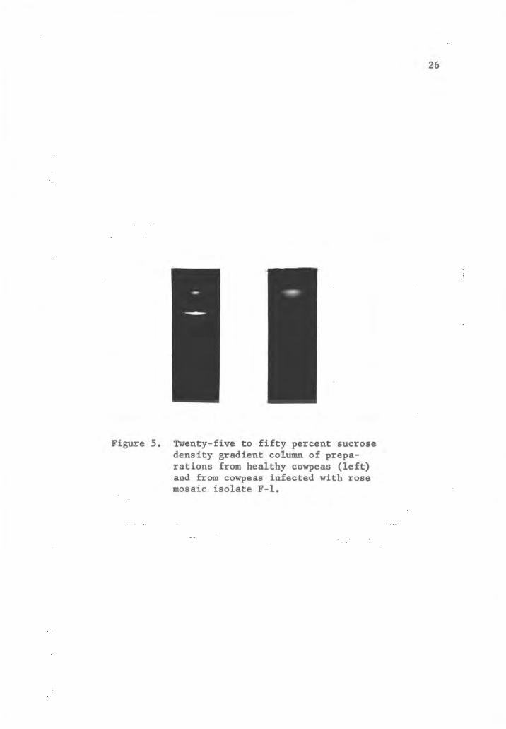
Figure 5. Twenty-five to fifty percent sucrosedensity gradient column of preparations from healthy cowpeas (left)and from cowpeas infected with rosemosaic isolate F-l.
26

27
Figure 6 illustrates the absorption spectrum of the various compon
ents separated from the partially purified preparations. The
E 260/280 ratio of the infectious particles was 1.23 and the
E max/min = 1.13. The ultraviolet absorption spectrum of this
component was characteristic of a nucleoprotein. The top zone,
which was comparable to that observed in healthy preparations, had
an absorption spectrum characteristic of proteins. The spectrum of
the material occurring in the second zone has not been characterized.
VI. Electron microscopy of rose mosaic virus.
Electron micrographs were taken of the RMV preparation components
separated in a density gradient column with an RCA EMU-2D. The
purified virus component, suspended in buffer, was sprayed on steel
grids coated with nitrocellulose and shadowed with platinum at a
grazing angle of 20 . A 1/8 dilution of the suspension showed good
particle distribution with little aggregation. A standardized con
centration of polystyrene latex of 1:1000 was used in each serial
dilution. Examination of electron micrographs of air-dried prepara
tions of purified RMV shows considerable flattening of the particles
(Figure 7). An average of 50 measurements of the particles from the
infectious preparation, calculated by comparison to polystyrene
latex spheres of 264 mu, was 40 mu. The height of the particles,
calculated from shadow measurements was 8.6 mu. If the virus parti
cles were considered spherical, and compensations made for the
flattening of the particles, a figure of 24 mu was derived.
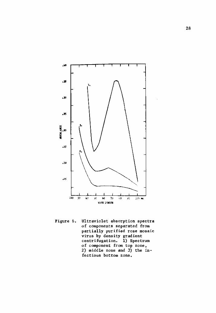
Figure 6. Ultraviolet absorption spectraof components separated frompartially purified rose mosaicvirus by density gradientcentrifugation. 1) Spectrumof component from top zone,2) middle zone and 3) the infectious bottom zone.
28
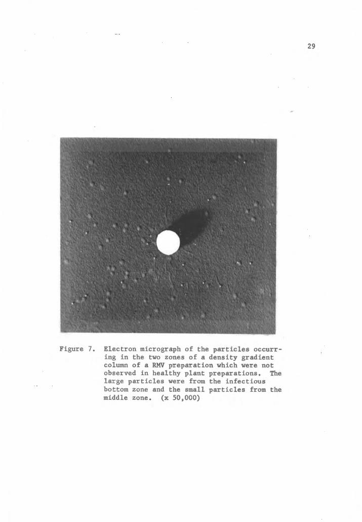
Figure 7. Electron micrograph of the particles occurring in the two zones of a density gradientcolumn of a RMV preparation which were notobserved in healthy plant preparations. Thelarge particles were from the infectiousbottom zone and the small particles from themiddle zone. (x 50,000)
29

30
The particles from the middle zone of a density gradient column
were approximately one-half the magnitude of the virus particles.
These particles appeared to be particularly rigid and not as subject
to flattening in air-dried preparations as was the RMV.
VII. Serological study of rose mosaic virus.
A. Preparation of antigen. Rose mosaic virus (F-l), extracted
from black cowpea by two high-speed differential centrifugation
cycles of the purification procedure, was prepared for injection
into two rabbits. The RMV antigen suspended in buffer was used for
intravenous injection, and for intramuscular injection the suspension
was emulsified with Freunds incomplete adjuvant at a ratio of ltl..
B. Immunization procedure. A combination of intramuscular and
intravenous injections were used. Weekly intramuscular injections
were generally followed every third or fourth day with an intravenous
injection. The antigen prepared for each injection was assayed for
activity on C. amaranticolor. Table I shows the immunization
schedule.
C. Serological tests. The titer of the rose mosaic virus
antiserum was determined by the reaction of adsorbed antiserum with
partially purified virus preparations in micro-precipitin tests (44).
The antiserum was adsorbed twice with partially purified preparations
from healthy cowpeas. The titer of the adsorbed antiserum was 1/32.

Table I. Immunization schedule used in the preparation of RMVantiserum.
Date of Date of Titer of
Type of injection injection test bleeding antiserum
Intramuscular (IM) 1-8-62
IM 1-16-62
Intravenous (IV) 1-18-62
IM 1-22-62
IV 1-25-62
IM 1-30-62
IV 2-2-62
IM 2-6-62 2-6-62 1/16
IV 2-9-62 2-12-62 1/64
Intramuscular injections were 3-4 ml of virus preparationemulsified with an equal volume of Freund adjuvant. Intravenous injections were 1-1.5 ml of virus preparation.
31
32
Crude sap preparations of RMV isolates P-4, P-2 and V-lB were
reacted with the F-l antiserum. Precipitation of the non-homologous
antigens with the adsorbed F-l antiserum had a dilution end point of
1/8. The low titer of the antiserum prohibited further tests to
determine strain relationships. Normal serum was not observed to
react with preparations from diseased or healthy cowpeas.
D. Serological relationship of rose mosaic virus to tomato ring
spot virus.
i. Preparation of the antigen. Tomato ring spot virus was
extracted from the inoculated primary leaves of Bountiful bean by
the same technique previously described for RMV. The activity of
TomRS from Bountiful bean was greater when the molarity of the ex
tracting and suspending buffer was 0.01 M; otherwise the procedure
was identical.
Infectious material used for injection received two high-speed
differential centrifugation cycles of the purification procedure.
ii. Immunization procedure. Tomato ring spot virus was
administered to two rabbits by a combination of intramuscular and
intravenous injections, similar to the schedule described by Cadman
et al. (5). The antigen for intramuscular use was emulsified at a
1:1 ratio with a 2:1 mixture of falba and mineral oil. Ivory soap
was used as an emulsifying agent. Table II shows the immunization
schedule.

33
Table II. Immunization schedule used in the preparation of TomRSantiserum.
Date of Date of Titer of
Type of injection injection test bleeding antiserum
Intramuscular (IM) 12-11-61
IM 12-16-61
IM 12-21-61
IM 12-29-61
IM 1-4-62
Intravenous (IV) 1-11-62
IV 1-20-62
1-15-62
1-24-62
1/64
1/256
Intramuscular injections were 3-4 ml of virus preparationemulsified with an equal volume of 2:1 mixture of falba andmineral oil. Intravenous injections were 1.5 ml of viruspreparation.
34
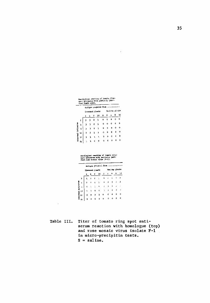
iii. Serological tests. The precipitin and micro-precipitin
tests were used to determine antiserum titer. The micro-precipitin
test, due to its relatively close check with the precipitin test,
was used exclusively in later tests.
TomRS antiserum was absorbed twice with healthy plant antigen
prepared by the same procedure as the virus antigen. Table III
shows the titer of the adsorbed TomRS antiserum when reacted with
the homologous antigen and non-homologous antigen (F-l).
The precipitation of the non-homologous antigen with the TomRS
antiserum was clearly less dense than the reaction observed with the
homologous antigen.
The dilution end point of the adsorbed antiserum when reacted
with TomRS was 1/64, and 1/16 when reacted with RMV isolate F-l.
E. Cross precipitation tests. Tomato ring spot virus was
reacted with the F-l RMV antiserum. As previously stated, the titer
of the RMV absorbed antiserum was 1/32 when reacted with the F-l
isolate. The dilution end point of the antiserum was 1/16 when
reacted with partially purified tomato ring spot type culture virus.
This again shows that TomRS virus and Fulton's RMV isolate are
serologically related.
Serological reaction of tomato ring-spot antiserum Kithpartially purified TomRS Time.
Antigen prepared
Diseased plants Healthy plants
2 u 8 16 3 2 k 8 16
2 3 3 2 1 0 0 0 0 0
1 u 3 3 2 1 0 0 0 0 0
S 8 3 3 2 1 0 0 0 0 0
•a 16
S 32
2 2 1 1 0 0 0 0 0
1 1 t t 0 0 0 0 0
*«- 1 t 0 0 0 0 0 0 0
S»roloKlo*l r»*otion of tommto rin^-•pot «ntii«nM with partUliy purified ro— «osai.o vim* (F-l).
Antigen prepaid fromi *
DiMU*d plmnti H.althy plaatt
8 16
z Z. 2 2 1 0 J 3 J
?« 2 2 L 1 0 0 0 0
I 82 1 1 t 3 J a J
„ 13 1 L t 0 3 J 0 0
|52 0 0 0 0 0 0 0 0
3« 84 0 0 0 0 0 0 0 0
Table III. Titer of tomato ring spot antiserum reaction with homologue (top)and rose mosaic virus isolate F-l
in micro-precipitin tests.S = saline.
35
36
DISCUSSION AND CONCLUSIONS
Mechanical transmission of rose mosaic virus from rose was
reported by Fulton (15) to be difficult and unreliable. He main
tained that the infectivity of RMV was affected by rose tissue
extracts, divalent ions and by the peculiarities of the virus. In
the present study, virus inhibitors were found primarily in the
extracts from young rose leaves. Mature and senescent leaves were
shown to have little inhibitive effect on the viruses tested; how
ever, RMV was not isolated from infected mature or senescent leaves.
Numerous chemicals and different techniques used in the extraction
process to counteract the effect of the virus inhibitors did not
prevent inactivation of RMV. Rose root extracts had a pronounced
stimulatory effect on the infectivity of alfalfa mosaic. This was
most likely due to an interference by a substance present in the rose
root with an inhibitor in the V. rosa or cowpea from which the AMV
was extracted. The effect of rose root extracts on the infectivity
of RMV was not investigated, but in view of the effect of this sub
stance on the infectivity of AMV, it would appear that rose roots
from infected plants would be the logical place from which to attempt
isolations for RMV.
Several investigators have graft-transmitted rose viruses to
peach, then have mechanically transmitted the viruses from peach
to herbaceous hosts. In this study rose mosaic was readily trans
mitted by grafting to peach seedlings and also to V. rosa during any
37
period of the year. This virus could be mechanically tranmitted
from both hosts to cowpea or cucumber with relative ease. RMV
transmission to V. rosa was not 100 percent reliable, but provided
an assay technique and a method of isolating RMV with a greater
percent of success than by previously reported methods.
Reports (6, 19) of viruses isolated from rose which resemble
Prunus ring spot virus are in all probability correct, but the con
tention that Prunus ring spot virus and RMV have a co-identity is
unlikely. The present investigation shows that in tests for PRSV
in 55 roses, 25 assayed positive for PRSV on Shiro-fugen. Forty-
four of the 55 roses displayed symptoms of RMV. Evidence of spread
of Prunus ring spot virus among roses was observed. Studies were
not conducted to determine the mode of spread, but it was presumed
to be by pollen transmission (11).
PRSV was transmitted by mechanical inoculation from rose to
cucumber, but this was only accomplished twice and only in the
spring of the year. This phenomenon was also observed by Fulton (15)
for RMV. A combination of factors, including the titer of virus
inhibitors, the virus titer and the balance between the two was
probably responsible. The virus titer was probably at its peak
during the rapid growth experienced during the spring months.
Jonathan apple, Hopa crab, Shiro plum, Prunus tomentosa and
Shiro-fugen were inoculated with rose buds infected with RMV and
PRSV. P. tomentosa and Shiro-fugen reacted characteristically to
the PRSV (13, 29). None of these hosts proved to be indicators for
RMV.
38
Oregon RMV isolates, Fulton's RMV isolate and tomato ring spot
virus type culture reactions were compared on black cowpea, C.
amaranticolor, Bountiful bean and cucumber. The host symptom re
actions of the three Oregon RMV isolates and Fulton's isolate
suggested strain relationships. The general spectrum of the host
symptoms of all the RMV isolates tested was similar to that produced
by TomRS virus. Serological reactions of Fulton's RMV with a TomRS
type culture antiserum, verified this relationship, and furthermore,
the TomRS culture reacted with a rose mosaic virus (F-l) antiserum
in cross precipitin tests. The Oregon RMV isolates were not tested
against the TomRS antiserum which reacted with Fulton's RMV, but did
react with the antiserum produced from Fulton's RMV isolate. This
indicates that the Oregon RMV isolates were also strains of tomato
ring spot virus.
Fulton's isolate of rose mosaic virus (F-l) was purified. The
clarification process, the most critical step in the procedure,
relied on the adsorption of the host material to hydrated calcium
phosphate (HCP). The ionic strength of 0.02 M of the extracting
buffer prevented the virus from being adsorbed to the HCP. The
addition of cysteine HCl to the extracting buffer effectively pro
tected the virus from inactivation during extraction and clarifica
tion. Other chelating and reducing agents were not as effective.
The virus was purified by density gradient centrifugation. Pre
parations from diseased plants contained two fractions not detected
in similar preparations from healthy plants. The bottom zone of a
39
density gradient column contained infectious virus and had an ultra
violet absorption spectrum typical of a nucleo-protein. Sedimentation
rates of the particles in the infectious zone were not determined.
The middle zone from the diseased plant preparations did not have any
of the characteristics of a virus and was not infectious. Investi
gations were not conducted to identify this component. The
ultraviolet absorption spectrum, and the comparison of electron
micrographs of this substance and the virus, did not indicate that
these particles were virus precursors. Electron micrographs of
TomRS published by Senseney et al. (36) contained small particles
similar to those found in the middle zone of RMV preparations. This
substance, occurring in RMV and perhaps TomRS infected plants, could
be a normal plant component, the synthesis of which was increased, or
a new component. In either case, the appearance of this product was
induced by RMV infection. The pellet from the 25-50 percent columns
contained some infectious material. This pellet, when re-suspended
and re-run through a 50-75 percent density gradient column did not
produce any new zones, and furthermore, the pellet at the bottom of
this column still contained some infectious particles. This can
probably be attributed to aggregated virus particles.
A density gradient separation of material prepared from healthy
plants showed a zone in a region not apparent in the preparations
from diseased plants. This zone had an absorption spectrum typical
of a protein. The absence of this protein fraction in the prepara
tions from the diseased plants was probably not due to the
40
clarification procedure or to an interference in the plant anabolic
processes by the virus, but more likely due to a degradation action
induced by the RMV infection.
Size of the RMV particles, determined by electron microscopy of
purified material, are comparable to the dimensions of TomRS virus
reported by Senseney et al. (35). He reports that TomRS was
43.0 x 13.5 mu. However, considerable flattening of the specimens
was observed, and if the particles were considered spherical when in
a normal state, they would measure approximately 27 mu. This figure
closely agrees with the dimensions of 40.0 x 8.6 mu for RMV.
However, if the RMV particles were considered to be spheres, a
figure of 24 mu is derived.

41
SUMMARY
1. The effects of virus inhibitors present in rose tissue extracts
have been studied. Data are presented to show that extracts
from young rose leaves reduced the infectivity of alfalfa mosaic
virus up to 58 percent, but those from mature and senescent
leaves had little effect. Extracts from rose root increased the
infectivity up to 97 percent.
2. A method for the transmission of rose mosaic virus by grafting
infected rose buds to Vinea rosa and then mechanically to
herbaceous hosts is discussed.
3. Rose mosaic virus isolates were compared on black cowpea,
Bountiful bean, cucumber and Chenopodium amaranticolor. Four
strains, including Fulton's isolate of RMV, were distinguished
on the basis of host and serological reaction of RMV isolates to
an antiserum prepared from Fulton's RMV.
4. Host reactions of the RMV isolates were compared with host
reactions of tomato ring spot virus. Host and serological re
actions of Fulton's RMV with TomRS antiserum were indicative of
strain relationship between the two viruses. This relationship
was further substantiated by cross-precipitin tests.
5. Fulton's RMV was purified by a method utilizing adsorption of
the host materials to hydrated calcium phosphate, differential
and density gradient centrifugation. Partially purified prepa
rations from RMV diseased cowpea contained a non-virus component
42
not observed in similar preparations from healthy cowpea.
Preparations from healthy cowpeas also contained a protein
fraction not detected in preparations from RMV diseased cow
peas. The purity of the virus preparation was verified by
electron microscopy and spectrophotometry. Electron micrographs
of purified RMV revealed particles with dimensions similar to
those reported for TomRS.
6. Prunus ring spot virus was detected in 45 percent of a plot of
hybrid tea roses, but most of the rose rootstocks tested were
free from this virus. Evidence of natural spread of PRSV among
the hybrid tea roses is presented.
7. Jonathan apple, Hopa crab, Shiro plum, Prunus tomentosa and
Shiro-fugen were surveyed as indicator hosts for RMV and PRSV.
PRSV infected rose buds caused a characteristic reaction on
P. tomentosa and Shiro-fugen. No reactions were observed on
the hosts inoculated with RMV.

43
BIBLIOGRAPHY
1. Baker, Kenneth F. and Earl H. Thomas. The effect of temperatureon symptom expression of rose mosaic. Phytopathology 32:321-326.1942.
2. Brierley, Philip and Floyd G. Smith. Streak, a virus disease ofroses. (Abstract) Phytopathology 24:7. 1935.
3. ° Mosaic and streak diseases of roses. Journalof Agricultural Research 61:625-660. 1940.
4. _____• Spread of rose virus diseases. AmericanNurseryman July 1, 1940. pp. 5-8.
5. Cadman, C. H. and R. M. Lister. Relationship between tomato ringspot and peach yellow bud mosaic viruses. Phytopathology 51:29-31. 1961.
6. Cochran, L. C. Infection of apple and rose with ring spot virus.(Abstract) Phytopathology 40:964. 1950.
7. Common names of virus diseases used in the Review of AppliedMycology. Vol. 35:Supplement. 1957.
8. Conners, I. L. Twentieth Annual Report of Canada. Plant DiseaseSurvey. 1940. p. 98.
9. _ _________• Twenty-first Annual Report of Canada. PlantDisease Survey. 1940. p. 98.
10. Current research and investigation. Orchard. N. Z. 31:307.1958. (Abstracted in Review of Applied Mycology. 38:239.1959.)
11. Das, C. R. and J. A. Milbrath. Plant-to-plant transfer of stonefruit ring spot virus in squash by pollination. Phytopathology51:489-490. 1961.
12. Diester, E. A. Plant Diseases. Rose wilt virus. Control ofblack spot of grape. Agric. Gaz. N. S. W. 64:487-489. 1958.(Abstracted in Review of Applied Mycology 38:283. 1959.)
13. Fink, H. C. Prunus tomentosa as an index plant for sour cherryvirus. Phytopathology 45:320-323. 1959.)
14. Fry, P. R. and J. R. Hunter. Rose mosaic virus in New Zealand.New Zealand Journal of Science and Technology Sec. A. 37:478-482. 1956.

44
15. Fulton, Robert W. Mechanical transmission and properties ofrose mosaic virus. Phytopathology 42:413-416. 1952.
16. . A rapid method for partial purification ofsome unstable plant viruses. (Abstract) Phytopathology 47:521.1957.
17. . Purification of sour cherry necrotic ringspot and prune dwarf viruses. Virology 9:522-535. 1959.
18. Gigante, R. Una nuova virosi della rosa in Italia. Boll.Staz. Pot. Veg. Roma. N. S. 16:76-94. 1936. (Abstracted inReview of Applied Mycology 16:179-180. 1937.)
19. Gilmer, R. M. Behavior of juice extracts of certain virusisolates from Prunus and from rose in cucumber plants.(Abstract) Phytopathology 46:241. 1956.
20. Grieve, B. J. Rose wilt and dieback, and virus diseases ofrose occurring in Australia. The Australian Journal of Experimental Biology and Medical Science 8:107-121. 1931.
21. . Further observations on rose wilt virus.
Proc. Roy. Soc. Victoria 54:229-238. 1942. (Abstracted inReview of Applied Mycology 23:18. 1942.)
22. Gualaccini, Franco. Una virosi della Rosa nuova per 1'Italia.Suoi rapporti con le virosi dei frutteferi. Bollettino dellastazione de Patologia Vegetale 15:79-88. 1957.
23. Holmes, Francis 0. Rose mosaic cured by heat treatments. PlantDisease Reporter 44:46-47. 1960.
24. Klastersky, I. A new morphogenic virosis on roses. Stud. bot.Cechosl. 10:1-13. 1949. (Abstracted in Review of AppliedMycology 29:28. 1950.)
25. . A cowl-forming virosis in roses, lime treesand elm trees. Stud. bot. Cechosl. 12:73-171. 1951.
(Abstracted in Review of Applied Mycology 31:38. 1952.)
26. Kristensen, H. R. and A. Thomsen. Visussygdomme hos roser I.Tidsskr planteavl. 63:369-393. 1959. (Abstracted in BiologicalAbstracts 35: no. 22349. 1959.)
27. Mushin, Rose. Serological studies on plant viruses. AustralianJournal of Experimental Biology and Medical Science 20:59-63.1942.

45
28. McWhorter, F. P. Further report on rose mosaic in Oregon.Plant Disease Reporter 15:1-3. 1931.
29. Milbrath, J. A. Selecting stone fruit trees free from virusdiseases. Oregon State University. Agricultural ExperimentStation. Technicial Bulletin 522. p. 1-27. 1952.
30. . The epinasty virus reaction of Kwanzan andShiro-fugen flowering cherry. Phytopathology 50:495-497. 1960.
31. . The relationship of stone fruit ring spotvirus to sour cherry yellows, prune dwarf and peach stunt. In:Proceedings of the Fourth Symposium on Virus Diseases of FruitTrees in Europe, Lyngby, 1960. Tidsskrift for Planteavl 125-133. 1961.
32. Milbrath, D. G. A discussion of the reported infectiouschlorosis of the rose. California State Department of Agriculture Monthly Bulletin 19:535-544. 1930.
33. . A discussion of the reported infectiouschlorosis of rose. Florist Exchange and Horticultural TradeWorld July 26, 1930. pp. 11 and 33.
34. Nelson, Ray. Infectious chlorosis of the rose. (Abstract)Phytopathology 20:130. 1930.
35. Newton, W. Chlorosis of rose. In: Report of DominionBotanist for year 1930. Division of Botany. Canada Department of Agriculture, p. 23. 1931. (Abstracted in Review ofApplied Mycology 11:245. 1932.)
36. Senseney, C. A., Robert P. Kahn and Paul R. Desjardins.Particle size and shape of purified tomato ring spot virus.Science 120:456-457. 1954.
37. Taubenhaus, J. J. Diseases of ornamentals. Plant DiseaseReporter. Supplement 29:.447. 1923.
38. . Diseases of ornamentals. Plant Disease
Reporter, Supplement 42:361. 1925.
39. Thomas, Earl H. Apple mosaic. Hilgardia 10:581-87. 1937.
40. and T. E. Rawlins. Some mosaic diseases ofPrunus species. Hilgardia 12:623-644. 1939.
41. ________________ a^d L. M. Massey. Mosaic diseases of the rosein California. Hilgardia 12:647-663. 1939.
46
42. and C. E. Scott. Rosette of rose.Phytopathology 43:218-219. 1953.
43. Valleau, W. D. A virus disease of plum and peach. KentuckyAgricultural Experiment Station. Bulletin 327:89-103. 1932.
44. Van Slogteren, D. H. M. Serological micro-reactions with plantviruses under paraffin oil. In: Proceedings of the SecondConference on Potato Virus Diseases, Wageningen, Netherlands,1945. Wageningen, H. Veenman & Zonin, 1955. pp. 51-54.
45. White, R. P. An infectious chlorosis of rose. Plant DiseaseReporter 12:33-34. 1928.
46. . An infectious chlorosis of rose. New JerseyAgricultural Experiment Station. Annual Report. Bulletin 42:42-43. 1929.
47. . An infectious chlorosis of rose. (Abstract)Phytopathology 20:130. 1930.
48. __. An infectious chlorosis of rose. TheAmerican Rose Annual 15:87-90. 1930.
49. . Infectious chlorosis of rose. FloristsExchange and Horticultural Trade World January 25, 1930.p. 46.
50. . Quarantines and rose chlorosis. FloristsExchange and Horticultural Trade World March 15, 1930.pp. 50a and 54.
51. . Chlorosis of the rose. Phytopathology22:53-69. 1932.
52. Weiss, F. and F. P. McWhorter. Pacific coast survey for rosemosaic. Plant Disease Reporter 14:203-205. 1930.
53. Wildon, C. E. Michigan rose men discuss infectious chlorosis.Florists Exchange and Horticultural Trade World March 8, 1930.p. 58.
54. Willison, R. S., M. Weintraub and J. D. Ferguson. Purificationand electron microscopy of viruses causing cherry yellows andrelated diseases. Canadian Journal of Botany 34:86-103. 1956.


