
REVIEW
In vivo models of brain tumors: roles of genetically engineeredmouse models in understanding tumor biology and usein preclinical studies
Iva Simeonova • Emmanuelle Huillard
Received: 21 January 2014 / Revised: 20 June 2014 / Accepted: 30 June 2014 / Published online: 10 July 2014
� The Author(s) 2014. This article is published with open access at Springerlink.com
Abstract Although our knowledge of the biology of
brain tumors has increased tremendously over the past
decade, progress in treatment of these deadly diseases
remains modest. Developing in vivo models that faithfully
mirror human diseases is essential for the validation of new
therapeutic approaches. Genetically engineered mouse
models (GEMMs) provide elaborate temporally and
genetically controlled systems to investigate the cellular
origins of brain tumors and gene function in tumorigenesis.
Furthermore, they can prove to be valuable tools for testing
targeted therapies. In this review, we discuss GEMMs of
brain tumors, focusing on gliomas and medulloblastomas.
We describe how they provide critical insights into the
molecular and cellular events involved in the initiation and
maintenance of brain tumors, and illustrate their use in
preclinical drug testing.
Keywords Mouse models � Glioma � Medulloblastoma �Cell of origin � Neural stem cells
Abbreviations
a-KG a-Ketoglutarate
AmpB Amphotericin B
BBB Blood brain barrier
BLI Bioluminescent imaging
D-2HG D-2-hydroxyglutarate
ECM Extracellular matrix
EGFR Epidermal growth factor receptor
ENU N-ethyl-N-nitrosourea
EP Ependymoma
FDA Food and drug administration (USA)
GEMM Genetically engineered mouse models
GBM Glioblastoma
GSC Glioma stem cells
HGA High grade astrocytoma
IDH1 Isocitrate dehydrogenase 1
IGF Insulin growth factor
MADM Mosaic analysis with double markers
MB Medulloblastoma
MHC Major histocompatibility complex
MNU N-methylnitrosourea
MRI Magnetic resonance imaging
NSC Neural stem cell
NPC Neural progenitor cell
OPC Oligodendrocyte precursor cell
PA Pilocytic astrocytomas
PI3K Phosphatidylinositide 3-kinase
PDGF Platelet-derived growth factor
PDGFRa Platelet-derived growth factor receptor alpha
SB Sleeping Beauty transposon
SVZ Subventricular zone
shRNA Short hairpin RNA
TERT Telomerase reverse transcriptase
WHO World health organization
Introduction
Primary brain tumors originate from the transformation of
neural stem cells (NSC) or cells committed to the neu-
ronal, astrocytic and oligodendrocytic lineages. In adults,
gliomas are the most common primary brain tumors,
accounting for about 30 % of all primary brain and cen-
tral nervous system tumors, and 80 % of malignant
I. Simeonova � E. Huillard (&)
Universite Pierre et Marie Curie (UPMC) UMR-S975, Inserm
U1127, CNRS UMR7225, Institut du Cerveau et de la Moelle
Epiniere, 47 boulevard de l’Hopital, 75013 Paris, France
e-mail: [email protected]
Cell. Mol. Life Sci. (2014) 71:4007–4026
DOI 10.1007/s00018-014-1675-3 Cellular and Molecular Life Sciences
123

tumors, according to the Central Brain Tumor Registry of
the United States [1]. High-grade gliomas, such as glio-
blastomas (GBM) and high-grade astrocytomas (HGA)
have a poor prognosis due to their resistance to conven-
tional radio- and chemotherapies. In children,
medulloblastomas (MB) are the most common brain
tumors. Although conventional therapies can cure subsets
of MB patients, the treated patients face long-term neu-
rological side effects [2].
The etiology of most brain tumors is not well under-
stood. As we will discuss below, thanks to recent large-
scale efforts on the molecular characterization of MB and
GBM, it is now clear that these tumors, initially thought
as unique entities, comprise distinct diseases at the clin-
ical and molecular levels. The molecular and cellular
mechanisms underlying tumor formation and heterogene-
ity are just starting to emerge. Better understanding of
brain tumor development and biology is critical for the
identification of new targets and the design of successful
therapies.
What is a good model of brain tumor?
The goal of a model is to reproduce the etiology and
biology of the corresponding human disease, to under-
stand its development and to identify adequate
treatments. Ideally, a good model should (1) display the
same genetic lesions, anatomical location, histopatholo-
gical features, and developmental time frame as the
human tumor; (2) recapitulate intertumoral and intratu-
moral heterogeneity; (3) be predictive of the patients’
response to treatment.
Cell lines derived from brain tumors are useful for the
characterization of the genetic lesions that occur in human
tumors, and to build primary hypothesis about gene
function. However, they cannot model effectively the key
aspects of tumorigenesis, such as microenvironment
contribution, invasion, angiogenesis or inflammatory
response. Therefore, they only have a limited predictive
value for the development of cancer therapies. In vivo
models provide a more accurate experimental system, as
they mimic tumor behavior in an entire mammalian
organism.
In vivo approaches for modeling brain tumors
Numerous in vivo models of brain tumors have been
developed, including carcinogen-induced rodent models,
xenograft and genetically engineered mouse models
(GEMMs) (Table 1). Rat models have been extensively
used since the mid-1970s. Gliomas could be induced in rats
injected with the alkylating agents N-methylnitrosourea
(MNU) or N-ethyl-N-nitrosourea (ENU). These models
have the advantage of developing tumors de novo, pre-
serving tumor–host interactions. However, in many of these
models, tumors grow as circumscribed tumors, instead of
being invasive like the human tumors [3]. In addition, rat
tumors have not been characterized at the molecular level
and thus it is not known to what extent their mutational and
transcriptional profiles match those of human tumors.
Xenograft models are generated by the transplantation of
biopsies or cultured cells derived from human brain tumors
into immunodeficient mice. Cell lines derived from gliomas
and grown in serum-containing medium have been used to
develop cytotoxic agents and tumor-specific agents. These
cells represent an unlimited source of material for drug testing
and engraft easily into immunocompromised mice. However,
tumors initiated from cells cultured in serum-containing
medium poorly resemble the genotype and phenotype of their
parental tumors: they grow as circumscribed, non-invasive
tumors and do not display the same transcriptomic profile and
genomic alterations as the parental tumors [4–6].
The past decade witnessed the identification, based on the
expression of the cell surface marker CD133, of brain tumor
stem cells (or brain tumor-initiating cells) in several brain
cancers [7, 8]. These cells are able to reconstitute a tumor upon
transplantation and display cardinal features of normal NSC,
such as the ability to self-renew and to give rise to the three
main cell types of the central nervous system (astrocytes, ol-
igodendrocytes, neurons). The cancer stem cell hypothesis
postulates a hierarchical tumor organization, where only a
small subpopulation of tumor cells drives tumorigenesis.
However, this hypothesis is being challenged. Indeed, sub-
sequent studies have shown that CD133-negative cells exhibit
similar properties [9, 10]. Actually, GBM tumors have been
shown to contain both CD133? and CD133-negative cell
types that generate highly aggressive tumors with different
growth kinetics, histology and gene expression profile [9].
These studies suggest that GBMs contain heterogeneous
populations of cells with distinct tumor-initiating properties.
By culturing cells freshly derived from GBM patients
under NSC serum-free conditions, researchers were able to
generate tumors that displayed the biology, genetics and
gene expression profiles of the corresponding human GBM
[4, 11]. In contrast, tumors generated from cells cultured in
serum failed to recapitulate the histopathological features of
the parental tumors. In a recent study, Joo et al. [11] estab-
lished a library of over 50 orthotopic xenografts generated
from GBM sphere cultures. They showed that the response of
the xenografts to in vivo irradiation and chemotherapy
matched the response of the parental GBM [11]. Xenografts
of serum-free GBM cultures thus appear to be promising
tools for recreating a human brain tumor in the mouse brain.
Nevertheless, a limitation of xenograft models is that they
4008 I. Simeonova, E. Huillard
123
usually require injection of a large amount of cells into the
host, which does not mirror the formation of human tumors
from a restricted number of cells. Moreover, the use of
immunodeficient mice does not take into account the con-
tribution of the immune system in tumor development. Some
brain tumor types have been more difficult to establish in
culture. For example, oligodendroglioma-derived cells are
difficult to propagate in vitro or do not engraft in host mice.
Only recently, the first oligodendroglioma cell cultures and
xenografts bearing typical genetic alterations have been
successfully established [12].
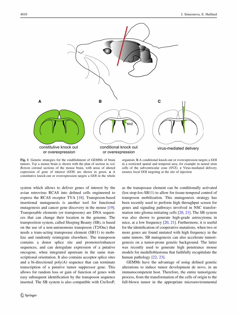
Genetically engineered mouse models of brain tumors
The availability of the complete sequence of the mouse
genome as well as powerful gene-targeting tools has led to
the development of GEMMs to investigate tumorigenesis
[13]. In these models, defined gene alterations identified in
human tumors (affecting specific oncogenes or tumor
suppressor genes) are introduced in the germline (knock-
out, knock-in, transgenic models) to allow for de novo
tumor formation (Fig. 1a) [14]. Although these models
may better mimic tumorigenesis in familial syndromes,
deletion of a tumor suppressor gene in the entire organism
is likely to lead to a wide spectrum of diseases, including
cancers, precluding correct analysis on its implication in
brain tumorigenesis. For example, mice constitutively
deficient for the tumor suppressor locus Cdkn2a (or Ink4a/
Arf) are viable and fertile but develop fibrosarcomas and
lymphomas [15]. As CDKN2A loss has been reported in
several brain tumors, a specific deletion on the locus in the
brain is needed to address its role in this pathology. The
development of conditional Cre/loxP systems has enabled
to specifically target defined cell populations (Fig. 1b). In
this system, the Cre recombinase permits the excision of a
gene flanked by two loxP sites. The induction of Cre
expression under the control of a tissue-specific promoter
allows for targeted deletion in a defined cell type. For
example, in the elegant MADM (Mosaic Analysis with
Double Markers) model, a mouse genetic mosaic system, it
is possible to induce sporadic mutations in a restricted cell
population and trace the fate of individual mutated cells
and their wild type siblings [16]. This system was used to
model high-grade glioma development from NSCs [17].
A Cre that can be induced temporally (Tamoxifen-induc-
ible Cre) further allows to introduce genetic alterations at a
given developmental time point, more faithfully modeling
somatic tumor development that occur in somatic cells at
later stages. Specific cell populations can also be directly
modified in a time-dependent manner using virus-mediated
gene delivery (Fig. 1c). An example is the RCAS–TVA
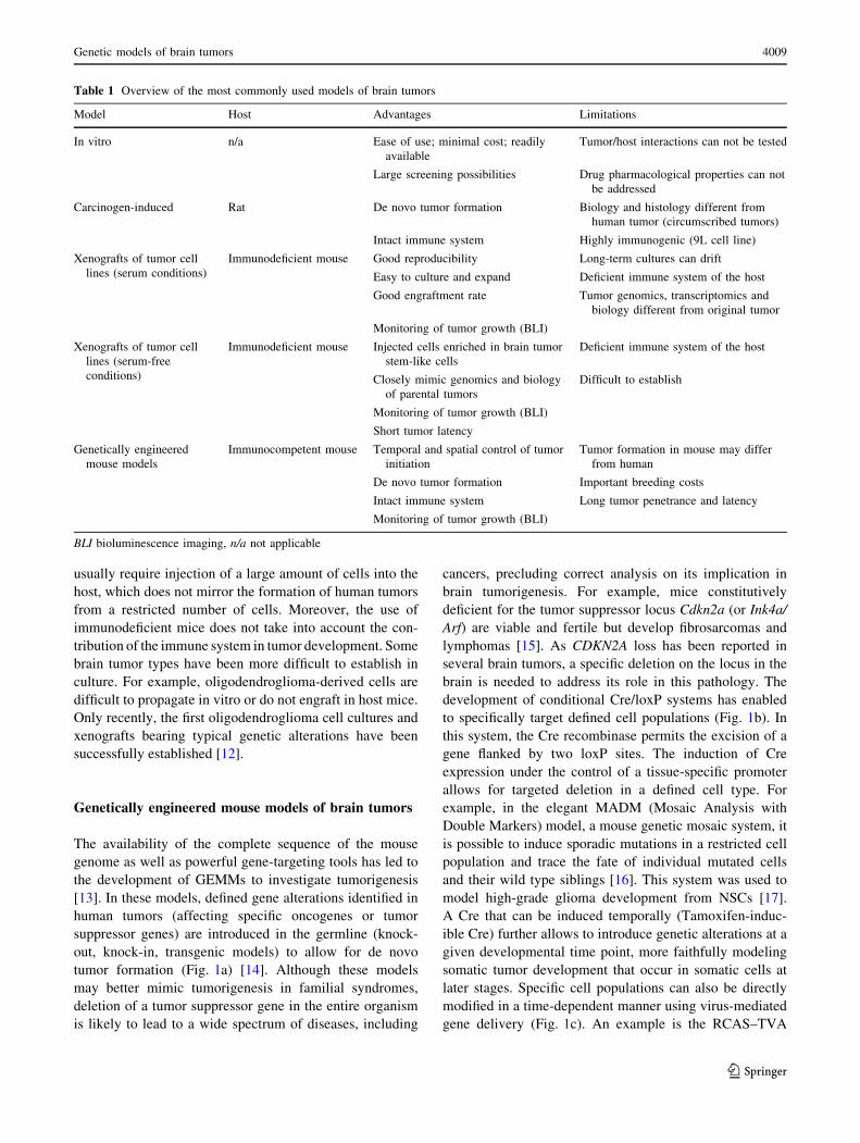
Table 1 Overview of the most commonly used models of brain tumors
Model Host Advantages Limitations
In vitro n/a Ease of use; minimal cost; readily
available
Tumor/host interactions can not be tested
Large screening possibilities Drug pharmacological properties can not
be addressed
Carcinogen-induced Rat De novo tumor formation Biology and histology different from
human tumor (circumscribed tumors)
Intact immune system Highly immunogenic (9L cell line)
Xenografts of tumor cell
lines (serum conditions)
Immunodeficient mouse Good reproducibility Long-term cultures can drift
Easy to culture and expand Deficient immune system of the host
Good engraftment rate Tumor genomics, transcriptomics and
biology different from original tumor
Monitoring of tumor growth (BLI)
Xenografts of tumor cell
lines (serum-free
conditions)
Immunodeficient mouse Injected cells enriched in brain tumor
stem-like cells
Deficient immune system of the host
Closely mimic genomics and biology
of parental tumors
Difficult to establish
Monitoring of tumor growth (BLI)
Short tumor latency
Genetically engineered
mouse models
Immunocompetent mouse Temporal and spatial control of tumor
initiation
Tumor formation in mouse may differ
from human
De novo tumor formation Important breeding costs
Intact immune system Long tumor penetrance and latency
Monitoring of tumor growth (BLI)
BLI bioluminescence imaging, n/a not applicable
Genetic models of brain tumors 4009
123
system which allows to deliver genes of interest by the
avian retrovirus RCAS into defined cells engineered to
express the RCAS receptor TVA [18]. Transposon-based
insertional mutagenesis is another tool for functional
mutagenesis and cancer gene discovery in the mouse [19].
Transposable elements (or transposons) are DNA sequen-
ces that can change their location in the genome. The
transposition system, called Sleeping Beauty (SB), is based
on the use of a non-autonomous transposon (T2/Onc) that
needs a trans-acting transposase element (SB11) to mobi-
lize and randomly reintegrate elsewhere. The transposon
contains a donor splice site and promoter/enhancer
sequences, and can deregulate expression of a putative
oncogene, when integrated upstream in the same tran-
scriptional orientation. It also contains acceptor splice sites
and a bi-directional poly(A) sequence that can terminate
transcription of a putative tumor suppressor gene. This
allows for random loss or gain of function of genes with
easy subsequent identification by the transposon sequence
inserted. The SB system is also compatible with Cre/loxP,
as the transposase element can be conditionally activated
(lox-stop-lox-SB11) to allow for tissue-temporal control of
transposon mobilization. This mutagenesis strategy has
been recently used to perform high throughput screen for
genes and signaling pathways involved in NSC transfor-
mation into glioma-initiating cells [20, 21]. The SB system
was also shown to generate high-grade astrocytoma in
mice, at a low frequency [20, 21]. Furthermore, it is useful
for the identification of cooperative mutations, when two or
more genes are found mutated with high frequency in the
same tumors. SB mutagenesis can also accelerate tumori-
genesis on a tumor-prone genetic background. The latter
was recently used to generate high penetrance mouse
models for medulloblastoma that faithfully recapitulate the
human pathology [22, 23].
GEMMs have the advantage of using defined genetic
alterations to induce tumor development de novo, in an
immunocompetent host. Therefore, the entire tumorigenic
process, from the transformation of the cells of origin to the
full-blown tumor in the appropriate microenvironmental
A B C
Fig. 1 Genetic strategies for the establishment of GEMMs of brain
tumors. Top a mouse brain is shown with the plan of section in red.
Bottom coronal sections of the mouse brain, with areas of altered
expression of gene of interest (GOI) are shown in green. a A
constitutive knock-out or overexpression targets a GOI in the whole
organism. b A conditional knock-out or overexpression targets a GOI
in a restricted spatial and temporal area, for example in neural stem
cells of the subventricular zone (SVZ). c Virus-mediated delivery
ensures local GOI targeting at the site of injection
4010 I. Simeonova, E. Huillard
123

stroma, can be appreciated. GEMMs have been used to
investigate the nature of the cell of origin and initiating
alterations, gene interactions in tumorigenesis, and the
contribution of the tumor microenvironment. Such issues
cannot be addressed with end-stage human tumors or using
immunodeficient hosts.
GEMMs to investigate the cell of origin and tumor
heterogeneity
The cell of origin can be defined as the cell type that is
initially transformed by genetic alterations to initiate tumor
formation. NSCs derive from neuroepithelial cells that line
the brain ventricles at early developmental stages. These
NSCs then divide asymmetrically to generate neural pro-
genitor cells (NPCs), which in turn give rise to neurons,
oligodendrocytes and astrocytes. NSCs persist in the adult,
and generate subsets of neurons and oligodendrocytes from
the subventricular zone (SVZ) and the dentate gyrus of the
hippocampus [24].
Identifying the lineage that is originally transformed
provides critical insights into understanding tumor mech-
anisms and for designing rational therapeutic strategies. As
discussed below, recent studies that integrate genomic
analysis and in vivo modeling approaches have yielded
important insights into the etiology and tumor heteroge-
neity of medulloblastomas and gliomas.
Medulloblastomas
Medulloblastomas are the most common pediatric brain
tumors that also rarely occur in adults. These tumors
affecting the cerebellum are heterogeneous at the clinical
level: they can be grouped into different subtypes that
display distinct histology, prognosis and demographics [2,
25]. At the molecular level, four MB subgroups have been
described. The most homogenous group is the Sonic
Hedgehog (SHH) Group 1, which is driven by aberrant
SHH signaling pathway. Overexpression of activated
Smoothened (SMO), inactivating mutations of PTCH1
(Ptc1 in mice), and MYCN amplification, are characteristic
for this group. The WNT Group 2 contains activating
mutations in CTNNB1, and frequent TP53 (the gene
encoding p53) mutations. MB Group 3 and 4 are less well
defined, although MYC amplification and aberrant expres-
sion is frequently observed in Group 3. Group 4 does not
display specific alterations; however, duplication of chro-
mosome arm 17q and loss of 17p, called isochromosome
17q (i17q), is observed in over 80 % of the cases [2, 25].
Studies in mouse models have shown that MB subtypes
arise from distinct cell populations. In models of Shh
pathway-induced Group 1 MB, tumors are generated from
Olig2?, GFAP? or Math-1? cells, which are related to
several stages of cerebellar granule neuron precursors
(GNP) development [26, 27]. Shh activation in a distinct
Nestin? quiescent progenitor population also committed to
the GNP lineage exhibited even more aggressive tumori-
genesis, due to intrinsically increased genomic instability
[28]. Conversely, no tumors formed when a non-granule
cerebellar lineage, such as Purkinje cells, is targeted [26,
27]. In striking contrast, a mouse model of WNT Group
2 MB that overexpresses an activated form of Ctnnb1 in
hindbrain neural progenitors, develop tumors from the
dorsal brainstem, and not from the cerebellum [29]. These
mouse models faithfully recapitulate human MB tumors, in
which SHH- and WNT-subtypes tend to be also anatomi-
cally distinct [29]. Indeed, nearly half of SHH-subtype
tumors are located within the cerebellar hemispheres,
whereas WNT-subtype, locate close to the dorsal surface of
the brainstem. The evidence of heterogeneity within sub-
groups adds an additional level of complexity. For
instance, Group 1 SHH-driven MB is most prevalent in
infants and adults, yet these tumors have distinct tran-
scriptional profiles [2]. Mouse models for Myc activation
Group 3 MB, the most aggressive type of MB, have
recently been reported [30, 31]. In these models, orthotopic
transplantation of GNP cells or cerebellar NSCs expressing
Myc and mutant p53 leads to the formation of MB that bear
histological and transcriptional features of human Group 3
tumors. In addition, the expression profiles of the mouse
tumors matched those of NSCs, suggesting that Group 3
tumors arise from NSCs or de-dedifferentiated GNP cells
upon Myc expression (Table 2).
Taken together, these data suggest that subgroups of MB
may represent different diseases with distinct origins and
distinct driver mutations, which have implications for the
efficacy of targeted therapies (see discussion below).
Gliomas
Gliomas are tumors that are named after the glial cell type
they show morphological similarities with. For example
oligodendrogliomas display features of oligodendrocytes,
whereas astrocytomas display features of astrocytes. The
most common glioma types include ependymomas, astro-
cytomas, oligodendrogliomas, and mixed gliomas.
Ependymomas
Ependymomas (EP) represent only 7 % of all gliomas [1]
yet are the third most frequent primary brain tumor in
children [32]. These tumors display morphological char-
acteristics of ependymal cells, the cells lining the ventricles
of the brain and the spinal canal. Tumors can arise from
different regions along the neuraxis: cerebral hemispheres
Genetic models of brain tumors 4011
123

Tab
le2
Hum
antu
mor
subgro
ups
and
pro
pose
dce
lls
of
ori
gin
bas
edon
GE
MM
studie
s
Corr
espondin
ghum
anbra
intu
mor
Tar
get
edce
llpopula
tion
Cre
dri
ver
Init
iati
ng
alte
rati
ons
Phen
oty
pe
Fre
quen
cy/l
aten
cyM
ain
contr
ibuti
on
of
the
model
Lim
itat
ions
Ref
eren
ces
Med
ull
obla
stom
as
Gro
up
1S
HH
pat
hw
ayG
ranule
neu
ron
pre
curs
ors
(cer
ebel
lum
)
hG
FA
P-C
re,
Oli
g2-t
va-
cre,
Mat
h1-C
reS
moM
2M
edull
obla
stom
a100
%/1
–2
month
sA
cquis
itio
nof
gra
nule
cell
linea
ge
iden
tity
isre
quir
edfo
rS
hh-d
riven
MB
form
atio
n
Tra
nsc
ripti
onal
mat
chw
ith
hum
anG
roup
1M
Bhas
yet
tobe
dem
onst
rate
d
[26
]
Gra
nule
neu
ron
pre
curs
ors
(cer
ebel
lum
)
Mat
h1-C
reE
RP
tcM
edull
obla
stom
a100
%/2
–3
month
sS
hh
pat
hw
ayac
tivat
ion
inst
emce
lls
pro
mote
sst
emce
llpro
life
rati
on
but
only
cause
stu
mors
afte
rco
mm
itm
ent
to,
and
expan
sion
of,
the
gra
nule
cell
linea
ge
Tra
nsc
ripti
onal
mat
chw
ith
hum
anG
roup
1M
Bhas
yet
tobe
dem
onst
rate
d
[27
]
Gro
up
2W
NT
pat
hw
ayN
SC
(hin
dbra
in)
BL
BP
-Cre
Ctn
nb
1,
p53
Med
ull
obla
stom
a,W
NT
-su
bty
pe
20
%/6
–9
month
sP
rogen
itor
cell
sw
ithin
the
dors
albra
inst
emar
esu
scep
tible
totr
ansf
orm
atio
nby
concu
rren
tm
uta
tion
inC
tnnb1
and
p53,
resu
ltin
gin
the
form
atio
nof
tum
ors
that
mim
icth
ean
atom
ical
feat
ure
sof
hum
anW
NT
-su
bty
pe
med
ull
obla
stom
a
Low
pen
etra
nce
and
long
late
ncy
[29
]
Gro
up
3M
yc
acti
vat
ion
Gra
nule
neu
ron
pre
curs
ors
(cer
ebel
lum
)
N/A
Cdkn
2c,
p53,
Myc
Med
ull
obla
stom
a,M
YC
subty
pe
NR
/1m
onth
This
model
sm
imic
shum
anM
YC
subgro
up
of
MB
and
signifi
cantl
ydif
fers
from
mouse
model
sof
the
Shh
and
WN
Tsu
bgro
ups
Tum
ors
do
not
aris
ede
novo
(ort
hoto
pic
tran
spla
nta
tion
model
)
[30
]
NS
C(c
ereb
ellu
m)
N/A
p53,
Myc
Med
ull
obla
stom
a,M
YC
subty
pe
NR
/1.5
–3
month
sS
tem
cell
san
dG
NP
sca
nboth
serv
eas
cell
sof
ori
gin
for
MY
C-d
riven
MB
,su
gges
ting
that
linea
ge
com
mit
men
tis
not
requir
edfo
rtr
ansf
orm
atio
n
Tum
ors
do
not
aris
ede
novo
(ort
hoto
pic
tran
spla
nta
tion
model
)
[31
]
Gro
up
4U
nknow
nN
D
Gli
om
as
Hig
h-g
rade
astr
ocy
tom
aor
Gli
obla
stom
a
ND
NS
CG
FA
P-C
rep53,
Nf1
Gra
de
IIto
IVas
trocy
tom
a100
%/
5–10
month
sP
resy
mpto
mat
icle
sions
resi
de
wit
hin
the
subven
tric
ula
rzo
ne
(SV
Z)
Cre
acti
ve
not
only
inad
ult
NS
Cbut
also
embry
onic
ally
,in
both
astr
ocy
tes
and
NS
C;
long
late
ncy
[52
]
GF
AP
-Cre
p53,
Nf1
,P
ten
Hig
h-g
rade
astr
ocy
tom
a100
%/5
–8
month
sH
eter
ozy
gosi
tyof
Pte
nca
use
sac
cele
rate
tum
or
form
atio
n[5
1,
126
]
Adult
NS
CA
den
ovir
us-
Cre
p53,
Pte
nL
ow
tohig
hgra
de
astr
ocy
tom
a30
%/7
–8
month
sN
eura
lst
emce
lls,
but
not
astr
ocy
tes,
giv
eri
seto
bra
intu
mors
Low
pen
etra
nce
[54
]
Nes
tin-C
reE
RT
2,
Aden
ovir
us-
Cre
p53,
Nf1
,P
ten
Hig
h-g
rade
astr
ocy
tom
a100%
/4–11
month
sA
dult
neu
ral
stem
/pro
gen
itor
cell
sca
ngiv
eri
seto
mal
ignan
tas
trocy
tom
as.
Tar
get
ing
non
neu
rogen
icre
gio
ns
does
not
induce
tum
ors
Mic
ein
ject
edat
4w
eeks
of
age
hav
ea
long
late
ncy
of
tum
or
form
atio
n(1
1m
onth
s)
[53
]
4012 I. Simeonova, E. Huillard
123

Ta
ble
2co
nti
nu
ed
Corr
espondin
ghum
anbra
intu
mor
Tar
get
edce
llpopula
tion
Cre
dri
ver
Init
iati
ng
alte
rati
ons
Phen
oty
pe
Fre
quen
cy/l
aten
cyM
ain
contr
ibuti
on
of
the
model
Lim
itat
ions
Ref
eren
ces
Pro
neu
ral
Oli
goden
dro
cte
pre
curs
or
cell
s(s
ubco
rtic
alw
hit
em
atte
r)
Ret
rovir
us-
Cre
p53,
Pte
n,
PD
GF
GB
M100
%/1
month
Gli
alpro
gen
itors
inth
ead
ult
subco
rtic
alw
hit
em
atte
rca
ngiv
eri
seto
bra
intu
mors
;tu
mors
rese
mble
hum
anpro
neu
ral
GB
Man
dex
pre
sssi
gnat
ure
sof
OP
Cs
Ret
rovir
alin
fect
ion
may
targ
etdiv
idin
gce
lls
oth
erth
anO
PC
s
[56
]
Oli
goden
dro
cyte
pre
curs
or
cell
sN
G2-C
rep53,
Nf1
Hig
h-g
rade
astr
ocy
tom
a86
%/7
–8
month
sO
PC
sas
the
cell
of
ori
gin
inth
ism
odel
,ev
enw
hen
init
ial
muta
tions
occ
ur
inN
SC
s.A
ber
rant
gro
wth
of
OP
Cs,
but
not
NS
C,
inpre
mal
ignan
tst
ages
Cre
dri
ver
isac
tive
inboth
embry
onic
and
adult
cell
s[1
7]
NS
CN
esti
n-C
reor
hG
FA
P-
Cre
p53,
Nf1
Hig
h-g
rade
astr
ocy
tom
a77–93
%/
4–5
month
s
Adult
NS
Cor
astr
ocy
tes
GF
AP
-Cre
ER
p53,
Pte
n,
Rb1
Hig
h-g
rade
astr
ocy
tom
a,pro
neu
ral
phen
toype
NR
/5m
onth
s22%
of
tum
ors
dev
elop
from
non-p
roli
fera
tive
regio
ns.
Exte
nsi
ve
anal
ysi
sof
mole
cula
r(g
enom
ic,
tran
scri
pto
mic
,R
TK
expre
ssio
n)
char
acte
rist
ics
of
the
tum
ors
show
sth
atse
ver
alsu
bgro
ups
are
gen
erat
edfr
om
the
sam
ein
itia
ting
alte
rati
ons
Alt
hough
Cre
isac
tive
only
inm
ature
bra
in,
its
expre
ssio
nis
not
spec
ific
toN
SC
(als
oac
tive
inpar
ench
ym
alas
trocy
tes)
.D
iffi
cult
yto
esta
bli
shth
eco
rrel
atio
nce
llof
ori
gin
/tum
or
subgro
up
phen
oty
pe
[58
]
Mes
ench
ym
alA
dult
NS
Cor
astr
ocy
tes
from
pons
and
bas
alhypoth
alam
us
GF
AP
-Cre
ER
p53,
Pte
n,
Rb1
Hig
h-g
rade
astr
ocy
tom
a,m
esen
chym
alphen
oty
pe
NR
/5m
onth
sN
SC
and/o
ras
trocy
tes
from
the
pons
and
bas
alhypoth
alam
us
are
mole
cula
rly
dis
tinct
from
NS
Can
das
trocy
tes
inoth
erbra
inre
gio
ns
Mat
ure
neu
rons
(cort
ex)
SynI-
Cre
p53,
Nf1
GB
M100
%/1
.5–2.5
month
sM
ature
neu
rons
can
gen
erat
ehig
hgra
de
gli
om
asD
edif
fere
nti
atio
nof
mat
ure
cell
types
may
occ
ur
ina
min
ori
tyof
GB
Min
hum
ans
[57
]
Mat
ure
astr
ocy
tes
(cort
ex)
GF
AP
-Cre
p53,
HR
asV
12
GB
M,
mes
ench
ym
alphen
oty
pe
100
%/
1.5
–2.5
month
sM
ature
astr
ocy
tes
can
gen
erat
ehig
hgra
de
gli
om
as
Adult
NS
Can
das
trocy
tes
hG
FA
P-C
reE
RT
2R
b/p
107/p
130
,K
RasG
12D
,P
ten
GB
M90
%/2
month
sT
his
model
reco
nst
itute
sth
ese
quen
ceof
even
tsnec
essa
ryfo
rG
BM
tum
or
init
iati
on
and
pro
gre
ssio
n:
loss
of
Rb
init
iate
sgli
om
agen
esis
,K
Ras
signal
ing
dri
ves
tum
or
pro
gre
ssio
n,
and
loss
of
Pte
ndri
ves
pro
gre
ssio
nto
gra
de
IV
Can
not
dis
tinguis
hbet
wee
nN
SC
or
astr
ocy
tece
llof
ori
gin
(both
expre
ssG
FA
P)
[59
]
Neu
ral
NS
C/N
PC
GF
AP
-Cre
,N
esti
n-C
re,
Sox2-C
rep53,
HR
asV
12
GB
M,
neu
ral
phen
oty
pe
100
%/
1.5
–2.5
month
sT
arget
ing
mat
ure
astr
ocy
tes
gen
erat
etu
mors
sim
ilar
toth
em
esen
chym
alsu
bgro
up
of
GB
M,
whil
eta
rget
ing
NS
Cs
wit
hth
esa
me
muta
tions
resu
ltin
tum
ors
wit
ha
neu
ral
signat
ure
Cre
dri
ver
isac
tive
inboth
embry
onic
and
adult
cell
s[5
7]
Genetic models of brain tumors 4013
123
Ta
ble
2co
nti
nu
ed
Corr
espondin
ghum
anbra
intu
mor
Tar
get
edce
llpopula
tion
Cre
dri
ver
Init
iati
ng
alte
rati
ons
Phen
oty
pe
Fre
quen
cy/l
aten
cyM
ain
contr
ibuti
on
of
the
model
Lim
itat
ions
Ref
eren
ces
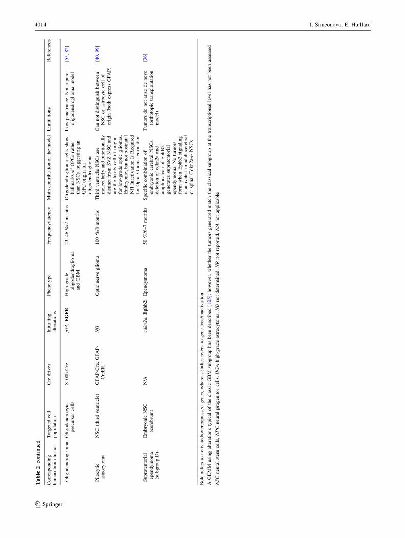
Oli
goden
dro
gli
om
aO
ligoden
dro
cyte
pre
curs
or
cell
sS
100b-C
rep53,
EG
FR
Hig
h-g
rade
oli
goden
dro
gli
om
aan
dG
BM
23–46
%/2
month
sO
ligoden
dro
gli
om
ace
lls
show
hal
lmar
ks
of
OP
Cs
rath
erth
anN
SC
s,su
gges
ting
anO
PC
ori
gin
for
oli
goden
dro
gli
om
a
Low
pen
etra
nce
.N
ot
apure
oli
goden
dro
gli
om
am
odel
[55
,82
]
Pil
ocy
tic
astr
ocy
tom
aN
SC
(thir
dven
tric
le)
GF
AP
-Cre
,G
FA
P-
Cre
ER
Nf1
Opti
cner
ve
gli
om
a100
%/8
month
sT
hir
dven
tric
leN
SC
sar
em
ole
cula
rly
and
funct
ional
lydis
tinct
from
SV
ZN
SC
and
are
the
likel
yce
llof
ori
gin
for
low
-gra
de
opti
cgli
om
as.
Em
bry
onic
,but
not
post
nat
alN
f1In
acti
vat
ion
IsR
equir
edfo
rO
pti
cG
liom
aF
orm
atio
n
Can
not
dis
tinguis
hbet
wee
nN
SC
or
astr
ocy
tece
llof
ori
gin
(both
expre
ssG
FA
P)
[40
,99
]
Supra
tento
rial
epen
dym
om
a(s
ubgro
up
D)
Em
bry
onic
NS
C(c
ereb
rum
)N
/Acd
kn2a,
Ep
hb
2E
pen
dym
om
a50
%/6
–7
month
sS
pec
ific
com
bin
atio
nof
embry
onic
cere
bra
lN
SC
s,del
etio
nof
cdkn2a
and
ampli
fica
tion
of
EphB
2gen
erat
essu
pra
tento
rial
epen
dym
om
a,N
otu
mors
form
when
Ephb2
signal
ing
isac
tivat
edin
adult
cere
bra
lor
spin
alC
dkn2a-
/-N
SC
s
Tum
ors
do
not
aris
ede
novo
(ort
hoto
pic
tran
spla
nta
tion
model
)
[36
]
Bold
refe
rsto
acti
vat
ed/o
ver
expre
ssed
gen
es,
wher
eas
ital
ics
refe
rsto
gen
elo
ss/i
nac
tivat
ion
AG
EM
Musi
ng
alte
rati
ons
typic
alof
the
clas
sic
GB
Msu
bgro
up
has
bee
ndes
crib
ed[1
25
];how
ever
,w
het
her
the
tum
ors
gen
erat
edm
atch
the
clas
sica
lsu
bgro
up
atth
etr
ansc
ripti
onal
level
has
not
bee
nas
sess
ed
NSC
neu
ral
stem
cell
s,N
PC
neu
ral
pro
gen
itor
cell
s,H
GA
hig
h-g
rade
astr
ocy
tom
a,N
Dnot
det
erm
ined
,N
Rnot
report
ed,
N/A
not
appli
cable
4014 I. Simeonova, E. Huillard
123

(supratentorial EP), cerebellum (posterior fossa EP) and
spinal cord (spinal EP). Although ependymomas are his-
tologically similar, they have disparate prognosis, gene
expression and genetic alterations profiles [33, 34]. In
2005, the group of R. Gilbertson revealed that human
supratentorial and spinal EP, which are histologically
identical, exhibit gene expression profiles similar to murine
NSCs of the cerebral ventricle and spinal cord, respectively
[35]. In 2010, the same authors identified molecular sub-
groups of ependymomas, characterized by distinct
chromosomal alterations, focal amplifications and deletions
[36]. Importantly, they identified previously unknown
potential ependymoma oncogenes, such as EPHB2, which
is selectively amplified and overexpressed in the supra-
tentorial subgroup of EP. By comparing the transcriptome
of EP from different regions with that of mouse NSCs of
different location and developmental stage, further showed
that subgroups of ependymoma match regionally, devel-
opmentally and genetically distinct NSCs. For example, the
transcriptome of a subgroup of supratentorial ependymoma
with amplified EPHB2 and CDKN2A loss (subgroup D)
closely matched that of embryonic cerebral Cdkn2a-/-
NSCs, whereas spinal ependymomas resembled adult wild-
type NSCs from the spinal cord. Activation of Ephb2 sig-
naling in embryonic cerebral Cdkn2a-/- NSCs generated
tumors when these cells were transplanted into immuno-
deficient hosts. This became the first model of EP,
accurately recapitulating the tumor histology and gene
expression profiles of the EPHB2/CDKN2A subgroup. It
also revealed high enrichment for regulators of neural
differentiation and maintenance, particularly ion transport
and synaptogenesis, suggesting a previously unsuspected
role for this pathway in this particular ependymoma sub-
group formation [36]. Contrary to embryonic cerebral
NSC, no tumors were generated when Ephb2 signaling was
activated in adult cerebral or spinal Cdkn2a-/- NSCs [36].
Interestingly, supratentorial tumors tend to occur more
frequently in children, whereas spinal EP mostly occurs in
adults. These results suggest that supratentorial EP may
derive from the transformation of NSCs during embryonic
development, whereas spinal EP may originate from the
transformation of NSCs during adulthood. Models for other
ependymal subgroups, for instance adult spinal ependy-
moma, remain to be established.
Pediatric astrocytomas
The molecular alterations underlying the development of
astrocytomas in children are not as characterized as in
adults. Molecular alterations in high-grade pediatric
astrocytomas (anaplastic astrocytoma and GBM) are
starting to be described [37, 38], but in the case of low-
grade tumors, the cellular origins and molecular alterations
are less well understood. Pilocytic astrocytomas (PA) are
low-grade tumors that often develop in the optic pathway
and cerebellum, and are frequently observed in patients
affected with neurofibromatosis type 1 (NF1) genetic dis-
order [39]. In a mouse model of pediatric astrocytoma
deficient for the Nf1 gene, Lee da et al. [40] recently
showed that for optic glioma to develop, Nf1 loss must
occur in a restricted cell population within a restricted
developmental window. Indeed, inactivation of Nf1 in
embryonic NSCs from the third ventricle resulted in optic
glioma formation, but inactivation in adult NSCs failed to
trigger gliomagenesis. Interestingly, only NSCs from the
3rd ventricle (and not those of the lateral ventricle) are
sensitive to specific mutations of PA, such as
KIAA1549:BRAF, a gene fusion found in the majority of
PA located in the hypothalamus/optic pathway regions [40,
41]. In accordance with this data, third ventricle NSCs are
molecularly distinct from NSCs of the subventricular zone,
the latter being the supposed cells of origin for adult gli-
omas [40]. Together, these observations point NSC from
the third ventricle as putative cells of origin for pediatric
optic gliomas.
High-grade astrocytomas and glioblastomas
Glioblastomas (GBM or high-grade astrocytomas WHO
grade IV) represent 54 % of all gliomas [1]. Most GBM in
the adult arise de novo, in the absence of a preexisting
tumor (primary GBM). Secondary GBM progress from a
low-grade astrocytoma, and occur in younger patients. In
2008, the Cancer Genome Atlas Research Network repor-
ted a comprehensive genomic and transcriptomic
characterization of over 200 GBM cases. This study
showed that the vast majority of GBM harbor alterations in
three core signaling pathways: receptor tyrosine kinase
(RTK)/Ras/phosphoinositide 3-kinase (PI3K), p53 and RB
pathways [42]. Subsequent studies have classified human
GBM into several subgroups (classical, mesenchymal,
neural, proneural), based on their genomic alterations, gene
expression and DNA methylation profiles [43–46]. Corre-
lations with clinical data revealed that the proneural
subgroup is associated with better survival, whereas the
mesenchymal subgroup has the worst prognosis and is
more resistant to conventional therapies. Recently, Sturm
et al. [47] have further refined this classification by sub-
classifying GBM into six groups, with respect to
characteristics in global DNA methylation and transcrip-
tome patterns, hotspot mutations, DNA copy-number
alterations, patient age and tumor location.
The majority of GEMMs of GBM have used combina-
tions of tumor suppressors p53 and/or Rb inactivation
(directly or through Cdkn2a deletion), and the activation of
pro-survival RTK and Ras signaling (through Pten and Nf1
Genetic models of brain tumors 4015
123

deletion or RTK/Ras activation) [6, 48–50]. These models
have provided essential clues on the identity of cell of
origin for GBM and HGA. The group of L. Parada has
developed a series of mouse strains harboring conditional
alleles for Nf1, p53 and Pten [51, 52]. These models
demonstrated that adult neural stem/progenitor cells can
give rise to malignant astrocytomas in vivo, whereas more
mature cell types cannot [53]. This finding was confirmed
in a different model developed by Jacques et al. [54]. The
authors used adenovirus-mediated Cre delivery to delete
Pten and p53 in adult NSCs of the subventricular zone and
in mature parenchymal astrocytes. They found that specific
deletion of both genes in adult NSCs, but not astrocytes,
gave rise to brain tumors. Other studies suggest that oli-
godendrocyte precursor cells (OPCs) can serve as tumor-
initiating cells [17, 55, 56]. Liu et al. used the MADM
system to generate high-grade astrocytomas by initiating
p53 and Nf1 deletion specifically in NSCs. Interestingly,
they found that the population that massively expanded at
premalignant stages was OPC —not NSCs or other lin-
eages— and the resulting tumors displayed many features
of this cell type. Moreover, introducing the p53 and Nf1
deletion directly in the OPC population resulted in the
formation of gliomas indistinguishable from NSC-initiated
tumors [17]. These studies suggest a model in which NSCs
may be the cells in which the genetic alterations initially
occur, but oligodendrocyte progenitor cells may be the
glioma-initiating cells of origin, in which the genetic
alterations have a functional impact. In contrast with this
hypothesis, the Verma group was able to generate high-
grade gliomas from the transformation of mature neurons
and astrocytes [57]. Lentivirus-mediated knock-down of
both p53 and Nf1 in mature neurons of the cortex led to the
formation of gliomas with GBM features. Likewise, tar-
geting of cortical mature astrocytes with an activated form
of Ras (H-RasV12) combined with an shRNA against p53
induced tumor formation. As tumors progressed, the
transduced cells eventually lost expression of the astrocytic
marker GFAP and turned on progenitor/stem cell markers.
These findings suggest that differentiated cells, by under-
going dedifferentiation or trans-differentiation, can also be
the cells of origin for gliomas. Although it is possible
experimentally to induce gliomas from differentiated cell
types, this mechanism may concern a minority of tumors.
Indeed, it seems more likely that NSCs, which cycle
throughout the lifespan of the individual, may be more
prone to acquire mutations leading to tumor formation.
By comparing the gene expression profiles of GBM sub-
groups to different murine neural cell types, Verhaak et al.
[43, 45] showed that subgroups harbor distinct predominant
alterations and match distinct neural cell types. These data
suggest that GBM subgroups may arise from distinct cell
populations, which are susceptible to distinct genetic
alterations. Several GEMMs have been used to test whether
combinations of different genetic alterations with different
cells of origin generate the different subgroups (Table 2).
Knocking-down p53 and activating Ras signaling in mature
astrocytes generated tumors that resembled the mesenchy-
mal subgroup of GBM at the transcriptomic level, while
targeting NSCs with the same mutations yielded tumors with
a neural signature [57]. In a different model, targeting p53
and Nf1 mutations to OPCs led to tumors that only resembled
the proneural subtype of human GBM [17]. Finally, inacti-
vating p53, Pten and Rb1 alleles in both adult NSCs and
mature astrocytes generated HGA that could be segregated
into three distinct subgroups resembling human molecular
subgroups of GBM [58]. These results suggest that sub-
groups do not necessarily correlate with the nature of the
initiating alterations, but rather depend on the identity of the
targeted cell type. Interestingly, Chow et al. [58] found that
most tumors with mesenchymal subgroup gene signature
arose from the pons or the basal hypothalamus. This implies
that adult NSCs and astrocytes from these regions are distinct
at the molecular level from NSCs and astrocytes from other
brain regions. Alternatively, these studies suggest that the
regional microenvironment may influence NSC to generate a
given subgroup (see section on microenvironment below).
As GEMMs become more sophisticated, they start to
provide insights into the different stages of gliomagenesis.
By comparing tumor formation in mice inactivated for Rb
family members, constitutively activated K-Ras
(KRasG12D), Pten loss or combinations of these alterations,
Song et al. [59] were able to reconstitute the sequence of
events necessary for GBM tumor initiation and progression.
Inactivation of Rb family proteins was required to initiate
tumorigenesis, and activation of K-Ras signaling induced
tumor progression from low-grade to high-grade. This
transition was accompanied by the spontaneous occurrence
of p53 mutations. Additional Pten loss (engineered or
spontaneous) drove progression to grade IV tumors [59].
Oligodendrogliomas
Oligodendrogliomas are the second most frequent gliomas
in population [60]. Several genomic alterations are com-
monly found in oligodendrogliomas [61, 62] and frequently
associated, indicating that they may play a key role in the
initiation and/or maintenance of oligodendrogliomas [63].
The most frequent alteration is a combined loss of one copy
of chromosome arms 1p and 19q (1p19q codeletion), which
has been observed in 60–90 % of oligodendrogliomas and
is associated with a better outcome for patients [61].
Although this alteration was discovered nearly 20 years
ago, very little is known on its functional impact on the
biology of the tumor. Another common alteration, a het-
erozygote mutation of isocitrate dehydrogenase 1 (IDH1)
4016 I. Simeonova, E. Huillard
123

on Arg132 (R132H), is found in about 80 % of oligoden-
drogliomas [64]. The mutated IDH enzyme reduces a-
ketoglutarate (a-KG) to D-2-hydroxyglutarate (D-2HG)
[65]. D-2HG acts as an oncometabolite, leading to pro-
found cell modifications including histone and DNA
hypermethylation, inhibition of cell differentiation and
increased proliferation [66, 67]. Interestingly, virtually all
1p19q codeleted gliomas are mutated for IDH1 [68]. More
recently, mutations within the core promoter of telomerase
reverse transcriptase (TERT) have been found in 100 % of
oligodendrogliomas with 1p19q codeletion and exclusively
in this group of tumors [69]. These mutations confer
increased transcriptional activity from the TERT promoter
[70], and ultimately an increased telomerase activity,
which is an important step in the immortalization process
[71]. Finally, two recent whole exome studies have
reported that the Capicua transcriptional repressor gene
(CIC) is frequently and specifically mutated in 1p19q
codeleted oligodendrogliomas [72, 73]. In addition to these
genomic alterations, increased EGFR and PDGF/PDGFR
expression is frequently observed in oligodendrogliomas
[74, 75]. Subgroups are being defined based on these
molecular alterations and appear to have distinct prognosis
and response to chemotherapy (reviewed in [76]). How-
ever, the precise sequence of molecular alterations leading
to the oligodendrogliomas formation is not completely
understood.
Only a few models for oligodendrogliomas have been
developed. These models nonetheless gave critical insights
into the nature of the cell of origin for oligodendrogliomas.
In most models, activation of the PDGF signaling pathway
was used to generate oligodendrogliomas. Retroviral
mediated delivery of PDGF-B, a ligand for PDGFRa, in
NPCs or OPCs of mouse embryos or neonates induced
oligodendroglioma formation [77–79]. Adult progenitor
cells seem to require additional alterations to be trans-
formed: infusion of PDGF-B in the lateral ventricles of the
adult mouse brain is not sufficient to promote tumorigen-
esis [80]. Likewise, infection of subcortical white matter
OPCs with a PDGF-B retrovirus does not lead to tumor
formation, and tumors are generated only in a Pten-null;
p53-null background [56]. However, these tumors resemble
GBM and not oligodendrogliomas. Therefore, the nature of
the genetic alterations and/or the developmental stage of
the targeted cells may determine the phenotype—oligo-
dendroglioma vs GBM—of the tumor. Another model of
oligodendroglioma is the GEMM expressing an activated
allele of EGFR (v-erbB). Transgenic mice expressing
v-erbB in glial cells develop oligodendrogliomas [81]. v-
erbB mice carrying deletion of Cdkn2a or p53 develop
tumors with an increased penetrance and grade. In the v-
erbB;p53-null model, tumor cells show characteristics of
OPCs, similar to human oligodendrogliomas, and it was
shown that cells with features of OPCs, rather than NSCs,
drive oligodendroglioma formation in mice [55, 81, 82]. In
accordance with these data, human oligodendrogliomas
were reported to associate with white matter tracts, where
OPCs reside, rather than lateral ventricles, suggesting an
origin from white matter progenitor cells [55]. Taken
together, these studies suggest that OPCs may be the cells
of origin for oligodendrogliomas. A GEMM for the 1p19q
codeletion has yet to be developed. Furthermore, the recent
identification of IDH1, CIC and TERT mutations will lead
to new models that should give more insights into the
mechanisms of oligodendroglioma genesis.
All together, studies using GEMMs of brain tumors
suggest that molecular heterogeneity may be due to sub-
groups originating from distinct cell types in different brain
locations. In addition, populations of neural progenitors
appear to be susceptible to particular genetic lesions, sug-
gesting a synergistic effect between these alterations and
signaling pathways specific of the cell type of origin [25].
The final goal being the development of effective therapies,
a better understanding of the molecular alterations and the
role of the microenvironment can lead to the identification
of relevant therapeutical targets and to understanding of the
mechanisms of therapy resistance.
GEMMs to investigate interactions between genetic
alterations and lineage-specific factors
For their growth, tumor cells rely on cell-specific factors
that are normal regulators of the lineage of origin [83].
Tumor growth is driven by cancer stem cells, which share
features with normal adult stem cells, such as the ability to
self-renew and the potential to differentiate into distinct
lineages. Therefore, genes controlling the proliferation and
differentiation of normal stem cells may also regulate the
biology of cancer stem cells. Mouse models have provided
insights into the role of these developmental genes in the
tumorigenic process.
Olig2 is a transcriptional repressor that is a key regulator
of glial cell fate during CNS development [84]. Olig2 is
exclusively expressed in the central nervous system, where
it plays distinct roles depending on the developmental
stage. Early during CNS development, Olig2 controls the
replication-competent state of neural progenitor cells. Later
on, it is required for the specification of oligodendrocytes
and subsets of neurons [85]. Olig2 protein is expressed in
almost all gliomas [86]. Importantly, OLIG2 is expressed
in virtually all CD133? tumor-initiating cells and in the
vast majority of Ki67? proliferating cells in GBM [87],
suggesting that it may promote tumor formation. To test
this hypothesis, we used an orthotopic mouse model of
high-grade astrocytoma that combines loss of the Cdkn2a
Genetic models of brain tumors 4017
123

tumor suppressor and expression of a constitutively acti-
vated form of EGFR (EGFRvIII). We found that no tumors
formed when Olig2 was absent. We further showed that
Olig2 is required for proliferation of tumorigenic neural
progenitors, as well as normal progenitor cells. This action
is partly mediated through repression of the cell cycle
inhibitor p21, an effector of the p53 pathway [87]. We also
found that Olig2 affects a key posttranslational modifica-
tion of p53 in both normal and malignant neural
progenitors, thereby antagonizing the interaction of p53
with promoter elements of multiple target genes [88].
Interestingly, Olig2 tumorigenic potential and antagonistic
action on p53 is dependent upon its phosphorylation on a
triple serine motif: absence of phosphorylation in this
region impairs Olig2 pro-tumorigenic activity [89]. These
studies identify Olig2 as a regulator of p53 activity in the
central nervous system and suggest that Olig2 may con-
tribute to p53 inactivation in the subset of GBM with wild-
type p53.
Atoh1 (or Math1) is a proneural basic helix-loop-helix
(bHLH) transcription factor highly expressed in GNP
cells of the cerebellum [90]. Atoh1 is a key factor in
cerebellar development, acting downstream of Shh sig-
naling to regulate GNP proliferation [91]. Importantly,
Atoh1 is highly expressed in the Shh-dependent MB
subset [92], suggesting that it may act as a lineage
dependency transcription factor in these tumors. Indeed,
studies using GEMMs have shown that Atoh1 is required
for MB formation [91, 93]. Recent work by Forget et al.
[94] now provide insights into the mechanisms by which
Shh regulate Atoh1 function. Using Huwe1-deficient
mice, the authors show that Shh regulates Atoh1 stability
by preventing its phospho-dependent degradation by the
E3 ubiquitin ligase Huwe1 [94]. Atoh1 accumulate in
Huwe1-deficient GNPs, leading to migration and differ-
entiation defects. Importantly, Huwe1 is strongly down
regulated in tumor-prone Ptch1?/- heterozygous mice,
and low HUWE1 expression is associated with poor
prognosis only within the SHH subgroup of human MB
[94]. This study identifies the developmental Huwe1-
Atoh1 module as a critical regulator of SHH-subtype MB.
GEMMs to study tumor–stroma interactions
The tumor microenvironment represents the non-neoplastic
cell types that are embedded in or adjacent to the tumor. It
is composed of numerous cell types and molecules, among
which immune cells (microglia, the resident immune cells
of the brain, peripheral macrophages, infiltrating lympho-
cytes), extracellular matrix (ECM) components, non-
neoplastic neural cells (astrocytes, oligodendrocytes, neu-
rons) and the specialized vasculature structure known as
the blood brain barrier (BBB), which is composed of
endothelial cells, pericytes, astrocytes). All these cell
populations interact with tumor cells to modulate, posi-
tively or negatively, tumor growth. GEMMs have provided
insights into the contribution of stromal elements, such as
microglia and non-neoplastic NPCs, in brain tumor
development.
Microglial contribution to tumorigenesis
Glioma-infiltrating macrophages and microglia constitute a
large proportion of tumor mass [95]. Current evidence
based on rodent experimental models indicates pro-
tumorigenic action of microglia on tumor cells. For
instance, GEMMs engineered to express the Herpes Sim-
plex Virus TK specifically in the C11b ? microglia/
macrophage lineage transplanted with glioma cells and
infused with ganciclovir led to an 80 % decrease in tumor
volume [96]. Soluble factors released from glioma stimu-
late microglial toll-like receptors TLRs, resulting in
microglial MT1-MMP expression via the TLR downstream
signaling molecules MyD88 and p38 MAPK. In turn, MT1-
MMP expression and activity in these immune cells pro-
motes glioma cell invasion and tumor expansion [96].
Additional work has shown that microglia may promote
glioma cell migration and invasiveness through release of
interleukin (IL)-6, IL-18 [97, 98].
Another demonstration of the contribution of microglia
to tumor formation came from studies using the Nf1
GEMM. Mice heterozygous for Nf1 (Nf1?/- mice) or
lacking Nf1 expression in astroglial cells alone (GFAPCre;
Nf1flox/flox mice) do not develop brain tumors. Nf1 ± mice
with conditional Nf1 inactivation in astroglial progenitors
(GFAPCre; Nf1flox/- mice) develop low-grade optic nerve
gliomas, similar to children affected with NF1 syndrome
[99]. This study shows that non-neoplastic Nf1?/- cells
provide a permissive environment required for glioma
formation. In fact, subsequent studies have revealed that
microglia was an important contributor to tumor growth in
the Nf1 model [100]. Genetic ablation of microglia, using a
CD11b-TK transgenic mouse reduced Nf1 optic glioma
proliferation during both tumor maintenance and tumor
development [100]. Nf1?/- microglia express high levels
of meningioma-expressed antigen-5 (MGEA5) and
CXCL12, and these were shown to act as glioma-promot-
ing molecules [101].
Recent studies highlight the importance of microglia in
defining tumor subgroups. Indeed, gliomasphere cultures of
the proneural subgroup can differentiate into a mesenchy-
mal subgroup. This transition is dependent upon activation
of the TNF-a/NK-jB pathway and is accompanied by
increased resistance to radiation. Interestingly, TNF-a is
secreted by microglia [102]. Microglia has been shown to
4018 I. Simeonova, E. Huillard
123

promote glioma migration and tumor growth and to pre-
dominantly infiltrate highly malignant tumors [95]. These
observations suggest a role of microglia in a proneural to
mesenchymal transition. Importantly, GBM patients with a
mesenchymal gene signature and NF-jB activation show a
poor response to radiation therapy and have a shorter sur-
vival. In this regard, GEMMs will be useful to further
explore the influence of regional microglia in mesenchymal
GBM development and progression.
Role of endogenous neural progenitor cells
and astrocytes
Mouse models have shown that endogenous NPCs are
recruited to the tumor site, where they exert tumor-sup-
pressive activities [95]. GEMMs were used to label
specifically NPCs, either through retroviral injection,
which labels dividing cells, or through the use of transgenic
mice with reporter-gene activity in endogenous NPCs (i.e.
Nestin-GFP mouse) by injection of retrovirus [103–105]. In
a syngenic glioma model, Walzlein et al. demonstrated that
endogenous NPCs engineered to express GFP are recruited
from the SVZ to the tumor, where they induce cell death.
The same group later demonstrated that NPCs secrete en-
dovanilloids that activate the TRPV1 receptor expressed by
glioma cells, triggering the ATF3-dependent ER stress
pathway resulting in cell death [106]. Importantly, an en-
dovanilloid agonist was effective against xenografted
human GBM cells and prolonged survival [106].
The group of Eric Holland has taken advantage of its
PDGF-driven GEMM to characterize non-neoplastic
astrocytes in the glioma microenvironment. By combining
this model to a GFAP-GFP mouse line to label reactive
astrocytes, they showed that tumor-associated astrocytes
have increased expression of MHC class II molecules and
components of antigen presentation pathway [107]. They
identified a gene signature for glioblastoma-associated
astrocytes; these genes were mainly expressed in the stro-
mal compartment of the tumor, and were associated with
survival in the proneural subtype of human glioma [107].
GEMMs have also been used to show that non-cell-of-
origin derived cells within glioma environment in the
mouse can be corrupted to become bona fide tumor cells. In
a PDGF-driven rat glioma model, Assanah et al. [103]
showed that injection of a retrovirus encoding PDGF and
GFP induced tumors composed of both GFP? and GFP-
negative cells. In this model, most of the proliferating
Ki67? cells were GFP-negative. Using a PDGF-driven
GEMM, Fomchenko et al., also showed that tumors were
composed of GFP? and GFP-negative cells, comprising
Olig2? proliferating NPCs and displaying a gene expres-
sion profile similar to that of tumor cells. GFP-
negative « recruited » cells were able, upon
retransplantation in mice, to initiate gliomas [105]. Whe-
ther this applies to human GBM (or GBM of other
subgroups) is unknown, as it is almost impossible to dis-
tinguish GBM cells from recruited progenitor cells in
human tumors, because of their phenotypic similarities.
Nonetheless these studies will have to be repeated in other
GEMMs and xenograft models.
GEMMs to study the blood–tumor barrier (BTB)
The blood brain barrier is a specialized vascular structure
tightly regulating homeostasis of the central nervous sys-
tem. It is composed of specialized endothelial cells
connected by tight junctions, a capillary basement mem-
brane, astrocyte end-feet ensheathing the vessels, and
pericytes [108]. The BBB tightly regulates the influx/efflux
of nutrients, endogenous compounds such as hormones and
immune cells between systemic circulation and the brain
parenchyma. Complex interplay between endothelial cells,
ECM, tight junction and astrocyte polarity has to be pre-
cisely controlled for barrier integrity. However, the
mechanisms involved between the different cell types, as
well as their respective role on BBB integrity, are not
known. GEMMs have been used to define the contribution
of tight junction proteins, transporters, or ECM compo-
nents in BBB development and biology [109]. GEMMs
have been used to demonstrate the role of key signaling,
such as VEGF, Notch, Wnt pathways, in BBB development
and maintenance. Shh signaling was recently shown to
have a protective role at the BBB [110]. Shh is secreted by
astrocytes and its receptor Ptc1 is expressed on endothelial
cells. Blocking the Hh pathway with cyclopamine injected
into mice induced BBB disruption, as demonstrated by the
increased extravasation of exogenous dextran and blood-
derived leukocytes. Furthermore, specific depletion of Smo
in endothelial cells, using a Tie2-Cre;Smofl/fl mouse line
significantly increased BBB permeability [110].
In GBM, the BBB is disorganized, displaying alterations
in ECM, tight junctions and basement membrane. The
water channel aquaporin-4 (AQP-4), which is specifically
localized at the astrocytic endfoot membranes in physio-
logical BBB, is upregulated in GBM and redistributed over
the cellular surface. Upregulation of AQP4 in GBM is
associated with loss of Agrin, a component of the ECM.
Accordingly, in Agrin-deficient mice, the BBB is intact but
AQP4 is no longer restricted vessel-directed membrane
domains [111, 112].
Modeling the blood–tumor barrier is important for
delivery of therapeutic substances to malignant brain
tumors. It is believed that 98 % of small-molecule drugs do
not cross the BBB [113]. For a small-molecule drug to
cross the BBB in pharmacologically significant amounts,
the drug must fit the dual molecular characteristics for
Genetic models of brain tumors 4019
123
lipid-mediated free diffusion across the BBB: molecular
mass \400-Da and high lipid solubility [113]. Drugs that
fulfill these criteria may still not be able to reach thera-
peutic levels in the CNS, as they may be substrates of the
efflux transporters at the BBB [114].
Although there are numerous GEMMs that have been
used to study the formation and biology of BBB under
physiological conditions, to our knowledge there is no
reported GEMM that investigates the biology of the BBB
in the tumoral context. Agarwal and colleagues recently
used a xenograft glioma mouse model to study the action of
the BBB on the brain distribution of Erlotinib, a small
molecule EGFR inhibitor. These authors found that co-
treatment of tumor-bearing mice with Erlotinib and phar-
macological inhibitors of the BBB efflux transporters P-gp
and Bcrp, increased Erlotinib concentration in the brain
parenchyma [114].
It is not known whether and how tumor cells contribute
to BBB permeability in the context of a brain tumor. There
is a need for GEMMs modeling the blood–tumor barrier to
understand the contribution of astrocytes and tumor cells to
the dysregulation of the BBB.
Role of GEMMs as preclinical models
Preclinical trials have been mostly performed using mouse
xenografts of human brain tumor cell lines. However, these
models did not translate into successful results in sub-
sequent clinical trials, probably due to fundamental
differences between cell-line derived models and patients’
tumors (see the section on ‘‘In vivo approaches for mod-
eling brain tumors’’).
GEMMs combined to non-invasive imaging techniques
(i.e. MRI, bioluminescence) that allow monitoring of tumor
development longitudinally [115], are starting to be used
for testing targeted therapies. An example is the Rosa26
ODD-Luciferase mouse that can be used to monitor tissue
hypoxia in vivo [116]. Since solid tumors often display
hypoxic regions, this mouse line can be used to monitor
spontaneous tumor development, as has been shown for
mammary gland tumors [117]. In another study, Sonabend
et al. developed a proneural GBM mouse model by
injecting PDGF-IRES-Cre retrovirus into the subcortical
white matter of adult mice bearing the floxed tumor sup-
pressors p53 and Pten and a luciferase reporter preceded by
a floxed transcriptional stop. The addition of the Cre
recombinase in this system inactivates p53 and Pten and
simultaneously activates the luciferase reporter. Biolumi-
nescence monitoring of tumors in vivo allowed for therapy
benefit evaluation in this mouse model [118].
Recently, a comprehensive in vitro and in vivo high-
throughput screen used a mouse model of the Ephb2-
amplified ependymoma subgroup to identify potential
therapies with predicted toxicity against normal NSCs
[119]. Importantly, this study found kinases of the insulin
growth factor (IGF) signaling and centrosome cycle path-
ways as regulators of this subtype of ependymoma.
Furthermore, this screening model was used to evaluate the
activity of the Food and Drug Administration (FDA)-
approved anticancer drug 5-fluorouracil (5-FU) against
ependymoma cells. Intravenous injection of 5-FU pro-
longed the survival of tumor-bearing mice, with minimal
toxicity against normal NSCs.
In medulloblastoma, the Ptc1?/-p53-null mouse model
was used to assess the activity of a small molecule inhibitor
of the Shh pathway. This resulted in reduced tumor growth
and increased tumor-free survival [120]. These findings
were translated into a clinical trial, in which treatment with
the SMO inhibitor GDC-0449 resulted in rapid, although
transient, regression of the tumor [121]. However, the
tumor recurred and analysis of the patient’s tumor cells
revealed the presence, in addition to the initial PTCH1
mutation, of a mutant SMO (SMO-D473H) [122]. This
mutant was shown to be insensitive to inhibition by GDC-
0449. Mouse models are now offering ways to explore the
mechanisms of resistance to this SMO inhibitor. A GDC-
0449-resistant mouse model was created, in which
Ptc1?/-p53-null mice intermittently treated with GDC-
0449 eventually stopped responding to the drug [122].
Interestingly, the SMO-D473H mutation identified in the
patient was found in one cell line derived from this model.
However, in two other cell lines, SMO mutations were not
detected, suggesting the existence of additional mecha-
nisms to GDC-0449 resistance.
GEMMs are key to understand the mechanisms of tumor
progression and dissemination. In the sleeping beauty-dri-
ven medulloblastoma model, addition of SB transposition
to the Ptc1 model results in metastatic dissemination
through the cerebrospinal fluid system, similar to the pat-
tern seen in children [22]. The patterns of genomic
alterations and DNA methylation in this model were con-
firmed in human tumor-metastases pairs, and further
revealed that metastases shared similarities with each
other, but were distinct from the primary tumor. These
findings suggested that metastases evolve from rare popu-
lations of cells in the primary tumors. Signaling pathways
that are enriched in both primary tumor and metastases
may represent promising therapeutical targets. Targeting
the insulin signaling pathway, the most significantly
deregulated in metastases, may be a promising strategy,
because it is also involved in primary MB tumors [22].
PI3K has been proposed to contribute to the resistance of
MB to Shh inhibitors. Therefore, combined inhibition of
Shh and Akt/PI3K pathways may represent a promising
therapeutical opportunity.
4020 I. Simeonova, E. Huillard
123
It is important to note that not all primary MB respond
to treatment with Shh inhibitors, in agreement with the
existence of distinct molecular subgroups. Two indepen-
dent studies showed that a Myc-driven mouse model of
MB does not respond to Shh inhibitors [30, 31]. Moreover,
Pei et al. showed that small molecule inhibitors of the
PI3K/mTOR pathway inhibited tumor cell growth in vitro
and extended survival of tumor-bearing mice, identifying
this signaling pathway as a potential therapeutic target for
this MB subgroup.
In pediatric gliomas, we recently described the genera-
tion of a high-grade astrocytoma model that recapitulates
the genetics of a subset of tumors with mutated BRAFV600E
and CDKN2A deletion. We found that treatment with a
combination of the BRAFV600E inhibitor PLX4720 and the
CDK4/6 inhibitor PD0332991 had synergistic activity
against intracranial tumors in vivo [123]. We observed a
similar effect in mouse xenografts of genetically relevant
human glioma cell lines. Because these inhibitors yield
encouraging clinical results in other cancers, our findings
indicate a rational therapeutic strategy for treating a subset
of pediatric astrocytomas with BRAFV600E mutation and
CDKN2A deficiency.
In adult gliomas, a few GEMMs have been used as
preclinical models. In a PDGF-driven glioma model, Mo-
mota et al. [124] showed that the combination of the
chemotherapeutic agent temozolomide with the Akt
inhibitor perifosine was more effective than temozolomide
treatment alone. This therapeutic strategy may be relevant
for the proneural GBM subgroup that harbors the highest
number of PDGFRa amplifications [42, 43]. More recently,
Zhu et al. [125] used an EGFR-driven GEMM of GBM to
test the action of a small molecule inhibitor of the
molecular chaperone Hsp90 that promotes cell prolifera-
tion. Treated mice displayed increased survival without
toxicity. Because Hsp90 inhibition targets multiple sig-
naling pathways, such as EGFR, AKT, CDK4 and
CyclinD1, it represents an attractive therapeutic candidate
for the treatment of GBMs.
Recently, Chen et al. [126] have created a GEMM to
investigate the cellular mechanisms of glioma recurrence.
By breeding hGFAP-Cre;Nf1?/-;p53fl/fl;Ptenfl/? glioma-
prone mice to a Nestin-DThymidine kinase-GFP transgenic
mouse line, in which GFP? NSCs and glioma stem cells
(GSCs) can be specifically ablated by ganciclovir, they
showed that survival is prolonged upon ganciclovir treat-
ment. In this model, treatment with temozolomide, the first
line chemotherapy in GBM, ablated proliferating cells but
not quiescent GSCs. This treatment resulted in tumor
recurrence. Conversely, combining temozolomide and
ganciclovir eliminated both proliferating cells and GSCs,
abrogating tumor formation. This study provides the first
in vivo evidence of the existence of GSCs, which are
responsible for tumor re-growth after treatment. It also
highlights the benefits of combination therapies that target
different cellular compartments and functions. Recently,
Sarkar et al. [127] demonstrated that microglia activation
by the FDA-approved drug Amphotericin B (AmpB)
reduces GSC tumorigenicity by preventing their prolifera-
tion and promoting their differentiation. AmpB treatment
of mice bearing xenogeneic or syngeneic GSCs extended
survival without substantial toxicity. Importantly, mono-
cytes and macrophages from GBM patients were unable to
reduce GSC sphere formation unless they were exposed to
AmpB. This observation was confirmed with a case report,
where a high-grade astrocytoma patient treated with AmpB
for a fungal infection went into apparent remission [127].
These data suggest that AmpB has the potential of treating
gliomas and open encouraging new approaches to glioma
therapy.
All together, these preclinical studies have provided
key aspects on our understanding of tumor development
and progression. Studies from Chen et al. and Sarkar
et al., demonstrating glioma maintenance by GSC and
their targeting by AmpB, and from Wu et al. demon-
strating divergence of metastases from primary
medulloblastoma and their resistance to therapeutics
against the primary tumor, have important clinical
implications [22, 126]. They explain why generalized
cancer treatments fail, and open new therapeutic avenues
to target distinct cellular populations. Tumor subtypes do
not respond to the same treatments, therefore, there is a
real need to use genetically relevant models during pre-
clinical drug development [30, 31, 123]. However,
although the frequency of animals acquiring tumors in
GEMMs of brain tumors is high in general (see Table 2),
a model with a low tumor frequency would not be suited
for preclinical testing, due to the lack of animals.
Limitations of GEMMs
In the previous sections, we have described how GEMMs
can provide critical insights into tumor formation and be
used as preclinical models. However, there are several
disadvantages to consider when using GEMMs. Distinct
genetic background of mouse strains can modify tumor
onset and spectrum, complicating the interpretation of a
mutation’s effects [128]. For instance, mice heterozygous
for Nf1 and p53 develop brain gliomas at a higher fre-
quency and grade on the C57Bl/6J background compared
to the 129S4/SvJae background [129]. Therefore, the
genetic background may affect gene function and tumor
susceptibility. Because GEMMs are often analyzed on one
genetic background, caution should be exerted when
extrapolating results from mice to humans.
Genetic models of brain tumors 4021
123

Fundamental differences between mouse and human
cells may affect tumor development [130]. For instance,
there are differences between mouse and human immune
systems, including components of the T cell and B cell
signaling pathways, cytokine and chemokine expression
[131]. Moreover, the basal metabolic rate is higher in mice
and humans, and rodents diverge in the spectrum of car-
cinogens they are susceptible to [130]. In addition, murine
cells have active telomerase, whereas telomerase expres-
sion is repressed in most mature cells in humans—but
activated in embryonic and adult stem cells. In addition,
mice have longer telomeres than humans [132]. All these
biological differences may have an impact on tumor
development.
Importantly, the tumor spectrum in mice may be differ-
ent from human pathology. Mice tend to spontaneously
develop sarcomas (tumors of mesodermal origin), whereas
humans are more prone to carcinomas (epithelial tumors)
[130]. Telomere dynamics may explain this difference, as
shown by the combined telomerase- and p53-deficient mice
that display a shift in the tumor spectrum towards epithelial
carcinomas [133]. Some GEMMs of tumor suppressor
genes often display a tumor spectrum differing from the
human pathology. In humans, inheritance of one mutant RB
allele predisposes to retinoblastoma but also to osteosar-
comas later in life. In contrast, mice hemizygous for Rb
develop pituitary gland tumors but no retinoblastoma.
Retinoblastomas develop in these mice only when a related
Rb family member, p107, is deleted [134]. In addition,
tumor development in mouse may be different from tumor
development in human, due to differences in transcriptional
networks. For example, we recently found that the rapid
evolution of tandem repeated sequences containing p53
response elements might shape differences in p53 tran-
scriptional networks among mammalian species. The
characterization of species-specific p53 target genes may
provide a key to improve human cancer modeling in mice
[135].
Brain development and organization are globally similar
between humans and mice. However, there are anatomical
and cellular differences. Beside the presence of convolu-
tions (gyrencephalic brain) in the human cerebral cortex
that are absent in the rodent brain, some brain areas show
distinct architecture. For instance, in the adult mouse brain,
the SVZ harbors NSCs that are tightly associated with
ependymal cells, which give rise to transit-amplifying
cells; in turn, these cells generate neuroblasts that migrate
to the olfactory bulb [24]. In contrast, the human SVZ
displays a large band of astrocytes with stem cell proper-
ties, separated from the ependymal by a hypocellular gap
region [136]. In human brain specimens, neuroblasts
migrating from the SVZ are not always observed in the
adult [136, 137], but have been detected in infants, where
the structure of the SVZ resembles that of the mouse [138].
Other brain cells also show specific functional character-
istics in humans. Protoplasmic astrocytes in the human
brain are larger and extend more processes than in mice. In
addition, human astrocytes propagate calcium signals
several fold faster than do rodent astrocytes [139]. Fur-
thermore, the human neocortex also harbors several
subclasses of astrocytes, such as interlaminar astrocytes,
whose function is unknown and that are not represented in
rodents [139]. Whether these species-specific aspects may
affect tumor development needs further investigation.
Finally, current models do not recapitulate the intratu-
moral heterogeneity seen in patients. A recent study used
single-cell RNA sequencing to profile individual cells from
the same GBM tumor [140]. The authors observed exten-
sive intratumoral heterogeneity at the transcriptional level,
including mosaic expression and/or mutational status of
RTKs and other signaling molecules. Furthermore, they
found that individual tumors contain a spectrum of glio-
blastoma subtypes [140]. Creating GEMMs that model the
intratumoral heterogeneity will be the next challenge in
experimental neuro-oncology.
Conclusions
In vivo models provide unique and valuable information
regarding the way brain tumors develop. Comprehensive
molecular and cellular characterization of GEMMs will
lead to the identification of biomarkers for early stages of
tumor development. The generation of molecularly defined
GEMMs matching human brain tumor subgroups holds
encouraging prospects. Nevertheless, differences in mouse
genetic background modifiers, as well as possible differ-
ences in tumor development and drug response between
mice and humans, should be taken into account in the
appreciation of drug efficiency. Therefore, it is essential to
integrate accurate GEMMs of brain tumors with xenograft
models, genomic and developmental studies, to provide
powerful platforms for target identification and drug
testing.
Acknowledgments The authors wish to thank M. Kalamarides, F.
Fernandes and F. Toledo for comments on the manuscript. Research
in Huillard lab is supported by Fondation ARC pour la Recherche sur
le Cancer, Ligue Nationale Contre le Cancer, Institut National de la
Sante et de la Recherche Medicale (INSERM), European Union (FP7-
PEOPLE-CIG-2012). Research in Huillard lab has received funding
from the program ‘‘Investissements d’avenir’’ ANR-10-IAIHU-06. IS
is a recipient of a fellowship from the Ligue Nationale Contre le
Cancer.
Open Access This article is distributed under the terms of the
Creative Commons Attribution License which permits any use, dis-
tribution, and reproduction in any medium, provided the original
author(s) and the source are credited.
4022 I. Simeonova, E. Huillard
123

References
1. Dolecek TA, Propp JM, Stroup NE, Kruchko C (2012) CBTRUS
statistical report: primary brain and central nervous system
tumors diagnosed in the United States in 2005–2009. Neuro
Oncol 14(Suppl 5):v1–v49
2. Northcott PA, Korshunov A, Pfister SM, Taylor MD (2012) The
clinical implications of medulloblastoma subgroups. Nat Rev
Neurol 8:340–351
3. Barth RF, Kaur B (2009) Rat brain tumor models in experi-
mental neuro-oncology: the C6, 9L, T9, RG2, F98, BT4C, RT-2
and CNS-1 gliomas. J Neurooncol 94:299–312
4. Lee J, Kotliarova S, Kotliarov Y, Li A, Su Q et al (2006) Tumor
stem cells derived from glioblastomas cultured in bFGF and
EGF more closely mirror the phenotype and genotype of pri-
mary tumors than do serum-cultured cell lines. Cancer Cell
9:391–403
5. Romer J, Curran T (2005) Targeting medulloblastoma: small-
molecule inhibitors of the Sonic Hedgehog pathway as potential
cancer therapeutics. Cancer Res 65:4975–4978
6. Huszthy PC, Daphu I, Niclou SP, Stieber D, Nigro JM, et al.
(2012) In vivo models of primary brain tumors: pitfalls and
perspectives. Neuro Oncol 14:979–993
7. Singh SK, Hawkins C, Clarke ID, Squire JA, Bayani J et al
(2004) Identification of human brain tumour initiating cells.
Nature 432:396–401
8. Venere M, Fine HA, Dirks PB, Rich JN (2011) Cancer stem
cells in gliomas: identifying and understanding the apex cell in
cancer’s hierarchy. Glia 59:1148–1154
9. Chen R, Nishimura MC, Bumbaca SM, Kharbanda S, Forrest
WF et al (2010) A hierarchy of self-renewing tumor-initiating
cell types in glioblastoma. Cancer Cell 17:362–375
10. Wang J, Sakariassen PO, Tsinkalovsky O, Immervoll H, Boe SO
et al (2008) CD133 negative glioma cells form tumors in nude
rats and give rise to CD133 positive cells. Int J Cancer
122:761–768
11. Joo KM, Kim J, Jin J, Kim M, Seol HJ et al (2013) Patient-
specific orthotopic glioblastoma xenograft models recapitulate
the histopathology and biology of human glioblastomas in situ.
Cell Rep 3:260–273
12. Klink B, Miletic H, Stieber D, Huszthy PC, Valenzuela JA et al
(2013) A novel, diffusely infiltrative xenograft model of human
anaplastic oligodendroglioma with mutations in FUBP1, CIC,
and IDH1. PLoS One 8:e59773
13. van Miltenburg MH, Jonkers J (2012) Using genetically engi-
neered mouse models to validate candidate cancer genes and test
new therapeutic approaches. Curr Opin Genet Dev 22:21–27
14. Cheon DJ, Orsulic S (2011) Mouse models of cancer. Annu Rev
Pathol 6:95–119
15. Serrano M, Lee H, Chin L, Cordon-Cardo C, Beach D et al
(1996) Role of the INK4a locus in tumor suppression and cell
mortality. Cell 85:27–37
16. Zong H, Espinosa JS, Su HH, Muzumdar MD, Luo L (2005)
Mosaic analysis with double markers in mice. Cell 121:479–492
17. Liu C, Sage JC, Miller MR, Verhaak RG, Hippenmeyer S et al
(2011) Mosaic analysis with double markers reveals tumor cell
of origin in glioma. Cell 146:209–221
18. Orsulic S (2002) An RCAS-TVA-based approach to designer
mouse models. Mamm Genome 13:543–547
19. Copeland NG, Jenkins NA (2010) Harnessing transposons for
cancer gene discovery. Nat Rev Cancer 10:696–706
20. Koso H, Takeda H, Yew CC, Ward JM, Nariai N et al (2012)
Transposon mutagenesis identifies genes that transform neural
stem cells into glioma-initiating cells. Proc Natl Acad Sci USA
109:E2998–E3007
21. Bender AM, Collier LS, Rodriguez FJ, Tieu C, Larson JD et al
(2010) Sleeping beauty-mediated somatic mutagenesis impli-
cates CSF1 in the formation of high-grade astrocytomas. Cancer
Res 70:3557–3565
22. Wu X, Northcott PA, Dubuc A, Dupuy AJ, Shih DJ et al (2012)
Clonal selection drives genetic divergence of metastatic
medulloblastoma. Nature 482:529–533
23. Genovesi LA, Ng CG, Davis MJ, Remke M, Taylor MD et al
(2013) Sleeping beauty mutagenesis in a mouse medulloblas-
toma model defines networks that discriminate between human
molecular subgroups. Proc Natl Acad Sci USA 110:E4325–
E4334
24. Kriegstein A, Alvarez-Buylla A (2009) The glial nature of
embryonic and adult neural stem cells. Annu Rev Neurosci
32:149–184
25. Gilbertson RJ (2011) Mapping cancer origins. Cell 145:25–29
26. Schuller U, Heine VM, Mao J, Kho AT, Dillon AK et al (2008)
Acquisition of granule neuron precursor identity is a critical
determinant of progenitor cell competence to form Shh-induced
medulloblastoma. Cancer Cell 14:123–134
27. Yang ZJ, Ellis T, Markant SL, Read TA, Kessler JD et al (2008)
Medulloblastoma can be initiated by deletion of patched in
lineage-restricted progenitors or stem cells. Cancer Cell
14:135–145
28. Li P, Du F, Yuelling LW, Lin T, Muradimova RE et al
(2013) A population of nestin-expressing progenitors in the
cerebellum exhibits increased tumorigenicity. Nat Neurosci
16:1737–1744
29. Gibson P, Tong Y, Robinson G, Thompson MC, Currle DS et al
(2010) Subtypes of medulloblastoma have distinct develop-
mental origins. Nature 468:1095–1099
30. Kawauchi D, Robinson G, Uziel T, Gibson P, Rehg J et al
(2012) A mouse model of the most aggressive subgroup of
human medulloblastoma. Cancer Cell 21:168–180
31. Pei Y, Moore CE, Wang J, Tewari AK, Eroshkin A et al (2012)
An animal model of MYC-driven medulloblastoma. Cancer Cell
21:155–167
32. Witt H, Korshunov A, Pfister SM, Milde T (2012) Molecular
approaches to ependymoma: the next step(s). Curr Opin Neurol
25:745–750
33. Witt H, Mack SC, Ryzhova M, Bender S, Sill M et al (2011)
Delineation of two clinically and molecularly distinct subgroups
of posterior fossa ependymoma. Cancer Cell 20:143–157
34. Modena P, Lualdi E, Facchinetti F, Veltman J, Reid JF et al
(2006) Identification of tumor-specific molecular signatures in
intracranial ependymoma and association with clinical charac-
teristics. J Clin Oncol 24:5223–5233
35. Taylor MD, Poppleton H, Fuller C, Su X, Liu Y et al (2005)
Radial glia cells are candidate stem cells of ependymoma.
Cancer Cell 8:323–335
36. Johnson RA, Wright KD, Poppleton H, Mohankumar KM,
Finkelstein D et al (2010) Cross-species genomics matches
driver mutations and cell compartments to model ependymoma.
Nature 466:632–636
37. Schwartzentruber J, Korshunov A, Liu XY, Jones DT, Pfaff E
et al (2012) Driver mutations in histone H3.3 and chromatin
remodelling genes in paediatric glioblastoma. Nature
482:226–231
38. Wu G, Broniscer A, McEachron TA, Lu C, Paugh BS et al
(2012) Somatic histone H3 alterations in pediatric diffuse
intrinsic pontine gliomas and non-brainstem glioblastomas. Nat
Genet 44:251–253
39. Gutmann DH, Parada LF, Silva AJ, Ratner N (2012) Neurofi-
bromatosis type 1: modeling CNS dysfunction. J Neurosci
32:14087–14093
Genetic models of brain tumors 4023
123

40. da Lee Y, Gianino SM, Gutmann DH (2012) Innate neural stem
cell heterogeneity determines the patterning of glioma formation
in children. Cancer Cell 22:131–138
41. Jacob K, Albrecht S, Sollier C, Faury D, Sader E et al (2009)
Duplication of 7q34 is specific to juvenile pilocytic astrocyto-
mas and a hallmark of cerebellar and optic pathway tumours. Br
J Cancer 101:722–733
42. The Cancer Genome Atlas Research Network (2008) Compre-
hensive genomic characterization defines human glioblastoma
genes and core pathways. Nature 455:1061–1068
43. Verhaak RG, Hoadley KA, Purdom E, Wang V, Qi Y et al
(2010) Integrated genomic analysis identifies clinically relevant
subtypes of glioblastoma characterized by abnormalities in
PDGFRA, IDH1, EGFR, and NF1. Cancer Cell 17:98–110
44. Brennan C, Momota H, Hambardzumyan D, Ozawa T, Tandon
A et al (2009) Glioblastoma subclasses can be defined by
activity among signal transduction pathways and associated
genomic alterations. PLoS One 4:e7752
45. Phillips HS, Kharbanda S, Chen R, Forrest WF, Soriano RH et al
(2006) Molecular subclasses of high-grade glioma predict
prognosis, delineate a pattern of disease progression, and
resemble stages in neurogenesis. Cancer Cell 9:157–173
46. Noushmehr H, Weisenberger DJ, Diefes K, Phillips HS, Pujara
K et al (2010) Identification of a CpG island methylator phe-
notype that defines a distinct subgroup of glioma. Cancer Cell
17:510–522
47. Sturm D, Witt H, Hovestadt V, Khuong-Quang DA, Jones DT
et al (2012) Hotspot mutations in H3F3A and IDH1 define
distinct epigenetic and biological subgroups of glioblastoma.
Cancer Cell 22:425–437
48. Chen J, McKay RM, Parada LF (2012) Malignant glioma: les-
sons from genomics, mouse models, and stem cells. Cell
149:36–47
49. Jones TS, Holland EC (2011) Animal models for glioma drug
discovery. Expert Opin Drug Discov 6:1271–1283
50. Wu X, Northcott PA, Croul S, Taylor MD (2011) Mouse models
of medulloblastoma. Chin J Cancer 30:442–449
51. Kwon CH, Zhao D, Chen J, Alcantara S, Li Y et al (2008) Pten
haploinsufficiency accelerates formation of high-grade astrocy-
tomas. Cancer Res 68:3286–3294
52. Zhu Y, Guignard F, Zhao D, Liu L, Burns DK et al (2005) Early
inactivation of p53 tumor suppressor gene cooperating with NF1
loss induces malignant astrocytoma. Cancer Cell 8:119–130
53. Alcantara Llaguno S, Chen J, Kwon CH, Jackson EL, Li Y et al
(2009) Malignant astrocytomas originate from neural stem/
progenitor cells in a somatic tumor suppressor mouse model.
Cancer Cell 15:45–56
54. Jacques TS, Swales A, Brzozowski MJ, Henriquez NV, Linehan
JM et al (2010) Combinations of genetic mutations in the adult
neural stem cell compartment determine brain tumour pheno-
types. EMBO J 29:222–235
55. Persson AI, Petritsch C, Swartling FJ, Itsara M, Sim FJ et al
(2010) Non-stem cell origin for oligodendroglioma. Cancer Cell
18:669–682
56. Lei L, Sonabend AM, Guarnieri P, Soderquist C, Ludwig T et al
(2011) Glioblastoma models reveal the connection between
adult glial progenitors and the proneural phenotype. PLoS One
6:e20041
57. Friedmann-Morvinski D, Bushong EA, Ke E, Soda Y, Marum-
oto T et al (2012) Dedifferentiation of neurons and astrocytes by
oncogenes can induce gliomas in mice. Science 338:1080–1084
58. Chow LM, Endersby R, Zhu X, Rankin S, Qu C et al (2011)
Cooperativity within and among Pten, p53, and Rb pathways
induces high-grade astrocytoma in adult brain. Cancer Cell
19:305–316
59. Song Y, Zhang Q, Kutlu B, Difilippantonio S, Bash R et al
(2013) Evolutionary etiology of high-grade astrocytomas. Proc
Natl Acad Sci USA 110:17933–17938
60. Rigau V, Zouaoui S, Mathieu-Daude H, Darlix A, Maran A et al
(2011) French brain tumor database: 5-year histological results
on 25 756 cases. Brain Pathol 21:633–644
61. Bromberg JE, van den Bent MJ (2009) Oligodendrogliomas:
molecular biology and treatment. Oncologist 14:155–163
62. Ducray F, Idbaih A, Wang XW, Cheneau C, Labussiere M et al
(2011) Predictive and prognostic factors for gliomas. Expert Rev
Anticancer Ther 11:781–789
63. Riemenschneider MJ, Jeuken JW, Wesseling P, Reifenberger G
(2010) Molecular diagnostics of gliomas: state of the art. Acta
Neuropathol 120:567–584
64. Yan H, Parsons DW, Jin G, McLendon R, Rasheed BA et al
(2009) IDH1 and IDH2 mutations in gliomas. N Engl J Med
360:765–773
65. Dang L, White DW, Gross S, Bennett BD, Bittinger MA et al
(2009) Cancer-associated IDH1 mutations produce 2-hydroxy-
glutarate. Nature 462:739–744
66. Turcan S, Rohle D, Goenka A, Walsh LA, Fang F et al (2012)
IDH1 mutation is sufficient to establish the glioma hyperme-
thylator phenotype. Nature 483:479–483
67. Rohle D, Popovici-Muller J, Palaskas N, Turcan S, Grommes
C et al (2013) An inhibitor of mutant IDH1 delays growth
and promotes differentiation of glioma cells. Science
340:626–630
68. Labussiere M, Idbaih A, Wang XW, Marie Y, Boisselier B et al
(2010) All the 1p19q codeleted gliomas are mutated on IDH1 or
IDH2. Neurology 74:1886–1890
69. Killela PJ, Reitman ZJ, Jiao Y, Bettegowda C, Agrawal N et al
(2013) TERT promoter mutations occur frequently in gliomas
and a subset of tumors derived from cells with low rates of self-
renewal. Proc Natl Acad Sci USA 110:6021–6026
70. Huang FW, Hodis E, Xu MJ, Kryukov GV, Chin L et al (2013)
Highly recurrent TERT promoter mutations in human mela-
noma. Science 339:957–959
71. Hanahan D, Weinberg RA (2011) Hallmarks of cancer: the next
generation. Cell 144:646–674
72. Bettegowda C, Agrawal N, Jiao Y, Sausen M, Wood LD et al
(2011) Mutations in CIC and FUBP1 contribute to human oli-
godendroglioma. Science 333:1453–1455
73. Yip S, Butterfield YS, Morozova O, Chittaranjan S, Blough MD
et al (2012) Concurrent CIC mutations, IDH mutations, and 1p/
19q loss distinguish oligodendrogliomas from other cancers.
J Pathol 226:7–16
74. Reifenberger J, Reifenberger G, Ichimura K, Schmidt EE,
Wechsler W et al (1996) Epidermal growth factor receptor
expression in oligodendroglial tumors. Am J Pathol 149:29–35
75. Robinson S, Cohen M, Prayson R, Ransohoff RM, Tabrizi N
et al (2001) Constitutive expression of growth-related oncogene
and its receptor in oligodendrogliomas. Neurosurgery
48:864–873 (discussion 873–864)
76. Alentorn A, Sanson M, Idbaih A (2012) Oligodendrogliomas:
new insights from the genetics and perspectives. Curr Opin
Oncol 24:687–693
77. Dai C, Celestino JC, Okada Y, Louis DN, Fuller GN et al (2001)
PDGF autocrine stimulation dedifferentiates cultured astrocytes
and induces oligodendrogliomas and oligoastrocytomas from
neural progenitors and astrocytes in vivo. Genes Dev
15:1913–1925
78. Appolloni I, Calzolari F, Tutucci E, Caviglia S, Terrile M et al
(2009) PDGF-B induces a homogeneous class of oligodendro-
gliomas from embryonic neural progenitors. Int J Cancer
124:2251–2259
4024 I. Simeonova, E. Huillard
123

79. Lindberg N, Kastemar M, Olofsson T, Smits A, Uhrbom L
(2009) Oligodendrocyte progenitor cells can act as cell of origin
for experimental glioma. Oncogene 28:2266–2275
80. Jackson EL, Garcia-Verdugo JM, Gil-Perotin S, Roy M, Qui-
nones-Hinojosa A et al (2006) PDGFR alpha-positive B cells are
neural stem cells in the adult SVZ that form glioma-like growths
in response to increased PDGF signaling. Neuron 51:187–199
81. Weiss WA, Burns MJ, Hackett C, Aldape K, Hill JR et al (2003)
Genetic determinants of malignancy in a mouse model for oli-
godendroglioma. Cancer Res 63:1589–1595
82. Sugiarto S, Persson AI, Munoz EG, Waldhuber M, Lamagna C
et al (2011) Asymmetry-defective oligodendrocyte progenitors
are glioma precursors. Cancer Cell 20:328–340
83. Garraway LA, Sellers WR (2006) Lineage dependency and
lineage-survival oncogenes in human cancer. Nat Rev Cancer
6:593–602
84. Rowitch DH (2004) Glial specification in the vertebrate neural
tube. Nat Rev Neurosci 5:409–419
85. Meijer DH, Kane MF, Mehta S, Liu H, Harrington E et al (2012)
Separated at birth? The functional and molecular divergence of
OLIG1 and OLIG2. Nat Rev Neurosci 13:819–831
86. Ligon KL, Alberta JA, Kho AT, Weiss J, Kwaan MR et al
(2004) The oligodendroglial lineage marker OLIG2 is univer-
sally expressed in diffuse gliomas. J Neuropathol Exp Neurol
63:499–509
87. Ligon KL, Huillard E, Mehta S, Kesari S, Liu H et al (2007)
Olig2-regulated lineage-restricted pathway controls replication
competence in neural stem cells and malignant glioma. Neuron
53:503–517
88. Mehta S, Huillard E, Kesari S, Maire CL, Golebiowski D et al
(2011) The central nervous system-restricted transcription factor
Olig2 opposes p53 responses to genotoxic damage in neural
progenitors and malignant glioma. Cancer Cell 19:359–371
89. Sun Y, Meijer DH, Alberta JA, Mehta S, Kane MF et al (2011)
Phosphorylation state of Olig2 regulates proliferation of neural
progenitors. Neuron 69:906–917
90. Hoshino M (2012) Neuronal subtype specification in the cere-
bellum and dorsal hindbrain. Dev Growth Differ 54:317–326
91. Flora A, Klisch TJ, Schuster G, Zoghbi HY (2009) Deletion of
Atoh1 disrupts Sonic Hedgehog signaling in the developing
cerebellum and prevents medulloblastoma. Science
326:1424–1427
92. Salsano E, Pollo B, Eoli M, Giordana MT, Finocchiaro G (2004)
Expression of MATH1, a marker of cerebellar granule cell
progenitors, identifies different medulloblastoma sub-types.
Neurosci Lett 370:180–185
93. Ayrault O, Zhao H, Zindy F, Qu C, Sherr CJ et al (2010) Atoh1
inhibits neuronal differentiation and collaborates with Gli1 to
generate medulloblastoma-initiating cells. Cancer Res
70:5618–5627
94. Forget A, Bihannic L, Cigna SM, Lefevre C, Remke M et al
(2014) Shh signaling protects atoh1 from degradation mediated
by the e3 ubiquitin ligase huwe1 in neural precursors Dev Cell
29:649–661
95. Charles NA, Holland EC, Gilbertson R, Glass R, Kettenmann H
(2011) The brain tumor microenvironment. Glia 59:1169–1180
96. Markovic DS, Vinnakota K, Chirasani S, Synowitz M, Raguet H
et al (2009) Gliomas induce and exploit microglial MT1-MMP
expression for tumor expansion. Proc Natl Acad Sci USA
106:12530–12535
97. Zhang J, Sarkar S, Cua R, Zhou Y, Hader W et al (2012) A
dialog between glioma and microglia that promotes tumor
invasiveness through the CCL2/CCR2/interleukin-6 axis. Car-
cinogenesis 33:312–319
98. Yeh WL, Lu DY, Liou HC, Fu WM (2012) A forward loop
between glioma and microglia: glioma-derived extracellular
matrix-activated microglia secrete IL-18 to enhance the migra-
tion of glioma cells. J Cell Physiol 227:558–568
99. Bajenaru ML, Hernandez MR, Perry A, Zhu Y, Parada LF et al
(2003) Optic nerve glioma in mice requires astrocyte Nf1 gene
inactivation and Nf1 brain heterozygosity. Cancer Res
63:8573–8577
100. Thangarajh M, Gutmann DH (2012) Review: low-grade gliomas as
neurodevelopmental disorders: insights from mouse models of
neurofibromatosis-1. Neuropathol Appl Neurobiol 38:241–253
101. Daginakatte GC, Gutmann DH (2007) Neurofibromatosis-1
(Nf1) heterozygous brain microglia elaborate paracrine factors
that promote Nf1-deficient astrocyte and glioma growth. Hum
Mol Genet 16:1098–1112
102. Bhat KP, Balasubramaniyan V, Vaillant B, Ezhilarasan R,
Hummelink K et al (2013) Mesenchymal differentiation medi-
ated by NF-kappaB promotes radiation resistance in
glioblastoma. Cancer Cell 24:331–346
103. Assanah M, Lochhead R, Ogden A, Bruce J, Goldman J et al
(2006) Glial progenitors in adult white matter are driven to form
malignant gliomas by platelet-derived growth factor-expressing
retroviruses. J Neurosci 26:6781–6790
104. Walzlein JH, Synowitz M, Engels B, Markovic DS, Gab-
rusiewicz K et al (2008) The antitumorigenic response of neural
precursors depends on subventricular proliferation and age.
Stem Cells 26:2945–2954
105. Fomchenko EI, Dougherty JD, Helmy KY, Katz AM, Pietras A
et al (2011) Recruited cells can become transformed and over-
take PDGF-induced murine gliomas in vivo during tumor
progression. PLoS One 6:e20605
106. Stock K, Kumar J, Synowitz M, Petrosino S, Imperatore R et al
(2012) Neural precursor cells induce cell death of high-grade
astrocytomas through stimulation of TRPV1. Nat Med
18:1232–1238
107. Katz AM, Amankulor NM, Pitter K, Helmy K, Squatrito M et al
(2012) Astrocyte-specific expression patterns associated with the
PDGF-induced glioma microenvironment. PLoS One 7:e32453
108. Alvarez JI, Katayama T, Prat A (2013) Glial influence on the
blood brain barrier. Glia 61:1939–1958
109. Sohet F, Daneman R (2013) Genetic mouse models to study
blood-brain barrier development and function. Fluids Barriers
CNS 10:3
110. Alvarez JI, Dodelet-Devillers A, Kebir H, Ifergan I, Fabre PJ et al
(2011) The Hedgehog pathway promotes blood-brain barrier
integrity and CNS immune quiescence. Science 334:1727–1731
111. Wolburg H, Noell S, Fallier-Becker P, Mack AF, Wolburg-
Buchholz K (2012) The disturbed blood-brain barrier in human
glioblastoma. Mol Asp Med 33:579–589
112. Rauch SM, Huen K, Miller MC, Chaudry H, Lau M et al (2011)
Changes in brain beta-amyloid deposition and aquaporin 4
levels in response to altered agrin expression in mice. J Neuro-
pathol Exp Neurol 70:1124–1137
113. Pardridge WM (2012) Drug transport across the blood-brain
barrier. J Cereb Blood Flow Metab 32:1959–1972
114. Agarwal S, Manchanda P, Vogelbaum MA, Ohlfest JR, Elm-
quist WF (2013) Function of the blood-brain barrier and
restriction of drug delivery to invasive glioma cells: findings in
an orthotopic rat xenograft model of glioma. Drug Metab Dispos
41:33–39
115. Gross S, Piwnica-Worms D (2005) Spying on cancer: molecular
imaging in vivo with genetically encoded reporters. Cancer Cell
7:5–15
116. Safran M, Kim WY, O’Connell F, Flippin L, Gunzler V et al
(2006) Mouse model for noninvasive imaging of HIF prolyl
hydroxylase activity: assessment of an oral agent that stimulates
erythropoietin production. Proc Natl Acad Sci USA
103:105–110
Genetic models of brain tumors 4025
123

117. Goldman SJ, Chen E, Taylor R, Zhang S, Petrosky W et al (2011)
Use of the ODD-luciferase transgene for the non-invasive imaging
of spontaneous tumors in mice. PLoS One 6:e18269
118. Sonabend AM, Yun J, Lei L, Leung R, Soderquist C et al (2013)
Murine cell line model of proneural glioma for evaluation of
anti-tumor therapies. J Neurooncol 112:375–382
119. Atkinson JM, Shelat AA, Carcaboso AM, Kranenburg TA,
Arnold LA et al (2011) An integrated in vitro and in vivo high-
throughput screen identifies treatment leads for ependymoma.
Cancer Cell 20:384–399
120. Romer JT, Kimura H, Magdaleno S, Sasai K, Fuller C et al
(2004) Suppression of the Shh pathway using a small molecule
inhibitor eliminates medulloblastoma in Ptc1(±)p53(-/-) mice.
Cancer Cell 6:229–240
121. Rudin CM, Hann CL, Laterra J, Yauch RL, Callahan CA et al
(2009) Treatment of medulloblastoma with hedgehog pathway
inhibitor GDC-0449. N Engl J Med 361:1173–1178
122. Yauch RL, Dijkgraaf GJ, Alicke B, Januario T, Ahn CP et al
(2009) Smoothened mutation confers resistance to a Hedgehog
pathway inhibitor in medulloblastoma. Science 326:572–574
123. Huillard E, Hashizume R, Phillips JJ, Griveau A, Ihrie RA et al
(2012) Cooperative interactions of BRAFV600E kinase and
CDKN2A locus deficiency in pediatric malignant astrocytoma
as a basis for rational therapy. Proc Natl Acad Sci USA
109:8710–8715
124. Momota H, Nerio E, Holland EC (2005) Perifosine inhibits
multiple signaling pathways in glial progenitors and cooperates
with temozolomide to arrest cell proliferation in gliomas in vivo.
Cancer Res 65:7429–7435
125. Zhu H, Woolfenden S, Bronson RT, Jaffer ZM, Barluenga S
et al (2010) The novel Hsp90 inhibitor NXD30001 induces
tumor regression in a genetically engineered mouse model of
glioblastoma multiforme. Mol Cancer Ther 9:2618–2626
126. Chen J, Li Y, Yu TS, McKay RM, Burns DK et al (2012) A
restricted cell population propagates glioblastoma growth after
chemotherapy. Nature 488:522–526
127. Sarkar S, Doring A, Zemp FJ, Silva C, Lun X et al (2014)
Therapeutic activation of macrophages and microglia to sup-
press brain tumor-initiating cells. Nat Neurosci 17:46–55
128. Reilly KM, Loisel DA, Bronson RT, McLaughlin ME, Jacks T
(2000) Nf1; Trp53 mutant mice develop glioblastoma with
evidence of strain-specific effects. Nat Genet 26:109–113
129. Reilly KM, Tuskan RG, Christy E, Loisel DA, Ledger J et al
(2004) Susceptibility to astrocytoma in mice mutant for Nf1 and
Trp53 is linked to chromosome 11 and subject to epigenetic
effects. Proc Natl Acad Sci USA 101:13008–13013
130. Rangarajan A, Weinberg RA (2003) Opinion: comparative
biology of mouse versus human cells: modelling human cancer
in mice. Nat Rev Cancer 3:952–959
131. Mestas J, Hughes CC (2004) Of mice and not men: differences
between mouse and human immunology. J Immunol
172:2731–2738
132. Calado RT, Dumitriu B (2013) Telomere dynamics in mice and
humans. Semin Hematol 50:165–174
133. Chang S, Khoo C, DePinho RA (2001) Modeling chromosomal
instability and epithelial carcinogenesis in the telomerase-defi-
cient mouse. Semin Cancer Biol 11:227–239
134. Robanus-Maandag E, Dekker M, van der Valk M, Carrozza ML,
Jeanny JC et al (1998) p107 is a suppressor of retinoblastoma
development in pRb-deficient mice. Genes Dev 12:1599–1609
135. Simeonova I, Lejour V, Bardot B, Bouarich-Bourimi R, Morin
A et al (2012) Fuzzy tandem repeats containing p53 response
elements may define species-specific p53 target genes. PLoS
Genet 8:e1002731
136. Sanai N, Tramontin AD, Quinones-Hinojosa A, Barbaro NM,
Gupta N et al (2004) Unique astrocyte ribbon in adult human
brain contains neural stem cells but lacks chain migration.
Nature 427:740–744
137. Curtis MA, Kam M, Nannmark U, Anderson MF, Axell MZ et al
(2007) Human neuroblasts migrate to the olfactory bulb via a
lateral ventricular extension. Science 315:1243–1249
138. Sanai N, Nguyen T, Ihrie RA, Mirzadeh Z, Tsai HH et al (2011)
Corridors of migrating neurons in the human brain and their
decline during infancy. Nature 478:382–386
139. Oberheim NA, Takano T, Han X, He W, Lin JH et al (2009)
Uniquely hominid features of adult human astrocytes. J Neuro-
sci 29:3276–3287
140. Patel AP, Tirosh I, Trombetta JJ, Shalek AK, Gillespie SM et al
(2014) Single-cell RNA-seq highlights intratumoral heteroge-
neity in primary glioblastoma. Science 344:1396–1401
4026 I. Simeonova, E. Huillard
123


