Heel-Line Hyperpigmentation: A Variant ofSock-Line Hyperpigmentation After the Use of
Heel-Length Socks
Heather Ciliberto, M.D.,* David Berk, M.D.,† Pankaj Salphale, M.D.,‡and Susan Bayliss, M.D.*
*Division of Dermatology, Department of Internal Medicine and Pediatrics, Washington University and St. LouisChildren’s Hospital, St. Louis, Missouri, †School of Medicine, Washington University and St. Louis Children’s
Hospital, St. Louis, Missouri, ‡Shushrusha Hospital, Chandrapur, India
Abstract: Two infants developed hyperpigmented curvilinear patcheson the posterior heel after wearing heel-length socks. Both of the patient’slesions improved after discontinuing the use of the heel-length socks.Hyperpigmented patches called sock-line or mitten-line hyperpigmenta-tion have been reported at sites of tight elastic bands from socks ormittens in infants on the calves and wrists. Recognizing this clinical entityis important to differentiate it from other causes of linear lesions such aschild abuse or amniotic band syndrome.
CASE REPORTS
Patient 1
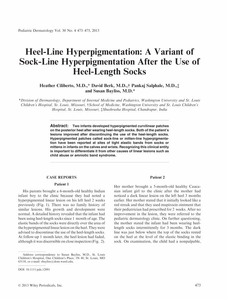
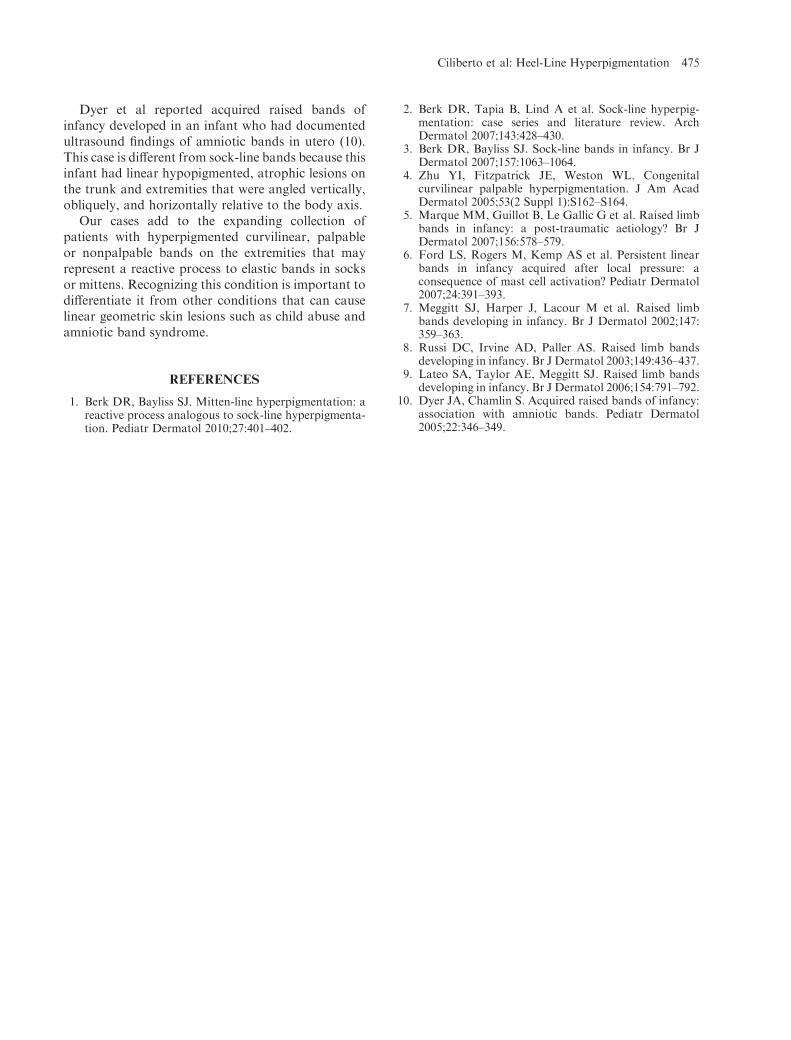
His parents brought a 6-month-old healthy Indianinfant boy to the clinic because they had noted ahyperpigmented linear lesion on his left heel 2 weekspreviously (Fig. 1). There was no family history ofsimilar lesions. His growth and development werenormal. A detailed history revealed that the infant hadbeen using heel-length socks since 1 month of age. Theelastic bands of the socks were directly over the area ofthe hyperpigmented linear lesionon theheel. Theywereadvised to discontinue the use of the heel-length socks.At follow-up 1 month later, the heel lesion had faded,although it was discernible on close inspection (Fig. 2).
Patient 2
Her mother brought a 5-month-old healthy Cauca-sian infant girl to the clinic after the mother hadnoticed a dark linear lesion on the left heel 3 monthsearlier. Her mother stated that it initially looked like ared streak and that they used mupirocin ointment thattheir pediatrician had prescribed for 2 weeks. After noimprovement in the lesion, they were referred to thepediatric dermatology clinic. On further questioning,the mother stated the infant had been wearing heel-length socks intermittently for 3 months. The darkline was just below where the top of the socks restedon the heel at the level of the elastic binding in thesock. On examination, the child had a nonpalpable,
Address correspondence to Susan Bayliss, M.D., St. LouisChildren’s Hospital, One Children’s Place, 3N 48, St. Louis, MO63110, or e-mail: [email protected].
DOI: 10.1111/pde.12091
© 2013 Wiley Periodicals, Inc. 473
Pediatric Dermatology Vol. 30 No. 4 473–475, 2013

curvilinear hyperpigmented patch along the left heel(Fig. 3). The lesion was not tender to palpation. Themother was advised to discontinue the use of the heel-length socks. On telephone follow-up the motherstated that the line was gone and had taken approx-imately 3 months to resolve.
DISCUSSION
Sock- or mitten-line hyperpigmentation has beendescribed in Caucasian, Asian, and African Americaninfants (1–3). It typically presents as hyperpigmentedpalpable or nonpalpable horizontal curvilinear patchesor plaques angled superiorly along the affected extrem-ity at the sites of contact of elastic bands from socks ormittens. Parents often report the presence of erythemaat the sites before the development of the hyperpigmen-tation. The lesions may be partially or fully circumfer-ential. In partially circumferential cases, the lesionstypically appear over the posterior calves as opposed tothe shins, perhaps because of a higher proportion of
subcutaneous fat in the calf or from greater pressure ofthe elastic band in this area. The lesions may beunilateral or bilateral andmay be single or multiple. Insome cases, parents are aware that the child had beenwearing socks or mittens that were too tight before thedevelopment of the lesions and have attributed themultiple lines on the same calf to be fromwearing socksof varying heights. Biopsy of these lesions has shownlentiginous melanocytic hyperplasia and basal layerhyperpigmentation that was thought to representpostinflammatory hyperpigmentation (2,4). Therewas no comment on the presence of adipocytes inthese biopsies. Further biopsies to analyze changes inthe fat, particularly in palpable lesions, may help todetermine whether the raised lesions are a form ofpanniculitis.
We believe that previous cases of sock- or mitten-line hyperpigmentation may have been reported underthe names congenital curvilinear palpable hyperpigmen-tation (neither of the cases in this article was congenital)(4) and acquired raised limb bands of infancy, orpersistent linear bands (5–9) in cases in which therewas no other evidence of amniotic bands. All of theprevious reports have been in infants or toddlers, withthe exception of one 18-year-old boy with mentalretardation who was reported to have raised hyper-pigmented linear lesions on his mid calves that weresaid to have been present since childhood (5). The timeto resolution of sock- or mitten-line hyperpigmenta-tion has been reported to vary between months andyears. The age of onset of these lesions in infancy couldbe a clue to the etiology being related to adipocytedamage due to the higher percentage of saturated fattyacids present in infantile fat. It may be that infants lackthe ability to move socks or mittens that are too tight,whereas older children or adults will take off the socksif the elastic bands are causing discomfort.
Figure 2. Nearly complete resolution of the lesion at 1-month follow-up.
Figure 3. Nonpalpable, curvilinear hyperpigmented patchon the left heel corresponding to a sock band.
Figure 1. Partially circumferential curvilinear hypopigmentedpatch on the left heel corresponding to a sock band.
474 Pediatric Dermatology Vol. 30 No. 4 July/August 2013
Dyer et al reported acquired raised bands ofinfancy developed in an infant who had documentedultrasound findings of amniotic bands in utero (10).This case is different from sock-line bands because thisinfant had linear hypopigmented, atrophic lesions onthe trunk and extremities that were angled vertically,obliquely, and horizontally relative to the body axis.
Our cases add to the expanding collection ofpatients with hyperpigmented curvilinear, palpableor nonpalpable bands on the extremities that mayrepresent a reactive process to elastic bands in socksor mittens. Recognizing this condition is important todifferentiate it from other conditions that can causelinear geometric skin lesions such as child abuse andamniotic band syndrome.
REFERENCES
1. Berk DR, Bayliss SJ. Mitten-line hyperpigmentation: areactive process analogous to sock-line hyperpigmenta-tion. Pediatr Dermatol 2010;27:401–402.
2. Berk DR, Tapia B, Lind A et al. Sock-line hyperpig-mentation: case series and literature review. ArchDermatol 2007;143:428–430.
3. Berk DR, Bayliss SJ. Sock-line bands in infancy. Br JDermatol 2007;157:1063–1064.
4. Zhu YI, Fitzpatrick JE, Weston WL. Congenitalcurvilinear palpable hyperpigmentation. J Am AcadDermatol 2005;53(2 Suppl 1):S162–S164.
5. Marque MM, Guillot B, Le Gallic G et al. Raised limbbands in infancy: a post-traumatic aetiology? Br JDermatol 2007;156:578–579.
6. Ford LS, Rogers M, Kemp AS et al. Persistent linearbands in infancy acquired after local pressure: aconsequence of mast cell activation? Pediatr Dermatol2007;24:391–393.
7. Meggitt SJ, Harper J, Lacour M et al. Raised limbbands developing in infancy. Br J Dermatol 2002;147:359–363.
8. Russi DC, Irvine AD, Paller AS. Raised limb bandsdeveloping in infancy. Br J Dermatol 2003;149:436–437.
9. Lateo SA, Taylor AE, Meggitt SJ. Raised limb bandsdeveloping in infancy. Br J Dermatol 2006;154:791–792.
10. Dyer JA, Chamlin S. Acquired raised bands of infancy:association with amniotic bands. Pediatr Dermatol2005;22:346–349.
Ciliberto et al: Heel-Line Hyperpigmentation 475


