Genetics
U.K. College of Nursing

Genes and Chromosomes
Each cell contains 23 pairs of matched chromosomes for a total of 46 chromosomes per cell.
One chromosome from each pair is inherited from each parent.
There are 22 pairs of autosomes, which control most traits in the body, and one pair of sex chromosomes, which determine gender and other traits.

Genes and Chromosomes
Some genes are dominant and their characteristics are expressed even if only on one chromosome.
Some genes are recessive and their characteristics will be expressed only if they are carried by both chromosomes in a pair.

Group Exercise
Human Variation

Punnet Squares
Visual representation of the principles of inheritance
Dominant trait--Capital letter
Recessive trait--Small letter
Male vs female trait/gene


Patterns of Inheritance
Autosomal DominantAutosomal RecessiveSex-Linked (or X-linked)
DominantSex-Linked (or X-linked)
RecessiveChromosomal
AbnormalitiesCongenital Anomalies

Autosomal Dominant
Trait appears in every generation (does not skip)
Both males and females are affected
Each pregnancy of an affected person has a 50% chance of producing an affected offspring

Autosomal Dominant Disorders
Disorders Huntington’s Disease Retinitis Pigmentosa Polycystic Kidney Disease Achodroplasia Marfan Syndrome

Autosomal Dominant Disorders- Marfan Syndrome

Autosomal Dominant Inheritance

Autosomal Dominant
Clinical Situation-- One parent is unaffected
One parent carries the defective gene for Marfan Syndrome
Draw the Punnet Square
Autosomal Dominant Punnett Square

Autosomal Recessive
Both parents are usually unaffected, but are carriers
Trait first appears only in siblings rather than in parents
Trait found equally in males and females
25% risk when both parents are carriers
Increased incidence with consanguinity

Autosomal Recessive
Disorders Phenylketonuria Fanconi’s Anemia Tay Sachs Disease Sickle Cell Anemia Cystic Fibrosis

Autosomal Recessive Inheritance

Autosomal Recessive
Clinical Situation Male carries the defective
gene for Tay Sachs disease
Female carries the defective gene for Tay Sachs disease
Draw the Punnett Square

Autosomal Recessive Punnett Square

X-linked Inheritance
Sex-Modified Traits - Dominant genes are expressed in both males & females but at differing frequencies Ex: Baldness - expressed as
dominant in males, but recessive in females, never as severe in females

X-linked Dominant
Very rareOften lethal in males
therefore few males present in the pedigree
Multiple miscarriages may be present
No carrier status, all individuals with the gene are affected
Trait appears in every generation

X-linked Dominant
Female children of affected males will all be affected (100% risk); no male to male transmission.
Homozygous females (both X chromosomes are affected) have a 100% chance of having an affected child of either sex.
Heterozygous females (only one X affected) have a 50% of having an affected child with each pregnancy.

X-linked Dominant Disorders
Hypophosphatemic Rickets
Fragile X Syndrome

Fragile X Syndrome

X-linked Dominant
Clinical Situation Male is affected with
hypophosphatemic rickets
Female is unaffected
Draw the Punnet square

X-linked Dominant Punnet Square

X-linked Recessive
Incidence of trait much higher among males in a kinship than among females
Trait cannot be transmitted from father to son
An affected male will pass the carrier status to all his daughters
Female carriers have a 50% risk of transmitting the gene to their offspring with each pregnancy

X-linked Recessive
Disorders Hemophilia A Duchenne’s Muscular
Dystrophy Color-Blindness

Duscenne’s Muscular Dystrophy

X-Linked Recessive Inheritance

X-linked Recessive
Clinical Situation Male is affected with
Hemophilia A
Female is normal (non-carrier)
Draw the Punnett SquareUse X1 for chomosome with
normal allele and X2 for chromosome with disease allele

X-linked Recessive Punnet Square
X-linked Recessive
Clinical Situation Male is normal
Female is a carrier of color-blindness
Draw the Punnett Square

X-linked Recessive Punnett Square

X-linked Recessive
Clinical Situation Male is affected with
Duschenne Muscular Dystrophy
Female is carrier of Duschenne Muscular Dystrophy
Draw the Punnett Square

X-linked Recessive Punnett Square

Genotype - The actual gene constitution of a given person.
Phenotype - The observable characteristics of a given person

Traits can be environmentally modified type 2 diabetes PKU
Traits can be medically modified Sickle cell disease (bone marrow
transplant) Polycysitc kidney disease (kidney
transplant)
However, genotype stays the same so next generation are not saved from condition

Group Exercise
Punnet Squares and Patterns of Inheritance

KaryotypesThe arranged representation of the chromosomal make-up of a cell nucleus


Chromosomal Abnormalities
Abnormalities in number of chromosomes Caused by nondisjunction:
failure of homologous chromosomes or sister chromatids to separate properly into different progeny cells
Monosomy - condition in which one chromosome of a pair is missing from a somatic cell

Monosomy X - Turners Syndrome

Monosomy--Turner’s Syndrome

Chromosomal Abnormalities
Trisomy - condition in which one chromosome in the pair is pesent in three copies in a somatic cell
Down Syndrome (21), Trisomy 13 or 18
Klinefelter’s Syndrome - XXY

Abnormalities of Chromosome NumberTrisomy


Chromosomal Structural Abnormalities
Deletions - absence of normal chromosomal material; can be terminal or interstitial
Duplications - presence of an extra copy of a chromosomal segment
Inversions - Intrachromosomal re-arrangement such that the rearranged section is inverted
Ring Chromosome - Fusion of the ends of a chromosome that forms a circle or ring

Chromosomal Structural Abnormalities
Translocations - Interchromosomal rearrangement; can be balanced (all chromosomal material is present) or unbalanced (chromosomal material has been gained or lost); can be reciprocal or Robertsonian
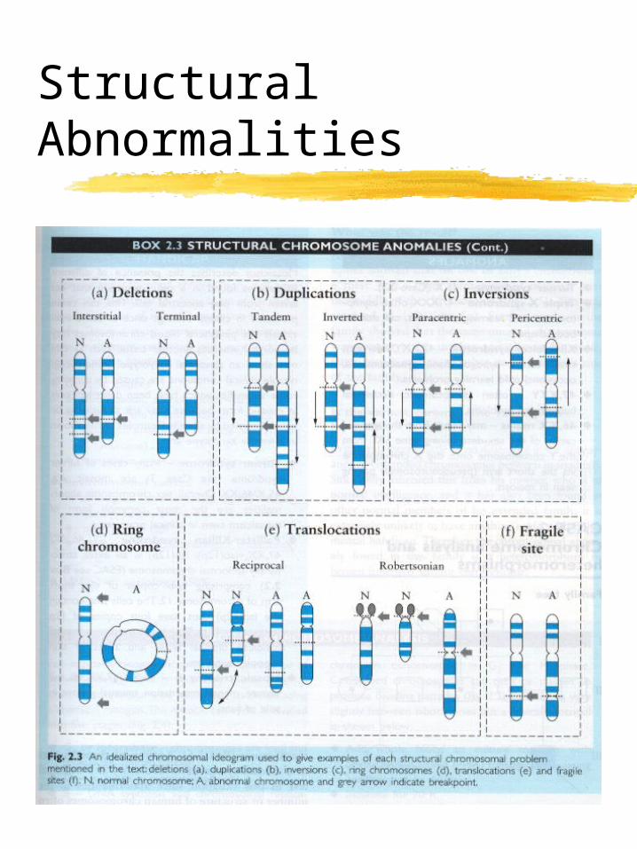
Structural Abnormalities

Group Exercise
Karyotype CD-Rom

Congenital Anomalies
Structural abnormalities present at birth
Are usually not identified with a known genetic cause
Cause may be a combination of genetic and environmental factors

Children at Risk for Congenital Anomalies
Positive family history of structual anomalies
Child with one known structural anomaly
The IUGR infantThe mentally retarded
childThe unusual appearing
child

Maternal Risk Factors
DiabetesPhenylketonuria (PKU)Seizure disorderAlcohol and substance
abuseRecurrent pregnancy loss

Teratogens
Environmental substances or exposures that result in functional or structural disability.
Any agent which when given to or ingested by a pregnant woman can produce a permanent morphologic or functional abnormality.
Agents Which Cause Teratogenesis
Drugs and Chemicals; Alcohol
Infections (viruses, TORCH)Radiation exposureFat-Soluble VitaminsNicotineHeat

Fetal Susceptibility to Teratogens
Gestational age at the time of exposure
Drug dosageRoute of administration of
agentGenetic predisposition of
fetus to respond to a particular agent

Gestational Susceptibility Factors
Days 1-17 Little effect
Days 18-60 Period of organogenesis Extreme sensitivity to major
structural abnormalities Days 61-270
Considerably reduced risks Functional abnormalities can still
occur

Pedigrees
A pictorial representation or diagram of the family history.
Allows visualization of relationships of affected individuals to other family members.
May indicate a pattern of inheritance
Helps pinpoint persons who should be examined or tested.

Pedigree Format
3 generations AT LEAST!!
Note name of informant
Roman numerals for generations
Number individuals on pedigree across families

Pedigree Pointers
Seek a balance between the need for asking specific versus general questions.
Ask specific questions about each individual as you construct the pedigree (birth defects, mental retardation, specific traits relevant to the diagnosis or concern)
Ask general questions about the whole family or section of a family. Can you think of any family characteristics (traits) or medical problems in more than one family member?

Pedigree Reminders
Multiple reproductive relationships “Have you had
children/pregnancies by anyone else?”
“Did you have any pregnancies prior to this relationship?”
Don’t forget half-sibs, abortions, miscarriages, stillbirths, previous marriages.

Pedigree Pointers
Indicate possible relationships “Sometimes when there is one
family member with cleft lip and palate, there are others in the family with little indentations in their lower lip, heart problems at birth, or poor vision and joint pain. Can you think of anyone in your family with anything like that?”
Use words the clients will understand seizures = fits = fainting spells


Group Exercise
Final Group Work


