Turkish Neurosurgery 6: 63 - 67, 1996 Yurt: Extradural Hnemntomn
Extradural Haematoma: Analysis of 190 Cases
Ekstradural Hematomlar: 190 Olgunun Analizi
ISMAIL YURT, HAM DI BEZIRCIOGLU, YUSUF ERSAHIN, FÜSUN DEMIRÇIVi,
MEHMET KAHRAMAN, SEVKET TEKTAS
Department of Neurosurgery, izmir State Hospita\ (IY, HB, FD, MK, STi, and Division of Paediatric Neurosurgery,Ege University Facu\ty of Medicine (YE), izmir, Turkey
Abstract: Traumatic extradural haematomas (EDHs)comprise 1 to 3% of all head trauma admissions. Theavailability of computed tomography has increased thediagnosis of extradural haematomas. From January 1,1986to December 31,1994,4,553 patients with head injury wereadmitted to the Department of Neurosurgery, Izmir StateHospitaL. Of 4,553 patients, 190 patients with surgicallytreated EDH were included in this study. There were 161males (85%) and 29 females (15%). The ages of the patientsranged from 4 to 70 years (mean 28 years). Seventy percentof the patients were between 15 and 40 years of age. Theoverall mortality was 15.7% (30 patients). All deathsoccurred in comatose patients, except for one case (p<0.0001). Comatose state, presence of focal motor signs,respiratory irregularities and bradycardia, pupillarychanges were determined as the bad prognostic factors. Amidline shift greater than 10 mm, haematoma volumegreater than 90 ml and haematoma thickness greater than30 mm significanHy increased the mortality. The primaryfactor on outcome is Glasgow Coma Scale scores of thepatients at the time of surgery. Therefore early surgery iscrucial in the managment of EDH which is a dynamicprocess.Key words: Computed tomography, extraduralhaematoma, head injury.
INTRODUCTION
Traumatic extradural haematomas (EDHs)comprise 1 to 3% of all head trauma admissions[2,7,10,12,16].Surgical mortality has rapidly decreasedsince Hutchinson described extradural haemorrhageas an emergency situation in 1867 (8).The availabilityof computed tomography (CT) has increased thediagnosis of extradural haematomas. We reviewed the
Özet: Travmatik ekstradural hematomlar tüm kafa travmasi
hastalarinin % 1 ile 3'ünü olusturur. Bilgisayarli tomografitani konan ekstradural hematom sayisini arttirmistir. IzmirDevlet Hastanesi Nörosirürji Klinigi'ne 1 Ocak 1986 ile 31Aralik 1994tarihleri arasinda 4553 kafa travmali hasta kabul
edilmistir. Bu gruptan cerrahi olarak tedavi edilen 190ekstradural hematom olgusu bu çalismada sunuldu.Hastalarin 161'i (% 85) erkek, 29'u (% 15) kadindir, yaslari4 ile 70 arasindadir (ortalama 28). Hastalari % 70'i 15 ile 40yas arasindadir. Toplam ölüm orani % 15.7'dir (30 hasta).Biri hariç ölümlerin tümü komadaki hastalardagörülmüstür (p<O.OOOl).Koma, fokal motor bulgularinvarligi, solunum bozukluklari ve bradikardi, pupildegisiklikleri olumsuz prognostik faktörler olarak belirlendi.On mm'den fazla orta hat kaymasi, hematom hacminin 90ml' den fazla olmasi, hematarnun 30 mm' den kalin olmasiölüm oranini anlamli olarak arttirmaktadir. Sonuç üzerindeetkili birincil unsur, ameliyata alinmadan önceki GlasgowKoma Ölçegi puanidir. Dinamik bir süreç olan ekstraduralhematomlarin tedavisinde erken cerrahi kritik önemesahiptir.Anahtar sözcükler: Bilgisayarli tomografi, ekstraduralhematom, kafa travmasi
records of patients with EDH in order to determinethe prognostic factors and the measures that can betaken to reduee the mortality rate.
MATERIALS AND METHOD S
From January I, 1986 to Deeember 31, 1994,4,553 patients with head injury were admitted to theDepartment of Neurosurgery, izmir State Hospita!.
63
Turkish Neurosurgery 6: 63 - 67, 1996
Of 4,553 patients, 190 patients with surgically treatedEDH were included in this study. Each patient wasevaluated in terms of age, type of injury, systemicand neurological findings. The patients with aClasgow Coma Scale (CCS) score 8 or less wereconsidered comatose and those with a CCS scoregreater than 8 were considered non-comatose.
Radiological evaluation of the patients such asplain skull X-ray films, angiograms and CT seanswere also reviewed. The site, thickness, densitypattern and volum e of EDHs were noted on the CTseans. The shift of the midline structures was
measured at the level of septum pellucidum. Theformula used to caleulate the volume of thehaematomas was as follows:
Haematoma volume (mi) = 0.5 x transversediameter (mm) x anteroposterior diameter (mm) xcraniocaudal diameter (mm) [14].
The origin of bleeding and characteristics ofhaematoma recorded in the operative notes of thepatients were also reviewed. Chi-square test wasused in the statistical analyses.
RESULTS
Incidence, Age and Sex
Of 4,553 patients with head injury, 270 (5.9%)had EDH and 190patients (4.1%) were operated uponfor EDH. There were 161 males (85%) and 29 females(15%). The female / male ratio was 1/ 6. The ages ofthe patients ranged from 4 to 70 years (mean 28years). Seventy percent of the patients were between15 and 40 years of age.
Types of Injury
Motor vehicle accidents were the most commoncause of injury in this series. One hundred and threepatients (54%) had traftic accidents and fall from aheight caused EDH in 58 (30.6%).
Symptoms and Signs
The examination on admission revealed either
a scalp laceration or cephalhaematoma in 158patients(83.2%) and 63 (39.8 % had pyramidal signs suchas hemiparesis and extensor plantar response. Sixtyfive patients (41.1%) were comatose. A lucid intervalwas described in 35 patients (18.4%). Pupillaryabnormalities were present in 64 (34%). Nineteen
64
Yurt: Extradural Haematoma
(29.2%) of the comatose patients had respiratoryirregularities. Bradycardia and arterial hypertensionwere noticed in 44 patients (23%).
Radiological Evaluation
Of 149 patients who had plain skull x-ray films,34 had anormal skull X-ray film and a fractureunrelated to EDH was seen on the skull X-ray filmsof 4 cases. The fractures crossing the middlemeningeal artery and a dural sinus were detected in29 and 9 patients, respectively. EDH was associatedwith adepressed skull fracture in 14 patients.Angiography was performed in 7 patients. Thediagnosis of EDH was based on exploratory burrholes in 22 patients, and CT in 161. The shape ofEDHs on CT sean was biconvex in 133, monoconvexin 17and bilenticular-biconvex in 11 patients. EDHsappeared hyperdense on CT seans performed within10 days af ter trauma. A hypodense EDH wasdetected in only two patients more than 10 days aftertrauma.
Site of EDHs
Temporo-parietal region was the most frequentsite of EDHs (35.7%). Parietal and frontal regionswere the second and third frequent locations,respectively. Three cases had bilateral EDHs and thehaematoma was localized in the vertex in one patient.There were 12 patients with posterior fossa EDH(6.3%).
Operative Findings
A clotted EDH was evacuated in the majorityof the cases (132 patients). The nature of EDH wasboth clot and active bleeding in 53 patients. it wasclot and partially liquefied in 4 and pure liquefiedhaematoma in one case. The origin of bleeding wasthe middle meningeal artery in 68 (35.7%), bone anddural oozing in 70 (36.8%), and dural veins in 15(7.8%) patients. In 37( 19.4%) patients, the origin ofbleeding could not be identified.
Dutcome
COS of the patients is show n in Table i. Theoverall mortality was 15.7% (30 patients). All deathsoccurred in comatose patients, except for one case(p < 0.000l). Patients with bradycardia andhypertension had a higher mortality rate (43%) thanthose without bradycardia and hypertension (7.5%)(X2 = 29.688, df = I, P < 0.0001). The presence ofrespiratory irregularities also worsened the outcome.

Tiirkish Neiirosiirgery 6: 63 - 67, 1996 Yurl: Exlradural Haemaloma
Table i: Glasgow outcome scale (GOS) scores of 190patients with EOH.
GOS No. Of patients (%)
80% 100%
Dsurvival
60%40%20%0%
<90m1Vol
<10 mm MS
>90m1Vol
>10 mm MS
DISCUSSION
Almost 6% of 4,553 patients with head injuryadmitted to our department had EDH. However,4.1% (190 patients) were operated upon for EOH.Traffic accidents and falls are the first and second
predominant types of injury in many series (1,3). In
Figure 2: CT findings were found to be significant inpredieting the outcome (Thk: thickness, MS:midline shift, Vol: volume).
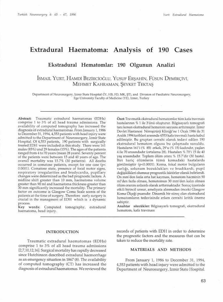
The mortality was 38% in patients in which anactive bleeding had been detected at surgery and7.5% in patients with clotted EOH. No deathoccurred in cases with partially liquefied or liquefiedEOHs. This difference was statistically significant (X2
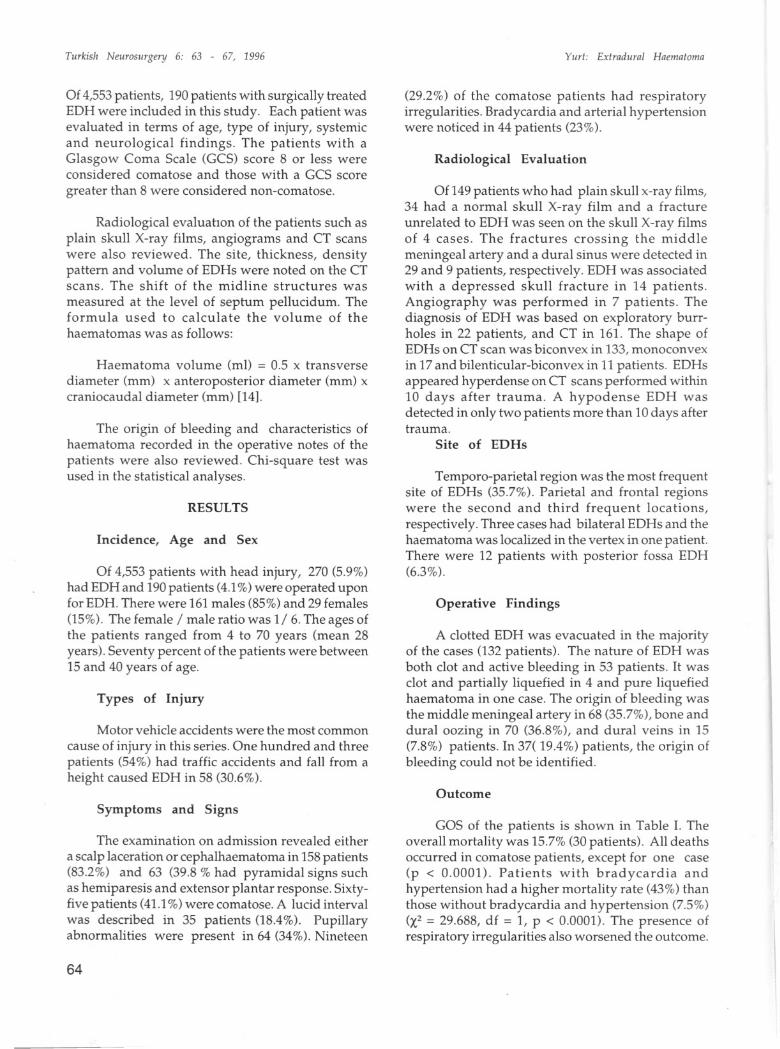
= 26.834, df = 3, P < 0.000l). Mortality was highest(35%) in patients in which the origin ofbleeding hadbeen the meningeal artery. The mortality ratessignificantly varied with the origin of bleeding ,i.e.,13.3% in venous bleeding, 4.2% in dural and boneaazing and 2.7% in unidentified origin (X2 = 31.257,df = 3, P < 0.000l) (Figure 3). The interval fromtrauma to surgery significantly influenced theoutcome. About one-third of the patients operatedupon within 6 hours after injury died, whereas fewerpatients died as the interval increased, there wereno mortalities in the patients undergoing surgerymore than 10 days after injury (X2= 26.821, df = 4,p< 0.0001) (Figure 4).
rate was only 4% in those with EOH of 90 ml or less(X2 = 53.696, df = I, P < 0.0001) (Figure 2).
<30 mm Thk
>30 mm Thk
73
93O
16100
13816
6O
30
190
GoodModerate deficitSevere deficit
Vegetative stateOeathTotal
abnormal pupll
GCS9-15
0% 20% 40% 60% 80% 100%
i- death DS urvivail
Figure 1: Symptoms and signs were found to be significantin predicting the outcome.
GCS3-8
lucid Interval
A midline shift greater than 10 mm andhaematoma thickness greater than 30 mmsignificantly increased the mortality (X2 = 28.934, df= I, P < 0.0001 and X2 = 7.597, df = I, P = 0.006,respectively). Fifty-nine percent of 27 patients havingEOH greater than 90 ml in volume died, whereas this
pyramldal .Ign.
no lucid Inteival
normal pupll
The mortality rates were 73% and 9.3% in those withand without respiratory irregularity, respectively (X2
= 48.490, df = I, P < 0.000l). Of 63 patients withpyramidal signs, 16 died and there were 14 deathsin 127 patients without pyramidal signs (X2 = 4.329,df = I, P = 0.037). Mortality rate was 42% in thepatients with pupillary changes and 2% in those withanormal pupil size and reaction (X2 = 47.630, df = I,P < 0.0001) (Figure 1).
no pyramidal signs
65
Tiirkis/i Neiirosiirgery 6: 63 - 67, 1996 Yiiri: Exlradiiral Haemaloma
?
d.oozing&bone
vetn
artery
Iiq.
liq&clol
clot
aclive bl.&clot
Twenty-nine (44.6%) of 65 comatose patients died,whereas onlyone death (0.8%) occurred among noncomatose patients (p < 0.0001). The level ofconsciousness significantly influenced the outcome.
Pyramidal syndrome and unilateral pupildilatation are invaluable signs in localizing the siteof EDH and are seen in 20% and 45% (3,9,11,16) ofpatients with EDH, respectively. Hemiparesis wasdetermined in 31.5% of our patients. Hypertension,bradycardia and respiratory irregularities are rarelyseen in patients with EDH. The presence of thesefindings was determined as a significant factor interms of mortality.
Figure 4: Mortality rates according to the injury to surgeryintervals (p < 0.0001).
this series, traffic accidents were the type of injuryin 55% of the patients and falls from height were thesecond frequent type of injury.
Figure 3: Significant operative findings in terms of theoutcome (?: unidentified bleeding origin, d.Gozing: dural oozing, liq.:liquefied hematoma,active bL.:active bleeding)
Although the most common site of EDHs hasbeen reported as the temporal region (1,10,12), EDHswere localized in the temporo-parietal or parietalregions in some recent papers (3,11,16), since CT hasenabled us to localize the haematoma precisely.Temporo-parietallocation was the most common siteof EDHs in our patients and was associated withhighest mortality. However the difference inmortality rates according to the site of haematomawas not statistically significant. Posterior fossa EDHscomprise 3.4 to 13% of all EDHs and have an 11 to22% mortality (4,5,13,18).There were 12 patients withposterior fossa EDH (6.3%).The origin of bleedingwas a dural vein in 2, a dural sinus in 6 patients andwas undetermined in 4. There was no death in
patients with posterior fossa EDH whose GCS scoreswere 9 or greater.
In some series, surgery was performed within6 hours, 6 to 24 hours and more than 24 hours afterinjury in 30-57%, 2-41% and 13-35% of the patientswith EDH, respectively [1,9,11,16].Trauma to surgeryinterval is an important factor in terms of outcome.The mortality rate has been 25-35% in patientsopera ted up on within 6 hours following trauma(4,11). Among 53 patients who underwent surgerywithin 6 hours after injury, the mortality rate was37.7%. The mortality rate in patients who underwentsurgery between 6 to 24 hours after injury was 7.8%in our series ( p < 0.001) . The majority of patientsundergoing early surgery were comatose. This mayaccount for the higher mortality rate.
100%
> 10 days
80%
D survival
60%40%
.death
20%
6·24 h,s 24·72 h,s 3·10 days
i .death Dsurvival
0%
0-6 h,sO
40
20
10
50
30
70
80
60
90
GCS scores of 125 patients were greater than 8and 65 (35%) were comatose. In some series, 35-40%of the patients were in the comatose group and 6065% in non-comatose group (1,3).The mortality rateshave been reported 18-44% and 1% in comatose andnon-comatose subjects, respectively (3,10,13,16).
There were 20 deaths in 53 patients (37.7%) inwhich an active bleeding and a dot were detected atsurgery. However, of 132 patients in which a dot wasevacuated, only 10 (7.5%) died. Active bleeding wassignificantly associated with the worst outcome
66

Turkis/i Neiirosiirgery 6: 63 - 67, 1996
(p< 0.0001). The number of deaths also variedsignificantly with the origin of EDH. The mortalityrates were 38%, 13.3% and 2.6% in arterial and
venous bleedings and in bleedings of unidentifiedorigin, respectively (p< 0.0001). it is conceivable thatthe development of EDH in a short time may beresponsible for a greater death rate in patients withan active bleeding.
In general, 73 % of patients with EDH have agood outcome. The outcome figure of our patientswas consistent with those in the literature (3,9,11).The experimental work of Ford and McLaurin (6)supports the hypothesis that the enlargement of acuteEDHs occurs shortly af ter the trauma. However, theIate enlargement of EDH in patients managedconservatively, and the development of delayedEDHs on serial CT scans are contrary to thishypothesis (15,17). The primary factor on outcomeis GCS scores of the patients at the time of surgery.Therefore early surgery is crucial in the managementof EDHs that are a dynamic process.
Correspondence: Or.Hamdi Bezircioglu1420 Sok. No:ll0-335220 Alsancak, Izmir, TurkeyPhone: (232) 463 49 10
REFERENCES
1. Bricolo A P, Pasut LM: Extradural hematoma: Towardzero mortality. Neurosurgery 14:8-12, 1984
2. Cordobes F, Lobato RO, Rivas H, Munoz MJ, ChillonD, Portillo MP, Lamas E: Observations on 82 patientswith extradural hematoma. J Neurosurg 54:179-186,1981
3. Cook RJ, Oorsch NWC, Fearnside MR, Chaseling R:Outcome prediction in extradural haematomas. ActaNeurochir (Wien) 95:90-94, 1988
4. Ersahin Y, Mutluer S, Güzelbag E: Extraduralhematoma analysis of 146 cases. Child's Nerv Syst9:96-99, 1993
Yurt: Extradiiral Haematoma
5. Ersahin Y, Mutluer S: Posterior fossa extraduralhematomas in Children. Pediatr Neurosurg 19:31-33,1993
6. Ford LE, Mc-Laurin RI: Meehanism of extraduralhematomas. J Neurosurg 20:760-769, 1963
7. Greenberg MS: Handbook of N eurosurgery, third ed.,Lakeland: Greenberg Graphics Inc., 1994,789 p.
8. Hutchinson J: Effusion of blood between bone anddura-mater. Lond Hosp Rep 4:51, 1867
9. Jamjoom A: The influenee of concomitant intraduralpathology on the presentation and outcome of patientswith aeute traumatic extradural haematoma. AetaNeuroehir (Wien) 155:86-89, 1992
10. Kvarnes TL, Trumpy JH: Extradural hematoma:Report of 132 cases. Aeta Neuroehir 41:223-231, 1978
11. Lobato RO, Rivas Jl, Cordobes F, Alted E, Perez C,Cabrera A, Oiez I, Gomez P, Lamas E: Aeute epiduralhematoma : An analysis of faetors influencing theoutcome of patients undergoing surgery in eoma. JNeurosurg 68:48-57, 1988
12. Mc.Kissoek W, Taylor]C, Bloom WH, Till K:Extradural hematoma: observations on 125 cases.Lancet 23:167-172,1960
13. Neubauer UJ: Extradural hematoma of the posteriorfossa. Twelve years experienees with CT-Sean. ActaNeuroehir (Wien) 87:105-111, 1987
14. Petersen OF, Espersen JO: Extradural hematomas:Measurement of size by volume summation on CTseanning. Neuroradiology 26:363-367, 1984
15. Rappaport ZH, Shaked I, Tadmor R: Oelayed epiduralhematoma demonstrated by computed tomography:Case report. Neurosurgery 10 : 487-488, 1982
16. Rivas H, Lobato RO, Sarabia R, Cordobes F, CabreraA, Gomez P: Extradural hematoma: Analysis of faetorinfluencing the eourses of 161 patients. Neurosurgery23:44-51, 1988
17. Sakai H, Takagi H, Ohtaka H, Tanabe T, Ohwada T,Yada K: Serial changes in aeute extradural hematomasize and associated changes in level of consciousnessand intraeranial pressure. J Neurosurg 68:566-570, 1988
18. Zueearello M, Pardateher K, AndrioH GC, Fiore OL,Iavieoli R: Epidural hematomas of the posterior eranialfossa. Neurosurgery 8:434-437, 1981
67


