ERT107ERT107MICROBIOLOGY MICROBIOLOGY
FOR BIOPROCESS FOR BIOPROCESS ENGINEERINGENGINEERING
Pn Syazni Zainul KamalPPK Bioprocess

Chapter 2: Microscopy Chapter 2: Microscopy TechniquesTechniques
CO2 :CO2 :
Ability to demonstrate practices in microscopy,Ability to demonstrate practices in microscopy,
staining, sterilization, isolation and identificationstaining, sterilization, isolation and identification
of bacteria and fungiof bacteria and fungi

Purpose of STAINING?Purpose of STAINING?

STAININGSTAINING
Microorganisms must be Microorganisms must be fixedfixed and and stainedstained prior prior examined under microscope to : examined under microscope to :
- increase visibility (increase contrast)- increase visibility (increase contrast)
- accentuate specific morphological features- accentuate specific morphological features
- preserve them for future study- preserve them for future study Staining – coloring specimens with stain (dyes)Staining – coloring specimens with stain (dyes)
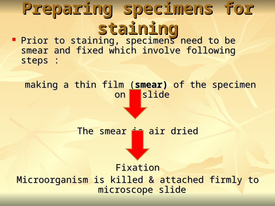
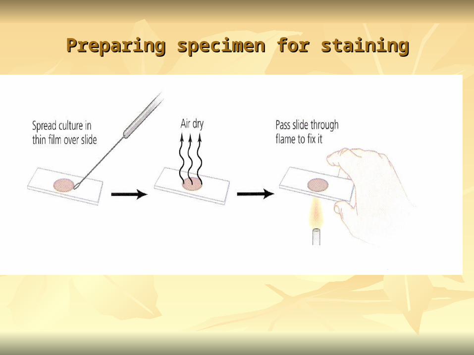
Preparing specimens for stainingPreparing specimens for staining Prior to staining, specimens need to be smear and fixed Prior to staining, specimens need to be smear and fixed
which involve following steps : which involve following steps :
making a thin film (making a thin film (smear) smear) of the specimen on a slideof the specimen on a slide
The smear is air driedThe smear is air dried
FixationFixationMicroorganism is killed & attached firmly to microscope Microorganism is killed & attached firmly to microscope
slideslide
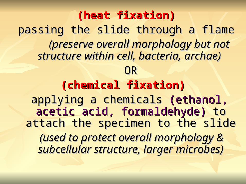
(heat fixation)(heat fixation)passing the slide through a flame passing the slide through a flame
(preserve overall morphology but not structure (preserve overall morphology but not structure within cell, bacteria, archae) within cell, bacteria, archae)
OROR(chemical fixation) (chemical fixation)
applying a chemicals applying a chemicals (ethanol, acetic acid, (ethanol, acetic acid, formaldehyde) formaldehyde) to attach the specimen to the to attach the specimen to the
slide slide (used to protect overall morphology & (used to protect overall morphology &
subcellular structure, larger microbes)subcellular structure, larger microbes)
Preparing specimen for stainingPreparing specimen for staining

DyesDyes make internal and external structures of cell make internal and external structures of cell
more visible by increasing contrast with more visible by increasing contrast with backgroundbackground
have two common features :have two common features :1) 1) chromophore groupschromophore groups
chemical groups with conjugated chemical groups with conjugated double bonds double bonds
give dye its colorgive dye its color2) 2) ability to bind cellsability to bind cells
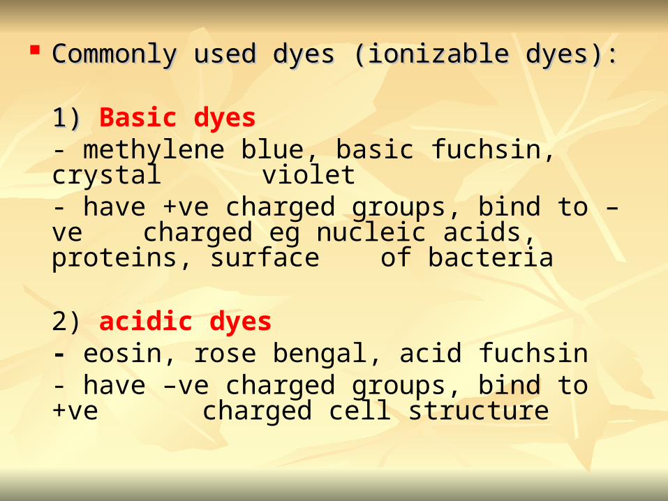
Commonly used dyes (ionizable dyes):Commonly used dyes (ionizable dyes):
1) 1) Basic dyes - methylene blue, basic fuchsin, crystal violet - have +ve charged groups, bind to –ve charged eg nucleic acids, proteins, surface of bacteria
2) acidic dyes- eosin, rose bengal, acid fuchsin - have –ve charged groups, bind to +ve charged cell structure
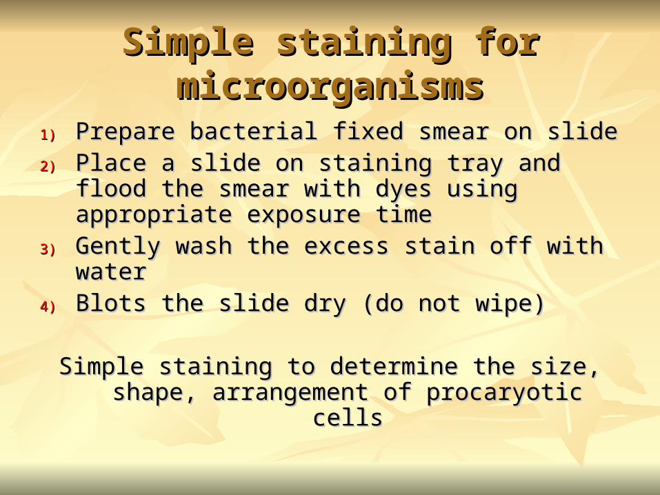
Simple staining for microorganismsSimple staining for microorganisms
1)1) Prepare bacterial fixed smear on slidePrepare bacterial fixed smear on slide2)2) Place a slide on staining tray and flood the Place a slide on staining tray and flood the
smear with dyes using appropriate exposure smear with dyes using appropriate exposure timetime
3)3) Gently wash the excess stain off with waterGently wash the excess stain off with water4)4) Blots the slide dry (do not wipe)Blots the slide dry (do not wipe)
Simple staining to determine the size, shape, Simple staining to determine the size, shape, arrangement of procaryotic cellsarrangement of procaryotic cells

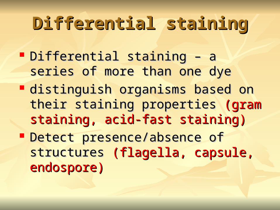
Differential stainingDifferential staining
Differential staining – a series of more than Differential staining – a series of more than one dyeone dye
distinguish organisms based on their staining distinguish organisms based on their staining properties properties (gram staining, acid-fast staining)(gram staining, acid-fast staining)
Detect presence/absence of structures Detect presence/absence of structures (flagella, (flagella, capsule, endospore)capsule, endospore)
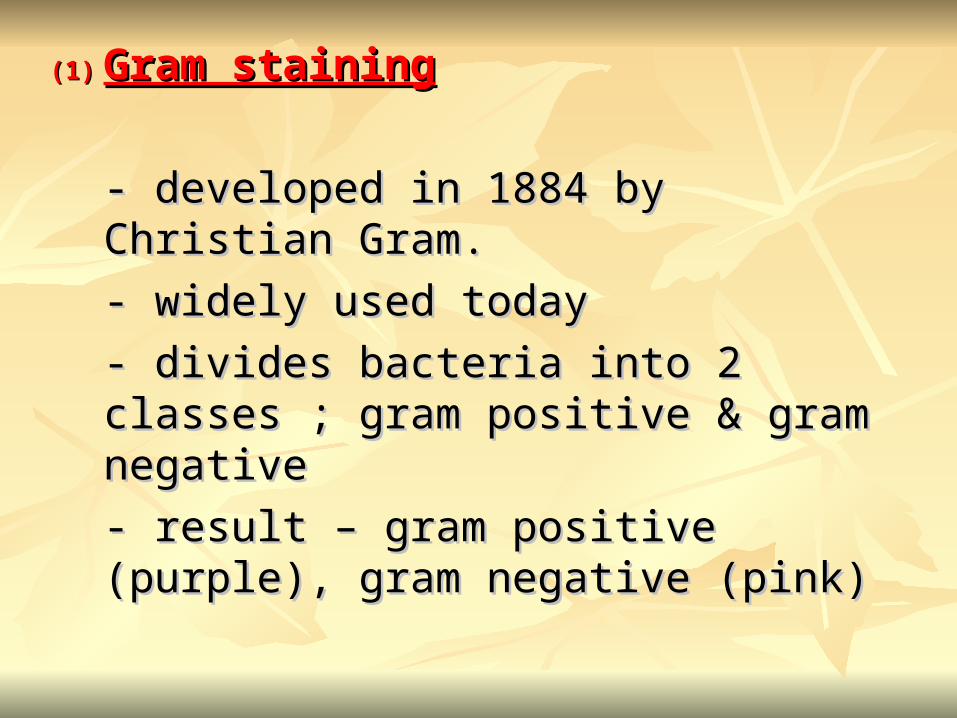
(1)(1) Gram stainingGram staining
- developed in 1884 by Christian Gram.- developed in 1884 by Christian Gram.
- widely used today- widely used today
- divides bacteria into 2 classes ; gram - divides bacteria into 2 classes ; gram positive & gram negativepositive & gram negative
- result – gram positive (purple), gram - result – gram positive (purple), gram negative (pink)negative (pink)
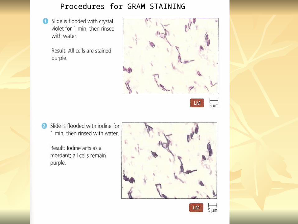
Procedures for GRAM STAINING


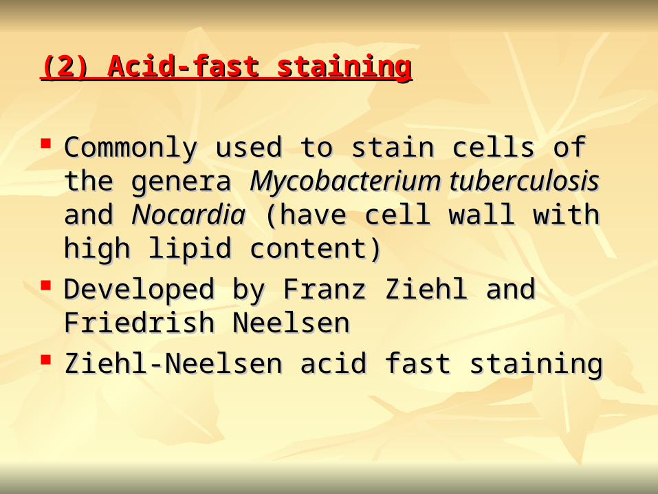
(2) Acid-fast staining(2) Acid-fast staining
Commonly used to stain cells of the genera Commonly used to stain cells of the genera Mycobacterium tuberculosisMycobacterium tuberculosis and and NocardiaNocardia (have cell wall with high lipid content)(have cell wall with high lipid content)
Developed by Franz Ziehl and Friedrish Developed by Franz Ziehl and Friedrish Neelsen Neelsen
Ziehl-Neelsen acid fast stainingZiehl-Neelsen acid fast staining

Acid-fast staining procedures ::
1)1) Flood the slide with the red primary stain, Flood the slide with the red primary stain, carbol fuchsincarbol fuchsin, for several minutes while , for several minutes while warming it over steaming water. warming it over steaming water. Heat is used to drive the stain through the Heat is used to drive the stain through the waxy wall & the cell, where it remains waxy wall & the cell, where it remains trapped. trapped.
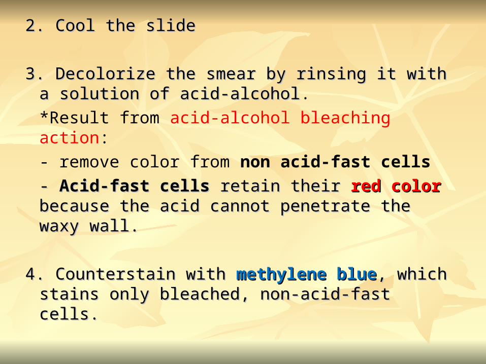
2. Cool the slide2. Cool the slide
3. Decolorize the smear by rinsing it with a 3. Decolorize the smear by rinsing it with a solution of acid-alcoholsolution of acid-alcohol.
*Result from acid-alcohol bleaching action:
- remove color from non acid-fast cells
- - Acid-fast cellsAcid-fast cells retain their retain their red color red color because because the acid cannot penetrate the waxy wall.the acid cannot penetrate the waxy wall.
4. Counterstain with 4. Counterstain with methylene bluemethylene blue, which stains , which stains only bleached, non-acid-fast cells. only bleached, non-acid-fast cells.
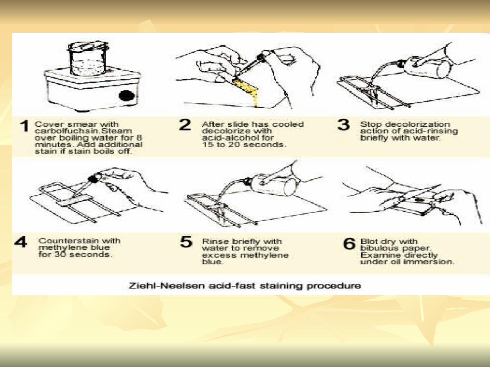

5. Rinse slide with water to remove excess 5. Rinse slide with water to remove excess methylene bluemethylene blue
6. Blot dry6. Blot dry

3. Capsule staining3. Capsule staining
Reveals the presence of capsules (made of Reveals the presence of capsules (made of polysaccharides), polysaccharides), B.anthracisB.anthracis
Dyes used : Dyes used : india ink, eosin, nigrosinindia ink, eosin, nigrosin (acidic (acidic dyes)dyes)
Mix dye with cell & make a smear over the Mix dye with cell & make a smear over the slideslide
Air driedAir dried Cell appear brighter again dark backgroundCell appear brighter again dark background

Acidic dyes stain the background & does not penetrate the capsuleThe bacteria cell had been counterstained with basic dyes
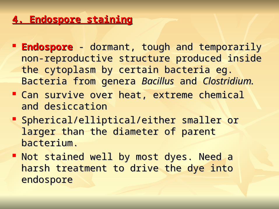
4. Endospore staining4. Endospore staining
EndosporeEndospore - dormant, tough and temporarily non- - dormant, tough and temporarily non-reproductive structure produced inside the cytoplasm reproductive structure produced inside the cytoplasm by certain bacteria eg. Bacteria from genera by certain bacteria eg. Bacteria from genera BacillusBacillus and and Clostridium.Clostridium.
Can survive over heat, extreme chemical and Can survive over heat, extreme chemical and desiccationdesiccation
Spherical/elliptical/either smaller or larger than the Spherical/elliptical/either smaller or larger than the diameter of parent bacterium.diameter of parent bacterium.
Not stained well by most dyes. Need a harsh treatment Not stained well by most dyes. Need a harsh treatment to drive the dye into endospore to drive the dye into endospore

Schaeffer-FultonSchaeffer-Fulton
1) Smear the organism and heat fix to a slide1) Smear the organism and heat fix to a slide
2) Place the slide over a steam bath and flood with 2) Place the slide over a steam bath and flood with Malachite GreenMalachite Green
3) Keep the stain over the bath for 3 - 5 minutes, 3) Keep the stain over the bath for 3 - 5 minutes, recovering the slide with Malachite Green if recovering the slide with Malachite Green if some evaporates. some evaporates.
4) Dump the Malachite Green off and allow to cool4) Dump the Malachite Green off and allow to cool
5) Rinse the slide with water to remove excess stain5) Rinse the slide with water to remove excess stain
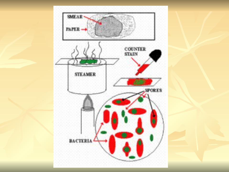

6) Counterstain the smear with Safranin for 2 6) Counterstain the smear with Safranin for 2 minutesminutes
7) Rinse the slide with water to remove excess 7) Rinse the slide with water to remove excess stainstain
8) Blot dry the stain and view under a 8) Blot dry the stain and view under a microscopemicroscope
Step FinishedColor of Vegetative Cell
Color of Endospore
Smear Colorless Colorless
Malachite Green Green Green
Cool/Wash Colorless Green
Safranin Safranin Green


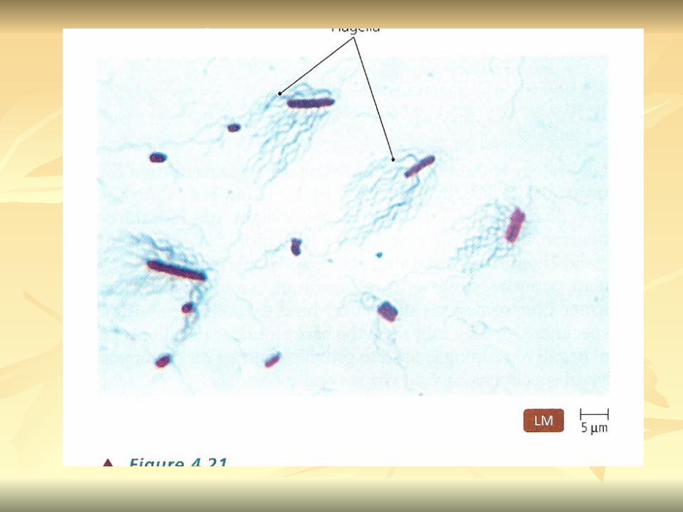
5. Flagella staining5. Flagella staining
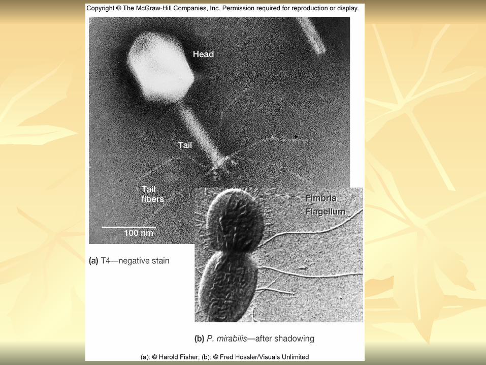
Provide information on the Provide information on the presence and distribution presence and distribution patternpattern of flagella on procaryote cells of flagella on procaryote cells
Procaryote flagella – fine, threadlike organelles of Procaryote flagella – fine, threadlike organelles of locomotion that are so slender (10-30mm). Can only locomotion that are so slender (10-30mm). Can only be seen directly using EMbe seen directly using EM
So, to observe using light microscope -increase So, to observe using light microscope -increase thickness of the flagellathickness of the flagella
Procedures :Procedures :1) coated with mordants eg. Tannic acid & 1) coated with mordants eg. Tannic acid & potassium alum (increase diameter)potassium alum (increase diameter)2) stain with pararosaniline or basic fuschin 2) stain with pararosaniline or basic fuschin (change colour & increase contrast)(change colour & increase contrast)
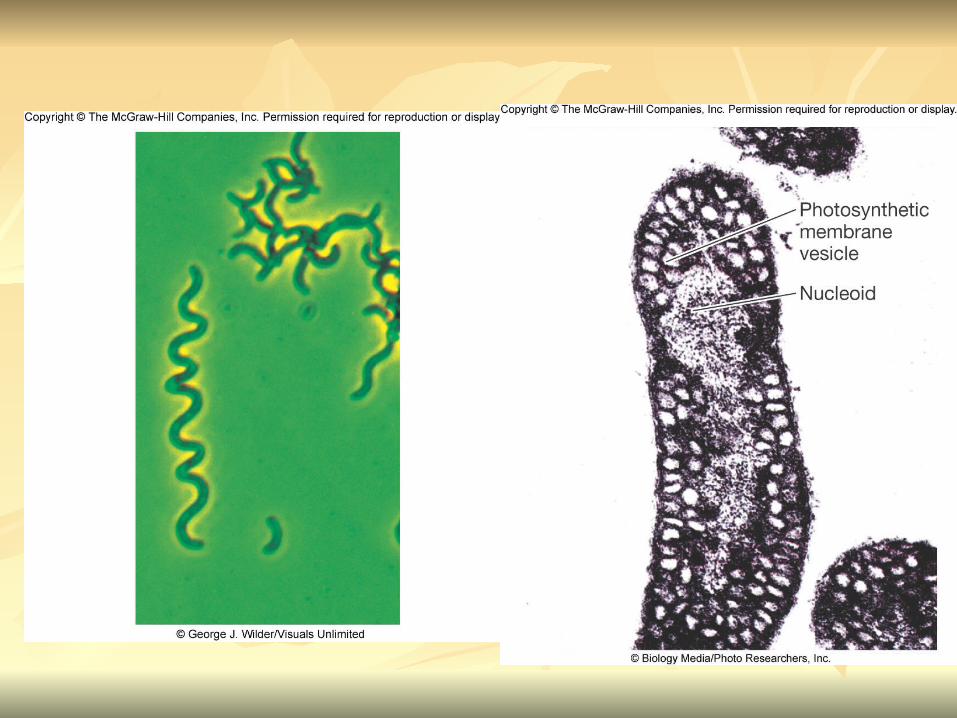
Electron MicroscopyElectron Microscopy
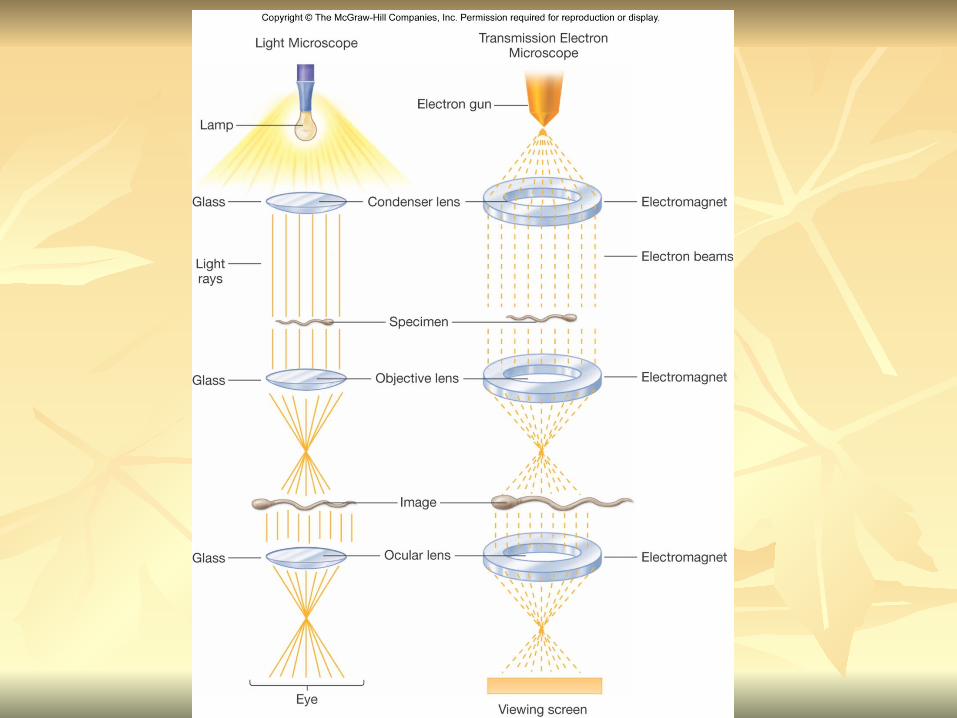
Electron microscopyElectron microscopy
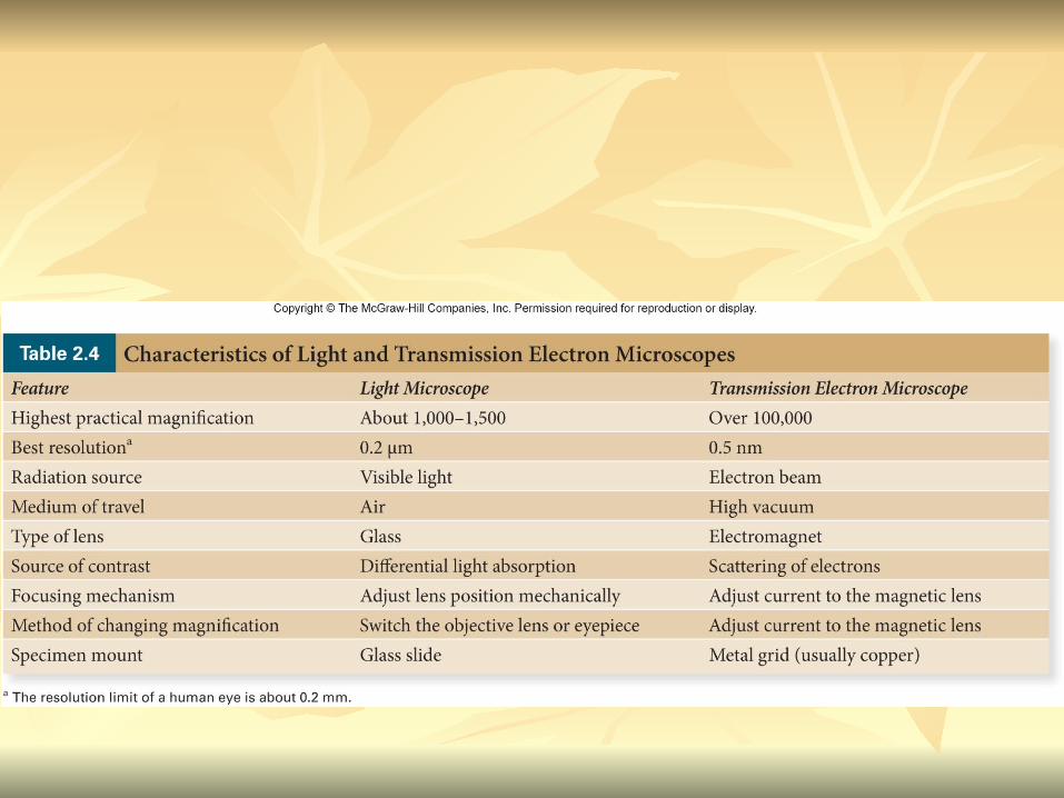
Light microscope Light microscope resolution limit of about resolution limit of about 0.20.2µmµm. .
Limits to detailed studies of many Limits to detailed studies of many microorganisms.microorganisms.
Eg. Viruses & internal structure of Eg. Viruses & internal structure of microorganismsmicroorganisms
shortest wavelength of visible light is about 400 shortest wavelength of visible light is about 400 nmnm
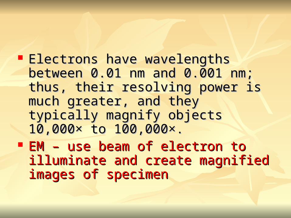
Electrons have wavelengths between 0.01 nm Electrons have wavelengths between 0.01 nm and 0.001 nm; thus, their resolving power is and 0.001 nm; thus, their resolving power is much greater, and they typically magnify much greater, and they typically magnify objects 10,000× to 100,000×. objects 10,000× to 100,000×.
EM – use beam of electron to illuminate and EM – use beam of electron to illuminate and create magnified images of specimencreate magnified images of specimen

There are two general types of electron There are two general types of electron microscope: microscope:
Transmission Electron Microscopes (TEM)Transmission Electron Microscopes (TEM)
Scanning Electron Microscopes (SEM)Scanning Electron Microscopes (SEM)

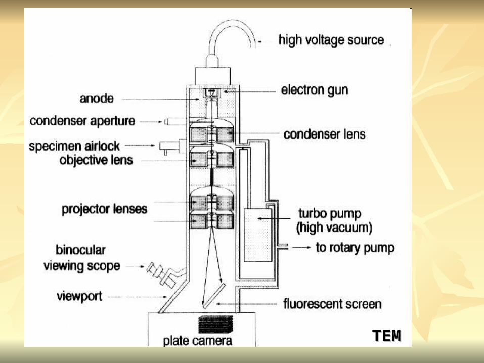
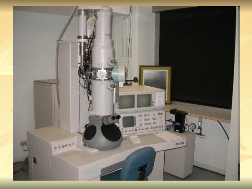
TEMTEM
A A TEM TEM generates a generates a beam of electronsbeam of electrons that passes that passes through a through a thinly sliced & dehydrated specimenthinly sliced & dehydrated specimen, , through through magnetic fieldsmagnetic fields that manipulate and focus the that manipulate and focus the beam, and then onto a beam, and then onto a fluorescent screenfluorescent screen that changes that changes the electron’s energy into visible light.the electron’s energy into visible light.
Column of the TEM must be vacuumColumn of the TEM must be vacuum- electrons are deflected by collisions with air - electrons are deflected by collisions with air moleculesmolecules
TEMTEM

TEMTEM
specimen must be very thin (20 to 100nm)specimen must be very thin (20 to 100nm) (solid matter easily absorb and deflect electron)(solid matter easily absorb and deflect electron)
- fixation of specimen using glutaraldehyde- fixation of specimen using glutaraldehyde- specimen need to be dehydrated- specimen need to be dehydrated- embedded in plastic- embedded in plastic- slice thinly- slice thinly
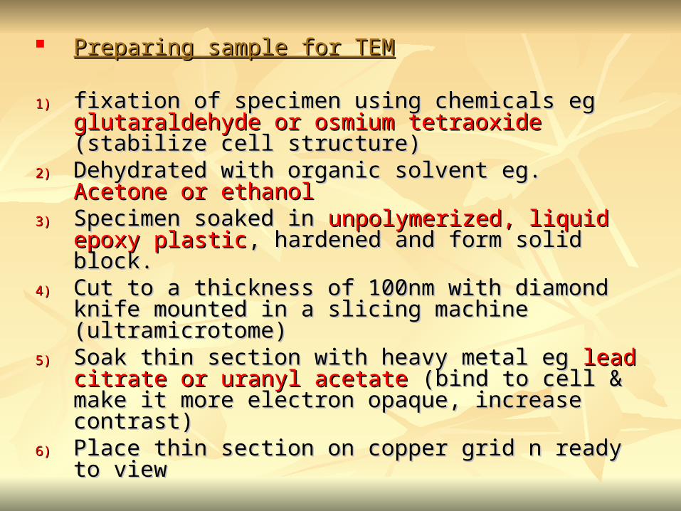
Preparing sample for TEMPreparing sample for TEM
1)1) fixation of specimen using chemicals eg fixation of specimen using chemicals eg glutaraldehyde or osmium tetraoxide glutaraldehyde or osmium tetraoxide (stabilize cell (stabilize cell structure)structure)
2)2) Dehydrated with organic solvent eg. Dehydrated with organic solvent eg. Acetone or Acetone or ethanolethanol
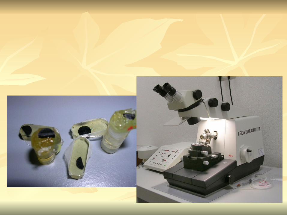
3)3) Specimen soaked in Specimen soaked in unpolymerized, liquid epoxy unpolymerized, liquid epoxy plasticplastic, hardened and form solid block. , hardened and form solid block.
4)4) Cut to a thickness of 100nm with diamond knife Cut to a thickness of 100nm with diamond knife mounted in a slicing machine (ultramicrotome)mounted in a slicing machine (ultramicrotome)
5)5) Soak thin section with heavy metal eg Soak thin section with heavy metal eg lead citrate or lead citrate or uranyl acetateuranyl acetate (bind to cell & make it more electron (bind to cell & make it more electron opaque, increase contrast)opaque, increase contrast)
6)6) Place thin section on copper grid n ready to view Place thin section on copper grid n ready to view

Other Preparation MethodsOther Preparation Methods
negative stainnegative stain heavy metals (phosphotungstic acid/ uranyl heavy metals (phosphotungstic acid/ uranyl
acetate) do not penetrate the specimen but render acetate) do not penetrate the specimen but render dark background dark background
used for study of viruses, bacterial gas vacuolesused for study of viruses, bacterial gas vacuoles shadowingshadowing
coating specimen with a thin film of a heavy metal coating specimen with a thin film of a heavy metal (platinum) only on one side(platinum) only on one side
useful for viral morphology, flagella, DNAuseful for viral morphology, flagella, DNA

freeze-etchingfreeze-etching freeze specimen using liquid nitrogenfreeze specimen using liquid nitrogen then fracture along lines of greatest weakness (e.g., then fracture along lines of greatest weakness (e.g.,
membranes)membranes) Shadowed & coated using platinum & carbonShadowed & coated using platinum & carbon allows for 3-D observation of shapes of allows for 3-D observation of shapes of
intracellular structuresintracellular structures

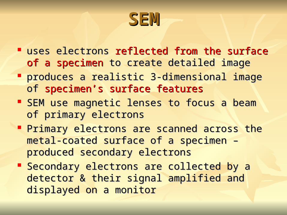
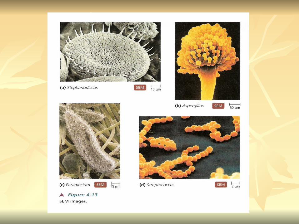
SEMSEM
uses electrons uses electrons reflected from the surface of a reflected from the surface of a specimen specimen to create detailed imageto create detailed image
produces a realistic 3-dimensional image of produces a realistic 3-dimensional image of specimen’s surface featuresspecimen’s surface features
SEM use magnetic lenses to focus a beam of primary SEM use magnetic lenses to focus a beam of primary electronselectrons
Primary electrons are scanned across the metal-coated Primary electrons are scanned across the metal-coated surface of a specimen – produced secondary electronssurface of a specimen – produced secondary electrons
Secondary electrons are collected by a detector & Secondary electrons are collected by a detector & their signal amplified and displayed on a monitortheir signal amplified and displayed on a monitor

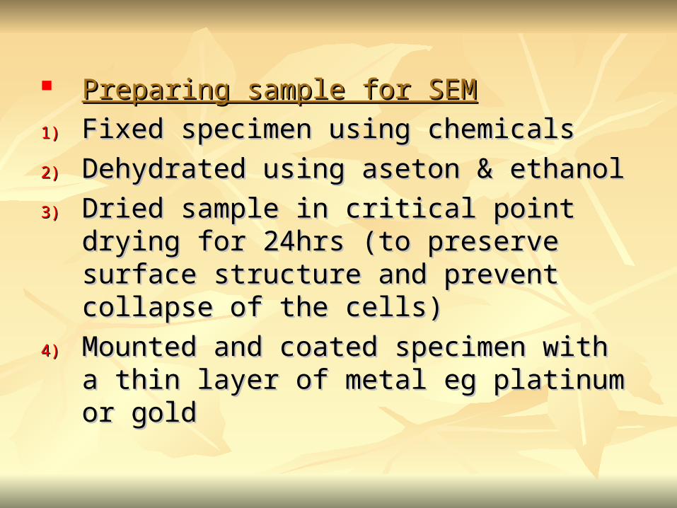
Preparing sample for SEMPreparing sample for SEM
1)1) Fixed specimen using chemicalsFixed specimen using chemicals
2)2) Dehydrated using aseton & ethanolDehydrated using aseton & ethanol
3)3) Dried sample in critical point drying for Dried sample in critical point drying for 24hrs (to preserve surface structure and 24hrs (to preserve surface structure and prevent collapse of the cells)prevent collapse of the cells)
4)4) Mounted and coated specimen with a thin Mounted and coated specimen with a thin layer of metal eg platinum or gold layer of metal eg platinum or gold
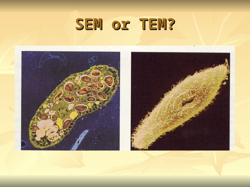
SEM or TEM?SEM or TEM?

ISOLATIONISOLATION

Isolation of microorganismsIsolation of microorganisms Natural habitat, microorganism grow in complex, Natural habitat, microorganism grow in complex,
mixed populations with many spp.mixed populations with many spp. Need a Need a pure culturepure culture to study and characterize an to study and characterize an
individual speciesindividual species Pure culture =contain one type of microorganismsPure culture =contain one type of microorganisms Techniques to prepare pure culture:Techniques to prepare pure culture:
-spread plate-spread plate
-streak plate-streak plate
-Pour plate-Pour plate
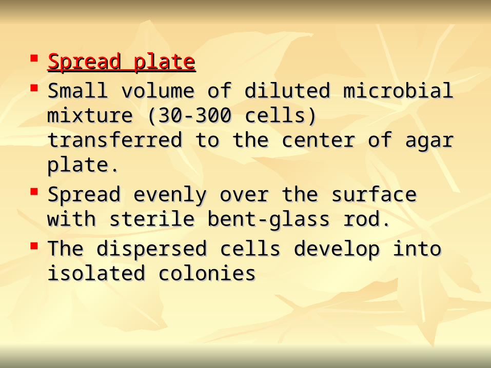
Spread plateSpread plate Small volume of diluted microbial mixture Small volume of diluted microbial mixture
(30-300 cells) transferred to the center of agar (30-300 cells) transferred to the center of agar plate.plate.
Spread evenly over the surface with sterile Spread evenly over the surface with sterile bent-glass rod.bent-glass rod.
The dispersed cells develop into isolated The dispersed cells develop into isolated coloniescolonies
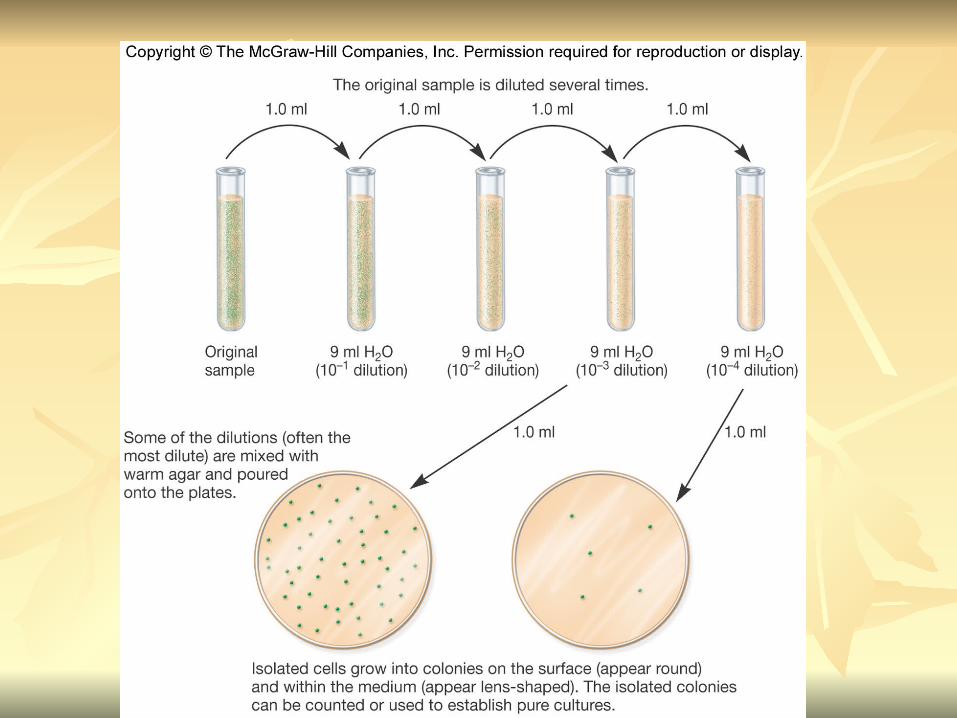
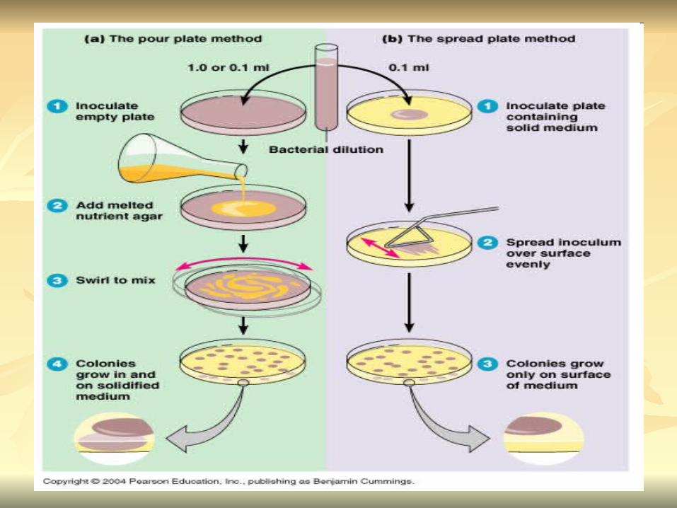

Pour platePour plate Original sample is diluted several times – Original sample is diluted several times –
reduce microbial populationreduce microbial population Small vol. of diluted sample mixed with Small vol. of diluted sample mixed with
melted agarmelted agar After agar had hardened, each cell is fixed in After agar had hardened, each cell is fixed in
place and form individual colonyplace and form individual colony
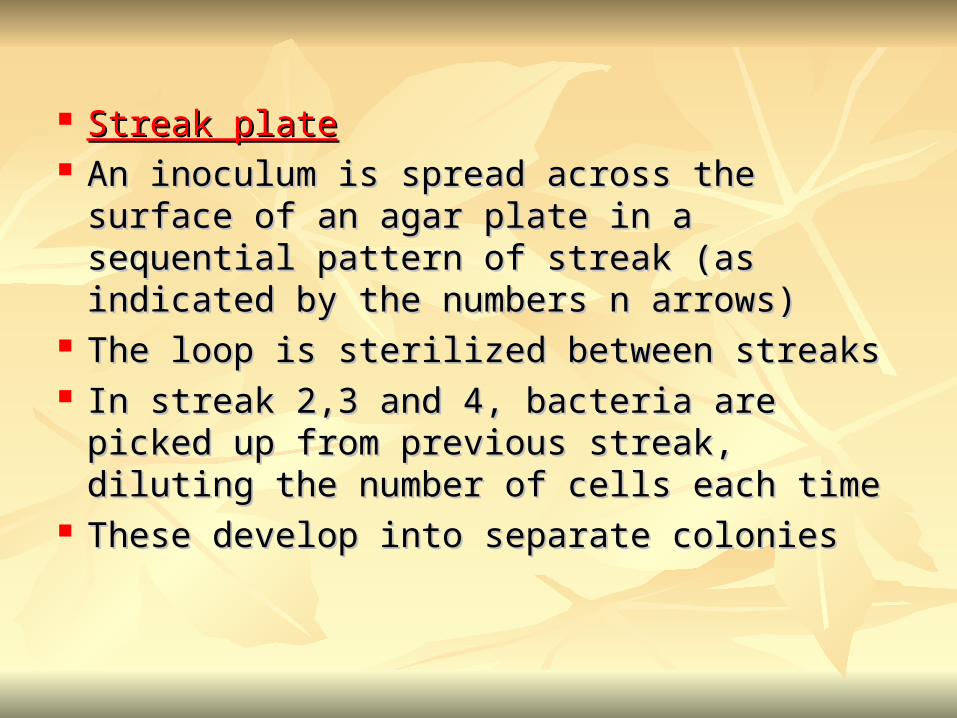
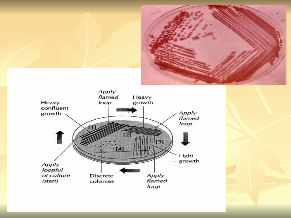
Streak plateStreak plate An inoculum is spread across the surface of an An inoculum is spread across the surface of an
agar plate in a sequential pattern of streak (as agar plate in a sequential pattern of streak (as indicated by the numbers n arrows)indicated by the numbers n arrows)
The loop is sterilized between streaksThe loop is sterilized between streaks In streak 2,3 and 4, bacteria are picked up In streak 2,3 and 4, bacteria are picked up
from previous streak, diluting the number of from previous streak, diluting the number of cells each timecells each time
These develop into separate coloniesThese develop into separate colonies
Microbial Growth on Solid Microbial Growth on Solid SurfacesSurfaces
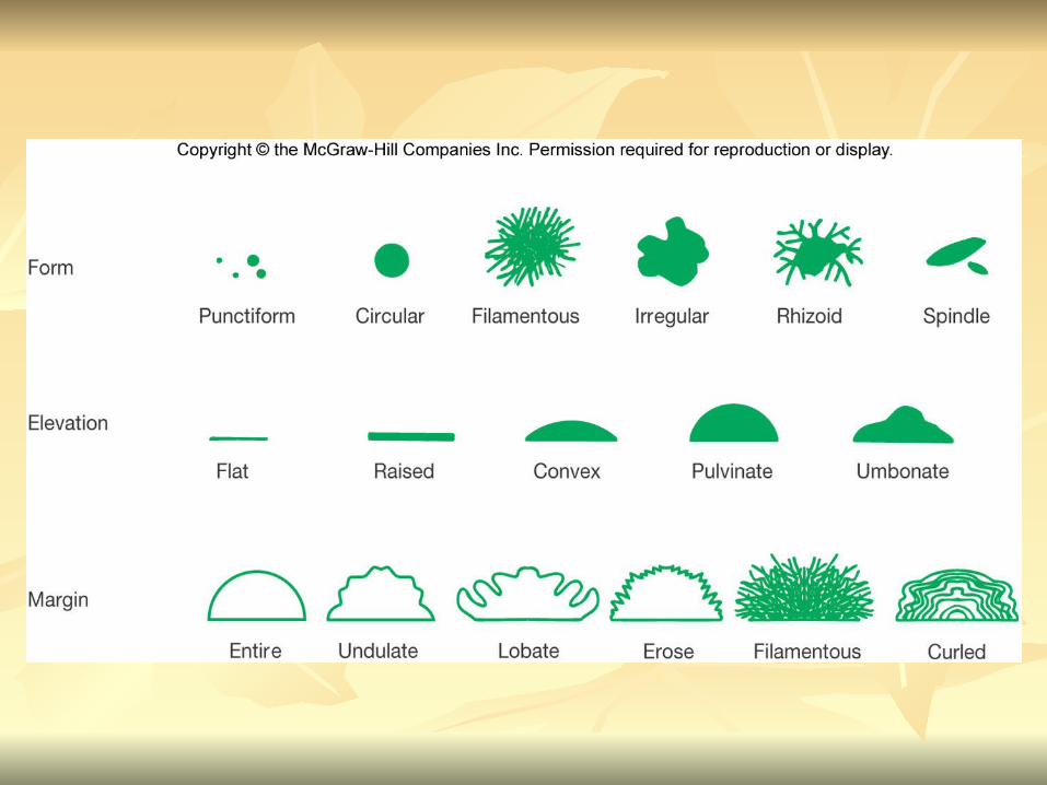
colony characteristicscolony characteristics that develop when that develop when microorganisms are grown on agar surfaces aid microorganisms are grown on agar surfaces aid in identificationin identification
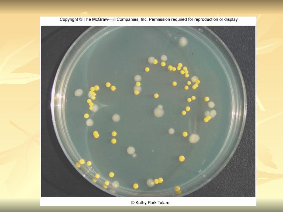
Colony – a macroscopically visible cluster of Colony – a macroscopically visible cluster of microorganisms microorganisms
differences in growth rate from edges to center differences in growth rate from edges to center is due to is due to oxygen, nutrients, and toxic productsoxygen, nutrients, and toxic products cells may be dead in some areascells may be dead in some areas

1. Form – The form refers to the shape of 1. Form – The form refers to the shape of the colony. the colony.
2. Elevation – This describes the “side view” 2. Elevation – This describes the “side view” of a colony. of a colony.
3. Margin – The margin or edge of a colony 3. Margin – The margin or edge of a colony (or any growth) may be an important (or any growth) may be an important characterisic in identifying an organisms. characterisic in identifying an organisms.