Tetralogia di Fallot Atresia polmonare con DIV Agenesia della polmonare
D Marini Cardiologia Pediatrica, Citta’ della Salute, Torino
Email:[email protected]

Tetralogia di Fallot
• 6% delle c.c. • 1/3 anomalie cromosomiche

Definizione
• DIV da malallineamento del setto conale
• aorta a cavaliere del difetto, generalmente dilatata
• stenosi polmonare/infundibolare
• ipertrofia del VD

Embriologia
• Anomalia conotruncale
• Ipotesi embriologiche:
o deviazione del setto conale
o ipoplasia infundibulare

Classificazione
• Spettro
• Fallot regolare e irregolare

• DIV alto, non restrittivo, perimembranoso associato a stenosi polmonare • Uguaglianza di pressioni Ao-VS-VD. • Shunt bidirezionale con cianosi +/-importante • Tendenza a progressione dell’ostruzione
Clinica
• Soffio eiettivo • Se soffio sistolico e/o continuo sottoclaveare o dorsale: flusso polmonare di origine sistemica • Tendenza alla cianosi • Possibili crisi anossiche • Rx: ombra cardiaca di normali dimensioni, II arco sx concavo, ipovascolarizzazione polmonare • ECG: sovraccarico dx isolato • Segni tardivi: ippocratismo digitale - squatting

Trattamento
• Palliazione: Anastomosi di Blalock
• Correzione: Chiusura del DIV, allargamento dell’infundibolo polmonare + tronco + valvola

Risultati
• Guarigione
• Possibile insufficienza polmonare con dilatazione-disfunzione ventricolare dx
• Complicanze tardive

ECO deve rispondere alle domande:
• DIV abituale? (non restrittivo, alto, perimembranoso, unico)
• Morfologia della via polmonare • Confluenza e diametro dei rami polmonari • Anatomia coronarica? (IVA dalla coronaria dx,
che incrocia l’anello polmonare nel 5%)

Aorta a cavaliere sul DIV The image cannot be displayed. Your computer may not have enough memory to open the image, or the image may have been corrupted. Restart your computer, and then open the file again. If the red x still appears, you may have to delete the image and then insert it
again.
The image cannot be displayed. Your computer may not have enough memory to open the image, or the image may have been corrupted. Restart your computer, and then open the file again. If the red x still appears, you may have to delete the image and then insert it again.

DIV anteriore
The image cannot be displayed. Your computer may not have enough memory to open the image, or the image may have been corrupted. Restart your computer, and then open the file again. If the red x still appears, you may have to delete the image and then insert it again.
The image cannot be displayed. Your computer may not have enough memory to open the image, or the image may have been corrupted. Restart your computer, and then open the file again. If the red x still appears, you may have to delete the image and then insert it again.

Shunt destro-sinistro e sinistro-destro The image cannot be displayed. Your computer may not have enough memory to open the image, or the image may have been corrupted. Restart your computer, and then open the file again. If the red x still appears, you may have to delete the image and then insert it again.
The image cannot be displayed. Your computer may not have enough memory to open the image, or the image may have been corrupted. Restart your computer, and then open the file again. If the red x still appears, you may have to delete the image and then insert it again.

Via d’efflusso destra: Asse corto
The image cannot be displayed. Your computer may not have enough memory to open the image, or the image may have been corrupted. Restart your computer, and then open the file again. If the red x still appears, you may have to delete the image and then insert it again.
The image cannot be displayed. Your computer may not have enough memory to open the image, or the image may have been corrupted. Restart your computer, and then open the file again. If the red x still appears, you may have to delete the image and then insert it again.

Deviazione del setto conale

Anello della valvola polmonare

Pattern Doppler

Tronco e rami polmonari

Aorta

Coronarie: CDx dalla CSin

Coronarie: IVA dalla CD
Atresia polmonare + DIV

Definizione
• atresia polmonare + DIV da malallineamento
• l’albero polmonare può essere completo o possono essere presenti collaterali aorto-
polmonari

Classificazione
• Tipo 1: Presenza di tronco polmonare e arterie polmonari confluenti, vascolarizzate dal Botallo • Tipo 2: Assenza di tronco polmonare ma arterie polmonari confluenti, vascolarizzate dal Botallo • Tipo 3: Presenza di arterie polmonari centrali (alimentate da collaterali sistemico-polmonari) e di collaterali sistemico-polmonari (MAPCAs) • Tipo 4: Assenza di arterie polmonari centrali

Embriologia
• Anomalia conotruncale
• Associazione 22q11

• Possibile dotto-dipendenza del circolo polmonare (tipo I-II) • Cianosi + importante • Possibile iperafflusso e ipertensione polmonare se MAPCAs non stenotici (tipo III-IV)

Clinica
• Simile nei 4 tipi • Cianosi +/- presente a seconda della qualita’ di flusso polmonare sistemico-dipendente • 2T unico • Soffio continuo sottoclaveare (da dotto) o dorsale (da collaterali) • Rx: cuore piccolo, ipovascolarizzazione, aorta a destra in 1/3 dei casi • ECG: segni di sovraccarico ventricolare

Trattamento
• Palliazione: anastomosi di Blalock o condotto tra VD e rami polmonari +/- unifocalizzazione dei collaterali
• Correzione: chiusura DIV + ricostruzione del tronco polmonare con patch di allargamento oppure con inserzione di condotto tra il VD e il tronco polmonare
Risultati
• Le forme che hanno un albero polmonare completo hanno una buona prognosi • Le forme in cui è stata necessaria l’inserzione di un condotto protesico, spesso evolvono verso una stenosi e/o insufficienza • Le forme con rami polmonari ipoplasici e MAPCAS hanno un prognosi riservata, con eventuale impossibilità di chiusura del DIV

ECO deve rispondere alle domande:
• DIV abituale? (non restrittivo, alto, perimembranoso, unico)
• Presenza di rami polmonari centrali, loro confluenza e diametro
• Presenza di collaterali sistemico-polmonari (MAPCAS)

Atresia della valvola polmonare o dell'infundibolo

Aorta a cavaliere sul DIV
The image cannot be displayed. Your computer may not have enough memory to open the image, or the image may have been corrupted. Restart your computer, and then open the file again. If the red x still appears, you may have to delete the image and then insert it again.
The image cannot be displayed. Your computer may not have enough memory to open the image, or the image may have been corrupted. Restart your computer, and then open the file again. If the red x still appears, you may have to delete the image and then insert it again.

MAPCAS

Agenesia della valvola polmonare

Definizione
• Presenza di valvola polmonare rudimentale, con annulus ipoplasico
• Si associa ad ampio DIV perimembranoso
• I rami polmonari sono ectasici

Classificazione
• NO

Embriologia
• Anomalia conotruncale
• Ipotesi embriologiche:
o Agenesia del Botallo?
o Variante di Fallot?

• Se DIV, DIV tipo Fallot • Protezione polmonare • Insufficienza polmonare + severa • I rami polmonari ectasici possono determinare compressione bronchiale, con disturbi respiratori gravi

Forma neonatale: grave • Ipoplasia polmonare • Ostruzione bronchiale • Disventilazione • Atelettasia • Enfisema • Associazione: stenosi
periferiche APs

Forma piu’ tardiva
• Come Fallot (+/- DIV) • Dilatazione delle APs +/- importante
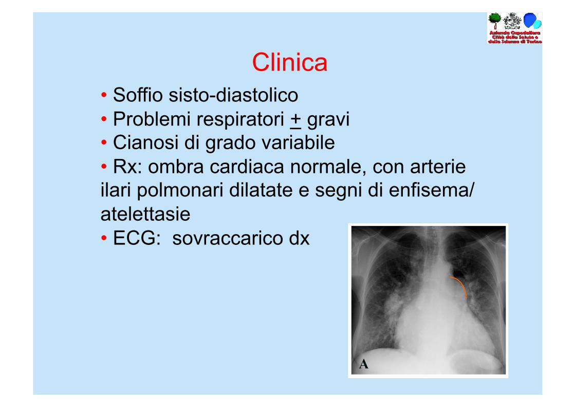
Clinica • Soffio sisto-diastolico • Problemi respiratori + gravi • Cianosi di grado variabile • Rx: ombra cardiaca normale, con arterie ilari polmonari dilatate e segni di enfisema/atelettasie • ECG: sovraccarico dx

Trattamento
• Miglioramento in decubito prono • Nel primo anno di vita trattamento medico • Correzione: allargamento dell’annulus polmonare, chiusura del DIV e plastica di riduzione dei rami

Risultati
• Buona prognosi se l’intervento è effettuato dopo l’anno di vita

ECO deve rispondere alle domande:
• DIV abituale? (non restrittivo, alto, perimembranoso, unico)
• Grado di ectasia dei rami polmonari • Presenza di ostruzione sui rami polmonari? • Compromissione ventricolare dx

Nel feto

Ectasia dei rami polmonari

Ectasia dei rami polmonari


