
DISSOCIATION OF A WEAK ELECTROLYTE Ostwald’s dilution law
Weak acetic acid: CH3COOH H+ + CH3COO- ; Kdis = = 1.75•10-5 H
H OOCH3C
OOCH3C
[ ]+ -[ ].
[ ]nondis
Dissociated molecules concentration is [H+]=[CH3COO-]=Cdis=α•C.
Concentration total is C=[CH3COOH]nondis+[H+] so [CH3COOH]nondis=C-[H+] and
[CH3COOH]nondis=C - α•C=C(1- α)
Inserting all these results we have: Kdis = = CC
C
22 2
1
Weak ammonium hydroxide base: NH4OH NH4+ + OH- ; Kdis = = 1.79•10-5
HONH4
ONH4
H
[ ]+ -[ ].
[ ]nondis
As week electrolyte dissociation degree tends to zero α =>0 and (1- α)=1 : Kdis = α2•C .
Ostwald’s dilution law α= is on 1886 in Riga proved discovery if concentration
C decreases dissociation degree α increases and remains pH=const .
Henderson Haselbalh buffer solution equation give that just on1908 as well 22 Years later .
C
Kdis
Ostwald dilution law side product is calculation pH for weak acid and weak base concentration:
Weak acid hydrogen ion concentration [H+]= = 10-pH Molarity ;
Weak base hydroxyl ion concentration [OH-]= = 10-pOH Molarity .
CKa
CKb
4th page : http://aris.gusc.lv/BioThermodynamics/H2ODissociation.doc
pH calculation in solutions of acids, bases and salts
Strong acid
H2SO4 => 2 H+ + SO42-, where z = 2eq, strong acid H+ ions concentration [H+] = zCdis,
[H+] = CM•α•z or, since CM•z = CN, [H+] = CN•α
Taking logarithm from both sides and changing signs we obtain pH expression:
pH = -log [H+] = -logCM•α•z or pH=-logCN•α .
Weak acid concentration as Ostwald’s dilution law side product [H+] = .
Taking a logarithm from both sides with minus pH = -log [H+] = - =
Strong base
Ca(OH)2 => Ca2+ + 2 OH- , where z = 2eq, strong base concentration of OH- ions [OH-] = zCdis,
[OH-] = α•z•CM or, since CM•z = CN, [OH-] = CN•α Taking logarithm from both sides and changing signs we obtain pOH expression:
pOH= -log [OH-]= -logCM•α•z=-logCN•α or pH=14-pOH=14+logCM•α•z
CKa
2
log CKa
2
log CpK a
Weak base concentration as Ostwald’s dilution law side product [OH-] = .
Taking a logarithm from both sides with minus pOH = -log [OH-] = - =
or pH= 14 - pOH =14 -
CKb
2
log CKb
2
log CpK b
2
log CpK b
Strong acid and strong base concentration for dissociated molecules Cdis = α•CM .
4th page : http://aris.gusc.lv/BioThermodynamics/H2ODissociation.doc
Hydrolysis of salts Brensted protolytic reaction with water
1. Strong acid and base salts are not hydrolyzed.
Strong acid anion is very weak base 0< Kb <1.
Strong base cation is very weak acid 0< Ka <1.
Na2SO4 is not hydrolyzed, as SO42- anion is a very weak base and Na+ cation is a very weak acid..
2. Weak acid and strong base salt Na2CO3 give CO32- anion is strong base Kb >> 1, but Na+
cation is a very weak acid. The CO32- reacts with water-weak acid - hydrolysis just in 1 stage:
stage 1 CO32- + H2O => HCO3
- + OH-.
Salt solution of weak acid and strong base due to OH- ions environment in is basic pH > 7.
stage 2: HCO3- + H2O => H2CO3 + OH- not happen without acid addition.
For this reason, if we have to write the hydrolysis reaction in molecular form according 1st stage:
Na2CO3 + H2O => NaHCO3 + NaOH
Hydrolysis will be increased by adding acid to solution and oppressed by adding base.
3. Weak base and strong acid salt NH4Cl give NH4+ cation strong acid Ka >> 1, but Cl- anion is
very weak base react with water-weak base. The NH4+ cation hydrolysed in one stage:
NH4+ + H2O => NH4OH + H+
Due to H+ ions environment in weak base salt solution is strong acid. As pH < 7 is acidic.
Hydrolysis reaction in molecular form is: NH4Cl + H2O => NH4OH + HCl
acid a H+ + b base
If acid a is weak
coupled salt is strong base b
Ka = ]a[
]b[]H[ Kb =
]b[]H[
]a[
; Ka = ; bK
1 b base + H+ acid a
If base b is weak
coupled salt is strong acid a
1st page : http://aris.gusc.lv/BioThermodynamics/H2ODissociation.doc
4. Hydrolysis of a weak acid and weak base salt CH3COONH4. Anion CH3COO- is strong base
and cation NH4+ is strong acid, which react with water weak acid and weak base:
CH3COO- + H2O => CH3COOH+OH- ; NH4+ + H2O => NH4OH + H+ .
Hydrolysis in great extent as H+ and OH- ions form water: H++OH- =>H2O
Weak acid its salt protolytic Equilibrium
BUFFER SOLUTIONS Brønsted Acid Base protolytic pair
Ka = = = ]a[
]b[]H[
acid
salt
C
C]H[
pH=pKa+log ; acid
salt
n
n
acid
salt
C
CpH=pKa+log
Vbuf
n acid
n salt
Vbuf
Cacid
Csalt
A BUFFER SOLUTION always consists
of both protolytic pair forms ratio b / a
of the same protolytic pair,
or, in other words, of an salt base b and
its conjugated weak acid a
ratio b / a.
Amount ratio of acid
[a]acid, Cacid , nacid and
its conjugated base
[b]salt, Csalt , nsalt
is amount in common
buffer solution volume
Vbuf.
since 1908 publicated Henderson Haselbalh expressions for pH calculation
weak acid a H+ + b salt (base)
Concentration of weak acid [a]
Concentration of hydrogen [H+] ion
Concentration of conjugated base [b] salt
pH=pKa+log ;
acid
salt
n
nH ][
Vbuf
a acid
b salt[ ]
[ ]
acid
salt
a
b
][
][
a H+ + b acid H+ + salt
nacid = Cacid * Vbuf
nsalt = Csalt * Vbuf
3rd page : http://aris.gusc.lv/BioThermodynamics/BufferSolution.doc
Factors, that affect the pH value of a buffer system
The pH value, that is kept constant by a buffer, depends :
1) on weak acid strength, included in buffer system (Ka is the measure of acid strength) and
2) on the ratio between salt and acid amount nsalt/nacid of buffer solution Vbuf.
3) not on dilution of buffer solution. Drinking the water leave safe the blood pH=7.36 constant.
4) Fourth factor, that affects pH of a buffer system, is temperature
for weak acids;
- temperature growth increase the Ka value and this shifts pH to lower values
(as pKa = -log Ka, the greater is acid Ka, the smaller is pKa);
- temperature decrease this shifts pH to greater values as Ka value decreases and
(as pKa = -log Ka greater is, the smaller is acid Ka).
for weak base;
- temperature growth increase the Kb value and this shifts pH to higher values
(as pKb = -log Kb, the greater is base Kb, the smaller is pKb);
- temperature decrease this shifts pH to smaller values as Kb value decreases and
(as pKb = -log Kb greater is, the smaller is base Kb).
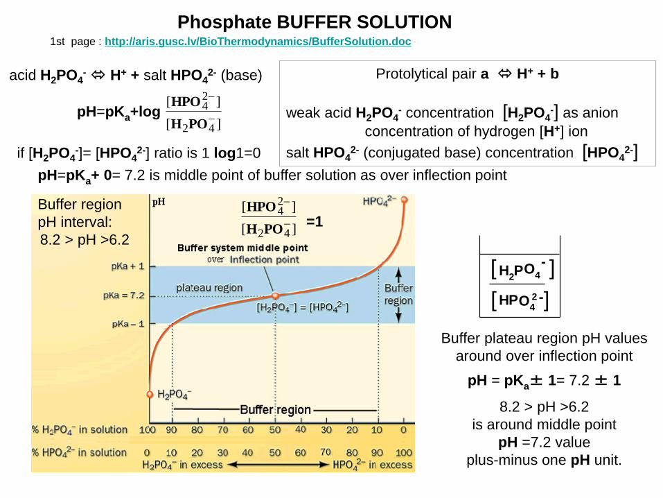
Phosphate BUFFER SOLUTION
H2P
HPO4
O4
2 -
-[ ]
[ ]
acid H2PO4- H+ + salt HPO4
2- (base)
pH=pKa+log ][
][
42
24
POH
HPO
if [H2PO4-]= [HPO4
2-] ratio is 1 log1=0
pH=pKa+ 0= 7.2 is middle point of buffer solution as over inflection point
Protolytical pair a H+ + b
weak acid H2PO4- concentration [H2PO4
-] as anion
concentration of hydrogen [H+] ion
salt HPO42- (conjugated base) concentration [HPO4
2-]
=1 ][
][
42
24
POH
HPOBuffer region
pH interval:
8.2 > pH >6.2
Buffer plateau region pH values
around over inflection point
pH = pKa± 1= 7.2 ± 1
8.2 > pH >6.2
is around middle point
pH =7.2 value
plus-minus one pH unit.
1st page : http://aris.gusc.lv/BioThermodynamics/BufferSolution.doc
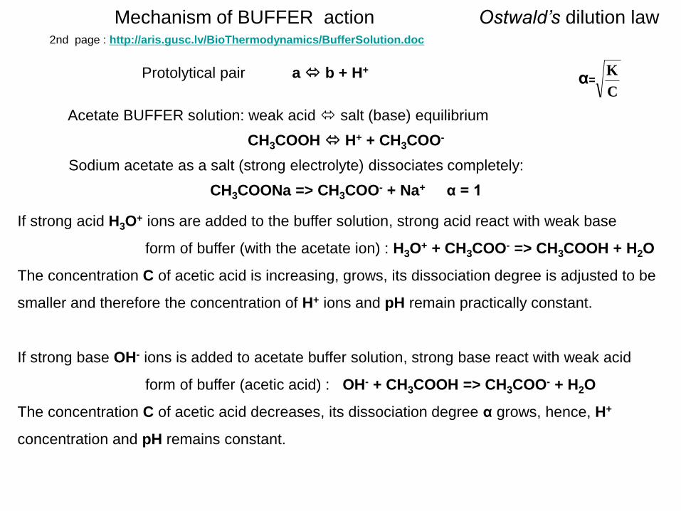
Mechanism of BUFFER action Ostwald’s dilution law
Acetate BUFFER solution: weak acid salt (base) equilibrium
CH3COOH H+ + CH3COO-
Sodium acetate as a salt (strong electrolyte) dissociates completely:
CH3COONa => CH3COO- + Na+ α = 1
If strong acid H3O+ ions are added to the buffer solution, strong acid react with weak base
form of buffer (with the acetate ion) : H3O+ + CH3COO- => CH3COOH + H2O
The concentration C of acetic acid is increasing, grows, its dissociation degree is adjusted to be
smaller and therefore the concentration of H+ ions and pH remain practically constant.
If strong base OH- ions is added to acetate buffer solution, strong base react with weak acid
form of buffer (acetic acid) : OH- + CH3COOH => CH3COO- + H2O
The concentration C of acetic acid decreases, its dissociation degree α grows, hence, H+
concentration and pH remains constant.
α= C
KProtolytical pair a b + H+
2nd page : http://aris.gusc.lv/BioThermodynamics/BufferSolution.doc

Weak base and its salt Ostwald’s dilution law
Ammonium BUFFER solution: weak base salt (acid) equilibrium
NH4OH NH4+ + OH- ; Kb = = ; pOH=pKb+log
Ammonium chloride as a salt (strong electrolyte) dissociates completely:
NH4Cl => NH4++ Cl- α = 1
nondis4
4
]OHNH[
]NH[]OH[
base
salt
C
COH ][
base
salt
C
C
pOH=pKb+log base
salt
n
npOH=pKb+log pOH=pKb+log
pOH=pKb+log
Vn
Vn
base
salt
/
/
''
''
basebase
saltsalt
VC
VC
acbase
acsalt
nn
nn
pOH=pKb+log
bbase
bsalt
nn
nn
If strong acid H3O+ ions are added to buffer solution strong acid is transformed to a salt (NH4
+):
H3O+ + NH4OH => NH4
+ + 2 H2O
The concentration C of weak base decreases, its dissociation degree α grows, hence,
OH- concentration remains constant and pH=const.
If strong base is added to buffer solution, the OH- ions are transformed into weak base NH4OH:
OH- + NH4+ => NH4OH
The concentration C of this weak base is increasing, grows, its dissociation degree is adjusted to
be smaller and therefore
the concentration of OH- ions remain practically constant and pH=const.
α= C
K
2nd page : http://aris.gusc.lv/BioThermodynamics/BufferSolution.doc

DIFFERENT FORMS OF pH Henderson Haselbalh EQUATION
=
pH=pKa+log acid
salt
C
CpH=pKa+log
acid
salt
n
n pH=pKa+log V/n
V/n
acid
salt pH=pKa+log ''
''
acidacid
saltsalt
VC
VC
pHac=pKa+log
acacid
acsalt
nn
nn
pHb=pKa+log
bacid
bsalt
nn
nn
The Δnac=Cas•Vac is number of strong acid moles, for example HCl, added to buffer
solution, which decreases the buffer system salt as Brensted base amount nsalt - Δnac and
increases the buffer system weak acid amount nacid + Δnac, thus change the buffer
system pH value about ΔpH= pH - pHac to decrease that.
The Δnb=Cb•Vb is number of strong base moles, for example NaOH, added to buffer
solution, which increases the buffer system salt as Brensted base amount nsalt + Δnb and
decreases the buffer system acid amount nacid - Δnb, thus change the buffer system pH
value about ΔpH= pHb - pH to increase that.
3rd page : http://aris.gusc.lv/BioThermodynamics/BufferSolution.doc
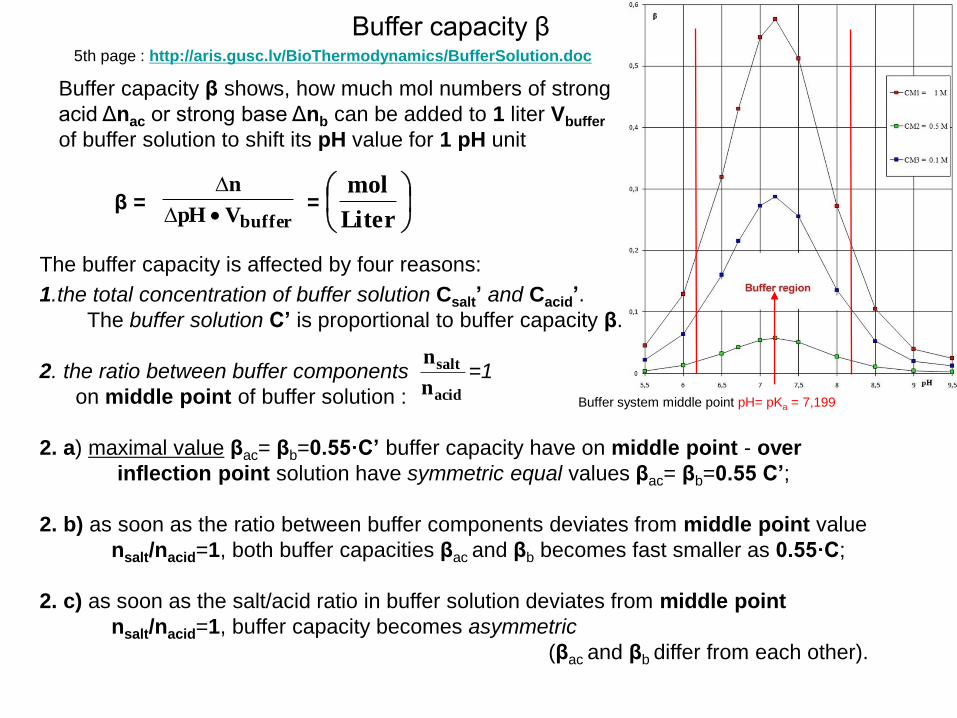
Buffer system middle point pH= pKa = 7,199
Buffer capacity β
Buffer capacity β shows, how much mol numbers of strong
acid Δnac or strong base Δnb can be added to 1 liter Vbuffer
of buffer solution to shift its pH value for 1 pH unit
bufferVpH
n
Liter
molβ = =
The buffer capacity is affected by four reasons:
1.the total concentration of buffer solution Csalt’ and Cacid’.
The buffer solution C’ is proportional to buffer capacity β.
2. the ratio between buffer components =1
on middle point of buffer solution :
2. a) maximal value βac= βb=0.55·C’ buffer capacity have on middle point - over
inflection point solution have symmetric equal values βac= βb=0.55 C’;
2. b) as soon as the ratio between buffer components deviates from middle point value
nsalt/nacid=1, both buffer capacities βac and βb becomes fast smaller as 0.55·C;
2. c) as soon as the salt/acid ratio in buffer solution deviates from middle point
nsalt/nacid=1, buffer capacity becomes asymmetric
(βac and βb differ from each other).
acid
salt
n
n
5th page : http://aris.gusc.lv/BioThermodynamics/BufferSolution.doc
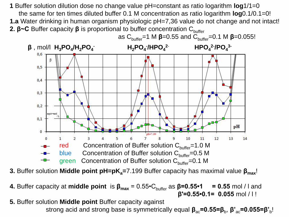
1 Buffer solution dilution dose no change value pH=constant as ratio logarithm log1/1=0
the same for ten times diluted buffer 0.1 M concentration as ratio logarithm log0.1/0.1=0!
1.a Water drinking in human organism physiologic pH=7,36 value do not change and not intact!
2. β~C Buffer capacity β is proportional to buffer concentration Cbuffer
as Cbuffer=1 M β=0.55 and Cbuffer=0.1 M β=0.055!
β , mol/l H3PO4/H2PO4- H2PO4
-/HPO42- HPO4
2-/PO43-
red Concentration of Buffer solution Cbuffer=1.0 M
blue Concentration of Buffer solution Cbuffer=0.5 M
green Concentration of Buffer solution Cbuffer=0.1 M
3. Buffer solution Middle point pH=pKa=7.199 Buffer capacity has maximal value βmax!
4. Buffer capacity at middle point is βmax = 0.55•Cbuffer as β=0.55•1 = 0.55 mol / l and
β'=0.55•0.1= 0.055 mol / l !
5. Buffer solution Middle point Buffer capacity against
strong acid and strong base is symmetrically equal βac=0.55=βb, β’ac=0.055=β’b!

1 Buffer solution Dilution dose no change value pH=constant as ratio logarithm log1/1=0
the same for ten times diluted buffer 0.1 M concentration as ratio logarithm log0.1/0.1=0!
1.a Water drinking in human organism physiologic pH=7,36 value do not change and not intact!
2. β~C Buffer capacity β is proportional to buffer concentration Cbuffer
as Cbuffer=1 M β=0.55 and Cbuffer=0.1 M β=0.055!
β , mol/l CH3COOH/CH3COO-
red Concentration of Buffer solution Cbuffer=1.0 M
blue Concentration of Buffer solution Cbuffer=0.5 M
green Concentration of Buffer solution Cbuffer=0.1 M
3. Buffer solution Middle point pH=pKa=4.76 buffer capacity has maximal value βmax!
4. Buffer capacity at middle point is βmax = 0.55•Cbuffer as β=0.55•1 = 0.55 mol / l and
β'=0.55•0.1 = 0.055 mol / l !
5. Buffer solution Middle point Buffer capacity against
strong acid and strong base is symmetrically equal βac=0.55=βb, β’ac=0.055=β’b!
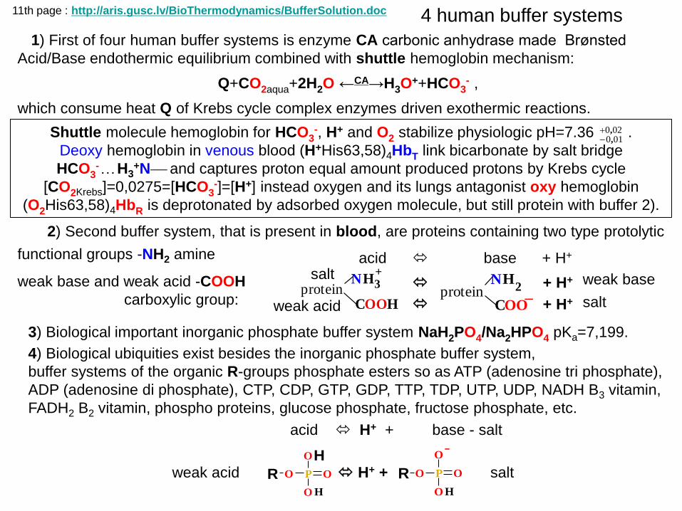
1) First of four human buffer systems is enzyme CA carbonic anhydrase made Brønsted
Acid/Base endothermic equilibrium combined with shuttle hemoglobin mechanism:
Q+CO2aqua+2H2O ←CA→H3O++HCO3
- ,
which consume heat Q of Krebs cycle complex enzymes driven exothermic reactions.
4 human buffer systems
2) Second buffer system, that is present in blood, are proteins containing two type protolytic
functional groups -NH2 amine
weak base and weak acid -COOH
carboxylic group:
Shuttle molecule hemoglobin for HCO3-, H+ and O2 stabilize physiologic pH=7.36 .
Deoxy hemoglobin in venous blood (H+His63,58)4HbT link bicarbonate by salt bridge
HCO3- H3
+N and captures proton equal amount produced protons by Krebs cycle
[CO2Krebs]=0,0275=[HCO3-]=[H+] instead oxygen and its lungs antagonist oxy hemoglobin
(O2His63,58)4HbR is deprotonated by adsorbed oxygen molecule, but still protein with buffer 2).
4) Biological ubiquities exist besides the inorganic phosphate buffer system,
buffer systems of the organic R-groups phosphate esters so as ATP (adenosine tri phosphate),
ADP (adenosine di phosphate), CTP, CDP, GTP, GDP, TTP, TDP, UTP, UDP, NADH B3 vitamin,
FADH2 B2 vitamin, phospho proteins, glucose phosphate, fructose phosphate, etc.
proteinNH
C
2
OO_
+ H+
+ H+ protein
NH
C
+
OO
3
H
acid base + H+ salt
weak acid salt
weak base
3) Biological important inorganic phosphate buffer system NaH2PO4/Na2HPO4 pKa=7,199.
O
O
O
P ORH
HO
O
O
P ORH
weak acid H+ + salt
acid H+ + base - salt
11th page : http://aris.gusc.lv/BioThermodynamics/BufferSolution.doc
020010
,,
4 6 7.0512=pKa 9 10 11→pH
7.36 = pH = pK+log =7.0512+log
]CO[
]HCO[
aqua2
3
]CO[
]HCO[
aqua2
3
β, mol/L buffer capacity
0.6
0.5
0.4
0.3
0.2
0.1
0
10
9
8
7
6
5
4
3
2
HCO3-0% 50% 100%
CO2+ 2H2O 100% 50% 0%
Human blood pH=7,36 Henderson Haselbalh CA equation - Shuttle hemoglobin
Main buffer system CA uses hemoglobin Shuttle stabilized pH=7,36 from tissues capturing
[H+]=0,02754 M instead desorbed oxygen for Krebs cycle use keeping venous [O2aqua] =1.85·10-5 M
but in lungs arteries adsorbed oxygen releases H+ keeping concentration [O2aqua] =6·10-5 M by
sensitive deoxy hemoglobin Tense state - oxy hemoglobin Relax state Shuttle equilibrium:
in lungs=> 4O2aqua + (H+His63,58)4HbT <= [O2aqua] =>(O2His63,58)4HbR +4 H+ <= tissues
Q+CO2aqua+2H2O ←CA→H3O++HCO3
- Alkaline reserve is
10(pH-pK)= 10(7.36-7.0512)= 100.3088 = = ][
][
2
3
aquaCO
HCO
1
2.0361
acidose alkalose
acidose alkalose
15th page : http://aris.gusc.lv/BioThermodynamics/BufferSolution.doc
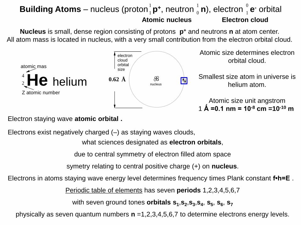
Building Atoms – nucleus (proton p+, neutron n), electron e- orbital
Electron staying wave atomic orbital .
Atomic nucleus
Nucleus is small, dense region consisting of protons p+ and neutrons n at atom center.
All atom mass is located in nucleus, with a very small contribution from the electron orbital cloud.
Electrons exist negatively charged (–) as staying waves clouds,
what sciences designated as electron orbitals,
due to central symmetry of electron filled atom space
symetry relating to central positive charge (+) on nucleus.
Electrons in atoms staying wave energy level determines frequency times Plank constant f•h=E .
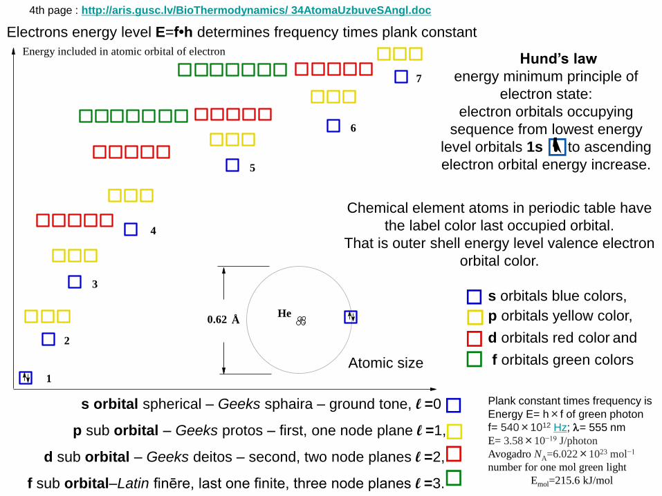
Periodic table of elements has seven periods 1,2,3,4,5,6,7
with seven ground tones orbitals s1,s2,s3,s4, s5, s6, s7
physically as seven quantum numbers n =1,2,3,4,5,6,7 to determine electrons energy levels.
2
4
He helium
1
1
0
1
1
0
0.62 Å+
+
nucleus
electron cloud orbitalsize
Atomic size determines electron
orbital cloud.
Smallest size atom in universe is
helium atom.
Atomic size unit angstrom
1 Ǻ =0.1 nm = 10-8 cm =10-10 m
Electron cloud
atomic mas
Z atomic number
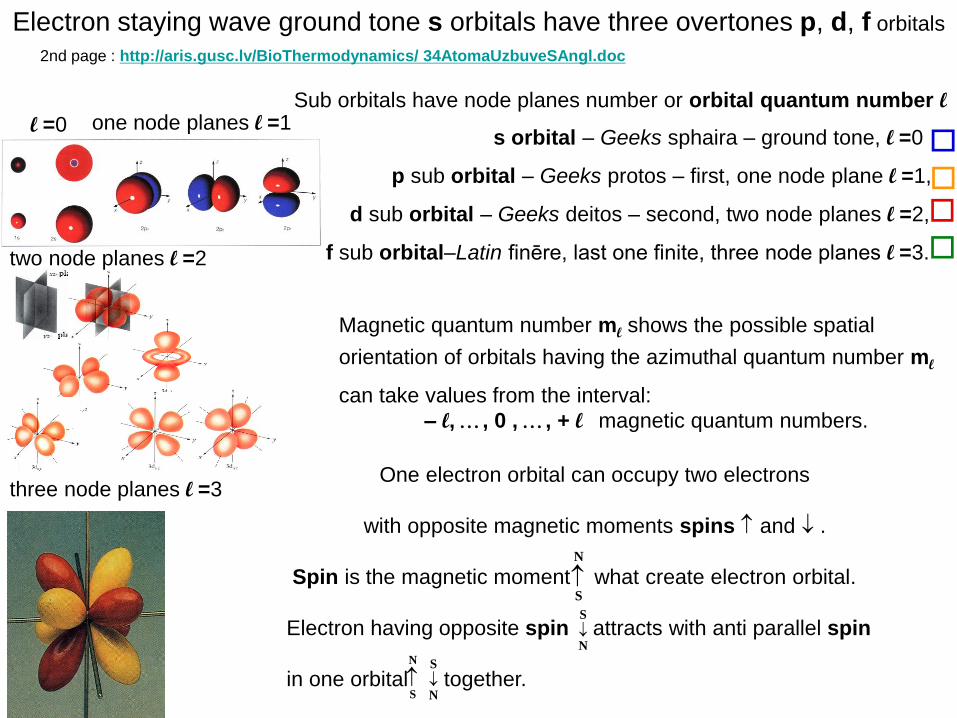
Electron staying wave ground tone s orbitals have three overtones p, d, f orbitals
s orbital – Geeks sphaira – ground tone, l =0
p sub orbital – Geeks protos – first, one node plane l =1,
d sub orbital – Geeks deitos – second, two node planes l =2,
f sub orbital–Latin finēre, last one finite, three node planes l =3.
Sub orbitals have node planes number or orbital quantum number l
Magnetic quantum number ml shows the possible spatial
orientation of orbitals having the azimuthal quantum number ml
can take values from the interval:
– l, , 0 , , + l magnetic quantum numbers.
One electron orbital can occupy two electrons
with opposite magnetic moments spins and .
Spin is the magnetic moment what create electron orbital.
Electron having opposite spin attracts with anti parallel spin
in one orbital together.
two node planes l =2
N
S
S
N
N
S
S
N
three node planes l =3
l =0 one node planes l =1
2nd page : http://aris.gusc.lv/BioThermodynamics/ 34AtomaUzbuveSAngl.doc
1
2
3
4
5
6
7
Energy included in atomic orbital of electron
He0.62 Å +
+
Atomic size
Hund’s law
energy minimum principle of
electron state:
electron orbitals occupying
sequence from lowest energy
level orbitals 1s to ascending
electron orbital energy increase.
Chemical element atoms in periodic table have
the label color last occupied orbital.
That is outer shell energy level valence electron
orbital color.
s orbitals blue colors,
p orbitals yellow color,
d orbitals red color and
f orbitals green colors
s orbital spherical – Geeks sphaira – ground tone, l =0
p sub orbital – Geeks protos – first, one node plane l =1,
d sub orbital – Geeks deitos – second, two node planes l =2,
f sub orbital–Latin finēre, last one finite, three node planes l =3.
Electrons energy level E=f•h determines frequency times plank constant
Plank constant times frequency is
Energy E= h×f of green photon
f= 540×1012 Hz; = 555 nm
E= 3.58×10⁻¹⁹ J/photon
Avogadro NA=6.022×10²³ mol−1
number for one mol green light
Emol=215.6 kJ/mol
4th page : http://aris.gusc.lv/BioThermodynamics/ 34AtomaUzbuveSAngl.doc

Chemical element atoms in periodic table have
the label color last occupied orbital.
That is outer shell energy level valence electron
orbital color.
s orbitals blue colors,
p orbitals yellow color,
d orbitals red color and
f orbitals green colors
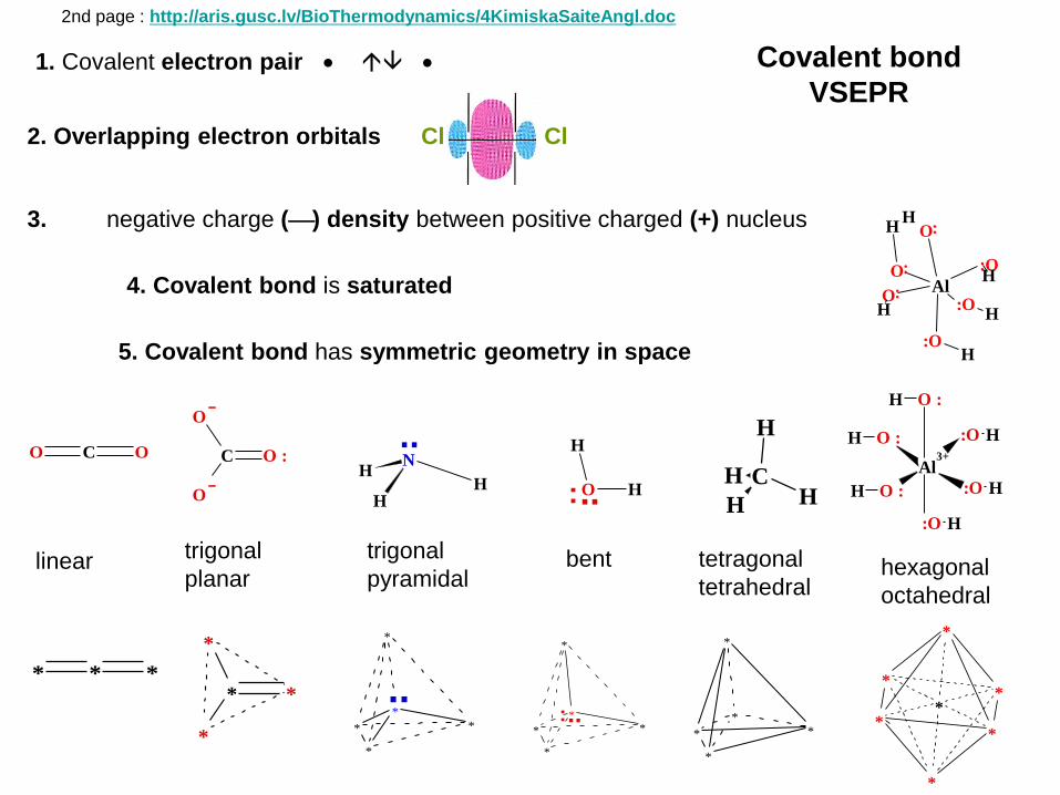
1. Covalent electron pair Covalent bond
VSEPR
2. Overlapping electron orbitals Cl Cl
3. negative charge () density between positive charged (+) nucleus
4. Covalent bond is saturated
5. Covalent bond has symmetric geometry in space
C O :
O
O
C OO
C
H
HH
H
N
HH
H
..
O
H
H..:
O :H
:O H
:O H
:O HO :H
Al3+
O :H
linear trigonal
planar
trigonal
pyramidal bent tetragonal
tetrahedral hexagonal
octahedral
*
**
**
*
*
*
*
*
*
*
*
*
*
*
*..:*
*
*
*
*
..* *
*
*
* * *
O
O
O
:O
Al
:O
:O
H
H
H
HH
H
2nd page : http://aris.gusc.lv/BioThermodynamics/4KimiskaSaiteAngl.doc
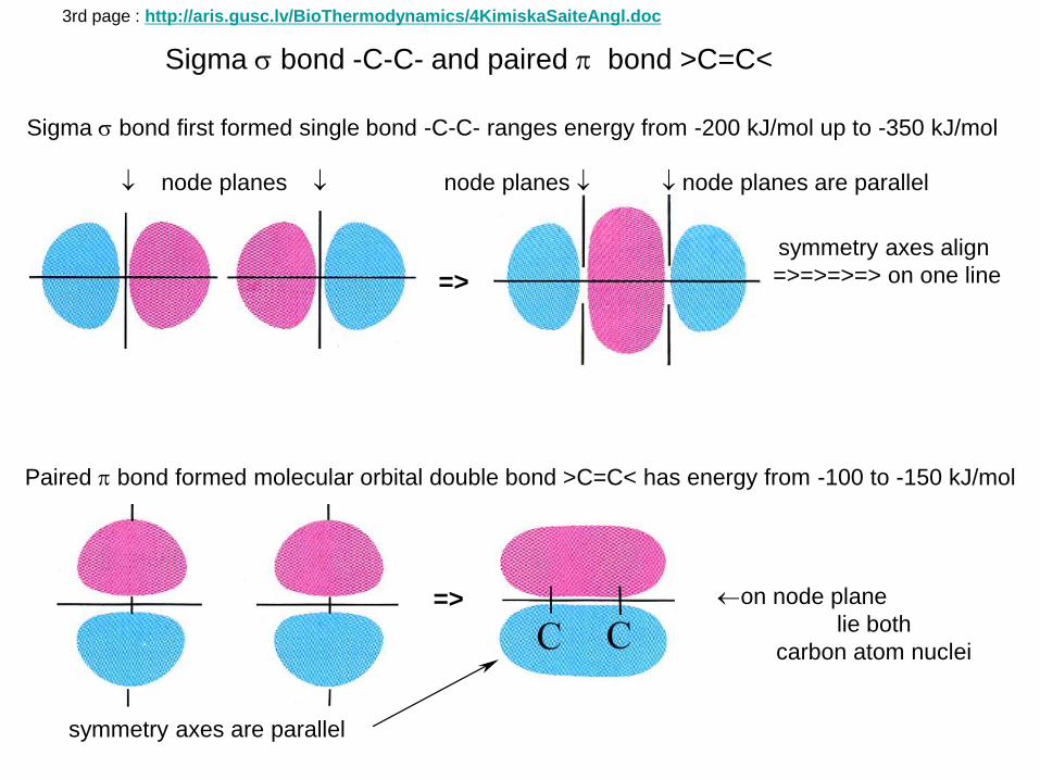
Sigma bond -C-C- and paired bond >C=C<
=>
node planes node planes node planes are parallel
symmetry axes align
=>=>=>=> on one line
Paired bond formed molecular orbital double bond >C=C< has energy from -100 to -150 kJ/mol
=> on node plane
lie both
carbon atom nuclei
Sigma bond first formed single bond -C-C- ranges energy from -200 kJ/mol up to -350 kJ/mol
3rd page : http://aris.gusc.lv/BioThermodynamics/4KimiskaSaiteAngl.doc
symmetry axes are parallel

Liquid water tetramer structure no free water molecules present
Hydrogen bond polar water molecule attraction force
H
H
O HH
H
OH
O
H
H
O0.98 A
1.6 A
104.8
104.7
-0.8+0.4
-0.8 1.6 A
104.7
+0.4
-0.8 1.6 A
104.7
+0.4
-0.8
-30 kJ/mol -30 kJ/mol -30 kJ/mol
0.98 A
0.98 A
OH( )42
Three hydrogen bonds as polar water interaction resulting in tetramer (H2O)4 three hydrogen
bonds energies sum ΔG= - 90 kJ/mol
Ice water molecules
hydrogen bonding hexagonal structure
Pure hydrogen bonding crystalline lattice
type structure OH
H
OHH
OH
H
OHH
OHH
OHH
O
H
H
O
H
H
OHH
O
H
H
OHH
OH
H
OHH
OH
H
OH
H
OH
H
0.96 Å
1.76 Å109,47°
MW 288.25 g/mol16 OH2
-0.8
+0.4+0.4
1st page : http://aris.gusc.lv/BioThermodynamics/4HydrogenBond.doc
O
H
H
..:2.61 A
-0.8 +0.8q= q=
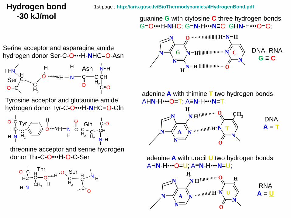
Serine acceptor and asparagine amide
hydrogen donor Ser-C-O•••H-NHC=O-Asn
Hydrogen bond
-30 kJ/mol
Tyrosine acceptor and glutamine amide
hydrogen donor Tyr-C-O•••H-NHC=O-Gln
O
HCO
NH
CHCH
2N H
C O
CHCH
2
CH
2
C
O
H
NHTyr Gln
1st page : http://aris.gusc.lv/BioThermodynamics/4HydrogenBond.pdf
O HNH
CO
CH
CH
2
H N
N CHCH
2
C
C OO
HH
Ser
Asn
threonine acceptor and serine hydrogen
donor Thr-C-O•••H-O-C-Ser
CO
NH
CH CH
O
CH3
H
NH
CO
CH
CH
2
OH
Thr Ser
guanine G with ciytosine C three hydrogen bonds
G=O•••H-NHC; G=N-H•••N≡C; GHN-H•••O=C;
adenine A with thimine T two hydrogen bonds
AHN-H•••O=T; A≡N-H•••N=T;
H
O
O
N
N
CH3
N
N
N N
N
H
H
AT
NH
H
O
N
N
N
N H
N HH
O
N
N
CG
adenine A with uracil U two hydrogen bonds
AHN-H•••O=U; A≡N-H•••N=U;
H
O
O
N
N
H
N
N
N N
N
H
H
AU
DNA, RNA
G ≡ C
DNA
A = T
RNA
A = U
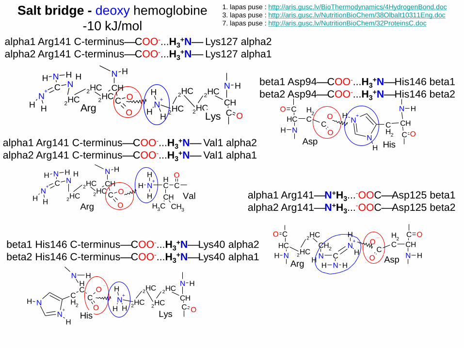
alpha1 Arg141 C-terminusCOO-...H3+N Lys127 alpha2
alpha2 Arg141 C-terminusCOO-...H3+N Lys127 alpha1
Salt bridge - deoxy hemoglobine
-10 kJ/mol
OC
O
N H
CH
2HC
2HC
2HC
NCN
+
N HHH
H H
N+
H
H
H
CH
C
O
CH
CH3 CH
3Arg
Val
N H
N+
H
CH2HC
2HC
C O
2HC
2HC
H
H
OC
O
N H
CH
2HC
2HC
2HC
NCN
+
N HHH
H HLys
Arg
NH
CO
CH C
H2
CO
ON H
C ON
H
N+
CHCH
2
H
Asp His
1. lapas puse : http://aris.gusc.lv/BioThermodynamics/4HydrogenBond.doc
3. lapas puse : http://aris.gusc.lv/NutritionBioChem/38Olbalt10311Eng.doc
7. lapas puse : http://aris.gusc.lv/NutritionBioChem/32ProteinsC.doc
alpha1 Arg141 C-terminusCOO-...H3+N Val1 alpha2
alpha2 Arg141 C-terminusCOO-...H3+N Val1 alpha1
beta1 Asp94COO-...H3+NHis146 beta1
beta2 Asp94COO-...H3+NHis146 beta2
OC
ON
+
H
N
CH
N
CH
2H
HN H
N+
H
CH2HC
2HC
C O
2HC
2HC
H
HLysHis
beta1 His146 C-terminusCOO-...H3+NLys40 alpha2
beta2 His146 C-terminusCOO-...H3+NLys40 alpha1 N H
C O
CHC
H2
CO
OCO
NH
CH2HC
2HC
CH2
N C
N+
NH
H H
H
H
AspArg
alpha1 Arg141N+H3...-OOCAsp125 beta1
alpha2 Arg141N+H3...-OOCAsp125 beta2
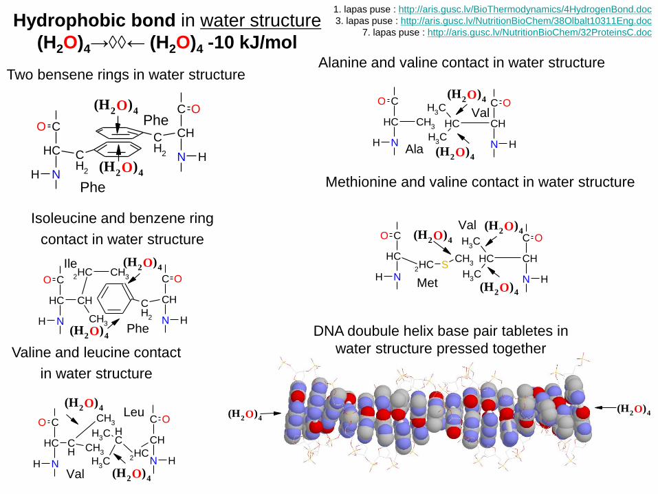
Two bensene rings in water structure
Hydrophobic bond in water structure
(H2O)4→← (H2O)4 -10 kJ/mol
CH CH
2HC CH
3
CH3
CO
NH
CH
C O
N H
CH
2
(H ) O 2 4
(H ) O 2 4
Ile
Phe
CH
CO
NH
CH
2
CH
C O
N H
CH
2
(H ) O 2 4
(H ) O 2 4
Phe
Phe CHCH
CH3
CH3
C O
N H
CH3
CH
CO
NH
(H ) O 2 4
(H ) O 2 4
Val
Ala
Isoleucine and benzene ring
contact in water structure
Alanine and valine contact in water structure
CH CH CH
3
CH3CO
NH
CHCH
3
CH3
CH
2HC
C O
N H
(H ) O 2 4
(H ) O 2 4Val
Leu
Valine and leucine contact
in water structure
CHCH
CH3
CH3
C O
N H
CH2HC S
CH3
CO
NH
(H ) O 2 4
(H ) O 2 4
Val
Met
(H ) O 2 4
Methionine and valine contact in water structure
DNA doubule helix base pair tabletes in
water structure pressed together
(H ) O 2 4(H ) O 2 4
1. lapas puse : http://aris.gusc.lv/BioThermodynamics/4HydrogenBond.doc
3. lapas puse : http://aris.gusc.lv/NutritionBioChem/38Olbalt10311Eng.doc
7. lapas puse : http://aris.gusc.lv/NutritionBioChem/32ProteinsC.doc
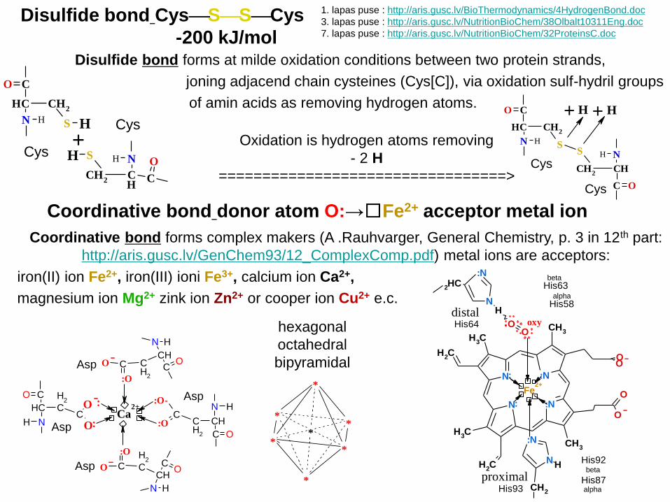
Disulfide bond forms at milde oxidation conditions between two protein strands,
joning adjacend chain cysteines (Cys[C]), via oxidation sulf-hydril groups
of amin acids as removing hydrogen atoms.
Disulfide bond CysSSCys
-200 kJ/mol
C
O
CH
H N
CH2
CO
CH
HN
CH2
HS
H S+
Cys
CysS
S
C O
CH
H N
CH2
CO
CH
HN
CH2
H H+ +
Cys
Cys
Oxidation is hydrogen atoms removing
- 2 H
=================================>
Coordinative bond forms complex makers (A .Rauhvarger, General Chemistry, p. 3 in 12th part:
http://aris.gusc.lv/GenChem93/12_ComplexComp.pdf) metal ions are acceptors:
iron(II) ion Fe2+, iron(III) ioni Fe3+, calcium ion Ca2+,
magnesium ion Mg2+ zink ion Zn2+ or cooper ion Cu2+ e.c.
Coordinative bond donor atom O:→□Fe2+ acceptor metal ion
NH
CO
CH C
H2
C
O
O
Ca2+ N H
C O
CHCH
2
C:O
:O-
N H
CO
CHCH
2
C
:O
O
N H
CO
CHC
H2
C
:O
O
Asp
Asp
Asp
Asp
N
:N
Fe2+
CH3
OO
N
CH3
CH2
CH3
CH2
:N
CH3
O
O
N
:N
CH2
H
NH
:N
2HC
OO
His93
His92beta
His87alpha
His63beta
His58alpha
His64 oxy
proximal
distal
hexagonal
octahedral
bipyramidal
*
**
**
*
*
1. lapas puse : http://aris.gusc.lv/BioThermodynamics/4HydrogenBond.doc
3. lapas puse : http://aris.gusc.lv/NutritionBioChem/38Olbalt10311Eng.doc
7. lapas puse : http://aris.gusc.lv/NutritionBioChem/32ProteinsC.doc

Ca2+
N+
HH
H
CAsn
O
N H
H
Asp
Ser
C
O
O
Lys
Phe
Glu
Phe
C
O
O
Cys
Glu
Cys S S
C
O
O
O
H(H ) O 2 4
(H ) O 2 4
1. Hydrogen bonds of
O and N atoms between
hydrogen atom H acceptor
and other side H donor
2. Salt bridge
5. Disulphide bonds
3. Hydrophobic bonds
4. Coordinative bonds AspCOO-...+H3NLys
N
:N
CH2
H
N
:N
Fe2+
CH3
OO
N
CH3
CH2
CH3
CH2
:N
CH3
O
O
NH
:NCH
2
OO
globinoxy
globin
CysSSCys
hydrogen acceptor
O
O
H
N
O
H
O
O
H
O
NH
N
NH
O
NH
hydrogen donor
coordinative donor acceptor bond calcium ion
with carboxyl groups -COO→□Ca2+ □←OOC-
or iron(II) ion on center of hem → □Fe2+ □←
(H2O)4→← (H2O)4
water press together
nonpolar residues
of amino acids or
base pairing tablets
in DNA and RNA
molecules
5 Intermolecular forces (intermolecular bonds) biomolecules
1st page : http://aris.gusc.lv/BioThermodynamics/4HydrogenBond.doc
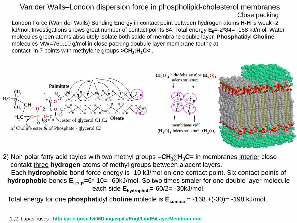
London Force (Wan der Walls) Bonding Energy in contact point between hydrogen atoms H-H is weak -2
kJ/mol. Investigations shows great number of contact points 84. Total energy EII=-2*84= -168 kJ/mol. Water
molecules green atoms absolutely isolate both saide of membrane double layer. Phosphatidyl Choline
molecules MW=760.10 g/mol in close packing doubule layer membrane touthe at
contact in 7 points with methylene groups >CH2:H2C< .
Close packing
Total energy for one phosphatidyl choline molecle is Esumma = -168 +(-30)= -198 kJ/mol.
2) Non polar fatty acid tayles with two methyl groups –CH3░H3C= in membranes interier close
contakt three hydrogen atoms of methyl groups between ajacent layers.
Each hydrophobic bond force energy is -10 kJ/mol on one contact point. Six contact points of
hydrophobic bonds Energy=6*-10= -60kJ/mol. So two times smaler for one double layer molecule
each side Ehydrophob=-60/2= -30kJ/mol.
Van der Walls–London dispersion force in phospholipid-cholesterol membranes
1.,2. Lapas puses : http://aris.gusc.lv/06Daugavpils/Engl/LipdBiLayerMembran.doc
C
H H
HH
H H
HH
H H
HH
H H
H H
H H
HH
H H
HH
HH
H H
HH
HH
H
C
HH
H H
HH
H H
HH
HH
H H
HH
H H
HH
H H
HH
H H
H H
HH
H
C
H
HN+
CH3
CH3
CH3
CH2
CH2
O P
O
O
O CHH
CH
O
O
O
O
Palmitate
1
2
3Oleateester of glycerol C1,C2
of Choline ester & of Phosphate - glycerol C3
7
CH
H H
C
HHH
CH
HH
CH
HH
(H ) O 2 4
(H ) O 2 4
(H ) O 2 4
(H ) O 2 4
hidrofoba- saistiba-
membranas vidu- -
udens struktura- - -
udens struktura- - -

Close packing
56 Å
↑↑↑↑
56 Å
↓↓↓↓
56 Å
Double layer membranes of phospho lipids 1.,2. Lapas puses : http://aris.gusc.lv/06Daugavpils/Engl/LipdBiLayerMembran.doc
Phosphatidyl cholin double layer membrane isolate two water compartments of solutions and
has electric double layer on surface with positive charged nitrogen N+ atoms blue and negative
charged phosphate groups PO4-. The average thickness mesurement value of membrane is
d =56Å or 5,6 nm. The size of water molecule 1,4 Å cover membrane thickness 56/1,4=40 times.
If wall thickness would be 40 man 1,7 m tall size, then wall thickness between rooms would be
68 m and home doures would be 68 m long tunnels in walls.
N+
PO4-
PO4-
N+
Waterless interier in membranes
are impermeable isolate cell membrane wall for water medium components molecules and
solutions: saļs and organic compound molecules. As the home walls separate rooms. That
entrance the rooms we use doores, but membranes equiped with transport and signaling
enzymes–proteins: aquaporins H2O,O2,CO,NO,C3H8O3, proton H+ channals, bicarbonate chanals
HCO3-, sodium Na+, potasium K+, magnesium Mg2+, calcium Ca2+, chloride Cl- ions, sugares Glc,
Gal, Man, 21 amino acids and other biologic water solutes. Destiny of studies is organic regulate
opened thermodynamics systemes, which through cell membranes organize the life organisms.

Composition of double layer membranes with Phospho lipids and Cholesterol 1.,2. Lapas puses : http://aris.gusc.lv/06Daugavpils/Engl/LipdBiLayerMembran.doc
1/3 mass fraction of cell and organell membranes constitute Phospho lipids as Phosphatidyl
choline. Binding energy Ebound=-198 kJ/mol make membranes liquid therefore can be mechanic
disordered, as liquids with pressure, gravitation and mechanic movement impact. Cholesterol
content makes membranes stronger and flexible evoid its destruction and following cell content
leaking which water solution contains salts and organic compounds molecules.
Second third part of membranes mass constitutes
hydrocarbon 27 carbon steric frame steroid
molecule: four rings are labeled A, B, C and D.
Angular methyl –CH3 groups 18 and 19 as well
tail fork, rod, splinter are good clutch fixing close
hydrocarbon chains. Double bond between carbon
atoms >C=C< 5 and 6. Alcohol HO- at C 3 forms
hydrogen bond >C=O...HO- with fatty acid
H HH
HHH
H
HH
H HH
H
O
H HH
O
C
H H
HH
H H
HH
H H
HH
H H
H H
H H
HH
H H
HH
HH
H H
HH
H H
H
C
HH
H H
HH
H H
HH
HH
H H
HH
H H
HH
H H
HH
H H
H H
HH
H
C
H
HN+
CH3
CH3
CH3
CH2
CH2
O P
O
O
O CHH
CH
O
O
O
1
2
3 18
19
abas angularas metil grupas
H HH
H
HH
H
HH
H HH
H HH
H
O
O
CO both angular methyl groups18
19
HH
H
H
H HH
H HH
H HH
HO
HH
Cholesterol
10
4
912
675
8
1211 13
14 1516
A
17
18
3B
C D
19
202122
23
24
2526
27
Oleate
carboxyl oxygen >C=O.
Cholesterol as Steroid
makes membranes unbroken, flexible and
so prevent following leaking of water molecules and of water solution components:
salts and water soluble organic molecules. The Cholesterol/Phospho Lipid C/PL mole ratio of human red blood cell membranes ranges from a normal value of 0.9–1.0
(since 1978 first publication as refference in Journal of Cellular Biochemistry 2004 V8, 4, p 413-430).
If Cholesterol amount decreases up to 0,5= C/PL , then membranes leak cell content out, but
If Cholesterol amount increases up to 1,5= C/PL , then membrane becomes solid, inflexible and
squeeze channels, aquaporins, but receptors becomes inactive due to absence conformational flexibility.

When a molecule is subjected to an outer electrical field,
the dipole momentum of the molecule is changed : = μo + μind, where
μ o is the dipole momentum of the molecule itself as well
μo = 0 for a non-polar molecule or μo > 0 for a polar one and
μ ind is the dipole momentum, induced by the outer field.
Non-polar molecules become polar and polar molecules
become more polar.
Polarization of molecules in outer electric field
Orientation forces can be established between two polar molecules.
Dispersion forces act between two non-polar molecules: :Aδ-Bδ+A:BA δ+Bδ-:
the common pair of electrons is shown as a pair of two dots :
Intra molecular polarization, when a hydrogen atom is replaced by a more
electro negative (EN) atoms (O, N, Cl). The Cl atom attracts electrons and
therefore shifts the electron density of the entire molecule:
As a result, the O-H bond in the carboxyl group becomes more polar and
therefore chloroacetic acid appears to be a much stronger acid than acetic
acid. Compare the acetic acid dissociation constant K = 1.75 • 10-5 and
chloroacetic acid K = 1.4 • 10-3 . If all the three hydrogen atoms in the methyl
group are substituted by Cl atoms, forming trichloro acetic acid one gets an
acid, the strength of which can be compared to hydrochloric acid.
Van der Waals’s interaction forces of biomolecules–London dispersion forces
4th page : http://aris.gusc.lv/BioThermodynamics/4HydrogenBond.doc
He
- +++
nucleus
electrons
outer electric field
Cl
Cl
Cl C C
O
O H
Cl C C
O
O H
H
H
C C
O
O H
H
H
H
- +
- +
- +
temperature
- +
He
-+ ++
nucleus
electrons
-+ -+ Induction forces exist between a polar and a non-polar molecules.
= q•l dipol H2O =0.8 el•2.61 Å=2.088 el•Å.
O
H
H
..:2.61 A
-0.8 +0.8q= q=


