Mycobiology 38(1) : 33-38 (2010) DOI:10.4489/MYCO.2010.38.1.033
© The Korean Society of Mycology
33
Direct Detection of Cylindrocarpon destructans, Root Rot Pathogen of Ginseng byNested PCR from Soil Samples
Chang Soon Jang, Jin Ha Lim, Mun Won Seo, Jeong Young Song and Hong Gi Kim*
Department of Agricultural Biology, Chungnam National University, Daejeon 305-764, Korea
(Received January 19, 2010. Accepted February 3, 2010)
We have successfully applied the nested PCR to detect Cylindrocarpon destructans, a major pathogen causing root rot disease
from ginseng seedlings in our former study. The PCR assay, in this study, was used to detect the pathogen from soils. The
nested PCR using internal transcribed spacer (ITS) 1, 4 primer set and Dest 1, 4 primer set maintained the specificity in
soils containing various microorganisms. For a soil DNA extraction method targeting chlamydospores, when several cell wall
disrupting methods were tested, the combination of lyophilization and grinding with glass beads, which broke almost all
the chlamydospores, was the strongest. The DNA extraction method which was completed based on the above was simple
and time-saving because of exclusion of unnecessary stages, and efficient to apply in soils. As three ginseng fields whose
histories were known were analyzed, the PCR assay resulted as our expectation derived from the field information. The
direct PCR method will be utilized as a reliable and rapid tool for detecting and monitoring C. destructans in ginseng fields.
KEYWORDS : Cylindrocarpon destructans, Diagnosis, Ginseng, Nested PCR, Root rot
Ginseng (Panax ginseng C. A. Meyer and Panax quin-
quefolius L.), belongs to Araliaceae of perennial plants,
and is an economically important cash crop in Korea and
North America [1]. Consumption of the crop is gradually
increasing in Southeast Asia and America while the yield
in Korea is decreasing currently because of an injury by
continuous cropping and exhaustion of the first planting
ginseng fields. In Korea, the production of ginseng
requires a 4- to 6-year cultivation period and, throughout
this time, soil borne pathogens may increase more in the
rhizosphere than in that of North America with a 3- to 4-
year cultivation period; Yield loss to disease is proportion
to the cultivation period [2, 3]. One of the major patho-
gens of ginseng is Cylindrocarpon destructans [4, 5], which
may be more serious in Korea with smaller size of land
than in other countries because the pathogen is impli-
cated in replant failure.
Research on ginseng root rot disease caused by C.
destructans has not been carried out for a long time in
Korea. It had been assumed about the disease that the
replanting failure had occurred because of the amassment
of poison and the deficiency of mineral elements in the
ginseng field. The difficulty of study on C. destructans is
because chlamydospores as main form in soil germ rarely,
mycelial growth is slow, and the host of C. destructans is
not economic crops except ginseng [6].
Diagnostic systems based on PCR have been developed
for plant pathogenic fungi [7-11]. The classical methods
of diagnosis are both time-consuming and laborious [12],
requiring isolation of the fungus from diseased tissue.
Moreover, because the fungus grows slowly, colonies aris-
ing from diseased tissue are often overgrown by more
rapidly growing fungi and rare germination of chlamy-
dospores makes the spread plate method of soil samples
unusable [5, 10].
A nested PCR-based assay was developed for the
detection of C. destructans in pine and spruce seedlings.
Preserving specificity, the PCR assay has detected the
pathogen from roots of the host plants [13]. We have uti-
lized the PCR to detect the pathogen from the roots for
selection of the non-infested one-year-old ginseng seed-
lings. To apply the technique to soil samples containing
various PCR inhibitors, DNA purification method to recover
the high quality and plenty DNA were required [14]. Our
objective of the study is to develop the DNA-based method
for the detection of C. destructans directly from the gin-
seng fields and ultimately for choice of the non-infected
fields for ginseng cultivation in the future.
Materials and Methods
Pathogen. C. destructans was collected from diseased
ginseng roots and infested soils located at major ginseng
cultivating areas in Korea. The pathogen was isolated
with single conidia on potato dextrose agar (PDA) con-
taining streptomycin sulfate at 15o
C, grown on PDA and
SNAY (supplemented nutrient agar plus yeast extract)
media at 20o
C in the dark for a month and observed with
× 100 and × 400 microscopes [15, 16].
Conidia were produced by culture on PDA media at*Corresponding author <E-mail : [email protected]>
34 Jang et al.
15o
C for 3 weeks and chlamydospores were produced by
culture on potato dextrose broth and V8 20% juice broth
media at 180 rpm, 15o
C for over one month in a shaking
incubator. Subsequently, hemacytometer was used to
determine spore concentrations.
Genomic DNA extraction. Genomic DNA was extracted
from fungal cultures grown on SNAY broth media for 2
weeks. Mycelia were harvested from liquid cultures by
filtration through cheesecloth, and DNA was extracted
with cetyltrimethylammonium bromide (CTAB) method
[17]. About 10 mg of lyophilized mycelia were ground in
1.5 mL effendolf tube by sterilized wooden sticks and
added 400 µL extraction buffer and 400 µL CTAB solu-
tion. The mixture was extracted by 600 µL chloroform :
isoamylalcohol (24 : 1), vortexed and centrifuged for 10
minutes at 10,000 × g. The aqueous phase was precipi-
tated with 0.7 volume of cold isopropanol and centri-
fuged (10,000 ×g, 10 min). The pellets were washed with
70% ethanol, air dried, re-suspended in 50 µL of H2O, and
stored at −20o
C until needed.
Cell wall disrupting test. Several methods for cell wall
disruption were tested to apply to soil DNA extraction
method for targeting chlamydospores. It is impossible to
separate the chlamydospores from the mycelia, so the cul-
tured micelial-chlamydospores were used in this experi-
ment. The broth culture was homogenized (1,300 ×g,
5 min) and filtrated through two layers of sterile cheese-
cloth. And the filtrates were concentrated by centrifuga-
tion, adjusted to a concentration of 1 × 10⁵
chlamydospores/
mL. Each 1 mL is placed into 1.5 mL effendolf tubes, and
four methods were carried out for cell wall disruption as
follows.
a) TENP solution [18]: 400 µL of TENP solution (50
mM Tris [pH 8.0], 20 mM EDTA, 100 mM NaCl, 1%
PVPP) was added to chlamydospores in 1.5 mL effendolf
tube.
b) TENP solution and glass bead homogenization:
400 µL of TENP solution and 0.3 g of glass bead (0.09~
0.15 mm diameter) were added and vortexed for 30 minutes.
This is current method using glass bead [12, 19-22].
c) 10% SDS and freeze-thawing: 300 µL of 10% SDS
was added and freeze-thawed three times.
d) Lyophilization and glass bead grinding (Fig. 1C):
grinded with glass bead in 1.5 mL effendolf tube by man-
ual grinder after lyophilization.
In the microscopic examination, hemacytometer was
used to determine concentrations of entire or broken
chlamydospores.
PCR amplification. Primers of nested PCR used in this
study were designed by Hamelin et al. [13] and PCR con-
ditions were modified a little. The species-specific primer
set, Dest 1 (5'-TTGTTGCCTCGGCGGTGCCTG-3') and
Dest 4 (5'-GGTTTAACGGCGTGGCCGCGCTGTT-3'),
was used in the second round of PCR to amplify about
400 bp fragment from 600 bp of the first round by internal
transcribed spacer (ITS) 1 (5'-TCCGTAGGTGAACCT-
GCGG-3’) and ITS 4 (5'-TCCTCCGCTTATTGATATGC-
3') primer set based on the ITS region.
PCR reactions of the first round were carried out in a
volume of 50 µL containing 5 µL of template DNA, 10
mM Tris-HCl (pH 8.3), 50 mM KCl, 1.5 mM MgCl2,
200 µM each of dNTP, 0.4 µM each of primer and 1 unit
of Taq DNA polymerase (Takara, Japan). The reactions
were carried out on a MJ-Research PTC-100TM
thermal
cycler (Watertown, MA, USA) and consisted of an initial
denaturation at 95o
C for 3 min, 30 cycles of 95o
C for 35 s,
55o
C for 1 min, and 72o
C for 2 min. The reactions were
completed by a 8 min extension at 72o
C.
The second round amplification with species-specific
internal primers, Dest 1 and Dest 4 was conducted using
as a template 1 µL of PCR product of the first round. The
PCR condition was the same as the previous amplifica-
tion, and reactions were carried out in volume of 50 µL
containing 10 mM Tris-HCl (pH 8.3), 50 mM KCl, 1.5
mM MgCl2, 200 µM each of dNTP, 0.8 µM each of
primer and 1 unit of Taq DNA polymerase.
Southern hybridization. Detection of C. destructans
was verified by DNA hybridization. PCR products were
analyzed by electrophoresis in a 1.5% agarose gel and the
gel was blotted on a Nylon transfer membrane (Schle-
icher & Schunell, Dassel, Germany) [23].
400 bp of species-specific fragments of C. destructans
amplified by Dest 1 and 4 as probe were used for certifi-
cation of specific detection in southern hybridization. Syn-
thesis of probe DNA and luminescent detection of target
DNA were experimented with DIG DNA Labeling and
Detection Kit following by the manufacturer's protocol
(Roche, Mannheim, Germany).
Results
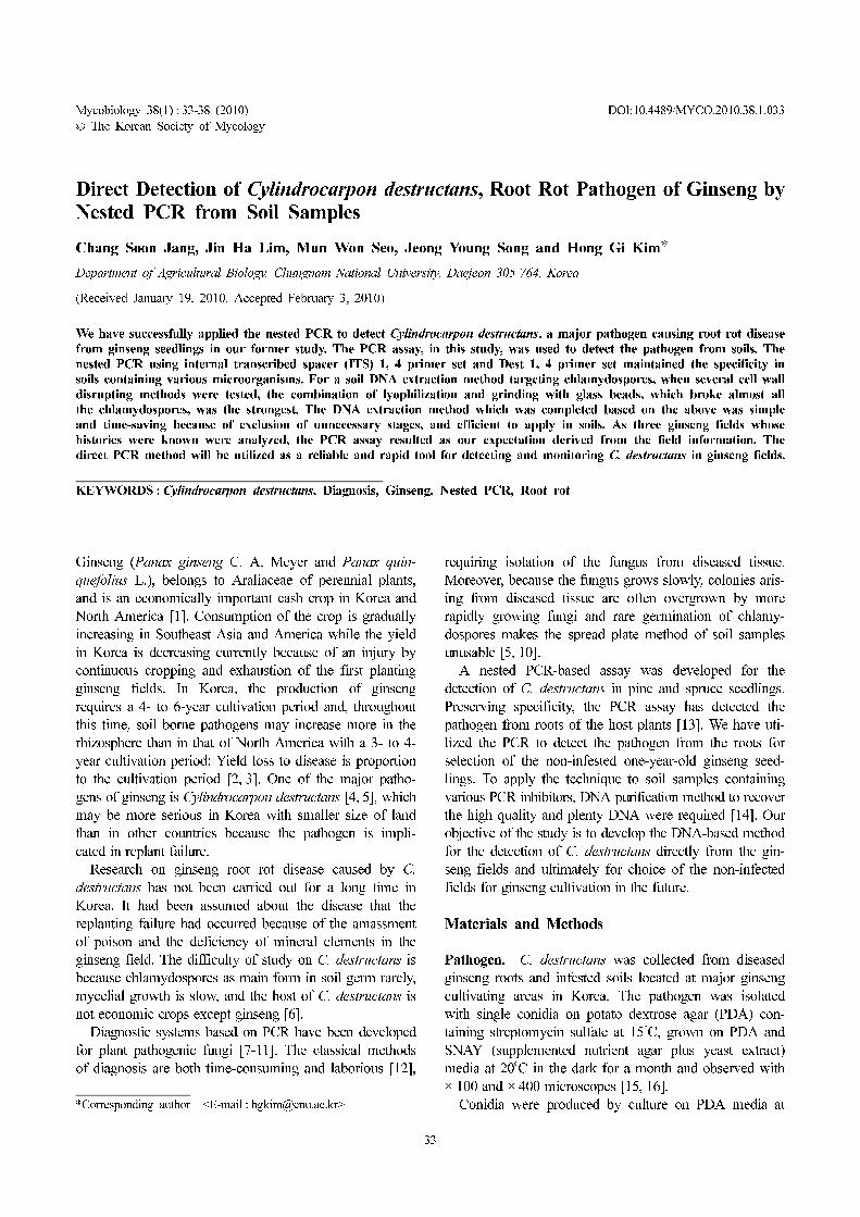
Cell wall disrupting test. Physical methods by direct
breaking (b, d) were more effective than the rest indirect
methods (a, c) on the whole. The best method, combina-
tion of lyophiliation and glass bead grinding (d) shows
cell wall disruption rate of 92% and method b was 67%.
The indirect methods, a and c were low, 1% and 2.3%,
respectively, and this result indicates that reagents associ-
ated with these two indirect methods were not effective
by themselves for thick cell wall of C. destructans at all
(Fig. 1A).
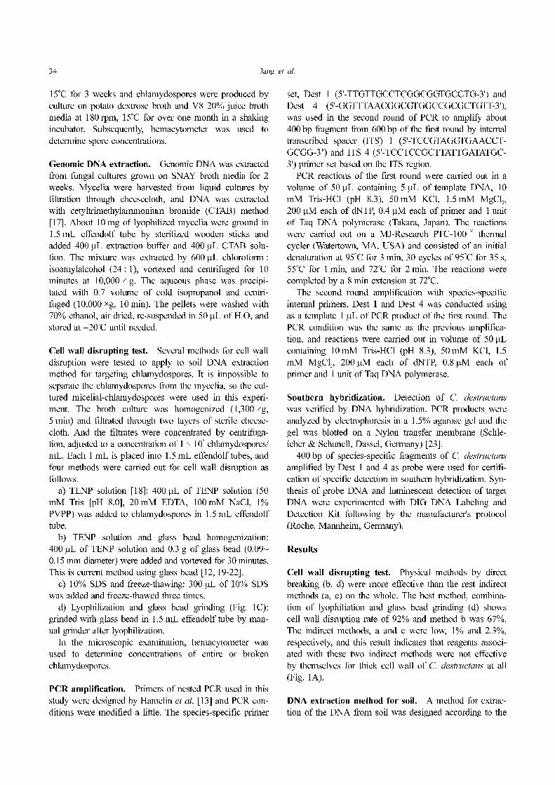
DNA extraction method for soil. A method for extrac-
tion of the DNA from soil was designed according to the
Direct Detection of Cylindrocarpon destructans, Root Rot Pathogen of Ginseng by Nested PCR from Soil Samples 35
above (Fig. 2). Each soil sample of 0.2 g from ginseng
fields was placed in 1.5 mL effendolf tubes, washed with
700 µL H2O, centrifuged (4,000 ×g, 5 min) and aqueous
phase was removed (× 3), then lyophilized.
Prepared soil samples were thoroughly ground with 0.2 g
glass beads by a manual grinder (Fig. 1C) and 400 µL
TENP solution was added. The mixture was extracted by
600 µL chloroform : isoamylalcohol (24 : 1), vortexed and
centrifuged for 10 minutes at 10,000 ×g.
The aqueous phases were purified by G-spinTM
Genomic
DNA extraction kit (Intron Biotechnology, Seongnam,
Korea). The samples were applied from as the step 7 of
the manual without precipitation phase by ethanol or iso-
propanol, and the purified DNA was eluted in 200 µL
H2O, and if needed, diluted ten-fold for PCR reactions of
soil DNAs in the final step (Fig. 2).
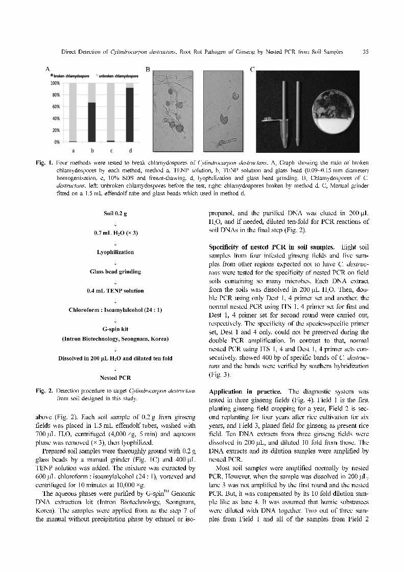
Specificity of nested PCR in soil samples. Eight soil
samples from four infested ginseng fields and five sam-
ples from other regions expected not to have C. destruc-
tans were tested for the specificity of nested PCR on field
soils containing so many microbes. Each DNA extract
from the soils was dissolved in 200 µL H2O. Then, dou-
ble PCR using only Dest 1, 4 primer set and another, the
normal nested PCR using ITS 1, 4 primer set for first and
Dest 1, 4 primer set for second round were carried out,
respectively. The specificity of the species-specific primer
set, Dest 1 and 4 only, could not be preserved during the
double PCR amplification. In contrast to that, normal
nested PCR using ITS 1, 4 and Dest 1, 4 primer sets con-
secutively, showed 400 bp of specific bands of C. destruc-
tans and the bands were verified by southern hybridization
(Fig. 3).
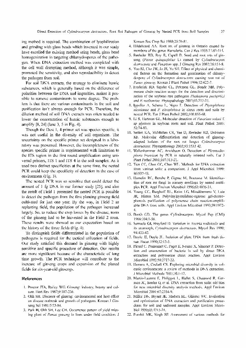
Application in practice. The diagnostic system was
tested in three ginseng fields (Fig. 4). Field 1 is the first
planting ginseng field cropping for a year, Field 2 is sec-
ond replanting for four years after rice cultivation for six
years, and Field 3, planed field for ginseng as present rice
field. Ten DNA extracts from three ginseng fields were
dissolved in 200 µL, and diluted 10 fold from those. The
DNA extracts and its dilution samples were amplified by
nested PCR.
Most soil samples were amplified normally by nested
PCR. However, when the sample was dissolved in 200 µL,
lane 3 was not amplified by the first round and the nested
PCR. But, it was compensated by its 10 fold dilution sam-
ple like as lane 4. It was assumed that humic substances
were diluted with DNA together. Two out of three sam-
ples from Field 1 and all of the samples from Field 2
Fig. 1. Four methods were tested to break chlamydospores of Cylindrocarpon destructans. A, Graph showing the ratio of broken
chlamydospores by each method, method a, TENP solution, b, TENP solution and glass bead (0.09~0.15 mm diameter)
homogenization, c, 10% SDS and freeze-thawing, d, lyophilization and glass bead grinding. B, Chlamydospores of C.
destructans, left: unbroken chlamydospores before the test, right: chlamydospores broken by method d. C, Manual grinder
fitted on a 1.5 mL effendolf tube and glass beads which used in method d.
Fig. 2. Detection procedure to target Cylindrocarpon destructans
from soil designed in this study.

36 Jang et al.
showed 400 bp of specific bands. No bands, however,
were shown in the planed field for ginseng cultivation,
Field 3. In Field 2 where replant failure occurred, the spe-
cific bands were detected more than in Field 1, the first
planting ginseng field.
Discussion
In Korea, ginseng has been usually cultivated as migra-
tion and return with a small size of land. The population
of the pathogen is increased in the soil and replant failure
occurred because of frequent cultivation in one site. Thus,
a diagnostic system was more required for the tactical uti-
lization of ginseng fields.
In PCR diagnosis to soil born fungi, it is difficult to get
a reliable results, which is associated with reproducibility.
Fungi have so hard cell wall, so the DNAs are not released
well from the cell. Accordingly, effective cell wall disrupt-
Fig. 3. Detection of Cylindrocarpon destructans from various soils with the variety of microorganisms. A, Amplification with only
Dest 1, 4 primer set twice. B, Normal nested PCR using Dest 1, 4 after the first round PCR using ITS 1, 4 primer set.
Both are hybridized with specific fragments amplified by Dest 1, 4 primer set as probe. M: 100 bp ladder, lane 1~8: soils
infested by C. destructans, lane 9~13: non-infested, lane 1, 2: a ginseng field from Eumsung, 3, 4: Seosan, 5, 6: Danyang,
7, 8: Jaechon, 9, 10: sand from playground in Chungnam National University, 11~13: rice fields in Chungnam National
University, n.c.: PCR mixture without template DNA.
Fig. 4. Detection of Cylindrocarpon destructans from three ginseng fields. Upper: first round PCR, below: second round PCR
from first. Field 1: the first planting ginseng field cropping for a year, Field 2: replanting field cropping ginseng for 4
years after rice cultivation for 6 years, Field 3: planed field for ginseng cultivation as present rice field. Lane M: 100 bp
ladder, lane 1, 3, 5, 7, 9, 11, 13, 15, 17, 19: DNA dissolved in 200 µL H2O, lane 2, 4, 6, 8, 10, 12, 14, 16, 18, 20: 10 fold
diluted DNA from DNA dissolved in 200 µL, respectively, n. c.: PCR mixture without template DNA.
Direct Detection of Cylindrocarpon destructans, Root Rot Pathogen of Ginseng by Nested PCR from Soil Samples 37
ing method is required. The combination of lyophilization
and grinding with glass beads which invented in our study
have excelled the existing method using beads, glass bead
homogenization in targeting chlamydospores of the patho-
gen. When DNA extraction method was completed with
the cell wall disrupting method, although it was simple,
promoted the sensitivity, and also reproducibility to detect
the pathogen from soil.
For soil DNA extracts, the strategy to eliminate humic
substances, which is generally based on the difference of
polarities between the DNA and impurities, makes it pos-
sible to remove contaminants to some degree. The prob-
lem is that there are various contaminants in the soil and
purification isn’t always enough for PCR. Therefore, the
dilution method of soil DNA extracts was often needed to
lower the concentration of humic substances enough to
amplify [8, 24] (lane 3, 5 in Fig. 4).
Though the Dest 1, 4 primer set was species specific, it
was not useful in the diversity of soil organisms. The
uncertainty on the specific primer set designed in a labo-
ratory was presumed. However, the incompleteness of the
species specific primer is supplemented with limitation to
the ITS region in the first round amplification using uni-
versal primers, ITS 1 and ITS 4 in the soil samples. As it
used two distinct specificities at the same time, the nested
PCR could keep the specificity of detection in the case of
environment (Fig. 3).
The nested PCR was so sensitive that could detect the
amount of 1 fg DNA in our former study [25], and also
the result of Field 1 presented the nested PCR is possible
to detect the pathogen from the first planting ginseng field
cultivated for just one year. By the way, in Field 2 as
replanting field, the population of the pathogen increased
largely. So, to reduce the crop losses by the disease, roots
of the ginseng had to be harvested in the Field 2 soon.
These results were showed as our expectation based on
the history of the three fields (Fig. 4).
To distinguish fields differentiated in the population of
pathogens is required for the tactical utilization of fields.
Our study satisfied this demand in ginseng with highly
sensitive and specific procedure of detection. Our results
are more significant because of the characteristic of long
time growth. The PCR technique will contribute to the
increase of ginseng crops and expansion of the planed
fields for six-year-old ginsengs.
References
1. Proctor JTA, Bailey WG. Ginseng: industry, botany and cul-
ture. Hort Rev 1987;9:187-236.
2. Ohh SH. Diseases of ginseng: environmental and host effect
on disease outbreak and growth of pathogens. Korean J Gin-
seng Sci 1981;5:73-84.
3. Park H, Ohh SH, Lee CH. Occurrence pattern of yield miss-
ing plant of Panax ginseng in lines under field condition. J
Korean Soc Crop Sci 1980;25:76-81.
4. Hildebrand AA. Root rot of ginseng in Ontario caused by
members of the genus Ramularia. Can J Res 1935;12:82-114.
5. Reeleder RD, Roy R, Capell B. Seed and root rots of gin-
seng (Panax quinquefolius L) caused by Cylindrocarpon
destructans and Fusarium spp. J Ginseng Res 2002;26:151-8.
6. Yoo SJ, Cho JW, Jo JS, Yu SH. Effect of physical and chemi-
cal factors on the formation and germination of chlamy-
dospore of Cylindrocarpon destructans causing root rot of
Panax ginseng. Korean J Plant Pathol 1996;12:422-7.
7. Frederick RD, Snyder CL, Peterson GL, Bonde MR. Poly-
merase chain reaction assays for the detection and discrimi-
nation of the soybean rust pathogens Phakopsora pachyrhizi
and P. meibomiae. Phytopathology 2002;92:217-27.
8. Ippolito A, Schena L, Nigro F. Detection of Phytophthora
nicotianae and P. citrophthora in citrus roots and soils by
nested PCR. Eur J Plant Pathol 2002;108:855-68.
9. Li S, Hartman GL. Molecular detection of Fusarium solani f.
sp glycines in soybean roots and soil. Plant Pathol 2003;
52:74-83.
10. Seifert KA, McMullen CR, Yee D, Reeleder RD, Dobinson
KF. Molecular differentiation and detection of ginseng-
adapted isolates of the root rot fungus Cylindrocarpon
destructans. Phytopathology 2003;93:1533-42.
11. Wallenhammar AC, Arwidsson O. Detection of Plasmodio-
phora brassicae by PCR in naturally infested soils. Eur J
Plant Pathol 2001;107:313-21.
12. Tien CC, Chao CC, Chao WL. Methods for DNA extraction
from various soils: a comparison. J Appl Microbiol 1999;
86:937-43.
13. Hamelin RC, Berube P, Gignac M, Bourassa M. Identifica-
tion of root rot fungi in nursery seedlings by nested multi-
plex PCR. Appl Environ Microbiol 1996;62:4026-31.
14. Young CC, Burghoff RL, Keim LG, Minakbernero V, Lute
JR, Hinton SM. Polyvinylpyrrolidone-agarose gel-electro-
phoresis purification of polymerase chain reaction-amplifi-
able DNA from soils. Appl Environ Microbiol 1993;59:1972-
4.
15. Booth CD. The genus Cylindrocarpon. Mycol Pap (CMI)
1966;104:1-56.
16. Samuels GJ, Brayford D. Variation in Nectria radicicola and
its anamorph, Cylindrocarpon destructans. Mycol Res 1990;
94:433-42.
17. Doyle JJ, Doyle JL. Isolation of plant DNA from fresh tis-
sue. Focus 1990;12:13-5.
18. Picard C, Ponsonnet C, Paget E, Nesme X, Simonet P. Detec-
tion and enumeration of bacteria in soil by direct DNA
extraction and polymerase chain reaction. Appl Environ
Microbiol 1992;58:2717-22.
19. Herrera A, Cockell CS. Exploring microbial diversity in vol-
canic environments: a review of methods in DNA extraction.
J Microbiol Methods 2007;70:1-12.
20. Martin-Laurent F, Philippot L, Hallet S, Chaussod R, Ger-
mon JC, Soulas G, et al. DNA extraction from soils: old bias
for new microbial diversity analysis methods. Appl Environ
Microbiol 2001;67:2354-9.
21. Miller DN, Bryant JE, Madsen EL, Ghiorse WC. Evaluation
and optimization of DNA extraction and purification proce-
dures for soil and sediment samples. Appl Environ Micro-
biol 1999;65:4715-24.
22. Purohit MK, Singh SP. Assessment of various methods for
38 Jang et al.
extraction of metagenomic DNA from saline habitats of
coastal Gujarat (India) to explore molecular diversity. Lett
Appl Microbiol 2009;49:338-44.
23. Sambrook J, Fritsch EF, Maniatis T. Molecular cloning: a
laboratory manual. New York: Cold Spring Harbor; 1989.
24. Kong P, Hong CX, Jeffers SN, Richardson PA. A species-
specific polymerase chain reaction assay for rapid detection
of Phytophthora nicotianae in irrigation water. Phytopathol-
ogy 2003;93:822-31.
25. Jang CS, Lee JJ, Kim SI, Song JY, Yoo SJ, Kim HG. Spe-
cific detection of root rot pathogen, Cylindrocarpon destruc-
tans, using nested PCR from ginseng seedlings. Res Plant
Dis 2005;11:48-55.


