Behavioral/Systems/Cognitive
Connectivity-Based Parcellation of Human Cingulate Cortexand Its Relation to Functional Specialization
Matthias Beckmann,1,3 Heidi Johansen-Berg,1 and Matthew F. S. Rushworth1,2
1Centre for Functional MRI of the Brain and 2Department of Experimental Psychology, University of Oxford, Oxford OX1 3UD, United Kingdom, and3Universitatsklinikum “Carl Gustav Carus,” Technische Universitat Dresden, 01307 Dresden, Germany
Whole-brain neuroimaging studies have demonstrated regional variations in function within human cingulate cortex. At the same time,regional variations in cingulate anatomical connections have been found in animal models. It has, however, been difficult to estimate therelationship between connectivity and function throughout the whole cingulate cortex within the human brain. In this study, magneticresonance diffusion tractography was used to investigate cingulate probabilistic connectivity in the human brain with two approaches.First, an algorithm was used to search for regional variations in the probabilistic connectivity profiles of all cingulate cortex voxels withthe whole of the rest of the brain. Nine subregions with distinctive connectivity profiles were identified. It was possible to characterizeseveral distinct areas in the dorsal cingulate sulcal region. Several distinct regions were also found in subgenual and perigenual cortex.Second, the probabilities of connection between cingulate cortex and 11 predefined target regions of interest were calculated. Cingulatevoxels with a high probability of connection with the different targets formed separate clusters within cingulate cortex. Distinct connec-tivity fingerprints characterized the likelihood of connections between the extracingulate target regions and the nine cingulate subre-gions. Last, a meta-analysis of 171 functional studies reporting cingulate activation was performed. Seven different cognitive conditionswere selected and peak activation coordinates were plotted to create maps of functional localization within the cingulate cortex. Regionalfunctional specialization was found to be related to regional differences in probabilistic anatomical connectivity.
Key words: cingulate cortex; DTI; tractography; human; premotor cortex; decision making; emotion
IntroductionHuman cingulate cortex has been implicated in many functions.Regional specialization within cingulate cortex is probably re-lated to its anatomical differentiation into cytoarchitectonicallydistinct subregions that, in macaque, have distinct connections(Matelli et al., 1991; Van Hoesen et al., 1993; Vogt et al., 1995;Zilles et al., 1995; Strick et al., 1998). The invasive nature of tech-niques for studying connectional anatomy has meant that little isknown directly of the anatomical connections of human cingu-late cortex despite the wealth of evidence for functional special-ization from neuroimaging studies. The dorsal cingulate sulcusincludes at least two and possibly more motor regions that areactive during movement (Picard and Strick, 1996; Paus, 2001).Anterior dorsal cingulate sulcus is active when the linking of anaction to reinforcement is critical, for example, during error de-tection or when action selection is guided by the prospect ofreward (Holroyd and Coles, 2002; Rushworth et al., 2007). Adja-cent regions are implicated in detecting when actions are poten-tially in conflict (Botvinick et al., 2004). The more ventral cingu-
late gyrus is active when subjects experience pain (Vogt, 2005)and during autonomic arousal (Critchley et al., 2000). Both ven-tral cingulate and anterior dorsomedial paracingulate regions areactive during emotion and social interaction (Vogt, 2005; Amo-dio and Frith, 2006). Posterior and retrosplenial areas are impli-cated in memory (Maguire, 2001). Given its importance in dis-eases such as Alzheimer’s disease, depression, schizophrenia, andobsessive compulsive disorder (Rosenberg et al., 2004; Gotlib etal., 2005; Kubicki et al., 2005; Naggara et al., 2006), it is desirableto identify human cingulate subregions on the basis of both ana-tomical connectivity and function in vivo.
Here, two complementary magnetic resonance imaging(MRI)-based strategies were used to identify regional differencesin anatomical connectivity in human cingulate cortex by meansof probabilistic diffusion tractography. First, we examined theprobabilistic connectivity profiles of all cingulate cortex voxels todetect inter-regional differences. Such differences define arealdivisions in premotor and ventral prefrontal cortex (Johansen-Berg et al., 2004; Rushworth et al., 2006; Anwander et al., 2007;Klein et al., 2007; Tomassini et al., 2007). This “blind,” a prioriapproach makes no assumptions about the identities of the inter-connecting regions driving the parcellation of the area underinvestigation. By calculating cross-correlations between the con-nectivity profiles of all cingulate voxels and reordering them byapplying k-means clustering (Klein et al., 2007), we characterizeda total of nine separate cingulate areas.
Second, a complementary a posteriori approach focused on 11
Received July 15, 2008; revised Nov. 13, 2008; accepted Dec. 2, 2008.This work was supported by the German National Academic Foundation (M.B.), the Medical Research Council
(M.F.S.R.), and the Wellcome Trust (H.J.-B.). We thank Saad Jbabdi, Johannes Klein, and P. M. Matthews for tech-nical assistance and useful discussions.
Correspondence should be addressed to Matthew F. S. Rushworth, Department of Experimental Psychology,University of Oxford, Oxford OX1 3UD, UK. E-mail: [email protected].
DOI:10.1523/JNEUROSCI.3328-08.2009Copyright © 2009 Society for Neuroscience 0270-6474/09/291175-16$15.00/0
The Journal of Neuroscience, January 28, 2009 • 29(4):1175–1190 • 1175
brain areas known to interconnect withcingulate cortex in other primate species.Each of these areas were used as targets forguided probabilistic tractography from thecingulate seed masks (CSMs) to estimatetheir probability of interconnection.
We created connectivity fingerprints(Passingham et al., 2002; Tomassini et al.,2007) to combine the results from bothapproaches and to visualize the likely ana-tomical connections of each identified cin-gulate cluster. In addition, to assess therelationship between anatomical connec-tivity and function, we also compared ourresults with a meta-analysis of functionalneuroimaging studies reporting cingulateactivation in eight different task conditions.
Materials and MethodsIn this study, diffusion tractography was used intwo complementary but distinct ways to characterize human cingulateprobabilistic connectivity. First, cingulate cortex was parcellated intodistinct subregions on the basis of regional differences in the probabilisticconnectivity profiles of each cingulate voxel. This approach made no apriori assumptions about the identity of the extracingulate regions thatwould drive the regional differences in connectivity profile within thecingulate cortex. Second, connections were estimated between cingulatecortex and 11 target regions known to be interconnected in anotherprimate species: the macaque. This part of the study therefore constituteda complementary a posteriori approach. As a third part of this study, weperformed a meta-analysis of previous reports of cingulate activation invarious behavioral conditions and examined the interrelationship offunctional activations to estimated anatomical connectivity.
Diffusion tractographySubjects, data acquisition, and analysisDiffusion-weighted (three repeats of 60 diffusion directions; b � 1000s/mm 2; 2 � 2 � 2 mm voxels) and T1-weighted images were acquired in11 healthy volunteers (seven male, four female; aged 20 –36 years) on a1.5 T Siemens Sonata MRI scanner in accordance with local ethical ap-proval. Data were analyzed using tools from the Oxford University Cen-tre for Functional MRI of the Brain (FMRIB) software library (http://www.fmrib.ox.ac.uk/fsl). Motion and eddy current correction as well asimage averaging were performed on the diffusion data. Before analysis,the structural volumes were registered to Montreal Neurological Insti-tute (MNI) standard space using the linear registration tool FLIRT byFMRIB.
Diffusion modeling applied a probabilistic diffusion model (Behrenset al., 2003), modified to allow for estimates of multiple fiber directions(Behrens et al., 2007b). For each subject, probabilistic tractography wasrun from voxels within the whole cingulate cortex seed area to assessconnectivity with every brain voxel. The approach draws a sample fromeach fiber orientation distribution at the current voxel and chooses thesample closest to the orientation of its previous step. For each subject, weinitiated 1000 samples from the connectivity distribution from each seedvoxel. The connection probability between a cingulate voxel and anothervoxel in the brain is defined as the sum of sample fiber lengths connectingthese two voxels. This value corresponds approximately to the connec-tion probability (i.e., the total number of samples connecting the tworegions) multiplied by the average connection path length. It corrects forthe fact that distant regions appear less connected than close regionswhen using the probability alone and is appropriate for parcellation-based studies in which distant connections are the principal focus andquantitative probability values are not required. The algorithm was lim-ited to estimating two fiber orientations at each voxel because of the bvalue and number of gradient orientations in the diffusion data (Behrenset al., 2007b).
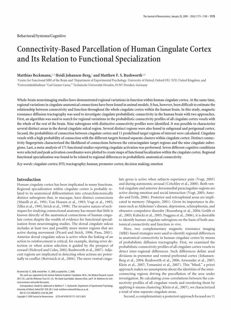
Definition of cingulate seed masksMasks were manually drawn on a single left-hemisphere sagittal slice(x � �4) on each individual’s T1-weighted anatomical scan, after regis-tration to standard (MNI 152) space, to include all tissue commonlyassigned to the cingulate cortex (Fig. 1). We refer subsequently to thismask as the whole CSM. This plane was appropriate because it coveredthe entire cingulate gyrus in all subjects. However, to include the cingu-late and, if applicable, the paracingulate sulci, the masks were chosen toextend laterally on to neighboring slices in these particular regions. Inpractice, this meant that, in the vicinity of the cingulate and paracingulatesulci, the mask extended as far as x � �10 in some subjects. Mask bound-aries were defined as follows. In the supracallosal portion of the cingulatecortex, the ventral boundary of the CSM was the corpus callosum. Therostral sulcus was taken as the ventral boundary of the cingulate cortex inwhich it lay anterior or ventral to the genu of the corpus callosum (Pauset al., 1996a,b). When present, the fundus of the paracingulate sulcus wastaken as the anterior and dorsal boundary of the CSM. The dorsal bank ofthe cingulate sulcus was taken as the dorsal limit of the CSM at moreposterior levels and also at anterior positions when the paracingulatesulcus was absent (Vogt et al., 1995; Paus et al., 1996a,b). Whenever thecingulate sulcus appeared broken, the dorsal limit of the mask followedan imaginary line between adjacent parts of the cingulate sulcus. Thesubparietal sulcus was the dorsal boundary of the CSM in the most pos-terior part of the cingulate cortex posterior to the marginal sulcus. In thisregion, the dorsal boundary of the mask was an imaginary line drawnbetween the posterior limit of cingulate sulcus and the anterior limit ofthe subparietal sulcus when these two sulci were not directly joined withone another. The posterior boundary of the mask was an imaginary linedrawn along the shortest route between the posterior boundary of thesubparietal sulcus and the nearest point on the corpus callosum. Care wastaken to ensure that the masks stayed clear of the corpus callosum itself aswell as of the cingulum bundle. In both approaches, the cingulate masksthen served as seeds for probabilistic diffusion tractography.
Connectivity-based parcellationThe blind connectivity-based parcellation of human cingulate cortex wasperformed iteratively. Connections were estimated between all voxels inthe CSM and all remaining voxels in the rest of the brain (which had beenstored at a lower resolution with a voxel size of 5 � 5 � 5 mm). After aninitial parcellation step had subdivided the cingulate into subregions,these subregions were then used as seed masks for additional iterations ofthe parcellation procedure.
In more detail, the following steps were used. First, probabilistic trac-tography was performed and values were obtained for all voxels in theCSM and their probability of connection to all remaining voxels in thebrain. Results were then stored in a two-dimensional matrix of cingulatecortex seed voxels by extracingulate brain voxels. From this initial, nativeconnectivity matrix, a cross-correlation matrix was calculated that rep-
Figure 1. A, The group average whole CSM superimposed on the group average structural MRI scan in MNI space on a sagittalsection (x ��4). B, Coronal section at y � 24 (as marked by the light blue line in A), showing the lateral extension of the maskinto paracingulate and sulcal areas.
1176 • J. Neurosci., January 28, 2009 • 29(4):1175–1190 Beckmann et al. • DTI-Parcellation of Human Cingulate Cortex
resented the correlation between connectivity profiles of cingulate seedvoxels. The cross-correlation matrix was then reordered using k-meansclustering (Klein et al., 2007; Tomassini et al., 2007), so that voxels weremore likely to be grouped together the more similar their connectivityprofiles. In this step, no spatial constraint was applied when identifyingclusters, and the grouping of voxels was entirely dependent on diffusioninformation. The number of component clusters was, however, chosenby the experimenter. It was set to be the highest number that produced aconsistent, comparable, and spatially similar pattern of clustering acrosssubjects (see Fig. 3). Some of the component clusters identified in thismanner were then subject to an additional iteration of analysis.
Initially, we found evidence for five distinct clusters. This is discussedin detail in Results. In brief, the largest cluster was a discontinuous regionthat included a large part of posterior cingulate cortex and a large part ofanterior cingulate cortex that were separated from one another by theother four clusters. A second parcellation iteration was therefore per-formed to further examine the connections of the large anterior andposterior cingulate cortex components separately. Additional analysisiterations did not lead to parcellations that were reliably similar acrosssubjects.
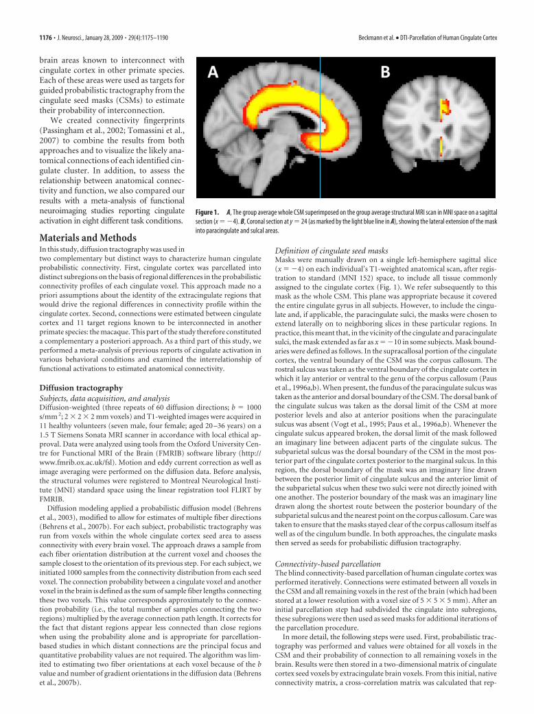
Probabilistic interconnections between cingulate cortex and 11target regionsIn the second part of the study, identical cingulate cortex seed masks wereused as in the initial stage of the parcellation analysis. On the basis of theknown connections of the macaque cingulate cortex, we also defined 11target regions of interest elsewhere in the brain as multislice three-dimensional masks (Fig. 2). Diffusion tractography was then used toestimate the probability of interconnection between voxels in the CSMand each of the other extracingulate regions within the same hemisphere.
For each subject, we drew 5000 samples from the connectivity distri-bution from each seed voxel in the CSM and computed the probability ofconnection with each of the 11 target masks. A connection probabilityvalue was recorded for every CSM voxel in relation to every target mask.
For each subject, the median probability of connection across voxelsbetween each of the nine cingulate subregions identified within the CSMand each extracingulate target region was computed. Subsequently, me-dian probability of connection across subjects was calculated. We thennormalized these values by dividing by the total sum of probability ofconnection between a given extracingulate region and all nine cingulateregions so that these values represent the connection probability of eachcluster as a proportion of total cingulate connections for a given extrac-ingulate region (Passingham et al., 2002; Croxson et al., 2005). Normal-
ized connection probabilities were then used to generate connectivityfingerprints.
The connectivity maps of cingulate voxels classified according to prob-ability of connection to target regions for each individual subject, thresh-olded at the 85th percentile of the highest connection probability valuefor each target, were then binarized. Overlays of the binarized resultsgenerated in all 11 subjects’ brain volumes were then created to visualizethe reproducibility of the results across subjects. Such a method of visu-alizing the results entails a very strict requirement: that the precise samevoxels, and not merely adjacent voxels within the same mask, be stronglyimplicated in a connection in several subjects. To provide a clear andcomprehensive summary of connections, two steps were therefore taken.First, group connectivity maps were thresholded at two subjects so thatcolored voxels indicate voxels that were strongly implicated in a connec-tion with a given area in at least two subjects. In addition, the yellowvoxels in the maps indicate that a connection was common to eight ormore subjects (see Fig. 5). Second, the results were also summarized asconnectivity fingerprints, which are independent of the thresholding ap-plied to the spatial maps. The fingerprints therefore give a threshold-freeindication of the average strength (across all subjects) of connectionbetween each cingulate cluster and each target region.
Definition of target masksWith the exception of the orbitofrontal cortex masks, the extracingulatecortical masks were adapted from studies [premotor cortex (Tomassiniet al., 2007), parietal cortex (Rushworth et al., 2006), precentral gyrus(Smith et al., 2004), and dorsal prefrontal cortex (Croxson et al., 2005)]that had defined these regions previously. The masks used to define thesubcortical target regions and the orbitofrontal cortex, however, weredrawn specifically for this study. All subcortical masks were delineated oneach individual’s structural images and only then registered to standardspace.
Amygdala and hippocampus. The probability of connections betweencingulate and amygdala was assessed because, in the monkey, the amyg-dala is interconnected with anterior cingulate cortex (Porrino et al., 1981;Amaral and Price, 1984; Carmichael and Price, 1995; Ghashghaei et al.,2007; Morecraft et al., 2007).
The hippocampal formation, particularly the subiculum, is intercon-nected with the ventromedial anterior cingulate and with retrosplenialcingulate cortex (Vogt et al., 1987; Carmichael and Price, 1995; Barbas etal., 1999; Parvizi et al., 2006). Care was taken to include the subiculumwithin the hippocampal mask. Because of the proximity of the whitematter subjacent to the subiculum and the white matter subjacent to the
Figure 2. Group average extracingulate target masks superimposed on the MNI 152 T1 standard brain. A, A coronal section ( y��8) showing parts of the group average hypothalamus (yellow),amygdalae (dark red), and hippocampus (dark blue) target masks. B, A coronal section ( y � 14) showing the group average dorsal striatum (yellow), ventral striatum (light blue), medial (green),and lateral (orange) orbitofrontal cortex target masks. C, A sagittal section (x ��18) showing the group average hippocampus (dark blue), lateral orbitofrontal (orange), dorsal prefrontal (yellow),premotor (light blue), precentral (red), and parietal (green) cortex target masks. D, An axial section (z � 56) showing the group average dorsal prefrontal (yellow), premotor (light blue), precentral(red), and parietal (green) cortex target masks.
Beckmann et al. • DTI-Parcellation of Human Cingulate Cortex J. Neurosci., January 28, 2009 • 29(4):1175–1190 • 1177
entorhinal cortex, there is a possibility that some entorhinal connectionswere also included. The parahippocampal gyrus was not included in themask, and its position lateral to the collateral sulcus (Pruessner et al.,2002) means that it is unlikely that hippocampal connectivity estimatesare confounded with parahippocampal connectivity estimates.
The amygdala mask was drawn onto six consecutive coronal slices; inthis way, the mask extended throughout the full anteroposterior extent ofthe amygdala. For the creation of the hippocampus mask, we also usedcoronal structural sections. The hippocampal mask extended from thehead of the hippocampus anteriorly to the tail of the hippocampus at thelevel of the last coronal slice on which the mesencephalon was still visible.Care was taken to ensure that amygdala and hippocampus masks did notoverlap, and, consequently, tissue at the boundary that could not beconfidently assigned to either the amygdala or hippocampus was ex-cluded from both masks.
Ventral and dorsal striatum. In the monkey, much of the striatum isinterconnected with the cingulate cortex. At a first approximation, ven-tral striatum is more strongly interconnected with ventral cingulate cor-tex, whereas more dorsal cingulate cortex, including the cingulate motorareas (CMAs) in the supracallosal cingulate sulcus, is more strongly con-nected with the dorsal striatum (Kunishio and Haber, 1994). The con-nections of the tissue in the cingulate motor area and immediately ante-rior, however, range over a wide extent of the anterior striatum,including both the caudate and putamen (Takada et al., 2001; Haber etal., 2006).
Although there are some projections from posterior cingulate to ven-tral striatum, the strongest projections are from ventral anterior cingu-late cortex (Kunishio and Haber, 1994; Haber et al., 1995; Parvizi et al.,2006). It is important to note, however, that even ventral anterior cingu-late cortex connections within the striatum are not limited to just itsventral part but extend to include dorsal caudate regions, too (Haber etal., 2006).
In the present study, the ventral striatum was defined in coronal slicesof the structural T1-weighted images. We only included gray matter thatcould be assigned unambiguously to the ventral striatum. The dorsalstriatum mask solely included parts of the caudate nucleus and the puta-men that were in, or dorsal to, the first axial MRI slice to include both thegenu and the splenium of the corpus callosum. This meant that an am-biguous intermediate region that was not easily assigned to either ventralor dorsal striatum was not included in either mask. A similar procedurehas been used previously by Croxson et al. (2005).
Hypothalamus. The ventromedial and anterior cingulate cortex andadjacent medial orbitofrontal cortex are distinguished from other pre-frontal regions by the density of their projections to the hypothalamus(Ongur et al., 1998; Freedman et al., 2000). Although the connections ofsubgenual cingulate cortex with the hypothalamus have received themost attention, it is clear that, in the monkey, the most ventral tier ofsupracallosal cingulate cortex also projects to hypothalamus (Ongur etal., 2003). Posterior cingulate cortex has some connections with adjacentregions of the zona incerta (Parvizi et al., 2006) that might also be de-tected with diffusion-weighted imaging (DWI) tractography.
The hypothalamus mask was drawn on T1 coronal sections and in-cluded all hypothalamic gray matter from �2 mm posterior to the coro-nal level of the optic chiasm to a coronal level �2 mm anterior to thecoronal level of the subthalamic nucleus. The anterior and posteriorborders of the hypothalamus mask were therefore at approximately y ��2 and y � �10, respectively.
Parietal cortex. In the monkey, posterior cingulate cortex is intercon-nected with an extended region of both superior and inferior posteriorlateral parietal cortex (Jones and Powell, 1970; Vogt and Pandya, 1987;Cavada and Goldman-Rakic, 1989; Morecraft et al., 2004). It is interest-ing to note that the posterior cingulate regions that are interconnectedwith parietal cortex primarily fail to overlap with the anterior cingulateregions in which interconnections with amygdala are prominent (VanHoesen et al., 1993). It has been suggested that, within cingulate cortex,there is a fundamental dichotomy between the functions of anterior andposterior regions, with posterior regions being more concerned withspatial representation and orientation and with memory (Baleydier andMauguiere, 1980; Vogt et al., 1992). The cingulate region most strongly
interconnected with the parietal cortex corresponds approximately tothe region commonly referred to as posterior cingulate cortex.
The connections of the human parietal cortex with the cingulate cortexwere, therefore, also estimated. The connections with a large region ofparietal cortex were estimated because of the distributed nature of pari-etal–posterior connections and because aspects of the correspondencebetween human and macaque parietal cortex remain contentious (Rush-worth et al., 2006; Husain and Nachev, 2007). The mask used was similarto the parietal cortex mask examined in a recent study of the subcorticalconnections of the parietal cortex (Rushworth et al., 2006). The parietalcortex masks drawn for individual subjects in that study were combinedin MNI space. The resulting combination image was thresholded so thatit included voxels identified as parietal cortex in at least half of the sub-jects; the resulting image therefore contained some discontinuous voxels.
Orbitofrontal cortex. In the macaque, the anterior cingulate cortex isconnected with the orbitofrontal cortex (Morecraft and Van Hoesen,1993; Cavada et al., 2000). The connections between the medial orbito-frontal cortex and the ventromedial anterior cingulate cortex are partic-ularly strong, and Price and Carmichael have argued that together theseareas comprise a medial frontal network. It is argued that this networkcan be distinguished from an “orbitofrontal” network that is centered onlateral orbitofrontal cortex (Carmichael and Price, 1996). The ventrome-dial and lateral orbitofrontal cortical networks are only sparsely inter-connected with one another (Carmichael and Price, 1996).
We used a combination of axial and sagittal views to create the orbito-frontal cortex masks. Both were drawn in the left hemisphere and acrosseight consecutive axial slices, ensuring that there was no overlap with theneighboring cingulate cortex mask. The drawing of the masks was guidedby the descriptions of orbitofrontal sulcal anatomy made by Chiavarasand colleagues (Chiavaras and Petrides, 2000; Chiavaras et al., 2001), andit was similar to the masks used by Croxson et al. (2005).
In the present study, two orbitofrontal masks were used. The lateralorbitofrontal mask extended from the horizontal ramus of the Sylvianfissure on the lateral surface to the medial orbital sulcus on the orbitalsurface. The anterior boundary was a line drawn between the anteriortips of the horizontal ramus of the Sylvian fissure, the lateral orbitofron-tal sulcus, and the medial orbitofrontal sulcus. The posterior boundarywas a line drawn between the posterior boundaries of the same sulci. Themedial orbitofrontal mask extended from the medial bank of the medialorbital sulcus to the rostral sulcus on the medial surface. The anteriorboundary was a line drawn between the anterior tips of the medial orbitalsulcus and the rostral sulcus, and the posterior boundary was a linedrawn between the posterior boundaries of the same two sulci.
Premotor cortex. In the macaque, the supracallosal cingulate sulcuscontains at least two and possibly three motor regions in which micro-stimulation leads to limb movement and which are connected to theventral horn of the spinal cord (Mitz and Wise, 1987; Hutchins et al.,1988; Dum and Strick, 1991, 1993, 1996; Luppino et al., 1991, 1994;Matelli et al., 1991; Shima et al., 1991; Morecraft and Van Hoesen, 1992;He et al., 1995; Morecraft et al., 1996). In the macaque, the most anteriorCMA is situated anterior to the coronal plane of the bow of the arcuatesulcus. It is often referred to as the rostral CMA (CMAr). It has beenargued that the caudal CMA can be subdivided into dorsal and ventral(CAMv) components. In the monkey, all of the cingulate motor areas areinterconnected with dorsal and ventral divisions of lateral premotor cor-tex (Barbas and Pandya, 1987; Luppino et al., 1998, 2003; Hatanaka et al.,2003; Takada et al., 2004). There is little evidence for connections be-tween the cingulate gyrus, ventral and posterior to the CMAs, and thelateral premotor cortex.
The probability of connection between the cingulate cortex mask andthe lateral premotor cortex was therefore investigated. The posteriorborder was approximately two-thirds of the way across the precentralgyrus (Alkadhi et al., 2002a,b), the inferior border was the ventral limit(Zhang et al., 2001) of the precentral gyrus, and the medial border was�5 mm from the medial surface of the brain. The anterior border waslocated �5 mm in front of the precentral sulcus (Tomassini et al., 2007)because it has been proposed that a part of human ventral premotorcortex might lie anterior to the inferior precentral sulcus (Geyer et al.,2000).
1178 • J. Neurosci., January 28, 2009 • 29(4):1175–1190 Beckmann et al. • DTI-Parcellation of Human Cingulate Cortex
Precentral cortex. In the macaque, the CMAs are also interconnectedwith the primary motor cortex, located immediately posterior to thelateral premotor cortex. Connections between the CMAr and primarymotor cortex are, however, weak compared with those between caudalCMAs and primary motor cortex (Wang et al., 2001; Hatanaka et al.,2003; Wang et al., 2004). The probability of connections between a pre-central cortex region posterior to the premotor cortex was therefore alsoexamined. The precentral gyrus masks were derived from the atlas incor-porated in the latest version of FSLview (Johansen-Berg et al., 2004)(http://www.cma.mgh.harvard.edu/manuals/).
Dorsal prefrontal cortex. In the macaque, dorsal prefrontal cortex isinterconnected with much of cingulate cortex (Bates and Goldman-Rakic, 1993; Lu et al., 1994). The region of interconnection includes, onthe one hand, much of dorsal prefrontal cortex and, on the other hand,tissue in the dorsal supracallosal cingulate cortex, including that in oradjacent to the CMAs, particularly CMAr and CMAv, but extends toinclude more anterior cingulate sulcus and parts of the cingulate gyrus.The dorsal prefrontal mask was based on one used previously (Croxsonet al., 2005) and extended from the inferior frontal sulcus on the lateralsurface to �1 cm above the paracingulate sulcus (or, when this wasabsent, cingulate sulcus) on the medial surface. The mask did not includethe frontal pole. Care was taken to ensure that there was no overlapbetween the mask and the adjacent cingulate cortex mask.
Meta-analysis of functional studiesThe following conditions were considered for the meta-analysis: pain,motor function, emotion, error, conflict, reward, and memory. ThePubMed database (http://www.ncbi.nlm.nih.gov/entrez/) was searchedfor functional MRI (fMRI) and positron emission tomography studies,published between January 1, 2002 and April 15, 2007, containing boththe term “cingulate cortex” or “medial prefrontal cortex” and one of theconditions specified above. The search was limited to the following jour-nals: Neuroimage, The Journal of Neuroscience, Cerebral Cortex, TheProceedings of the National Academy of Sciences of the U S A, BrainResearch, Neuron, Neuropsychologia, The Journal of Cognitive Neuro-science, Brain, Experimental Brain Research, The European Journal ofNeuroscience, Trends in Cognitive Sciences, The Journal of ComparativeNeurology, Nature Neuroscience, Nature Reviews Neuroscience, Hu-man Brain Mapping, and Biological Psychiatry.
We retrieved the peak activation coordinates of a total of 171 originalpublications. To visualize the activation centers, all study coordinates re-ferred to the MNI (Collins et al., 1994) template or were converted to MNIspace (http://wwwneuro03.uni-muenster.de/ger/t2tconv/conv3d.html)when only Talairach coordinates were provided. The coordinates werethen mapped onto the MNI 152 standard template, one voxel per study(in cases in which more than one cingulate peak was reported, only theset of coordinates with the highest degree of significance was included).To allow for comparison with the tractography results, all of these acti-vation sites were eventually projected onto one single sagittal plane (x ��4).
ResultsConnectivity-based parcellationThe first iteration of the parcellation of the whole cingulate seedmask identified five clusters with substantially distinct connectiv-ity profiles in both subjects lacking a well defined paracingulatesulcus and subjects with an identifiable paracingulate sulcus.Four of the clusters were found in the midcingulate region. Mostof the voxels assigned to each of these four clusters were spatiallyadjacent to one another. The rest of the cingulate comprised onelarge discontinuous cluster with an anterior and a posterior com-ponent. As explained below, the anterior and posterior compo-nents of this large cluster resembled one another in that theyshared a high probability of interconnection with hippocampusand hypothalamus. Although the investigation of intracingulateconnections is beyond the scope of the current report, it shouldalso be noted that these regions are known to be directly inter-
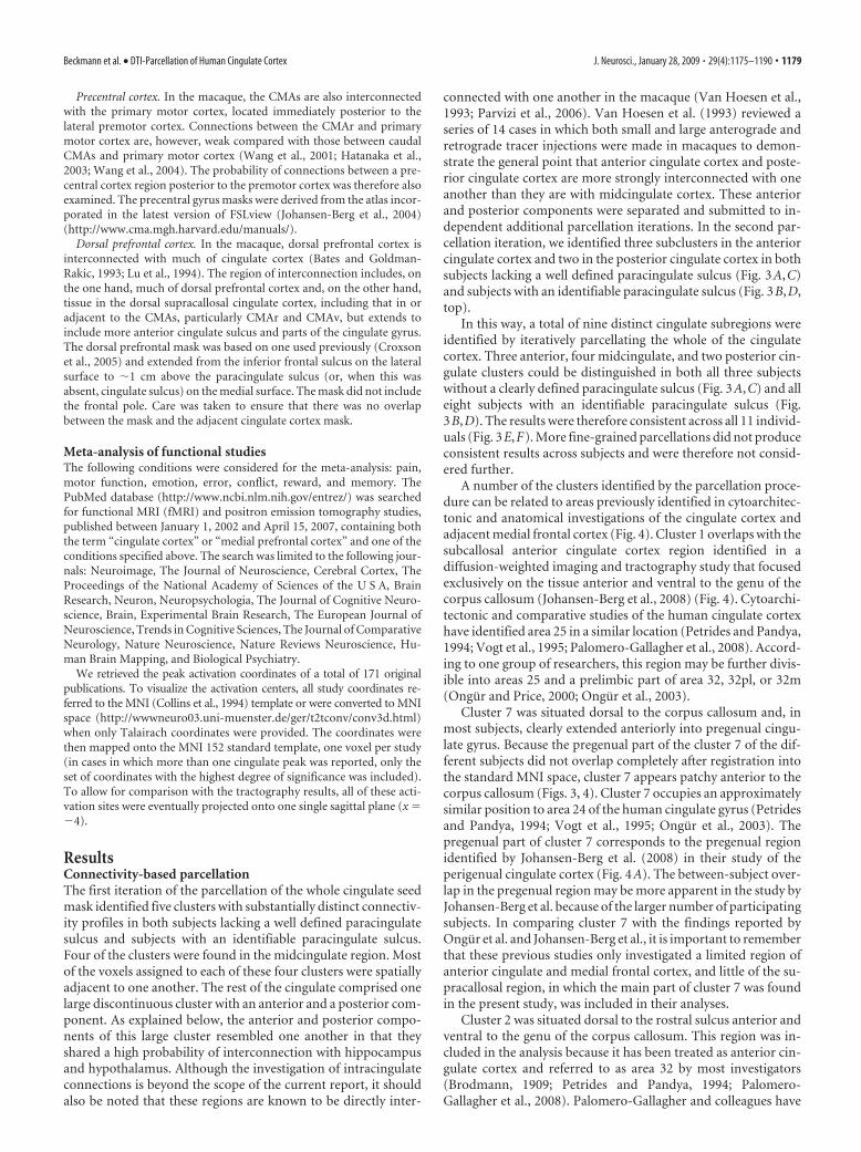
connected with one another in the macaque (Van Hoesen et al.,1993; Parvizi et al., 2006). Van Hoesen et al. (1993) reviewed aseries of 14 cases in which both small and large anterograde andretrograde tracer injections were made in macaques to demon-strate the general point that anterior cingulate cortex and poste-rior cingulate cortex are more strongly interconnected with oneanother than they are with midcingulate cortex. These anteriorand posterior components were separated and submitted to in-dependent additional parcellation iterations. In the second par-cellation iteration, we identified three subclusters in the anteriorcingulate cortex and two in the posterior cingulate cortex in bothsubjects lacking a well defined paracingulate sulcus (Fig. 3A,C)and subjects with an identifiable paracingulate sulcus (Fig. 3B,D,top).
In this way, a total of nine distinct cingulate subregions wereidentified by iteratively parcellating the whole of the cingulatecortex. Three anterior, four midcingulate, and two posterior cin-gulate clusters could be distinguished in both all three subjectswithout a clearly defined paracingulate sulcus (Fig. 3A,C) and alleight subjects with an identifiable paracingulate sulcus (Fig.3B,D). The results were therefore consistent across all 11 individ-uals (Fig. 3E,F). More fine-grained parcellations did not produceconsistent results across subjects and were therefore not consid-ered further.
A number of the clusters identified by the parcellation proce-dure can be related to areas previously identified in cytoarchitec-tonic and anatomical investigations of the cingulate cortex andadjacent medial frontal cortex (Fig. 4). Cluster 1 overlaps with thesubcallosal anterior cingulate cortex region identified in adiffusion-weighted imaging and tractography study that focusedexclusively on the tissue anterior and ventral to the genu of thecorpus callosum (Johansen-Berg et al., 2008) (Fig. 4). Cytoarchi-tectonic and comparative studies of the human cingulate cortexhave identified area 25 in a similar location (Petrides and Pandya,1994; Vogt et al., 1995; Palomero-Gallagher et al., 2008). Accord-ing to one group of researchers, this region may be further divis-ible into areas 25 and a prelimbic part of area 32, 32pl, or 32m(Ongur and Price, 2000; Ongur et al., 2003).
Cluster 7 was situated dorsal to the corpus callosum and, inmost subjects, clearly extended anteriorly into pregenual cingu-late gyrus. Because the pregenual part of the cluster 7 of the dif-ferent subjects did not overlap completely after registration intothe standard MNI space, cluster 7 appears patchy anterior to thecorpus callosum (Figs. 3, 4). Cluster 7 occupies an approximatelysimilar position to area 24 of the human cingulate gyrus (Petridesand Pandya, 1994; Vogt et al., 1995; Ongur et al., 2003). Thepregenual part of cluster 7 corresponds to the pregenual regionidentified by Johansen-Berg et al. (2008) in their study of theperigenual cingulate cortex (Fig. 4A). The between-subject over-lap in the pregenual region may be more apparent in the study byJohansen-Berg et al. because of the larger number of participatingsubjects. In comparing cluster 7 with the findings reported byOngur et al. and Johansen-Berg et al., it is important to rememberthat these previous studies only investigated a limited region ofanterior cingulate and medial frontal cortex, and little of the su-pracallosal region, in which the main part of cluster 7 was foundin the present study, was included in their analyses.
Cluster 2 was situated dorsal to the rostral sulcus anterior andventral to the genu of the corpus callosum. This region was in-cluded in the analysis because it has been treated as anterior cin-gulate cortex and referred to as area 32 by most investigators(Brodmann, 1909; Petrides and Pandya, 1994; Palomero-Gallagher et al., 2008). Palomero-Gallagher and colleagues have
Beckmann et al. • DTI-Parcellation of Human Cingulate Cortex J. Neurosci., January 28, 2009 • 29(4):1175–1190 • 1179
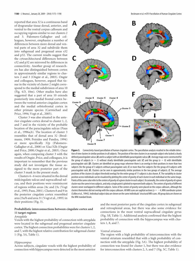
reported that area 32 is a continuous bandof dysgranular tissue dorsal, anterior, andventral to the rostral corpus callosum andoccupying regions similar to our clusters 2and 3. Palomero-Gallagher and col-leagues, however, emphasize a number ofdifferences between more dorsal and ven-tral parts of area 32 and subdivide theminto subgenual and pregenual areas s32and p32. The current results suggest thatthe cytoarchitectural differences betweens32 and p32 are mirrored by differences inconnectivity. Another group of research-ers has also distinguished between cortexin approximately similar regions to clus-ters 2 and 3 (Ongur et al., 2003). Ongurand colleagues, however, argued that tis-sue in the vicinity of cluster 2 might corre-spond to the medial subdivision of area 10(Fig. 4D, 10m). Other studies have alsosuggested that a part of area 10 extendsposteriorly into medial frontal cortex be-tween the ventral anterior cingulate cortexand the medial orbitofrontal cortex inother primate species (Carmichael andPrice, 1994; Vogt et al., 2005).
Cluster 3 was also situated in the ante-rior cingulate cortex dorsal to cluster 1, 2,and 7 and in the vicinity of the probablelocation of the paracingulate sulcus (Pauset al., 1996a,b). The location of cluster 3resembles that of dorsal area 32 (Brod-mann, 1909; Petrides and Pandya, 1994)or more specifically 32p (Palomero-Gallagher et al., 2008) or 32ac/32h (Ongurand Price, 2000; Ongur et al., 2003). Onceagain, when comparing cluster 3 with theresults of Ongur, Price, and colleagues, it isimportant to remember that the previousstudy did not investigate the tissue as-signed to the more posterior part of thecluster 3 mask in the present study.
Clusters 4 – 6 were situated in the dorsalmidcingulate sulcus and supracallosal sul-cus, and their positions were reminiscentof regions within areas 24c and 23c (Vogtet al., 1995; Paus, 2001). Clusters 8 and 9 inthe posterior cingulate cortex resembledarea 23a/b and area 31 (Vogt et al., 1995) intheir positions (Fig. 3).
Probabilistic interconnections between cingulate cortex and11 target regionsAmygdalaVoxels with the highest probability of connection with amygdalawere located in the subgenual and pregenual anterior cingulatecortex. The highest connection probabilities were for clusters 1, 2,and 7, with the highest relative contribution for subgenual cluster1 (Fig. 5A, Table 1).
HippocampusAcross subjects, cingulate voxels with the highest probability ofconnection with hippocampus were detected in the most anterior
and the most posterior parts of the cingulate cortex in subgenualand retrosplenial areas, but there was also some evidence forconnections in the most ventral supracallosal cingulate gyrus(Fig. 5B, Table 1). Additional analysis confirmed that the highestprobability of connection with the hippocampus was with clus-ters 1, 8, and 7.
Ventral striatumThe region with a high probability of interconnection with theventral striatum resembled that with a high probability of con-nection with the amygdala (Fig. 5A). The highest probability ofconnection was found for cluster 1, but there was also evidencefor interconnection with clusters 2, 3, and 7 (Fig. 5C, Table 1).
Figure 3. Connectivity-based parcellation of human cingulate cortex. The parcellation analysis resulted in the reliable detec-tion of nine clusters in similar positions in all subjects. The position of the nine clusters in an example subject who lacked a clearlydefined paracingulate sulcus (A) and in a subject with an identifiable paracingulate sulcus (B). Average maps were constructed forthe group of subjects (n � 3) without clearly identifiable paracingulate sulci (C) and the group (n � 8) with identifiableparacingulate sulci (D). Clusters are identified on group maps wherever there is an overlap in their positions in more than twosubjects (for the group of 3 subjects without paracingulate sulci; C) or more than five subjects (for the group of 8 subjects withparacingulate sulci; D). Clusters can be seen in approximately similar positions in the two groups of subjects. E, The averagepositions of the clusters (6 subject threshold overlap) for the entire group of 11 subjects is also shown. F, The variability in clusterposition across individuals can be visualized by plotting the center of gravity of each cluster in each individual on the same image.Points of the same color refer to the centers of gravity of a given cluster in each subject. Occasionally, the center of gravity of a givencluster was the same in two subjects, and only a single point is plotted to represent both subjects. The centers of gravity of differentclusters never overlapped in different subjects. Some of the centers of gravity were placed on the corpus callosum, although theclusters themselves did not overlap with the corpus callosum. All MRI scans are sagittal sections [x ��4, MNI coordinate system(Collins et al., 1994)]. Individual subject data are shown on the same individuals’ structural MRI scans. All group data are shown onthe MNI standard brain.
1180 • J. Neurosci., January 28, 2009 • 29(4):1175–1190 Beckmann et al. • DTI-Parcellation of Human Cingulate Cortex
Dorsal striatumThe dorsal striatum mask, situated either side of the internalcapsule, appeared to be predominantly interconnected with thedorsal supracallosal anterior cingulate cortex. Additional analysisconfirmed a high probability of connection with the cingulateclusters on the anterior cingulate sulcus, clusters 3– 6, but showedthat there was a relatively high probability of connection in atleast some voxels of several other clusters, especially subgenualcluster 1 (Fig. 5D, Table 1).
HypothalamusThere was strong evidence of interconnection between subgenualand perigenual cingulate cortex and the hypothalamus. Therewas, however, also a weaker suggestion of interconnection withthe ventral supracallosal region. Additional analysis suggestedthat clusters 1 and 2 had the highest probability of connectionwith the hypothalamus but that there was also some evidence ofconnection between the hypothalamus and ventral supracallosalclusters 7 and 8 (Fig. 5E, Table 1).
Parietal cortexVoxels of high probability with the lateral parietal cortex werefound spread across the midcingulate and posterior cingulate
regions. Additional analysis suggestedsome overlap with the connections withthe motor regions (precentral gyrus andpremotor cortex): the parietal cortex alsohad a reasonably high probability of con-nection with cluster 6 in the posterior partof the anterior cingulate sulcus, and therewas some evidence of connections withcluster 5 that lay immediately anteriorly.There was also evidence for connectionswith the clusters 8 and 9 and posteriorparts of cluster 7 (Fig. 5F, Table 1).
Orbitofrontal cortexLike the ventral striatum, medial orbito-frontal cortex connected with the high-est probability to subgenual and pre-genual anterior cingulate cortex. Thehighest connection probabilities werefor clusters 1 and 2, with the highest rel-ative contribution for cluster 2 (Fig. 5G,Table 1). The estimated pattern of con-nectivity between lateral orbitofrontalcortex and cingulate cortex was similarto that found between medial orbito-frontal cortex and cingulate cortex (Fig.5H ). Despite the similarity in connec-tion pattern, there was a noticeable dif-ference in connection strength; the me-dian number of connection streamlinesestimated for each voxel in each cingu-late cluster with the medial orbital re-gion was �40 times greater than theequivalent number for the lateral orbitalregion. In general, we refrain from com-paring the connections of different ex-tracingulate regions with one anotherbecause the comparison will be partlydependent on the size of the extracingu-late target region and the route by whichit interconnects with cingulate cortex.Nevertheless, it is of note that, despite
the fact that these two extracingulate masks have very similarvolumes, the medial orbitofrontal cortex was estimated to bemuch more strongly interconnected with the anterior cingu-late cortex than was the adjacent lateral orbitofrontal cortex.
Premotor cortexThe premotor cortex had the highest probability of connectionwith dorsal supracallosal anterior cingulate cortex. Additionalanalysis demonstrated that the highest probability of intercon-nection was with the dorsal anterior cingulate clusters. The high-est probability of connection was found for cluster 5, followed bycluster 6 and then cluster 4 (Fig. 5I, Table 1).
Precentral gyrusThe precentral gyrus mask, which was located just posterior tothe premotor mask, also had a high probability of interconnec-tion with the dorsal anterior cingulate sulcus region. In the case ofthe precentral gyrus, however, the focus of connection probabil-ity was more posterior than had been the case for the premotorcortex. The highest probability of connection was with cluster 6,followed by cluster 5 (Fig. 5J, Table 1).
Figure 4. The parcellation clusters resemble regions identified in previous studies. A, Cluster 1 occupies a similar position to thesubcallosal cingulate region identified by Johansen-Berg et al. (2007). Johansen-Berg and colleagues performed a diffusion-weighted imaging tractography parcellation analysis of the region targeted in deep brain stimulation for depression (area coloredblue or yellow within the oval). They identified two component clusters (shown in blue and in yellow), one of which (yellow)corresponds in location to cluster 1 in the current study. Cluster 1 also occupies a similar position to area 25 as identified in previouscytoarchitectonic analyses of the human brain conducted by Petrides and Pandya (1994), figure adapted by mirroring to matchbrain orientation (B), and Vogt (2008), figure adapted by mirroring to match brain orientation (C). Note that, in this recentdiagram, the label 24 is assigned to tissue sometimes divided into subareas 24a and 24b, and the labels 23d, 23v, and v23 areassigned to tissue sometimes divided into 23a and 23b. D, According to some researchers (Ongur et al., 2003), this region may befurther subdivided into areas 25 and 32pl. The positions of clusters 2 and 3 resemble those of the medial extension of area 10, 10m,and 32ac (D). Vogt et al. (2005) have also identified area 10m in a similar position ventral to cingulate cortex and dorsal toorbitofrontal cortex in the macaque (data not shown). It is important to note that Ongur and colleagues only analyzed the regionoutlined by the light blue dotted line, and so they did not report on the posterior extension of area 32ac. The position of clusters4 – 6 resemble those of cingulate sulcus regions 24c and 23c (C). The position of cluster 7 resembles that of 24a/b (B–D). Thepositions of clusters 8 and 9 resemble those of areas 23a/b and of area 31, respectively (B, C).
Beckmann et al. • DTI-Parcellation of Human Cingulate Cortex J. Neurosci., January 28, 2009 • 29(4):1175–1190 • 1181
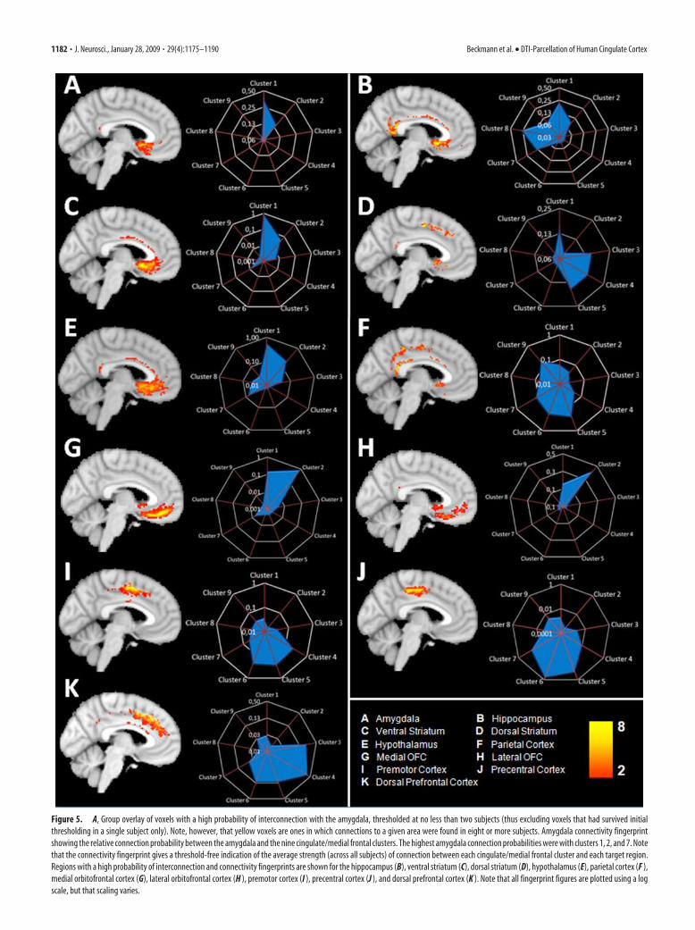
Figure 5. A, Group overlay of voxels with a high probability of interconnection with the amygdala, thresholded at no less than two subjects (thus excluding voxels that had survived initialthresholding in a single subject only). Note, however, that yellow voxels are ones in which connections to a given area were found in eight or more subjects. Amygdala connectivity fingerprintshowing the relative connection probability between the amygdala and the nine cingulate/medial frontal clusters. The highest amygdala connection probabilities were with clusters 1, 2, and 7. Notethat the connectivity fingerprint gives a threshold-free indication of the average strength (across all subjects) of connection between each cingulate/medial frontal cluster and each target region.Regions with a high probability of interconnection and connectivity fingerprints are shown for the hippocampus (B), ventral striatum (C), dorsal striatum (D), hypothalamus (E), parietal cortex (F ),medial orbitofrontal cortex (G), lateral orbitofrontal cortex (H ), premotor cortex (I ), precentral cortex (J ), and dorsal prefrontal cortex (K ). Note that all fingerprint figures are plotted using a logscale, but that scaling varies.
1182 • J. Neurosci., January 28, 2009 • 29(4):1175–1190 Beckmann et al. • DTI-Parcellation of Human Cingulate Cortex
Dorsal prefrontal cortexEvidence for dorsal prefrontal cortex connections was foundthroughout the cingulate cortex. The highest probability of con-nection, however, was with clusters 3 and 4 (Fig. 5K, Table 1).
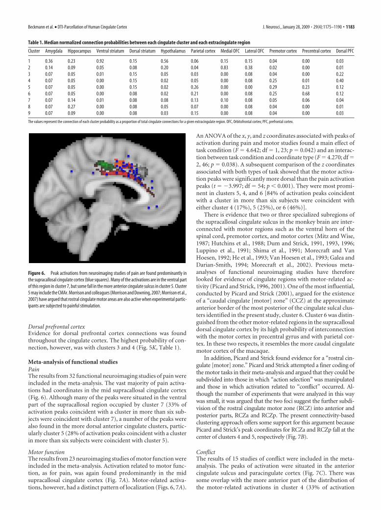
Meta-analysis of functional studiesPainThe results from 32 functional neuroimaging studies of pain wereincluded in the meta-analysis. The vast majority of pain activa-tions had coordinates in the mid supracallosal cingulate cortex(Fig. 6). Although many of the peaks were situated in the ventralpart of the supracallosal region occupied by cluster 7 (33% ofactivation peaks coincident with a cluster in more than six sub-jects were coincident with cluster 7), a number of the peaks werealso found in the more dorsal anterior cingulate clusters, partic-ularly cluster 5 (28% of activation peaks coincident with a clusterin more than six subjects were coincident with cluster 5).
Motor functionThe results from 23 neuroimaging studies of motor function wereincluded in the meta-analysis. Activation related to motor func-tion, as for pain, was again found predominantly in the midsupracallosal cingulate cortex (Fig. 7A). Motor-related activa-tions, however, had a distinct pattern of localization (Figs. 6, 7A).
An ANOVA of the x, y, and z coordinates associated with peaks ofactivation during pain and motor studies found a main effect oftask condition (F � 4.642; df � 1, 23; p � 0.042) and an interac-tion between task condition and coordinate type (F � 4.270; df �2, 46; p � 0.038). A subsequent comparison of the z coordinatesassociated with both types of task showed that the motor activa-tion peaks were significantly more dorsal than the pain activationpeaks (t � �3.997; df � 54; p � 0.001). They were most promi-nent in clusters 5, 4, and 6 [84% of activation peaks coincidentwith a cluster in more than six subjects were coincident witheither cluster 4 (17%), 5 (25%), or 6 (46%)].
There is evidence that two or three specialized subregions ofthe supracallosal cingulate sulcus in the monkey brain are inter-connected with motor regions such as the ventral horn of thespinal cord, premotor cortex, and motor cortex (Mitz and Wise,1987; Hutchins et al., 1988; Dum and Strick, 1991, 1993, 1996;Luppino et al., 1991; Shima et al., 1991; Morecraft and VanHoesen, 1992; He et al., 1993; Van Hoesen et al., 1993; Galea andDarian-Smith, 1994; Morecraft et al., 2002). Previous meta-analyses of functional neuroimaging studies have thereforelooked for evidence of cingulate regions with motor-related ac-tivity (Picard and Strick, 1996, 2001). One of the most influential,conducted by Picard and Strick (2001), argued for the existenceof a “caudal cingulate [motor] zone” (CCZ) at the approximateanterior border of the most posterior of the cingulate sulcal clus-ters identified in the present study, cluster 6. Cluster 6 was distin-guished from the other motor-related regions in the supracallosaldorsal cingulate cortex by its high probability of interconnectionwith the motor cortex in precentral gyrus and with parietal cor-tex. In these two respects, it resembles the more caudal cingulatemotor cortex of the macaque.
In addition, Picard and Strick found evidence for a “rostral cin-gulate [motor] zone.” Picard and Strick attempted a finer coding ofthe motor tasks in their meta-analysis and argued that they could besubdivided into those in which “action selection” was manipulatedand those in which activation related to “conflict” occurred. Al-though the number of experiments that were analyzed in this waywas small, it was argued that the two foci suggest the further subdi-vision of the rostral cingulate motor zone (RCZ) into anterior andposterior parts, RCZa and RCZp. The present connectivity-basedclustering approach offers some support for this argument becausePicard and Strick’s peak coordinates for RCZa and RCZp fall at thecenter of clusters 4 and 5, respectively (Fig. 7B).
ConflictThe results of 15 studies of conflict were included in the meta-analysis. The peaks of activation were situated in the anteriorcingulate sulcus and paracingulate cortex (Fig. 7C). There wassome overlap with the more anterior part of the distribution ofthe motor-related activations in cluster 4 (33% of activation
Table 1. Median normalized connection probabilities between each cingulate cluster and each extracingulate region
Cluster Amygdala Hippocampus Ventral striatum Dorsal striatum Hypothalamus Parietal cortex Medial OFC Lateral OFC Premotor cortex Precentral cortex Dorsal PFC
1 0.36 0.23 0.92 0.15 0.56 0.06 0.15 0.15 0.04 0.00 0.032 0.14 0.09 0.05 0.08 0.20 0.04 0.83 0.38 0.02 0.00 0.013 0.07 0.05 0.01 0.15 0.05 0.03 0.00 0.08 0.04 0.00 0.224 0.07 0.05 0.00 0.15 0.02 0.05 0.00 0.08 0.25 0.01 0.405 0.07 0.05 0.00 0.15 0.02 0.26 0.00 0.00 0.29 0.23 0.126 0.07 0.05 0.00 0.08 0.02 0.21 0.00 0.08 0.25 0.68 0.127 0.07 0.14 0.01 0.08 0.08 0.13 0.10 0.08 0.05 0.06 0.048 0.07 0.27 0.00 0.08 0.05 0.07 0.00 0.08 0.04 0.00 0.019 0.07 0.09 0.00 0.08 0.03 0.15 0.00 0.08 0.04 0.00 0.03
The values represent the connection of each cluster probability as a proportion of total cingulate connections for a given extracingulate region. OFC, Orbitofrontal cortex; PFC, prefrontal cortex.
Figure 6. Peak activations from neuroimaging studies of pain are found predominantly inthe supracallosal cingulate cortex (blue squares). Many of the activations are in the ventral partof this region in cluster 7, but some fall in the more anterior cingulate sulcus in cluster 5. Cluster5 may include the CMAr. Morrison and colleagues (Morrison and Downing, 2007; Morrison et al.,2007) have argued that rostral cingulate motor areas are also active when experimental partic-ipants are subjected to painful stimulation.
Beckmann et al. • DTI-Parcellation of Human Cingulate Cortex J. Neurosci., January 28, 2009 • 29(4):1175–1190 • 1183
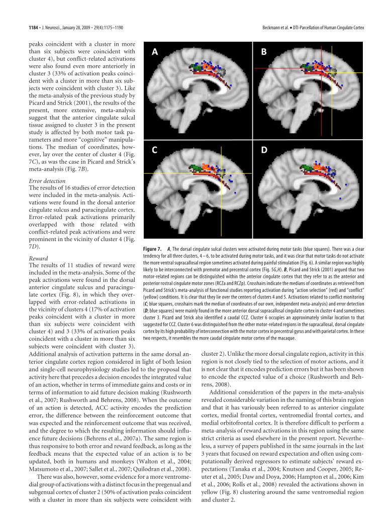
peaks coincident with a cluster in morethan six subjects were coincident withcluster 4), but conflict-related activationswere also found even more anteriorly incluster 3 (33% of activation peaks coinci-dent with a cluster in more than six sub-jects were coincident with cluster 3). Likethe meta-analysis of the previous study byPicard and Strick (2001), the results of thepresent, more extensive, meta-analysissuggest that the anterior cingulate sulcaltissue assigned to cluster 3 in the presentstudy is affected by both motor task pa-rameters and more “cognitive” manipula-tions. The median of coordinates, how-ever, lay over the center of cluster 4 (Fig.7C), as was the case in Picard and Strick’smeta-analysis (Fig. 7B).
Error detectionThe results of 16 studies of error detectionwere included in the meta-analysis. Acti-vations were found in the dorsal anteriorcingulate sulcus and paracingulate cortex.Error-related peak activations primarilyoverlapped with those related withconflict-related peak activations and wereprominent in the vicinity of cluster 4 (Fig.7D).
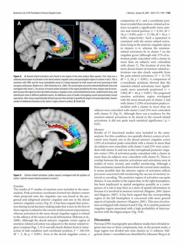
RewardThe results of 11 studies of reward wereincluded in the meta-analysis. Some of thepeak activations were found in the dorsalanterior cingulate sulcus and paracingu-late cortex (Fig. 8), in which they over-lapped with error-related activations inthe vicinity of clusters 4 (17% of activationpeaks coincident with a cluster in morethan six subjects were coincident withcluster 4) and 3 (33% of activation peakscoincident with a cluster in more than sixsubjects were coincident with cluster 3).Additional analysis of activation patterns in the same dorsal an-terior cingulate cortex region considered in light of both lesionand single-cell neurophysiology studies led to the proposal thatactivity here that precedes a decision encodes the integrated valueof an action, whether in terms of immediate gains and costs or interms of information to aid future decision making (Rushworthet al., 2007; Rushworth and Behrens, 2008). When the outcomeof an action is detected, ACC activity encodes the predictionerror, the difference between the reinforcement outcome thatwas expected and the reinforcement outcome that was received,and the degree to which the resulting information should influ-ence future decisions (Behrens et al., 2007a). The same region isthus responsive to both error and reward feedback, as long as thefeedback means that the expected value of an action is to beupdated, both in humans and monkeys (Walton et al., 2004;Matsumoto et al., 2007; Sallet et al., 2007; Quilodran et al., 2008).
There was also, however, some evidence for a more ventrome-dial group of activations with a distinct focus in the pregenual andsubgenual cortex of cluster 2 (50% of activation peaks coincidentwith a cluster in more than six subjects were coincident with
cluster 2). Unlike the more dorsal cingulate region, activity in thisregion is not closely tied to the selection of motor actions, and itis not clear that it encodes prediction errors but it has been shownto encode the expected value of a choice (Rushworth and Beh-rens, 2008).
Additional consideration of the papers in the meta-analysisrevealed considerable variation in the naming of this brain regionand that it has variously been referred to as anterior cingulatecortex, medial frontal cortex, ventromedial frontal cortex, andmedial orbitofrontal cortex. It is therefore difficult to perform ameta-analysis of reward activations in this region using the samestrict criteria as used elsewhere in the present report. Neverthe-less, a survey of papers published in the same journals in the last3 years that focused on reward expectation and often using com-putationally derived regressors to estimate subjects’ reward ex-pectations (Tanaka et al., 2004; Knutson and Cooper, 2005; Re-uter et al., 2005; Daw and Doya, 2006; Hampton et al., 2006; Kimet al., 2006; Rolls et al., 2008) revealed the activations shown inyellow (Fig. 8) clustering around the same ventromedial regionand cluster 2.
Figure 7. A, The dorsal cingulate sulcal clusters were activated during motor tasks (blue squares). There was a cleartendency for all three clusters, 4 – 6, to be activated during motor tasks, and it was clear that motor tasks do not activatethe more ventral supracallosal region sometimes activated during painful stimulation (Fig. 6). A similar region was highlylikely to be interconnected with premotor and precentral cortex (Fig. 5G,H). B, Picard and Strick (2001) argued that twomotor-related regions can be distinguished within the anterior cingulate cortex that they refer to as the anterior andposterior rostral cingulate motor zones (RCZa and RCZp). Crosshairs indicate the medians of coordinates as retrieved fromPicard and Strick’s meta-analysis of functional studies reporting activation during “action selection” (red) and “conflict”(yellow) conditions. It is clear that they lie over the centers of clusters 4 and 5. Activations related to conflict monitoring(C; blue squares, crosshairs mark the median of coordinates of our own, independent meta-analysis) and error detection(D; blue squares) were mainly found in the more anterior dorsal supracallosal cingulate cortex in cluster 4 and sometimescluster 3. Picard and Strick also identified a caudal CCZ. Cluster 6 occupies an approximately similar location to thatsuggested for CCZ. Cluster 6 was distinguished from the other motor-related regions in the supracallosal, dorsal cingulatecortex by its high probability of interconnection with the motor cortex in precentral gyrus and with parietal cortex. In thesetwo respects, it resembles the more caudal cingulate motor cortex of the macaque.
1184 • J. Neurosci., January 28, 2009 • 29(4):1175–1190 Beckmann et al. • DTI-Parcellation of Human Cingulate Cortex
EmotionThe results of 37 studies of emotion were included in the meta-analysis. Peak activation coordinates formed two distinct centerswhen projected onto the cingulate: one was found in the pre-genual and subgenual anterior cingulate and one in the dorsalanterior cingulate cortex (Fig. 9). It has been argued that activa-tion during social decision-making tasks that is seen in the first ofthese regions is related to the current reinforcement expectations,whereas activation in the more dorsal cingulate region is relatedto the saliency of the source of social information (Behrens et al.,2008). Although the dorsal anterior cingulate cortex region ofemotion-related activation was adjacent to the motor-related re-gion (compare Figs. 7, 9) it was still clearly distinct from it (inter-action of task condition and coordinate position, F � 106.529;df � 2, 26; p � 0.001). Even in the dorsal cingulate cortex, a
comparison of y- and z-coordinate posi-tions revealed that emotion-related activa-tions occupied a significantly more ante-rior and ventral position (t � 8.181, df �18, p � 0.001; and t � 17.186, df � 36, p �0.001, respectively). Such a separation isconsistent with the motor-related activa-tions lying in the anterior cingulate sulcusin clusters 4 – 6, whereas the emotion-related activations lie in cluster 7 on thecingulate gyrus (although only 17% of ac-tivation peaks coincident with a cluster inmore than six subjects were coincidentwith cluster 7). The location of even themore dorsal cluster of emotion-related ac-tivations was also clearly separable fromthe pain-related activations (F � 51.779;df � 2, 26; p � 0.001). A comparison ofy-coordinate positions showed that theemotion-related activations were signifi-cantly more anteriorly positioned (t �5.882; df � 44; p � 0.001). The pregenualemotion activation region overlappedwith the most dorsal part of cluster 2 andwith cluster 3 (29% of activation peaks co-incident with a cluster in more than six
subjects were coincident with cluster 2 and 25% were coincidentwith cluster 3) (Fig. 9). Although there was a tendency for theemotion-related activations to lie dorsal to the reward-relatedactivations, it did not quite reach statistical significance ( p �0.05).
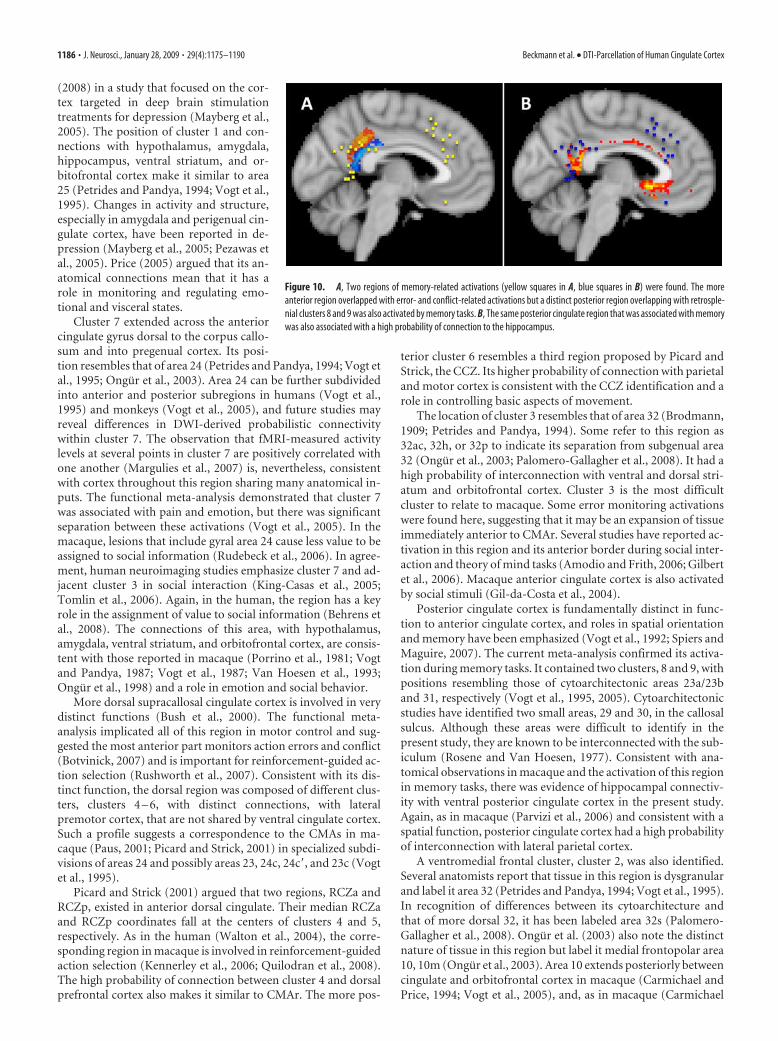
MemoryResults of 27 functional studies were included in the meta-analysis. For this condition, two spatially distinct centers of acti-vation were found, one in the dorsal anterior cingulate cortex(19% of activation peaks coincident with a cluster in more thansix subjects were coincident with cluster 3 and 25% were coinci-dent with cluster 4) and one in the retrosplenial posterior cingu-late cortex (19% of activation peaks coincident with a cluster inmore than six subjects were coincident with cluster 9). There isoverlap between the anterior activations and activations seen instudies of error, reward, and conflict monitoring. Although afirm conclusion cannot be drawn from the current meta-analysis,it seems possible that the anterior region of activation reflectsprocesses concerned with monitoring the success of memory re-trieval or encoding. The posterior region of activation was quitedistinct. It was similar to the region that, in the right hemisphere,has been implicated in spatial navigation possibly as a conse-quence of a role it may have as a store of spatial information orbecause it is involved in memory retrieval (Maguire, 2001; Spiersand Maguire, 2007). It has been argued that the correspondingregion in the left hemisphere is concerned with more generalaspects of episodic memory (Maguire, 2001). This area of activa-tion overlapped with clusters 8 and 9 (Fig. 10A) and the posteriorcingulate region associated with a high probability of intercon-nection with the hippocampus (Fig. 10B).
DiscussionPrevious DWI-tractography parcellation studies have divided re-gions into two or three components, but, in the present study, alarge region was divided into nine clusters in 11 subjects. Sub-genual cluster 1 resembled one identified by Johansen-Berg et al.
Figure 8. A, Reward-related activations were found in two regions in the meta-analysis (blue squares). First, there was arelatively dorsal region of activation in the dorsal anterior cingulate sulcus and paracingulate regions in clusters 4 and 3. This isconsistent with CMAr and the tissue immediately rostral to it being important for both reward and error processing in bothmacaques and humans (Walton et al., 2004; Kennerley et al., 2006). The second region was in the ventromedial frontal cortex andoverlapped with cluster 2. The picture of reward-related activation in this region provided by the meta-analysis may be incom-plete because this region has been described variously as cingulate cortex, ventromedial frontal cortex, medial frontal cortex, andorbitofrontal cortex in different published reports. An additional survey of studies investigating reward and particularly rewardexpectation, often using computationally derived regressors that attempt to quantify the degree of reward expectation, found anumber of additional activations in the cluster 2 region (shown in yellow). B, Detail of A.
Figure 9. Emotion-related activations (yellow squares) overlapped with the positions ofcluster 1 and the reward-related activations in cluster 2.
Beckmann et al. • DTI-Parcellation of Human Cingulate Cortex J. Neurosci., January 28, 2009 • 29(4):1175–1190 • 1185
(2008) in a study that focused on the cor-tex targeted in deep brain stimulationtreatments for depression (Mayberg et al.,2005). The position of cluster 1 and con-nections with hypothalamus, amygdala,hippocampus, ventral striatum, and or-bitofrontal cortex make it similar to area25 (Petrides and Pandya, 1994; Vogt et al.,1995). Changes in activity and structure,especially in amygdala and perigenual cin-gulate cortex, have been reported in de-pression (Mayberg et al., 2005; Pezawas etal., 2005). Price (2005) argued that its an-atomical connections mean that it has arole in monitoring and regulating emo-tional and visceral states.
Cluster 7 extended across the anteriorcingulate gyrus dorsal to the corpus callo-sum and into pregenual cortex. Its posi-tion resembles that of area 24 (Petrides and Pandya, 1994; Vogt etal., 1995; Ongur et al., 2003). Area 24 can be further subdividedinto anterior and posterior subregions in humans (Vogt et al.,1995) and monkeys (Vogt et al., 2005), and future studies mayreveal differences in DWI-derived probabilistic connectivitywithin cluster 7. The observation that fMRI-measured activitylevels at several points in cluster 7 are positively correlated withone another (Margulies et al., 2007) is, nevertheless, consistentwith cortex throughout this region sharing many anatomical in-puts. The functional meta-analysis demonstrated that cluster 7was associated with pain and emotion, but there was significantseparation between these activations (Vogt et al., 2005). In themacaque, lesions that include gyral area 24 cause less value to beassigned to social information (Rudebeck et al., 2006). In agree-ment, human neuroimaging studies emphasize cluster 7 and ad-jacent cluster 3 in social interaction (King-Casas et al., 2005;Tomlin et al., 2006). Again, in the human, the region has a keyrole in the assignment of value to social information (Behrens etal., 2008). The connections of this area, with hypothalamus,amygdala, ventral striatum, and orbitofrontal cortex, are consis-tent with those reported in macaque (Porrino et al., 1981; Vogtand Pandya, 1987; Vogt et al., 1987; Van Hoesen et al., 1993;Ongur et al., 1998) and a role in emotion and social behavior.
More dorsal supracallosal cingulate cortex is involved in verydistinct functions (Bush et al., 2000). The functional meta-analysis implicated all of this region in motor control and sug-gested the most anterior part monitors action errors and conflict(Botvinick, 2007) and is important for reinforcement-guided ac-tion selection (Rushworth et al., 2007). Consistent with its dis-tinct function, the dorsal region was composed of different clus-ters, clusters 4 – 6, with distinct connections, with lateralpremotor cortex, that are not shared by ventral cingulate cortex.Such a profile suggests a correspondence to the CMAs in ma-caque (Paus, 2001; Picard and Strick, 2001) in specialized subdi-visions of areas 24 and possibly areas 23, 24c, 24c�, and 23c (Vogtet al., 1995).
Picard and Strick (2001) argued that two regions, RCZa andRCZp, existed in anterior dorsal cingulate. Their median RCZaand RCZp coordinates fall at the centers of clusters 4 and 5,respectively. As in the human (Walton et al., 2004), the corre-sponding region in macaque is involved in reinforcement-guidedaction selection (Kennerley et al., 2006; Quilodran et al., 2008).The high probability of connection between cluster 4 and dorsalprefrontal cortex also makes it similar to CMAr. The more pos-
terior cluster 6 resembles a third region proposed by Picard andStrick, the CCZ. Its higher probability of connection with parietaland motor cortex is consistent with the CCZ identification and arole in controlling basic aspects of movement.
The location of cluster 3 resembles that of area 32 (Brodmann,1909; Petrides and Pandya, 1994). Some refer to this region as32ac, 32h, or 32p to indicate its separation from subgenual area32 (Ongur et al., 2003; Palomero-Gallagher et al., 2008). It had ahigh probability of interconnection with ventral and dorsal stri-atum and orbitofrontal cortex. Cluster 3 is the most difficultcluster to relate to macaque. Some error monitoring activationswere found here, suggesting that it may be an expansion of tissueimmediately anterior to CMAr. Several studies have reported ac-tivation in this region and its anterior border during social inter-action and theory of mind tasks (Amodio and Frith, 2006; Gilbertet al., 2006). Macaque anterior cingulate cortex is also activatedby social stimuli (Gil-da-Costa et al., 2004).
Posterior cingulate cortex is fundamentally distinct in func-tion to anterior cingulate cortex, and roles in spatial orientationand memory have been emphasized (Vogt et al., 1992; Spiers andMaguire, 2007). The current meta-analysis confirmed its activa-tion during memory tasks. It contained two clusters, 8 and 9, withpositions resembling those of cytoarchitectonic areas 23a/23band 31, respectively (Vogt et al., 1995, 2005). Cytoarchitectonicstudies have identified two small areas, 29 and 30, in the callosalsulcus. Although these areas were difficult to identify in thepresent study, they are known to be interconnected with the sub-iculum (Rosene and Van Hoesen, 1977). Consistent with ana-tomical observations in macaque and the activation of this regionin memory tasks, there was evidence of hippocampal connectiv-ity with ventral posterior cingulate cortex in the present study.Again, as in macaque (Parvizi et al., 2006) and consistent with aspatial function, posterior cingulate cortex had a high probabilityof interconnection with lateral parietal cortex.
A ventromedial frontal cluster, cluster 2, was also identified.Several anatomists report that tissue in this region is dysgranularand label it area 32 (Petrides and Pandya, 1994; Vogt et al., 1995).In recognition of differences between its cytoarchitecture andthat of more dorsal 32, it has been labeled area 32s (Palomero-Gallagher et al., 2008). Ongur et al. (2003) also note the distinctnature of tissue in this region but label it medial frontopolar area10, 10m (Ongur et al., 2003). Area 10 extends posteriorly betweencingulate and orbitofrontal cortex in macaque (Carmichael andPrice, 1994; Vogt et al., 2005), and, as in macaque (Carmichael
Figure 10. A, Two regions of memory-related activations (yellow squares in A, blue squares in B) were found. The moreanterior region overlapped with error- and conflict-related activations but a distinct posterior region overlapping with retrosple-nial clusters 8 and 9 was also activated by memory tasks. B, The same posterior cingulate region that was associated with memorywas also associated with a high probability of connection to the hippocampus.
1186 • J. Neurosci., January 28, 2009 • 29(4):1175–1190 Beckmann et al. • DTI-Parcellation of Human Cingulate Cortex
and Price, 1995; Petrides and Pandya, 2007), there was evidenceof cluster 2 interconnection with amygdala, orbitofrontal cortex,and hypothalamus. The area was associated with reward and re-ward expectation in the functional meta-analysis. Although acti-vations in this region are often related to orbitofrontal cortex, thepresent results are consistent with it being a distinct area.
A number of findings now attest to the reliability of DWItractography. DWI-derived boundaries are replicable acrossscanning sessions both within and across individuals (Klein et al.,2007; Tomassini et al., 2007). Parcellation clusters are correlatedwith regional differences in functional activation in the samesubjects (Johansen-Berg et al., 2004). Moreover, statistical differ-ences in connectivity profiles in meta-analyses of animal tracttracing studies are related to differences in function (Stephan etal., 2000; Kotter et al., 2001; Passingham et al., 2002; Averbeckand Seo, 2008).
The DWI-tractography procedure is, however, probabilisticand insensitive to several features of axonal projections, such aspolarity, that can be examined with tract tracing techniques inanimals, which remain the gold standard for studying anatomicalconnectivity in the primate brain. Nevertheless, in macaques,DWI tractography has identified several frontal cortical connec-tions known from tract tracing studies (Croxson et al., 2005), andindividual differences in DWI-estimated connection strength arecorrelated with neurophysiological indices of connectivity(Boorman et al., 2007; Wahl et al., 2007).
Although tract tracing techniques yield unambiguous infor-mation about connections, the probability values derived fromDWI tractography are influenced not only by the true underlyingprobability of an anatomical connection existing but also byother factors, such as the size of the target mask, its distance fromthe seed mask, and the geometry of the pathways between theseed and target (more tortuous, or crossing, pathways will beharder to track). This is why we have been wary of reading toomuch into quantitative differences in absolute connectivity val-ues but rather have focused on differences in patterns of relativeconnection strength.
DWI tractography provides less information than cytoarchi-tecture. For example, during parcellation, small subcallosal sulcalareas were not identified. This may, however, just reflect thesesmall size of the areas, their proximity to the corpus callosum,and the care taken to avoid including callosal tissue in the cingu-late mask. In addition, although an area with a high amygdalaconnection probability was identified that resembled the coreamygdala-connecting region in macaque, there was no strongevidence for interconnection between dorsal cingulate motor re-gions and amygdala in the present study, although such connec-tions exist, albeit at a lower density, in the macaque (Morecraft etal., 2007). It should, however, be noted that the region identifiedas interconnected with amygdala in the present study resemblesthat found to vary in tandem with amygdala in morphometricanalyses of the human brain (Pezawas et al., 2005). Despite theselimitations, DWI tractography has the advantage that it can beused in vivo, and so it was possible to demonstrate that principalfeatures of cingulate anatomy established in other primates arealso present in the human.
ReferencesAlkadhi H, Crelier GR, Boendermaker SH, Hepp-Reymond MC, Kollias SS
(2002a) Somatotopy in the ipsilateral primary motor cortex. Neurore-port 13:2065–2070.
Alkadhi H, Crelier GR, Boendermaker SH, Golay X, Hepp-Reymond MC,Kollias SS (2002b) Reproducibility of primary motor cortex somatotopyunder controlled conditions. Am J Neuroradiol 23:1524 –1532.
Amaral DG, Price JL (1984) Amygdalo-cortical projections in the monkey(Macaca fascicularis). J Comp Neurol 230:465– 496.
Amodio DM, Frith CD (2006) Meeting of minds: the medial frontal cortexand social cognition. Nat Rev Neurosci 7:268 –277.
Anwander A, Tittgemeyer M, von Cramon DY, Friederici AD, Knosche TR(2007) Connectivity-based parcellation of Broca’s area. Cereb Cortex17:816 – 825.
Averbeck BB, Seo M (2008) The statistical neuroanatomy of frontal net-works in the macaque. PLoS Comput Biol 4:e1000050.
Baleydier C, Mauguiere F (1980) The duality of the cingulate gyrus in mon-key. Neuroanatomical study and functional hypothesis. Brain103:525–554.
Barbas H, Pandya DN (1987) Architecture and frontal cortical connectionsof the premotor cortex (area 6) in the rhesus monkey. J Comp Neurol256:211–228.
Barbas H, Ghashghaei H, Dombrowski SM, Rempel-Clower NL (1999) Me-dial prefrontal cortices are unified by common connections with superiortemporal cortices and distinguished by input from memory related areasin the rhesus monkey. J Comp Neurol 410:343–367.
Bates JF, Goldman-Rakic PS (1993) Prefrontal connections of medial motorareas in the rhesus monkey. J Comp Neurol 336:211–228.
Behrens TE, Johansen-Berg H, Woolrich MW, Smith SM, Wheeler-KingshottCA, Boulby PA, Barker GJ, Sillery EL, Sheehan K, Ciccarelli O, ThompsonAJ, Brady JM, Matthews PM (2003) Non-invasive mapping of connec-tions between human thalamus and cortex using diffusion imaging. NatNeurosci 6:750 –757.
Behrens TE, Woolrich MW, Walton ME, Rushworth MF (2007a) Learningthe value of information in an uncertain world. Nat Neurosci10:1214 –1221.
Behrens TE, Berg HJ, Jbabdi S, Rushworth MF, Woolrich MW (2007b)Probabilistic diffusion tractography with multiple fibre orientations:what can we gain? Neuroimage 34:144 –155.
Behrens TE, Hunt LT, Woolrich MW, Rushworth MF (2008) Associativelearning of social value. Nature 456:245–249.
Boorman ED, O’Shea J, Sebastian C, Rushworth MF, Johansen-Berg H(2007) Individual differences in white-matter microstructure reflectvariation in functional connectivity during choice. Curr Biol17:1426 –1431.
Botvinick MM (2007) Conflict monitoring and decision making: reconcil-ing two perspectives on anterior cingulate function. Cogn Affect BehavNeurosci 7:356 –366.
Botvinick MM, Cohen JD, Carter CS (2004) Conflict monitoring and ante-rior cingulate cortex: an update. Trends Cogn Sci 8:539 –546.
Brodmann K (1909) Vergleichende Lokalisationslehre der Grosshirnrindein ihren Prinzipien dargestellt auf Grund des Zellenbaues. Liepzig, Ger-many: J. A. Barth [translated as Localisation in the Cerebral Cortex, by L.J. Garey (1994). London: Smith-Gordon].
Bush G, Luu P, Posner MI (2000) Cognitive and emotional influences inanterior cingulate cortex. Trends Cogn Sci 4:215–222.
Carmichael ST, Price JL (1994) Architectonic subdivision of the orbital andmedial prefrontal cortex in the macaque monkey. J Comp Neurol346:366 – 402.
Carmichael ST, Price JL (1995) Limbic connections of the orbital and me-dial prefrontal cortex in macaque monkeys. J Comp Neurol 363:615– 641.
Carmichael ST, Price JL (1996) Connectional networks within the orbitaland medial prefrontal cortex of macaque monkeys. J Comp Neurol371:179 –207.
Cavada C, Goldman-Rakic PS (1989) Posterior parietal cortex in rhesusmonkey. I. Parcellation of areas based on distinctive limbic and sensorycorticocortical connections. J Comp Neurol 287:393– 421.
Cavada C, Company T, Tejedor J, Cruz-Rizzolo RJ, Reinoso-Suarez F (2000)The anatomical connections of the macaque monkey orbitofrontal cor-tex. A review. Cereb Cortex 10:220 –242.
Chiavaras MM, Petrides M (2000) Orbitofrontal sulci of the human andmacaque monkey brain. J Comp Neurol 422:35–54.
Chiavaras MM, LeGoualher G, Evans A, Petrides M (2001) Three-dimensional probabilistic atlas of the human orbitofrontal sulci in stan-dardized stereotaxic space. Neuroimage 13:479 – 496.
Collins DL, Neelin P, Peters TM, Evans AC (1994) Automatic 3D intersub-ject registration of MR volumetric data in standardized Talairach space.J Comput Assist Tomogr 18:192–205.
Critchley HD, Corfield DR, Chandler MP, Mathias CJ, Dolan RJ (2000)
Beckmann et al. • DTI-Parcellation of Human Cingulate Cortex J. Neurosci., January 28, 2009 • 29(4):1175–1190 • 1187
Cerebral correlates of autonomic cardiovascular arousal: a functionalneuroimaging investigation in humans. J Physiol 523:259 –270.
Croxson PL, Johansen-Berg H, Behrens TE, Robson MD, Pinsk MA, GrossCG, Richter W, Richter MC, Kastner S, Rushworth MF (2005) Quanti-tative investigation of connections of the prefrontal cortex in the humanand macaque using probabilistic diffusion tractography. J Neurosci25:8854 – 8866.
Daw ND, Doya K (2006) The computational neurobiology of learning andreward. Curr Opin Neurobiol 16:199 –204.
Dum RP, Strick PL (1991) The origin of corticospinal projections from thepremotor areas in the frontal lobe. J Neurosci 11:667– 689.
Dum RP, Strick PL (1993) Cingulate motor areas. In: Cingulate cortex andlimbic thalamus (Vogt BA, Gabriel M, eds), pp 415– 441. Boston:Birkhauser.
Dum RP, Strick PL (1996) Spinal cord terminations of the medial wall mo-tor areas in macaque monkeys. J Neurosci 16:6513– 6525.
Freedman LJ, Insel TR, Smith Y (2000) Subcortical projections of area 25(subgenual cortex) of the macaque monkey. J Comp Neurol 421:172–188.
Galea MP, Darian-Smith I (1994) Multiple corticospinal neuron popula-tions in the macaque monkey are specified by their unique cortical ori-gins, spinal terminations, and connections. Cereb Cortex 4:166 –194.
Geyer S, Matelli M, Luppino G, Zilles K (2000) Functional neuroanatomy ofthe primate isocortical motor system. Anat Embryol (Berl) 202:443– 474.
Ghashghaei HT, Hilgetag CC, Barbas H (2007) Sequence of informationprocessing for emotions based on the anatomic dialogue between pre-frontal cortex and amygdala. Neuroimage 34:905–923.
Gilbert SJ, Spengler S, Simons JS, Steele JD, Lawrie SM, Frith CD, Burgess PW(2006) Functional specialization within rostral prefrontal cortex (area10): a meta-analysis. J Cogn Neurosci 18:932–948.
Gil-da-Costa R, Braun A, Lopes M, Hauser MD, Carson RE, Herscovitch P,Martin A (2004) Toward an evolutionary perspective on conceptualrepresentation: species-specific calls activate visual and affective process-ing systems in the macaque. Proc Natl Acad Sci U S A 101:17516 –17521.
Gotlib IH, Sivers H, Gabrieli JD, Whitfield-Gabrieli S, Goldin P, Minor KL,Canli T (2005) Subgenual anterior cingulate activation to valencedemotional stimuli in major depression. Neuroreport 16:1731–1734.
Haber SN, Kunishio K, Mizobuchi M, Lynd-Balta E (1995) The orbital andmedial prefrontal circuit through the primate basal ganglia. J Neurosci15:4851– 4867.
Haber SN, Kim KS, Mailly P, Calzavara R (2006) Reward-related corticalinputs define a large striatal region in primates that interface with asso-ciative cortical connections, providing a substrate for incentive-basedlearning. J Neurosci 26:8368 – 8376.
Hampton AN, Bossaerts P, O’Doherty JP (2006) The role of the ventrome-dial prefrontal cortex in abstract state-based inference during decisionmaking in humans. J Neurosci 26:8360 – 8367.
Hatanaka N, Tokuno H, Hamada I, Inase M, Ito Y, Imanishi M, Hasegawa N,Akazawa T, Nambu A, Takada M (2003) Thalamocortical and intracor-tical connections of monkey cingulate motor areas. J Comp Neurol462:121–138.
He SQ, Dum RP, Strick PL (1993) Topographic organization of corticospi-nal projections from the frontal lobe: motor areas on the lateral surface ofthe hemisphere. J Neurosci 13:952–980.
He SQ, Dum RP, Strick PL (1995) Topographic organization of corticospi-nal projections from the frontal lobe: motor areas on the medial surface ofthe hemisphere. J Neurosci 15:3284 –3306.
Holroyd CB, Coles MG (2002) The neural basis of human error processing:reinforcement learning, dopamine, and the error-related negativity. Psy-chol Rev 109:679 –709.
Husain M, Nachev P (2007) Space and the parietal cortex. Trends Cogn Sci11:30 –36.
Hutchins KD, Martino AM, Strick PL (1988) Corticospinal projectionsfrom the medial wall of the hemisphere. Exp Brain Res 71:667– 672.
Johansen-Berg H, Behrens TE, Robson MD, Drobnjak I, Rushworth MF,Brady JM, Smith SM, Higham DJ, Matthews PM (2004) Changes inconnectivity profiles define functionally distinct regions in human medialfrontal cortex. Proc Natl Acad Sci U S A 101:13335–13340.
Johansen-Berg H, Gutman DA, Behrens TE, Matthews PM, Rushworth MF,Katz E, Lozano AM, Mayberg HS (2008) Anatomical connectivity of thesubgenual cingulate region targeted with deep brain stimulation fortreatment-resistant depression. Cereb Cortex 18:1374 –1383.
Jones EG, Powell TPS (1970) An anatomical study of converging sensorypathways within the cerebral cortex of the monkey. Brain 93:793– 820.
Kennerley SW, Walton ME, Behrens TE, Buckley MJ, Rushworth MF (2006)Optimal decision making and the anterior cingulate cortex. Nat Neurosci9:940 –947.
Kim H, Shimojo S, O’Doherty JP (2006) Is avoiding an aversive outcomerewarding? Neural substrates of avoidance learning in the human brain.PLoS Biol 4:e233.
King-Casas B, Tomlin D, Anen C, Camerer CF, Quartz SR, Montague PR(2005) Getting to know you: reputation and trust in a two-person eco-nomic exchange. Science 308:78 – 83.
Klein JC, Behrens TE, Robson MD, Mackay CE, Higham DJ, Johansen-BergH (2007) Connectivity-based parcellation of human cortex using diffu-sion MRI: Establishing reproducibility, validity and observer indepen-dence in BA 44/45 and SMA/pre-SMA. Neuroimage 34:204 –211.
Knutson B, Cooper JC (2005) Functional magnetic resonance imaging ofreward prediction. Curr Opin Neurol 18:411– 417.
Kotter R, Stephan KE, Palomero-Gallagher N, Geyer S, Schleicher A, Zilles K(2001) Multimodal characterisation of cortical areas by multivariateanalyses of receptor binding and connectivity data. Anat Embryol (Berl)204:333–350.
Kubicki M, McCarley R, Westin CF, Park HJ, Maier S, Kikinis R, Jolesz FA,Shenton ME (2005) A review of diffusion tensor imaging studies inschizophrenia. J Psychiatr Res 41:15–30.
Kunishio K, Haber SN (1994) Primate cingulostriatal projection: limbicstriatal versus sensorimotor striatal input. J Comp Neurol 350:337–356.
Lu MT, Preston JB, Strick PL (1994) Interconnections between the prefron-tal cortex and the premotor areas in the frontal lobe. J Comp Neurol341:375–392.
Luppino G, Matelli M, Camarda RM, Gallese V, Rizzolatti G (1991) Multi-ple representations of body movements in mesial area 6 and the adjacentcingulate cortex: an intracortical microstimulation study in the macaquemonkey. J Comp Neurol 311:463– 482.
Luppino G, Matelli M, Camarda R, Rizzolatti G (1994) Corticospinal pro-jections from mesial frontal and cingulate areas in the monkey. Neurore-port 5:2545–2548.
Luppino G, Govoni P, Matelli M (1998) Prefrontal and cingulate afferentsto the rostral premotor areas in the macaque monkey. Soc Neurosci Abstr24:257.211.
Luppino G, Rozzi S, Calzavara R, Matelli M (2003) Prefrontal and agranularcingulate projections to the dorsal premotor areas F2 and F7 in the ma-caque monkey. Eur J Neurosci 17:559 –578.
Maguire EA (2001) The retrosplenial contribution to human navigation: areview of lesion and neuroimaging findings. Scand J Psychol 42:225–238.
Margulies DS, Kelly AM, Uddin LQ, Biswal BB, Castellanos FX, Milham MP(2007) Mapping the functional connectivity of anterior cingulate cortex.Neuroimage 37:579 –588.
Matelli M, Luppino G, Rizzolatti G (1991) Architecture of superior andmesial area 6 and the adjacent cingulate cortex in the macaque monkey.J Comp Neurol 311:445– 462.
Matsumoto M, Matsumoto K, Abe H, Tanaka K (2007) Medial prefrontalcell activity signaling prediction errors of action values. Nat Neurosci10:647– 656.
Mayberg HS, Lozano AM, Voon V, McNeely HE, Seminowicz D, Hamani C,Schwalb JM, Kennedy SH (2005) Deep brain stimulation for treatment-resistant depression. Neuron 45:651– 660.
Mitz AR, Wise SP (1987) The somatotopic organization of the supplemen-tary motor area: intracortical microstimulation mapping. J Neurosci7:1010 –1021.
Morecraft RJ, Van Hoesen GW (1992) Cingulate input to the primary andsupplementary motor cortices in the rhesus monkey: evidence for soma-totopy in areas 24c and 23c. J Comp Neurol 322:471– 489.
Morecraft RJ, Van Hoesen GW (1993) Frontal granular cortex input to thecingulate (M3), supplementary (M2) and primary (M1) motor cortices inthe rhesus monkey. J Comp Neurol 337:669 – 689.
Morecraft RJ, Schroeder CM, Keifer J (1996) Organization of face represen-tation in the cingulate cortex of the rhesus monkey. Neuroreport7:1343–1348.
Morecraft RJ, Herrick JL, Stilwell-Morecraft KS, Louie JL, Schroeder CM,Ottenbacher JG, Schoolfield MW (2002) Localization of arm represen-tation in the corona radiata and internal capsule in the non-human pri-mate. Brain 125:176 –198.
1188 • J. Neurosci., January 28, 2009 • 29(4):1175–1190 Beckmann et al. • DTI-Parcellation of Human Cingulate Cortex
Morecraft RJ, Cipolloni PB, Stilwell-Morecraft KS, Gedney MT, Pandya DN(2004) Cytoarchitecture and cortical connections of the posterior cingu-late and adjacent somatosensory fields in the rhesus monkey. J CompNeurol 469:37– 69.
Morecraft RJ, McNeal DW, Stilwell-Morecraft KS, Gedney M, Ge J, SchroederCM, van Hoesen GW (2007) Amygdala interconnections with the cingulatemotor cortex in the rhesus monkey. J Comp Neurol 500:134–165.
Morrison I, Downing PE (2007) Organization of felt and seen pain re-sponses in anterior cingulate cortex. Neuroimage 37:642– 651.
Morrison I, Peelen MV, Downing PE (2007) The sight of others’ pain mod-ulates motor processing in human cingulate cortex. Cereb Cortex17:2214 –2222.
Naggara O, Oppenheim C, Rieu D, Raoux N, Rodrigo S, Dalla Barba G, MederJF (2006) Diffusion tensor imaging in early Alzheimer’s disease. Psychi-atry Res 146:243–249.
Ongur D, Price JL (2000) The organization of networks within the orbitaland medial prefrontal cortex of rats, monkeys and humans. Cereb Cortex10:206 –219.
Ongur D, An X, Price JL (1998) Orbital and medial prefrontal cortical pro-jections to the hypothalamus in macaque monkeys. J Comp Neurol401:480 –505.
Ongur D, Ferry AT, Price JL (2003) Architectonic subdivision of the humanorbital and medial prefrontal cortex. J Comp Neurol 460:425– 449.
Palomero-Gallagher N, Mohlberg H, Zilles K, Vogt B (2008) Cytology andreceptor architecture of human anterior cingulate cortex. J Comp Neurol508:906 –926.
Parvizi J, Van Hoesen GW, Buckwalter J, Damasio A (2006) Neural connec-tions of the posteromedial cortex in the macaque. Proc Natl Acad SciU S A 103:1563–1568.
Passingham RE, Stephan KE, Kotter R (2002) The anatomical basis of func-tional localization in the cortex. Nat Rev Neurosci 3:606 – 616.
Paus T (2001) Primate anterior cingulate cortex: where motor control, driveand cognition interface. Nat Rev Neurosci 2:417– 424.
Paus T, Tomaiuolo F, Otaky N, MacDonald D, Petrides M, Atlas J, Morris R,Evans AC (1996a) Human cingulate and paracingulate sulci: pattern,variability, asymmetry, and probabilistic map. Cereb Cortex 6:207–214.
Paus T, Otaky N, Caramanos Z, MacDonald D, Zijdenbos A, D’Avirro D,Gutmans D, Holmes C, Tomaiuolo F, Evans AC (1996b) In vivo mor-phometry of the intrasulcal gray matter in the human cingulate, paracin-gulate, and superior-rostral sulci: hemispheric asymmetries, gender dif-ferences and probability maps. J Comp Neurol 376:664 – 673.
Petrides M, Pandya DN (1994) Comparative architectonic analysis of thehuman and the macaque frontal cortex. In: Handbook of neuropsychol-ogy (Boller F, Grafman J, eds), pp 17–58. Amsterdam: Elsevier Science.
Petrides M, Pandya DN (2007) Efferent association pathways from the rostralprefrontal cortex in the macaque monkey. J Neurosci 27:11573–11586.
Pezawas L, Meyer-Lindenberg A, Drabant EM, Verchinski BA, Munoz KE,Kolachana BS, Egan MF, Mattay VS, Hariri AR, Weinberger DR (2005)5-HTTLPR polymorphism impacts human cingulate-amygdala interac-tions: a genetic susceptibility mechanism for depression. Nat Neurosci8:828 – 834.
Picard N, Strick PL (1996) Motor areas of the medial wall: a review of theirlocation and functional activation. Cereb Cortex 6:342–353.
Picard N, Strick PL (2001) Imaging the premotor areas. Curr Opin Neuro-biol 11:663– 672.
Porrino LJ, Crane AM, Goldman-Rakic PS (1981) Direct and indirect path-ways from the amygdala to the frontal lobe in rhesus monkeys. J CompNeurol 198:121–136.
Price JL (2005) Free will versus survival: brain systems that underlie intrin-sic constraints on behavior. J Comp Neurol 493:132–139.
Pruessner JC, Kohler S, Crane J, Pruessner M, Lord C, Byrne A, Kabani N,Collins DL, Evans AC (2002) Volumetry of temporopolar, perirhinal,entorhinal and parahippocampal cortex from high-resolution MR im-ages: considering the variability of the collateral sulcus. Cereb Cortex12:1342–1353.
Quilodran R, Rothe M, Procyk E (2008) Behavioral shifts and action valua-tion in the anterior cingulate cortex. Neuron 57:314 –325.
Reuter J, Raedler T, Rose M, Hand I, Glascher J, Buchel C (2005) Patholog-ical gambling is linked to reduced activation of the mesolimbic rewardsystem. Nat Neurosci 8:147–148.
Rolls ET, McCabe C, Redoute J (2008) Expected value, reward outcome,
and temporal difference error representations in a probabilistic decisiontask. Cereb Cortex 18:652– 663.
Rosenberg DR, Mirza Y, Russell A, Tang J, Smith JM, Banerjee SP, Bhandari R,Rose M, Ivey J, Boyd C, Moore GJ (2004) Reduced anterior cingulate glu-tamatergic concentrations in childhood OCD and major depression versushealthy controls. J Am Acad Child Adolesc Psychiatry 43:1146–1153.
Rosene DL, Van Hoesen GW (1977) Hippocampal efferents reach wide-spread areas of cerebral cortex and amygdala in the rhesus monkey. Sci-ence 198:315–317.
Rudebeck PH, Buckley MJ, Walton ME, Rushworth MF (2006) A role forthe macaque anterior cingulate gyrus in social valuation. Science313:1310 –1312.
Rushworth MF, Behrens TE (2008) Choice, uncertainty and value in pre-frontal and cingulate cortex. Nat Neurosci 11:389 –397.
Rushworth MF, Behrens TE, Johansen-Berg H (2006) Connection patternsdistinguish 3 regions of human parietal cortex. Cereb Cortex16:1418 –1430.
Rushworth MF, Behrens TE, Rudebeck PH, Walton ME (2007) Contrastingroles for cingulate and orbitofrontal cortex in decisions and social behav-iour. Trends Cogn Sci 11:168 –176.
Sallet J, Quilodran R, Rothe M, Vezoli J, Joseph JP, Procyk E (2007) Expec-tations, gains, and losses in the anterior cingulate cortex. Cogn AffectBehav Neurosci 7:327–336.
Shima K, Aya K, Mushiake H, Inase M, Aizawa H, Tanji J (1991) Twomovement-related foci in the primate cingulate cortex observed in signal-triggered and self-paced forelimb movements. J Neurophysiol65:188 –202.
Smith SM, Jenkinson M, Woolrich MW, Beckmann CF, Behrens TE,Johansen-Berg H, Bannister PR, De Luca M, Drobnjak I, Flitney DE,Niazy RK, Saunders J, Vickers J, Zhang Y, De Stefano N, Brady JM, Mat-thews PM (2004) Advances in functional and structural MR image anal-ysis and implementation as FSL. Neuroimage 23 [Suppl 1]:S208 –S219.
Spiers HJ, Maguire EA (2007) The neuroscience of remote spatial memory:a tale of two cities. Neuroscience 149:7–27.
Stephan KE, Hilgetag CC, Burns GA, O’Neill MA, Young MP, Kotter R(2000) Computational analysis of functional connectivity between areasof primate cerebral cortex. Philos Trans R Soc Lond B Biol Sci355:111–126.
Strick PL, Dum RP, Picard N (1998) Motor areas on the medial wall of thehemisphere. Novartis Foundation Symposium 218:64 –75; discussion 75–80, 104 –108.
Takada M, Tokuno H, Hamada I, Inase M, Ito Y, Imanishi M, Hasegawa N,Akazawa T, Hatanaka N, Nambu A (2001) Organization of inputs fromcingulate motor areas to basal ganglia in macaque monkey. Eur J Neurosci14:1633–1650.
Takada M, Nambu A, Hatanaka N, Tachibana Y, Miyachi S, Taira M, Inase M(2004) Organization of prefrontal outflow toward frontal motor-relatedareas in macaque monkeys. Eur J Neurosci 19:3328 –3342.
Tanaka SC, Doya K, Okada G, Ueda K, Okamoto Y, Yamawaki S (2004)Prediction of immediate and future rewards differentially recruitscortico-basal ganglia loops. Nat Neurosci 7:887– 893.
Tomassini V, Jbabdi S, Klein JC, Behrens TE, Pozzilli C, Matthews PM, Rush-worth MF, Johansen-Berg H (2007) Diffusion-weighted imagingtractography-based parcellation of the human lateral premotor cortexidentifies dorsal and ventral subregions with anatomical and functionalspecializations. J Neurosci 27:10259 –10269.
Tomlin D, Kayali MA, King-Casas B, Anen C, Camerer CF, Quartz SR, Mon-tague PR (2006) Agent-specific responses in the cingulate cortex duringeconomic exchanges. Science 312:1047–1050.
Van Hoesen GW, Morecraft RJ, Vogt BA (1993) Connections of the mon-key cingulate cortex. In: Neurobiology of cingulate cortex and limbicthalamus (Vogt BA, Gabriel M, eds). Boston: Birkhauser.
Vogt B (2008) Architecture, cytology and comparative organization of pri-mate cingulate cortex. In: Cingulate neurobiology and disease (Vogt B,ed). Oxford: Oxford UP.
Vogt BA (2005) Pain and emotion interactions in subregions of the cingu-late gyrus. Nat Rev Neurosci 6:533–544.
Vogt BA, Pandya DN (1987) Cingulate cortex of the rhesus monkey. II.Cortical afferents. J Comp Neurol 262:271–289.
Vogt BA, Pandya DN, Rosene DL (1987) Cingulate cortex of the rhesusmonkey. I. Cytoarchitecture and thalamic afferents. J Comp Neurol262:256 –270.
Beckmann et al. • DTI-Parcellation of Human Cingulate Cortex J. Neurosci., January 28, 2009 • 29(4):1175–1190 • 1189
Vogt BA, Finch DM, Olson CR (1992) Functional heterogeneity in cingulatecortex: the anterior executive and posterior evaluative regions. CerebralCortex 2:435– 443.
Vogt BA, Nimchinsky EA, Vogt LJ, Hof PR (1995) Human cingulate cortex:surface features, flat maps, and cytoarchitecture. J Comp Neurol359:490 –506.
Vogt BA, Vogt L, Farber NB, Bush G (2005) Architecture and neurocytologyof monkey cingulate gyrus. J Comp Neurol 485:218 –239.
Wahl M, Lauterbach-Soon B, Hattingen E, Jung P, Singer O, Volz S, Klein JC,Steinmetz H, Ziemann U (2007) Human motor corpus callosum: to-pography, somatotopy, and link between microstructure and function.J Neurosci 27:12132–12138.
Walton ME, Devlin JT, Rushworth MFS (2004) Interactions between deci-sion making and performance monitoring within prefrontal cortex. NatNeurosci 7:1259 –1265.
Wang Y, Shima K, Sawamura H, Tanji J (2001) Spatial distribution of cin-gulate cells projecting to the primary, supplementary, and pre-supplementary motor areas: a retrograde multiple labelling study in themacaque monkey. Neurosci Res 39:39 – 49.
Wang Y, Matsuzaka Y, Shima K, Tanji J (2004) Cingulate cortical cells pro-jecting to monkey frontal eye field and primary motor cortex. Neurore-port 15:1559 –1563.
Zhang Y, Brady M, Smith S (2001) Segmentation of brain MR imagesthrough a hidden Markov random field model and the expectation-maximization algorithm. IEEE Trans Med Imaging 20:45–57.
Zilles K, Schlaug G, Matelli M, Luppino G, Schleicher A, Qu M, DabringhausA, Seitz R, Roland PE (1995) Mapping of human and macaque sensori-motor areas by integrating architectonic, transmitter receptor, MRI andPET data. J Anat 187:515–537.
1190 • J. Neurosci., January 28, 2009 • 29(4):1175–1190 Beckmann et al. • DTI-Parcellation of Human Cingulate Cortex


