
Comparison of Biotinylated Monoclonal and PolyclonalAntibodies in an Evaluation of a Direct RapidImmunohistochemical Test for the Routine Diagnosis ofRabies in Southern AfricaAndre Coetzer1 Claude T Sabeta2 Wanda Markotter1 Charles E Rupprecht3 Louis H Nel1
1 Department of Microbiology and Plant Pathology University of Pretoria Gauteng South Africa 2 Agricultural Research Council-Onderstepoort Veterinary Institute
Rabies Division Gauteng South Africa 3 Ross University School of Veterinary Medicine Basseterre St Kitts West Indies
Abstract
The major etiological agent of rabies rabies virus (RABV) accounts for tens of thousands of human deaths per annum Themajority of these deaths are associated with rabies cycles in dogs in resource-limited countries of Africa and Asia Althoughroutine rabies diagnosis plays an integral role in disease surveillance and management the application of the currentlyrecommended direct fluorescent antibody (DFA) test in countries on the African and Asian continents remains quite limitedA novel diagnostic assay the direct rapid immunohistochemical test (dRIT) has been reported to have a diagnosticsensitivity and specificity equal to that of the DFA test while offering advantages in cost time and interpretation Priorstudies used the dRIT utilized monoclonal antibody (MAb) cocktails The objective of this study was to test the hypothesisthat a biotinylated polyclonal antibody (PAb) preparation applied in the dRIT protocol would yield equal or improvedresults compared to the use of dRIT with MAbs We also wanted to compare the PAb dRIT with the DFA test utilizing thesame PAb preparation with a fluorescent label The PAb dRIT had a diagnostic sensitivity and specificity of 100 which wasshown to be marginally higher than the diagnostic efficacy observed for the PAb DFA test The classical dRIT relying ontwo-biotinylated MAbs was applied to the same panel of samples and a reduced diagnostic sensitivity (8350 and 9078respectively) was observed Antigenic typing of the false negative samples indicated all of these to be mongoose RABVvariants Our results provided evidence that a dRIT with alternative antibody preparations conjugated to a biotin moietyhas a diagnostic efficacy equal to that of a DFA relying on the same antibody and that the antibody preparation should beoptimized for virus variants specific to the geographical area of focus
Citation Coetzer A Sabeta CT Markotter W Rupprecht CE Nel LH (2014) Comparison of Biotinylated Monoclonal and Polyclonal Antibodies in an Evaluation of aDirect Rapid Immunohistochemical Test for the Routine Diagnosis of Rabies in Southern Africa PLoS Negl Trop Dis 8(9) e3189 doi101371journalpntd0003189
Editor Jakob Zinsstag Swiss Tropical and Public Health Institute Switzerland
Received February 19 2014 Accepted August 14 2014 Published September 25 2014
Copyright 2014 Coetzer et al This is an open-access article distributed under the terms of the Creative Commons Attribution License which permitsunrestricted use distribution and reproduction in any medium provided the original author and source are credited
Funding The work published in study was partially funded by the National Research Foundation (NRF Grant number 66187) Poliomyelitis Research Foundation(PRF Grant number 1238 [MSc]) and the European Virus Archive project (EVA Grant number 0417c215) The funders had no role in study design datacollection and analysis decision to publish or preparation of the manuscript
Competing Interests The authors have declared that no competing interests exist
Email louisnelupacza
Introduction
Rabies is a neglected zoonosis that is responsible for the death of
tens of thousands of people per annum [1] The majority of
human rabies deaths are associated with canine rabies in resource-
limited countries Rabies is caused by multiple lyssaviruses (Genus
Lyssavirus Family Rhabdoviridae) of which the prototype is
rabies virus (RABV) While RABV is most important from a global
disease perspective there are more than 12 other lyssavirus
species most of which have been associated with infrequent cases
of human rabies [23] Although classical rabies has the highest
known case-fatality rate of any infectious disease and is
preventable by means of effective pre- and post-exposure
prophylaxis the disease is still widespread throughout developing
countries on the African and Asian continents [14ndash7] The
process of post-mortem diagnostic confirmation of rabies plays a
crucial role in general disease surveillance and is also involved in
disease management programs for animal populations (eg
identifying disease outbreaks within geographical regions where
dog vaccination campaigns are being implemented) as well as in
risk assessments for consideration of human prophylaxis
In the case of resource-limited developing countries where
limited or no diagnostic confirmation is undertaken very little
rabies data are reported to relevant authorities In some instances
it has also been found that even though limited diagnosis may
occur the diagnostic results are not reported to the relevant
authorities at all This appears to be due to various logistical
reasons such as a lack of record keeping limited communication
etc [8] As a result of the under estimation of the disease in animal
populations developing countries typically give little or no support
and rabies remains of low political priority [18] The demonstra-
tion of the disease burden is thus dependent on proper surveillance
and diagnostic activities to break this cycle of neglect
The gold standard assay for rabies diagnosis is the direct
fluorescent antibody (DFA) test [910] but proper application of
this method in much of the developing world remains limited This
is due in part to a lack of i) stable infrastructure -power supply
easy access to running water and good quality waste disposal ii)
PLOS Neglected Tropical Diseases | wwwplosntdsorg 1 September 2014 | Volume 8 | Issue 9 | e3189
preservation of cold chains during sample transport iii) well
equipped diagnostic laboratories and iv) a quality management
system [11] The development of diagnostic assays that are more
suitable for routine application in developing countries has
undergone major advances with the innovation of numerous
novel diagnostic assays [12ndash14] Among the rabies diagnostic
assays that are potentially advantageous for low-resource
settings the direct rapid immunohistochemical test (dRIT)
has to date shown promise in preliminary applications This
test has been shown to have a diagnostic sensitivity and
specificity equal to that of the DFA test but requires smaller
initial capital investment and may offer other significant
advantages [15] For example the dRIT can be performed on
fresh frozen or glycerol-preserved samples using basic equip-
ment such as a light microscope that does not need an external
power supply The dRIT can also be performed in a shorter
time than required routinely for the DFA and decentralized
implementation of such a method may be helpful in overcoming
crucial lack of sample submission due to poor infrastructure
and cost of transport [14ndash19]
To date studies on the dRIT method have utilized
monoclonal antibody (MAb) preparations (lsquolsquoanti-N 502rsquorsquo etc
[20]) [1416ndash19] The objective of this study was to test the
hypothesis that an alternative polyclonal antibody (PAb)
preparation could be biotinylated and applied in the dRIT
diagnostic reaction with equal or improved results compared to
the use of the MAbs The use of alternative antibody
preparations could also contribute to a more widespread
application of the dRIT
Our approach was to use a PAb preparation which is used
routinely for the DFA test (after fluorescein isothiocyanate
labelling) in South Africa and a number of southern African
countries [21] A dRIT assay using this biotinylated PAb was
compared with the dRIT using reference MAb 1 and MAb 2 as
used in other studies and the DFA (using the PAb) In our hands
the PAb dRIT was marginally more accurate than the DFA assay
using the same PAb preparation For this cohort of African viruses
the PAb preparation improved the diagnoses of some mongoose
rabies virus antigens in comparison to the dRIT assays in which
two MAbs were used
Methods
Biotinylated antibodies used in the studyBiotinylation of the anti-ribonucleoprotein polyclonal
antibody preparation The PAb preparation used in this
study was produced at the Agricultural Research Council-
Onderstepoort Veterinary Institute (ARC-OVI) Rabies Division
by immunizing goats with purified ribonucleoprotein (RNP)
antigens obtained from two lyssavirus species (a RABV laboratory
strain SAG-2 and Mokola virus (MOKV 22997)) according to
the standard operating procedure [22] The unlabelled PAb
preparation was biotinylated using an EZ-Link Sulfo-NHS-
Biotinylation Kit (Thermo Scientific) according to the manufac-
turerrsquos instructions Briefly the biotinylation process was per-
formed by mixing 10 mgml of the clarified PAb preparation with
268 ml reconstituted Sulfo-NHS biotin compound (10 mM
sulfosuccinimidyl-6-[biotin-amido]-hexanoate) The reaction mix-
ture was incubated on ice for two hours and then desalted with a
Zeba desalt spin column (Thermo Scientific) Subsequent to the
antibody biotinylation the quantification of the biotinylation was
determined using a HABAAvidin assay (Thermo Scientific) The
optical density values molecular weight and concentration of the
PAb preparation was applied in the lsquolsquoHABA calculatorrsquorsquo (http
wwwpiercenetcomhaba) to determine the molar ratio of biotin
to the polyclonal antibody
Biotinylated anti-nucleocapsid monoclonal antibodies To
date biotinylated MAbs have been used routinely as a cocktail of
highly concentrated antibodies During this study however the
diagnostic efficacy of two individual MAbs was investigated as
previously described [16] The MAbs binding to two different epitopes
on the nucleoprotein were supplied as two individual ready-to-use vials
(MAb 1 and MAb 2)
Sample size and selectionEthics statement Animal rabies is a notifiable disease in
South Africa State veterinarians had submitted central nervous
system (CNS) tissue samples to the OIE Rabies Reference
Laboratory the ARC-OVI Rabies Division for routine rabies
diagnosis based on suspicion of rabies As part of its mandate
the Reference Laboratory performs routine rabies diagnosis for
disease control and management of bite victims on behalf of the
Department of Agriculture Fisheries and Forestry (DAFF)
All the CNS samples for the rabies-related viruses used in this
study were from virus stocks that had been prepared according to
the diagnostic procedures of the Rabies Laboratory at the ARC-
OVI Rabies Division (ARC-OVI Ethical approval 154P001)
The other samples used in this study were selected from a much
larger set of archived samples submitted to the ARC-OVI Rabies
Division for routine rabies diagnosis over a period of two years
(year 2011ndash2012) while a sub-set of 30 archival samples (year
1999) were included This study thus only relied on archived CNS
tissues and did not require specific ethical clearance as no live
animal models were used but permission was granted by the
Director of the Institute
lsquolsquoThe animal experimental protocols animal caging and care as
well as end point for the animal experiments performed at the
ARC-OVI Rabies Division were approved by the Animal Ethics
Committee for the use of living vertebrates for research diagnostic
procedures and product development (Agricultural Research
Council-Onderstepoort Veterinary Institute South Africa) under
154P001rsquorsquo
Sample cohort The sample set used in this study consisted of
255 central nervous system (CNS) samples Of the 255 samples
249 were derived from the following mammalian species domestic
Author Summary
Rabies is a neglected disease that primarily affects poorrural communities of the developing world Lack ofsurveillance related to limited diagnostic capabilitiescontributes to the underestimation of the burden of thisdisease Here we report an evaluation of the directimmunohistochemical test (dRIT) as a method for routinerabies diagnosis in southern Africa The dRIT has potentialas a practical and cost-effective test that may improverabies diagnostic capacities where it is most needed andwith this work we hope to contribute to the advancementof the dRIT as a more generally accepted and appliedmethod For the first time we have evaluated a modifi-cation of the dRIT in which a polyclonal antibodypreparation was biotinylated and compared to themonoclonal antibodies used for the development of allsubsequent experimental applications of the dRIT to dateWe conclude that the dRIT is a superior test for rabiesdiagnosis that is easily adaptable to tolerate the use ofdifferent antibody preparations We further demonstratethat the assay should be optimized with respect to thevirus variants of the region where it is to be implemented
Diagnosis of Rabies in Southern Africa Using a dRIT
PLOS Neglected Tropical Diseases | wwwplosntdsorg 2 September 2014 | Volume 8 | Issue 9 | e3189
dog (Canis familiaris n = 132) domestic cat (Felis domesticusn = 27) black-backed jackal (Canis mesomelas n = 26) bat-eared
fox (Otocyon megalotis n = 11) yellow mongoose (Cynictispenicillata n = 26) and cattle (Bos taurus n = 27) (Table S1)
The species chosen for this study were selected based on their
importance as reservoirs maintenance hosts or indicator species
for rabies virus infection in southern Africa [23]
The remaining CNS tissue samples (n = 6) had been derived
from mouse models each infected with one of six southern
African representative lyssavirus isolates (Table S2) The
southern African lyssavirus isolates were proliferated in 2ndash3
day old suckling mice (ARC-OVI Ethical approval 154P001)
[24]
Prior to performing diagnosis on the chosen samples each of
the CNS samples was placed in a sterile petri dish and small
pieces of tissue were removed from multiple sites to ensure that
viral antigens were obtained from representative segments of
the CNS sample The composite samples were homogenized
using a mortar and pestle to facilitate an efficient supply of
viral antigens throughout each of the investigated samples
Direct fluorescent antibody testAll the samples included in this study (n = 255) were tested
initially with the DFA test to determine the immunoreactivity
scores associated with each of the samples [25] The DFA test
relying on the FITC-labelled anti-ribonucleoprotein PAb prepa-
ration (ARC-OVI Rabies Division) was performed according to
the standard operating procedure [10] To improve the contrast of
the image and to reduce the levels of observed background
staining all tissue impressions were counterstained with an Evans
blue counterstain (05 in PBS) before interpreting the final
immunoreactivity Negative results were based on a lack of apple-
green fluorescing inclusion bodies (Figure 1A) while rabies-
positive results were based on the presence of apple-green
fluorescent inclusions visible within reddish counterstained neuro-
nal tissue (Figure 2A)
Direct rapid immunohistochemical testThe dRIT diagnostic assay was performed in triplicate on all the
CNS samples according to the published standard operating
procedure using one of the three-biotinylated antibodies (MAb 1
MAb 2 and PAb) as described [16] The results of the dRIT were
produced while the DFA result was not known to the operator at
the time when the dRIT results were interpreted Negative results
were based on a lack of magenta inclusion bodies on the blue
neuronal background (Figure 1B) while positive results were
based on the presence of magenta inclusions visible on a blue
neuronal background (Figure 2B) For any samples that produced
a different result with any of the tests all the tests were repeated a
further three times to ensure that all tests were performed
correctly
Molecular investigation of false-positive resultsHemi-nested polymerase chain reaction The total RNA
extraction of any false RABV-positive CNS samples was
performed using the Trizol reagent (Sigma-Aldrich) according to
the manufacturerrsquos instructions and the subsequent viral cDNA
synthesis was performed according to a protocol published
previously [26] The subsequent hemi-nested-PCR (hn-PCR)
amplification of the RABV nucleoprotein gene was performed
according to a published protocol [26ndash28] The hn-PCR product
was extracted from the agarose gel and purified using the Wizard
SV Gel and PCR clean-up system (Promega) according to the
manufacturerrsquos instructions
Sequencing and phylogenetic analysis of the purified hn-
PCR product Both the forward and reverse strands of the
purified PCR amplicon were sequenced using the hn-PCR primers
and the BigDye Terminator v31 sequencing reaction kit
according to the manufacturerrsquos instructions (Applied Biosystems)
The sequencing reactions were precipitated according to the
manufacturerrsquos instructions (Applied Biosystems) and subsequently
sequenced using an ABI 3100 automated capillary sequencer
(Applied Biosystems University of Pretoria) The sequences
Figure 1 Touch impression of a rabies-negative domestic dog brain tested with the direct fluorescent antibody test (A) and directrapid immunohistochemical test (B) (A) No immunofluorescence observed in the brain processed by DFA Magnification 6400 (B) No magentainclusions are visible on the blue neuronal background of the brain processed by dRIT Magnification 6200doi101371journalpntd0003189g001
Diagnosis of Rabies in Southern Africa Using a dRIT
PLOS Neglected Tropical Diseases | wwwplosntdsorg 3 September 2014 | Volume 8 | Issue 9 | e3189
obtained from both the forward and reverse primers were used to
create a trimmed consensus sequence of 466 nt using CLC Main
Workbench (CLC bio Version 70) and subsequently subjected to
a BLAST analysis on the GenBank website
After the assembly of the consensus sequence an alignment
was created using the ClustalW subroutine of the BioEdit
software [29] A Maximum likelihood phylogenetic analysis was
subsequently performed using the lsquolsquoKimura-2rsquorsquo parameter
(determined by JModel test software Version 213) in MEGA
(Version 210) with an estimated bootstrap support of 1000
replicates
Antigenic and molecular investigation of false-negativeresults
All RABV-positive tissue samples that produced apparent false-
negative dRIT results with any of the biotinylated antibody
preparations were subjected to antigenic typing and a molecular
analysis to investigate the origin of the discrepancies in the
observed immunoreactivity
Antigenic typing of false-negative results All the afore-
mentioned samples were typed as either a canid or mongoose
RABV variant using a panel of sixteen MAbs supplied by the
Centre of Expertise for Rabies (Canadian Food Inspection
Agency Ottawa Canada) [3031]
Quantifying viral RNA using real-time PCR amplifica-
tion The total RNA extraction was performed using the Trizol
reagent (Sigma-Aldrich) according to the manufacturerrsquos instruc-
tions and an established lsquolsquoone-steprsquorsquo quantifying real-time PCR
assay [28] was performed to quantify the amount of viral RNA
present in all the samples
Data analysisThe determination of the diagnostic sensitivity specificity
Cohenrsquos kappa value and respective confidence intervals of the
dRIT diagnostic assays was determined using an exact binomial
distribution (MedCalc 12210 Ostend Belgium)
Results
Immunoreactivity scores associated with both the DFAand dRIT diagnostic assays
Of the 255 samples tested in the study the PAb dRIT diagnostic
assay produced one false-positive result in comparison to the DFA
test In contrast the dRIT diagnostic reaction relying on either of
the two-biotinylated MAbs (MAb 1 and MAb 2) apparently
produced several incorrect results The dRIT diagnostic assay
relying on MAb 1 produced 34 false-negative results and a single
false-positive result while the dRIT diagnostic assay relying on
MAb 2 produced 19 false-negative results and two false-positive
results
Investigation of false-positive resultsTwo samples that were characterised as lyssavirus-negative
according to the DFA test tested positive with the dRIT diagnostic
assay The first sample (sample 66412 Table S1) was collected
from a dog in the Limpopo province of South Africa and
produced a false-positive result with the dRIT diagnostic assay
with MAb 2 The second sample (sample 71112 Table S1) was
collected from a dog in the Mpumalanga province of South Africa
and produced a false-positive result with the dRIT diagnostic assay
using any of the three-biotinylated antibodies (MAb 1 MAb 2 or
PAb)
After applying the hn-PCR amplification sample 71112
contained viral nucleic acid that was amplified and used for
further analysis The BLAST analysis of the trimmed consensus
sequence indicated that the amplified nucleic acid had a maximum
identity of 99 with the nucleoprotein sequence of the RABV
56704 isolate (GenBank Accession number HM179505) The
Figure 2 Touch impression of a rabies-positive domestic dog brain tested with the direct fluorescent antibody test (A) and directrapid immunohistochemical test (B) (A) Apple-green immunofluorescent viral inclusions observed on the red neuronal tissue in the brainprocessed by DFA Magnification 6400 (B) Magenta viral inclusions are visible on the blue neuronal background of the brain processed by dRITMagnification 6200doi101371journalpntd0003189g002
Diagnosis of Rabies in Southern Africa Using a dRIT
PLOS Neglected Tropical Diseases | wwwplosntdsorg 4 September 2014 | Volume 8 | Issue 9 | e3189
RABV 56704 sequence belonged to the canid RABV variant
which was isolated from the same province of South Africa
(Mpumalanga province) in 2004 [32] The fact that the sample
contained a canid variant of the RABV was further supported by
phylogeny which differentiated the mongoose and canid RABV
variants (Figure 3) The molecular and phylogenetic evidence
obtained here indicated that the DFA had missed a lyssavirus-
positive sample (sample 71112) which was subsequently
confirmed by dRIT
Investigation of false-negative resultsFrom the sample cohort 36 samples (Canis familiaris n = 4
Felis domesticus n = 14 Canis mesomelas n = 1 Cynictis penicil-lata n = 14 Bos taurus n = 3) gave false-negative results with
either one or both of the dRIT assays using the MAbs (Table 1)
To further investigate we performed antigenic typing and
molecular characterisation of all these samples and also included
those mongoose samples that tested positive (n = 7) with both
MAbs (Table 1)
Antigenic typing The antigenic typing performed in this
study indicated that all the isolates in question belonged to a
mongoose RABV variant based on the observed immunoreactivity
patterns associated with the panel of 16 MAbs (Table 1)
Quantification of the viral RNA copy numbers of false
negative results To test a hypothesis that MAb 1 and MAb 2
could have been over-diluted and not optimized for application to
the mongoose RABV variant the viral RNA copy per nanogram
of total isolated RNA was calculated (Table 1) Since the viral
copy number of samples that tested positive with both MAbs was
lower than some of those that tested negative with either one or
both of the MAbs the observed false-negative results were not
explained exclusively by viral copy number (Table 1)
Statistical analysisThe number of true-positive (n = 206) and negative (n = 49)
samples was used to perform the statistical analysis of the final
results (Table 2) The DFA test had produced a single false-
negative result and had a slightly reduced diagnostic sensitivity of
9951 (Table 2) The PAb dRIT diagnostic assay had a
marginally higher diagnostic efficacy because all the samples
included in the sample set had been diagnosed correctly as either
RABV-positive or -negative As such the diagnostic sensitivity and
specificity of the PAb dRIT was calculated to be 100
respectively (Table 2)
In contrast to the findings of the pilot dRIT studies released to
date [1416ndash19] the dRIT diagnostic assay relying on either of the
two-biotinylated MAbs had a reduced diagnostic efficacy because
of the increased number of incorrect results (Table 2) The dRIT
assay using MAb 1 had a decreased diagnostic sensitivity of
8350 due to the higher number of false-negative results (n = 34)
while the dRIT assay using MAb 2 had a diagnostic sensitivity of
9078 and a slightly reduced diagnostic specificity of 9796 due
to a single false-positive case (Table 2)
The theoretical diagnostic sensitivity and specificity of the dRIT
assay using the MAb cocktail was also calculated based on the
assumption that false-negative results would only occur when both
MAbs produced false-negative results and false-positive results
would occur when a single MAb produced a false-positive result
(Table 2) The theoretical MAb cocktail thus had a diagnostic
sensitivity of 9175 and a diagnostic specificity of 9796
(Table 2)
Discussion
The general applicability of a novel diagnostic assay in terms of
providing a reliable test that can supplement or replace the gold
standard DFA test is primarily determined by the diagnostic
sensitivity and specificity of the suggested alternative Regardless
that the diagnostic effectiveness is one of the most important
factors assays must be easy to perform and interpreted in a
qualitative manner In addition the test should rely on reagents
and equipment that are inexpensive and easy to procure and
maintain
Previous published applications of the dRIT [1416ndash19] have
shown that this assay produced rapid immunoreactivity patterns
that were easily observed on a compound light microscope while
maintaining a diagnostic efficacy that is equal to that of the DFA
test One potential hindrance associated with the widespread
application of the dRIT diagnostic assay in developing countries
Figure 3 Phylogenetic representation of the genetic relationship between the rabies virus-positive sample (71112) andrepresentative canine and mongoose rabies virus variants circulating in southern Africadoi101371journalpntd0003189g003
Diagnosis of Rabies in Southern Africa Using a dRIT
PLOS Neglected Tropical Diseases | wwwplosntdsorg 5 September 2014 | Volume 8 | Issue 9 | e3189
Table 1 Summary of the viral RNA concentration and antigenic variants of the rabies-positive neuronal tissue samples thatproduced false-negative results subsequent to the application of the direct rapid immunohistochemical test using either one orboth of the biotinylated monoclonal antibody preparations (MAb 1 or MAb 2)
Reference number Host species Rabies virus variantViral RNA copy number per ng totalisolated RNA
CNS samples delivering false-negative results with Monoclonal antibody 1
75699 Canid Mongoose variant 5496102
60199 Feline Mongoose variant 1016107
62099 Feline Mongoose variant 1896104
11411 Feline Mongoose variant 4656104
37611 Feline Mongoose variant 3086102
66011 Feline Mongoose variant 9276104
26112 Feline Mongoose variant 1636105
38212 Feline Mongoose variant 5286106
54099 Yellow mongoose Mongoose variant 1686107
108799 Yellow mongoose Mongoose variant 5896106
15311 Yellow mongoose Mongoose variant 1666107
17711 Yellow mongoose Mongoose variant 6736103
44812 Yellow mongoose Mongoose variant 2386107
50212 Yellow mongoose Mongoose variant 2256107
59411 Black-backed jackal Mongoose variant 4986106
102999 Bovine Mongoose variant 4466104
108699 Bovine Mongoose variant 7686104
CNS samples delivering false-negative results with Monoclonal antibody 2
30612 Feline Mongoose variant 1156104
52999 Yellow mongoose Mongoose variant 1606104
CNS samples delivering false-negative results with both monoclonal antibodies
100399 Canid Mongoose variant 4356103
57911 Canid Mongoose variant 2706106
13312 Canid Mongoose variant 1396104
28311 Feline Mongoose variant 1716105
52011 Feline Mongoose variant 2736104
61311 Feline Mongoose variant 4016102
45712 Feline Mongoose variant 7896103
65012 Feline Mongoose variant 9376105
65112 Feline Mongoose variant 1256104
9111 Yellow mongoose Mongoose variant 5006104
9911 Yellow mongoose Mongoose variant 2876103
16911 Yellow mongoose Mongoose variant 1316105
01012 Yellow mongoose Mongoose variant 3136104
07212 Yellow mongoose Mongoose variant 1506105
10012 Yellow mongoose Mongoose variant 1636104
13112 Yellow mongoose Mongoose variant 8226104
10712 Bovine Mongoose variant 2816103
CNS samples delivering true-positive results with both Monoclonal antibodies
100099 Yellow mongoose Mongoose variant 1176103
09811 Yellow mongoose Mongoose variant 1986104
14911 Yellow mongoose Mongoose variant 7966102
26711 Yellow mongoose Mongoose variant 2856103
60511 Yellow mongoose Mongoose variant 1306103
15912 Yellow mongoose Mongoose variant 9426101
28612 Yellow mongoose Mongoose variant 2306104
Canid ndash Canis familiaris Feline - Felis domesticus Black-backed jackal - Canis mesomelas Yellow mongoose - Cynictis penicillata Bovine - Bos taurusdoi101371journalpntd0003189t001
Diagnosis of Rabies in Southern Africa Using a dRIT
PLOS Neglected Tropical Diseases | wwwplosntdsorg 6 September 2014 | Volume 8 | Issue 9 | e3189
may be a lack of a commercial supplier of the biotinylated MAb
cocktail Considering this our study endeavoured to determine
whether it would be straightforward and effective to prepare an
alternative biotinylated antibody as replacement for a biotinylated
MAb cocktail in the established dRIT diagnostic test
Thus an unlabelled PAb prepared in goats at the ARC-OVI in
South Africa was conjugated to a biotin moiety and the PAb
dRIT assay applied to a cohort of samples in parallel with the
recommended DFA assay The results of this study indicated that
the PAb dRIT had a diagnostic sensitivity and specificity of 100
which was marginally higher than that of the DFA Despite the
single false-negative result associated with the DFA test the
diagnostic efficacy of the assay was still within the acceptable limits
(98ndash100) recommended by the WHO [25] In contrast the
dRIT using two-biotinylated MAbs was found to be less effective
individually in terms of their diagnostic sensitivity once compared
to the DFA test under these protocol conditions Upon calculating
the diagnostic efficacy of a theoretical MAb cocktail the diagnostic
sensitivity increased slighly while the diagnostic efficacy remained
marginally lower than 100 Although the diagnostic efficacy of
the theoretical MAb cocktail was slightly higher than that of the
two individual MAbs it was lower than the efficacy of the PAb
dRIT Upon closer inspection of the immunoreactivity scores
observed in this study it was clear that PAb dRIT did not only
have a superior diagnostic efficacy in comparison to that of the
dRIT using the MAbs but that a higher immunoreactivity scores
(+3 and +4) were observed for some samples that had produced a
lower immunoreactivity score (+1) once the dRIT relying on either
of the MAbs was applied (Table S1) Although this finding did not
influence the diagnostic efficacy of the dRIT relying on any of the
antibody preparations the ease with which samples are interpreted
is an important point Samples with a higher immunoreactivity
score (+3 and +4) remain easier to diagnose correctly especially
when the test is performed by technicians with limited experience
or an ill-equipped laboratory
The two individual MAbs had a significantly lower diagnostic
sensitivity which was directly attributed to the high number of
false-negative results produced by the MAb-based dRIT diagnos-
tic reactions on some but not all of the mongoose RABV
specimens The antigenic typing of the RABV variant that
produced false-negative results as well as the remaining mongoose
samples that were correctly diagnosed as RABV-positive by both
MAbs were all found to be associated with the mongoose variant
of the RABV To our knowledge this was the first application of
the dRIT assay using either of the two-biotinylated MAbs (MAb 1
or MAb 2) to the mongoose RABV variant The inclusion of an
African mongoose RABV variant thus resulted in the first
demonstration whereby the classical dRIT has shown a reduced
diagnostic efficacy However the two MAbs used in the study were
supplied as lsquolsquoready-to-usersquorsquo solutions and as such the working
dilutions could not be optimized on the sample cohort included in
this study To investigate that the working dilution of each of the
MAbs might have been over-diluted a quantitative real-time PCR
analysis of the RABV RNA concentration in each of the samples
was performed Although not indicative of the number of
assembled RABV particles the RNA concentration was used to
determine whether a cut-off value could be determined whereby
the inconsistency of the dRIT assay using the MAbs could be
explained Because no clear cut-off values in terms of the viral
RNA copy number could be determined between the specimens
that produced true-positive and false-negative results the hypoth-
esis that the MAbs had been over-diluted prior to shipment could
not be easily addressed adequately We also argue that it is not
copy number alone that influences avidity but the relative
Ta
ble
2
Dia
gn
ost
icse
nsi
tivi
ty
spe
cifi
city
and
Co
he
nrsquos
Kap
pa
me
asu
reo
fag
ree
me
nt
of
the
dir
ect
rap
idim
mu
no
his
toch
em
ical
test
usi
ng
on
eo
fth
ree
bio
tin
ylat
ed
anti
bo
dy
pre
par
atio
ns
asw
ell
asth
eth
eo
reti
cal
mo
no
clo
nal
anti
bo
dy
cock
tail
eva
luat
ed
inth
isst
ud
y
DF
A
Bio
tin
yla
ted
An
tib
od
ies
Tru
eP
osi
tiv
eF
als
eP
osi
tiv
eT
rue
Ne
ga
tiv
eF
als
eN
eg
ati
ve
Dia
gn
ost
icS
en
siti
vit
y
Dia
gn
ost
icS
pe
cifi
city
K
ap
pa
Va
lue
Po
lycl
on
al
An
tib
od
y2
05
04
91
99
51
(9
73
1
ndash9
99
2
)1
00
(9
26
8
ndash1
00
)
mdash
dR
IT
Po
lycl
on
al
An
tib
od
y2
06
04
90
10
0
(98
21
ndash
10
0
)1
00
(9
26
8
ndash1
00
)
09
87
(09
63
ndash1
00
0)
Mo
no
clo
na
la
nti
bo
dy
1
17
20
49
34
83
50
(7
77
1
ndash8
82
9
)1
00
(9
26
8
ndash1
00
)
06
49
(05
48
ndash0
75
1)
Mo
no
clo
na
la
nti
bo
dy
2
18
71
48
19
90
78
(8
59
7
ndash9
43
5
)9
79
6
(89
10
ndash
99
66
)
07
67
(06
74
ndash0
86
1)
Th
eo
reti
cal
Mo
no
clo
na
la
nti
bo
dy
cock
tail
18
91
48
17
91
75
(8
71
1
ndash9
51
2
)9
79
6
(89
10
ndash
99
66
)
08
32
(07
51
ndash0
91
4)
Val
ue
sin
bra
cke
tsre
pre
sen
ted
the
95
co
nfi
de
nce
inte
rval
(CI)
d
oi1
01
37
1j
ou
rnal
pn
td0
00
31
89
t0
02
Diagnosis of Rabies in Southern Africa Using a dRIT
PLOS Neglected Tropical Diseases | wwwplosntdsorg 7 September 2014 | Volume 8 | Issue 9 | e3189
availability of antigenic sites recognized by a given MAb based
upon variant epitopes largely determined based on the panel of
viruses used for the optimization of the assay For the MAbs used
here the majority of the test panel had been primarily New World
wildlife variants including viruses isolated from mongoose
(Caribbean) which are unlike mongoose viruses from southern
Africa With this in mind any future application of the dRIT
assay using any biotinylated antibody preparations should be
preceded by the optimization of the antibody preparation for the
given assay based upon variants of public health importance to the
region Clearly this was an unequal advantage in the protocol
evaluated here because the MAb preparations were pre-diluted
based upon other RABV variants whereas the PAb was optimized
based upon local production
Another potential explanation for the observed false negative
results could be that the two-biotinylated MAb preparations interact
with single epitopes on antigenic sites that are less conserved in
certain mongoose variants present in sub-Saharan Africa The two-
MAbs used in this study each interact with a different epitope on the
nucleoprotein A single change at the nucleotide level could
theoretically result in a change in the translated amino acid
sequence of the virus resulting in altered epitopes with which the
MAbs cannot interact Despite the obvious advantages that MAb
preparations have in terms of the lot-to-lot consistency sustainable
supply from immortal cell lines without the further use of animals
and comparatively inexpensive costs [3334] PAb preparations
have the benefit of associating with multiple epitopes on various
antigenic sites [3334] This advantageous trait enables diagnostic
reactions to be influenced to a potentially lesser degree by the high
rate of mutation observed in lyssaviruses
The application of the dRIT diagnostic reaction produced no
false-negative results when applied to the six representative
southern African rabies-related isolates Although not all of the
currently known African lyssavirus isolates were included in this
study the successful diagnosis of the representative isolates was
used as an initial proof of concept to highlight the utility of the
dRIT diagnostic assay as applied to antigenically and genetically
distinct lyssavirus species
Although numerous novel diagnostic assays such as molecular
amplification and lateral flow immunochromatographic assays
(rapid test kits) are being developed and may show promise in
terms of becoming viable options for supplementing the DFA test
they are still under development Such diagnostic tools might have
potential future applications and the widespread implementation
of the highly reliable diagnostic assays that are currently available
should be encouraged In this study the dRIT diagnostic assay has
been shown to be one such option The dRIT test using the
biotinylated PAb preparation has a diagnostic sensitivity and
specificity that is marginally higher than that of the DFA This fact
thus justifies further evaluation of the dRIT diagnostic assay on a
global scale to ensure the widespread application of this diagnostic
assay corroborate our preliminary findings and continue to
compare the utility of MAbs in the dRIT when antibody
concentration and cocktail optimization is not a limiting factor
to experimental design
Supporting Information
Checklist S1 STARD checklist
(DOC)
Table S1 Sample details and relevant immunoreactivityscores associated with the direct fluorescent antibodytest and direct rapid immunohistochemical test asperformed on various southern African maintenancehosts
(DOCX)
Table S2 Sample details and relevant immunoreactivityscores associated with the direct fluorescent antibodytest and direct rapid immunohistochemical test asperformed on six representative rabies-related lyssa-viruses from southern Africa
(DOCX)
Acknowledgments
We thank the Canadian Food Inspection Agency Agricultural Research
Council-Onderstepoort Veterinary Institute and M Niezgoda from the US
Centers for Disease Control and Prevention for supplying the antibodies
used for diagnostic and typing purposes in this study We acknowledge the
work performed by the Laboratory for Microscopy and Microanalysis
University of Pretoria for the photo documentation of the neuronal tissue
impressions used in this paper
Author Contributions
Conceived and designed the experiments AC CER LHN Performed the
experiments AC Analyzed the data AC Contributed reagentsmaterials
analysis tools CTS CER Wrote the paper AC LHN Main supervisor of
the study LHN Co-supervisors of the study CTS WM Revising the
manuscript CTS WM CER LHN
References
1 Knobel DL Cleaveland S Coleman PG Fevre EM Meltzer MI et al (2005)
Re-evaluating the burden of rabies in Africa and Asia Bull World Health Organ83 360ndash368
2 Dietzgen R Calisher CH Kurath G Kuzmin I V Rodriguez LL et al
(2011) Rhabdoviridae In King AMQ Adams MJ Carstens EB LefkowitzEJ editors Virus taxonomy ninth report of the International Committee on
Taxonomy of Viruses San Diego (CA) Elsevier Ltd pp 654ndash6813 Nel LH Markotter W (2007) Lyssaviruses Crit Rev Microbiol 33 301ndash324
4 Hemachudha T Laothamatas J Rupprecht CE (2002) Human rabies a disease
of complex neuropathogenetic mechanisms and diagnostic challenges LancetNeurol 1 101ndash109
5 Coleman PG Fevre EM Cleaveland S (2004) Estimating the public healthimpact of rabies Emerg Infect Dis 10 140ndash142
6 Lembo T Hampson K Kaare MT Ernest E Knobel DL et al (2010) TheFeasibility of Canine Rabies Elimination in Africa Dispelling Doubts with Data
PLoS Negl Trop Dis 4 1ndash9
7 Fevre EM Kaboyo RW Persson V Edelsten M Coleman PG et al (2005) Theepidemiology of animal bite injuries in Uganda and projections of the burden of
rabies Trop Med Int Heal 10 790ndash7988 Nel LH (2013) Discrepancies in Data Reporting for Rabies Africa Emerg Infect
Dis 19 529ndash533 Available httpwwwnccdcgoveidarticle19412-0185_
articlehtm
9 Goldwasser RA Kissling RE (1958) Fluorescent antibody staining of street and
fixed rabies virus antigens Proc Soc Exp Biol Med 98 219ndash22310 Dean DJ Abelseth MK Atanasiu P (1996) The fluorescent antibody test In
Meslin F-X Kaplan MM Koprowski H editors Laboratory techniques in
rabies Geneva World Health Organization pp 88ndash8911 Weyer J Blumberg L (2007) Rabies Challenge of Diagnosis in Resource Poor
Countries Infect Dis J Pakistan Brief Comm 86ndash8812 Wacharapluesadee S Hemachudha T (2010) Ante- and post-mortem diagnosis
of rabies using nucleic acid-amplification tests Expert Rev Mol Diagnostics 10
207ndash21813 McElhinney LM Fooks AR Radford AD (2008) Diagnostic tools for the
detection of rabies virus EJCAP 18 224ndash23114 Durr S Naissengar S Mindekem R Diguimbye C Niezgoda M et al
(2008) Rabies diagnosis for developing countries PLoS Negl Trop Dis 2e206
15 Coetzer A Markotter W Sabeta CT Nel LH (2013) Comparison of biotinylated
monoclonal and polyclonal antibodies in an evaluation of a direct rapidimmunohistochemical test for the routine diagnosis of rabies in southern Africa
University of Pretoria16 Lembo T Niezgoda M Velasco-Villa A Cleaveland S Ernest E et al (2006)
Evaluation of a direct Rapid Immunohistochemical Test for rabies diagnosis
Emerg Infect Dis 12 310ndash313
Diagnosis of Rabies in Southern Africa Using a dRIT
PLOS Neglected Tropical Diseases | wwwplosntdsorg 8 September 2014 | Volume 8 | Issue 9 | e3189
17 Saturday GA King R Fuhrmann L (2009) Validation and operational
application of a rapid method for rabies antigen detection US Army Med
Dep J 42ndash45
18 Madhusudana SN Subha S Thankappan U Ashwin YB (2012) Evaluation of a
direct rapid immunohistochemical test (dRIT) for rapid diagnosis of rabies in
animals and humans Virol Sin 27 299ndash302
19 Tao XY Niezgoda M Du JL Li H Wang XG et al (2008) The primary
application of direct rapid immunohistochemical test to rabies diagnosis in
China Chinese J Exp Clin Virol 22 168ndash170
20 Dietzschold B Rupprecht CE Tollis M Lafon M Mattei J et al (1988)
Antigenic diversity of the glycoprotein and nucleocapsid proteins of rabies and
rabies-related viruses implications for epidemiology and control of rabies Rev
Infect Dis 10 Suppl 4 S785ndash98 Available httpwwwncbinlmnihgov
pubmed3206089
21 Sabeta CT Khaiseb S Ngeleja-Mpelumbe C Shumba W (2011) Harmonisation
of the fluorescent antibody test (FAT) protocol for use in the SADC countries
Southern and Eastern Africa Rabies Group (SEARG) meeting Maputo
Mozambique pp 1ndash28
22 Perrin P (1973) Techniques for the preparation of rabies conjugates In Meslin
FX Kaplan MM Kaprowski H editors Laboratory techniques in rabies
Geneva World Health Organization pp 433ndash441
23 Bishop GC Durrheim DN Kloeck PE Godlonton JD Bingham J et al (2003)
Rabies Guide for the medical veterinary and allied professions 2nd ed
Pretoria Department of agricultue and department of health
24 Koprowski H (1996) The mouse inoculation test In Meslin FX Kaplan MM
Koprowski H editors Laboratory techniques in rabies World Health
Organization pp 80ndash85
25 WHO (2005) WHO expert consultation on rabies WHO Tech Rep Ser 931 1ndash
12126 Markotter W Kuzmin I V Rupprecht CE Randles J Sabeta CT et al (2006)
Isolation of Lagos Bat Virus from Water Mongoose Emerg Infect Dis 12 1913ndash
191827 Heaton PR Johnstone P McElhinney LM Cowley R OrsquoSullivan E et al
(1997) Heminested PCR assay for detection of six genotypes of rabies and rabies-related viruses J Clin Microbiol 35 2762ndash2766
28 Coertse J Weyer J Nel LH Markotter W (2010) Improved PCR methods for
detection of African rabies and rabies-related lyssaviruses J Clin Microbiol 483949ndash3955
29 Hall TA (1999) BioEdit a user-friendly biological sequence alignment editor andanalysis program for Windows 9598NT Nucleic Acid Symp 41 95ndash98
30 Smith JS King AA (1996) Monoclonal antibodies for the identification of rabiesand non-rabies lyssaviruses In Meslin F-X Kaplan MM Koprowski H editors
Laboratory techniques in rabies Geneva World Health Organization pp 145ndash
15631 Ngoepe EC Fehlner-Gardiner C Wandeler AL Sabeta CT (2014) Antigenic
characterization of lyssaviruses in South Africa Onderstepoort J Vet Res 81artpound711 doihttpdxdoiorg104102ojvrv81i1711
32 Kgaladi J Markotter W Nel LH (2013) Comparison of pathogenic domains of
rabies and African rabies-related lyssaviruses and pathogenicity observed inmice Onderstepoort J Vet Res 80 1ndash13 doi104102ojvrv80i1511
33 Boenisch M (2009) Antibodies In Kumar GL Rudbeck L editors Educationguide Immunohistochemical (IHC) Staining Methods California DAKO
North America pp 1ndash934 Burry RW (2010) Antibodies Immunocytochemistry A Practical Guide for
Biomedical Research Ohio USA Springer pp 7ndash16
Diagnosis of Rabies in Southern Africa Using a dRIT
PLOS Neglected Tropical Diseases | wwwplosntdsorg 9 September 2014 | Volume 8 | Issue 9 | e3189
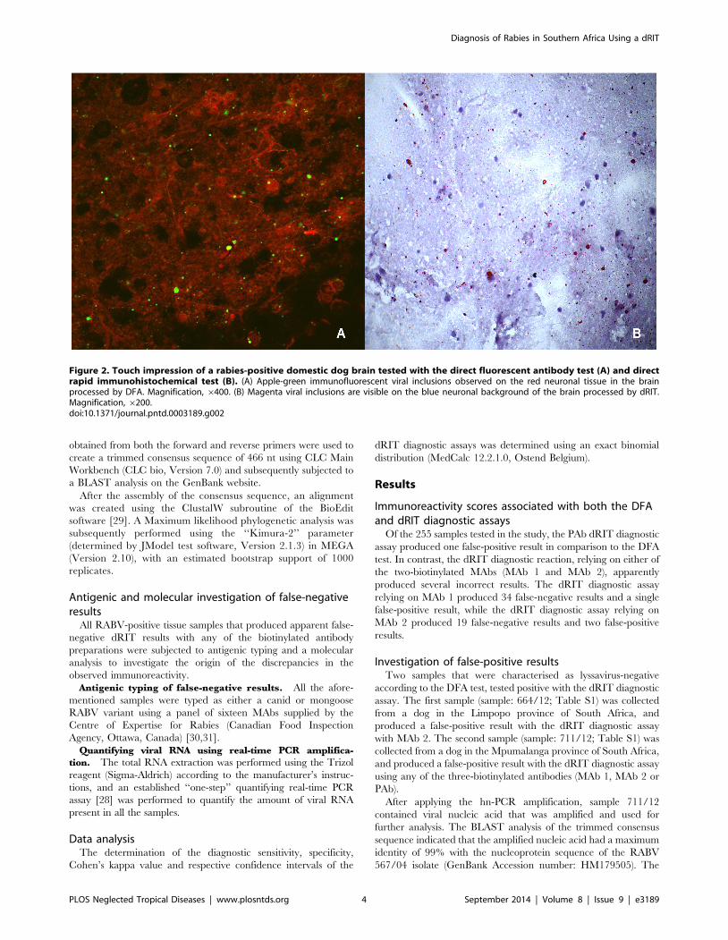
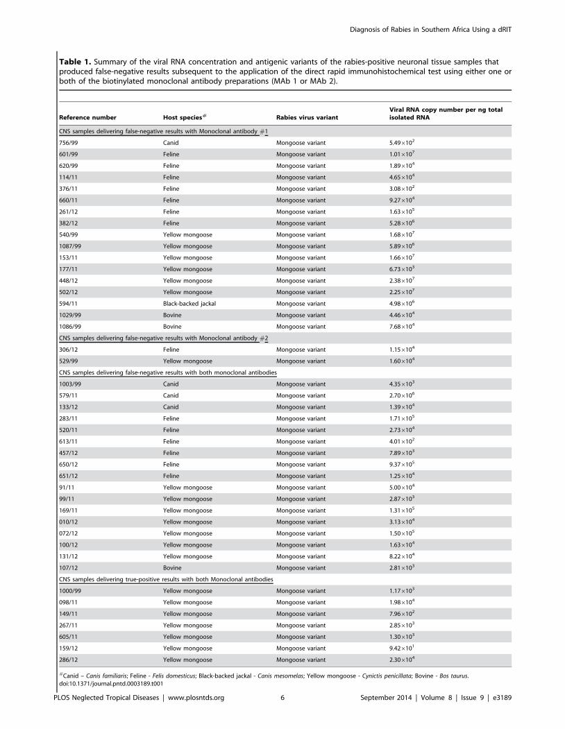


