
9/21/2018
1
Clinical Evaluation of the Bleeding Patient
Kathleen W. Phelan, M.D.
Division of Hematology/Oncology
September 25, 2017
Objectives
1. Articulate the differential diagnosis of a patient with a bleeding disorder.
2. Name several causes of a quantitative platelet disorders.
3. Describe a couple causes of qualitative platelet disorders.
4. Identify clues in the history, physical exam and laboratory data that suggest a bleeding disorder.
Objectives
5. Understand that in bleeding disorders stemming from defects in one or more of the coagulation factors, the prothrombin time (PT), partial thromboplastin time (PTT), or both can be prolonged.
6. Explain how to interpret a mixing study.
7. Formulate a clinical hypothesis from the history and physical exam for a patient with a bleeding disorder.
8. Choose appropriate laboratory testing for cases of bleeding disorders and interpret results.
9/21/2018
2
References
• * Powerpoint slides – focus of test questions
• Handouts
• Robbins Basic Pathology
Outline
I. Bleeding Disorder Review
II. Illustrated Case Studies
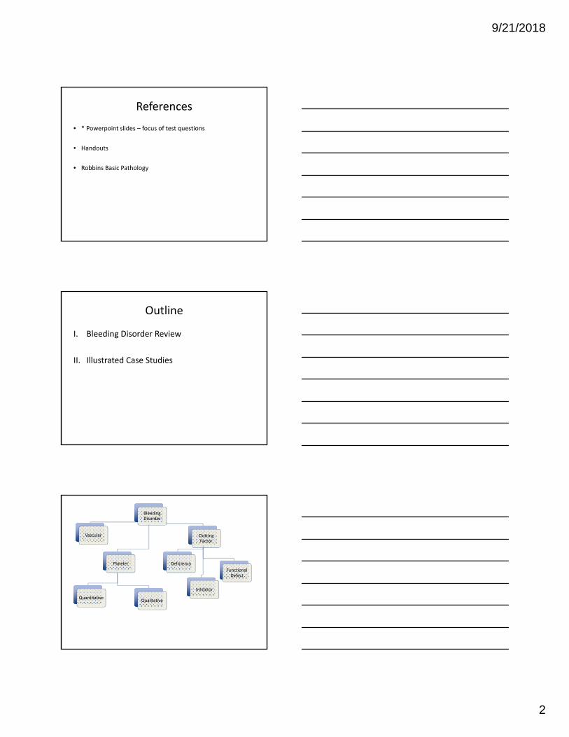
Bleeding Disorder
Vascular
Platelet
QuantitativeQualitative
Clotting Factor
Deficiency
Inhibitor
Functional Defect

9/21/2018
3
Bleeding Disorders: Differential Diagnosis
Platelet Disorders Clotting Factor Abnormalities
Thrombocytopenia (Quantitative Platelet Disorders)
• Idiopathic Thrombocytopenic Purpura (ITP)
• Drug‐induced thrombocytopenia• Hypersplenism• Marrow infiltration by neoplasia• Viral infections (e.g., HIV, EBV, Rubella)• DIC• TTP‐HUS• Autoimmune condition (e.g. Lupus)• Gestational thrombocytopenia
Qualitative Platelet Disorders• Inherited
• Von Willebrand’s Disease• Glanzmann Thrombasthenia• Bernard‐Soulier Syndrome
• Acquired• Drugs• Chronic renal failure
Inherited• Factor VIII deficiency (Hemophilia A)• Factor IX deficiency (Hemophilia B)• Other Factor deficiencies (less
common)Acquired
• Vitamin K deficiency• Factor inhibitors • Failure of Synthetic Function of the
Liver• Drugs• DIC
History is Key!
• Surgeries, tooth extraction, childbirth
• Bleeding that required surgical intervention, blood transfusion, or replacement therapy
• Positive family history (especially in children without a history of hemostatic challenge)
• Circumcision in boys
• Menorrhagia in women
• Medications (e.g. warfarin, heparin, aspirin, plavix, other anticoagulants)
• Viral illness, malnutrition, liver disease, malignancy
Physical Exam
• Epistaxis, bruising, petechiae, hematomas
• Oral cavity bleeding
• Hematemesis, hematochezia, melena
• Hemarthrosis
• CNS bleeding
• Lymphadenopathy
• Splenomegaly, hepatomegaly

9/21/2018
4
Some Clues
Disorder Clues
Platelet Disorders (Quantitative) Mucosal bleeding, bruising, petechia, or purpura
Platelet Disorders (Qualitative) Consider in a patient with a lifelong history of bleeding despite negative laboratory work‐up
Hemophilia type A or B (Factor VII or IX Deficiency) or other Factor Deficiencies
Classically presents with joint or soft‐tissue bleeding; family history of bleeding in men (skipped generations)
Factor Inhibitors Presentation similar to hemophilia, but onset is typically sudden with no patient or family history of bleeding
DIC Bleeding from multiple sites; prolonged PT and PTT
Vitamin K Deficiency More common causes include malabsorption (bacterial overgrowth, celiac disease, inflammatory bowel disease), poor diet (alcoholism, total parenteral nutrition) or drugs that bind vitamin K
Adapted from www.aafp.org
CASE #1
Case #1: A. K.
• “I have a rash”
• 66 yo female, generally in good health
• Noted rash 1‐2 weeks ago, seems a little worse now.
• I also had a cold – not too bad, but a sore throat – about a month ago
• Otherwise, no particular medical complaints

9/21/2018
5
Case #1: A. K.
• Characteristics– Mucosal vs. Deep Tissue
– Immediate vs Delayed
• Associated conditions– Epistaxis, hemoptysis, dark or tarry stool
– Skin – bruising, purpura, petechiae, telangiectasias
– Trauma/Accident
– Childbirth
– Circumcision
• Social Exacerbations/Danger
Case #1: A. K.
• Other bleeding?• Maybe a little bruising on her arms – doesn’t really think it’s too bad.
• Characteristics• No joint bleeds
• Associated conditions• Never had a history of easy bleeding, no blood transfusions
• Never with excessive menorrhagia
• 3 children, all vaginal deliveries, no complications
9/21/2018
6
• PMH– High cholesterol– Hypertension– Thyroid disease
• Medications– Simvastatin– Metoprolol– Levothyroxine– No OTCs
• Social History– Docent at local museum– Retired banker– Divorced
• Family History– Mother died of breast cancer
complications at 83– Father died of CAD at 68– Children are alive and well, no
illnesses– 2 siblings, no bleeding history
• ROS– Fever 4 weeks ago with this
“cold.” Only 1‐2 days– No chills, no nausea, no
vomiting, no change in energy level, no night sweats
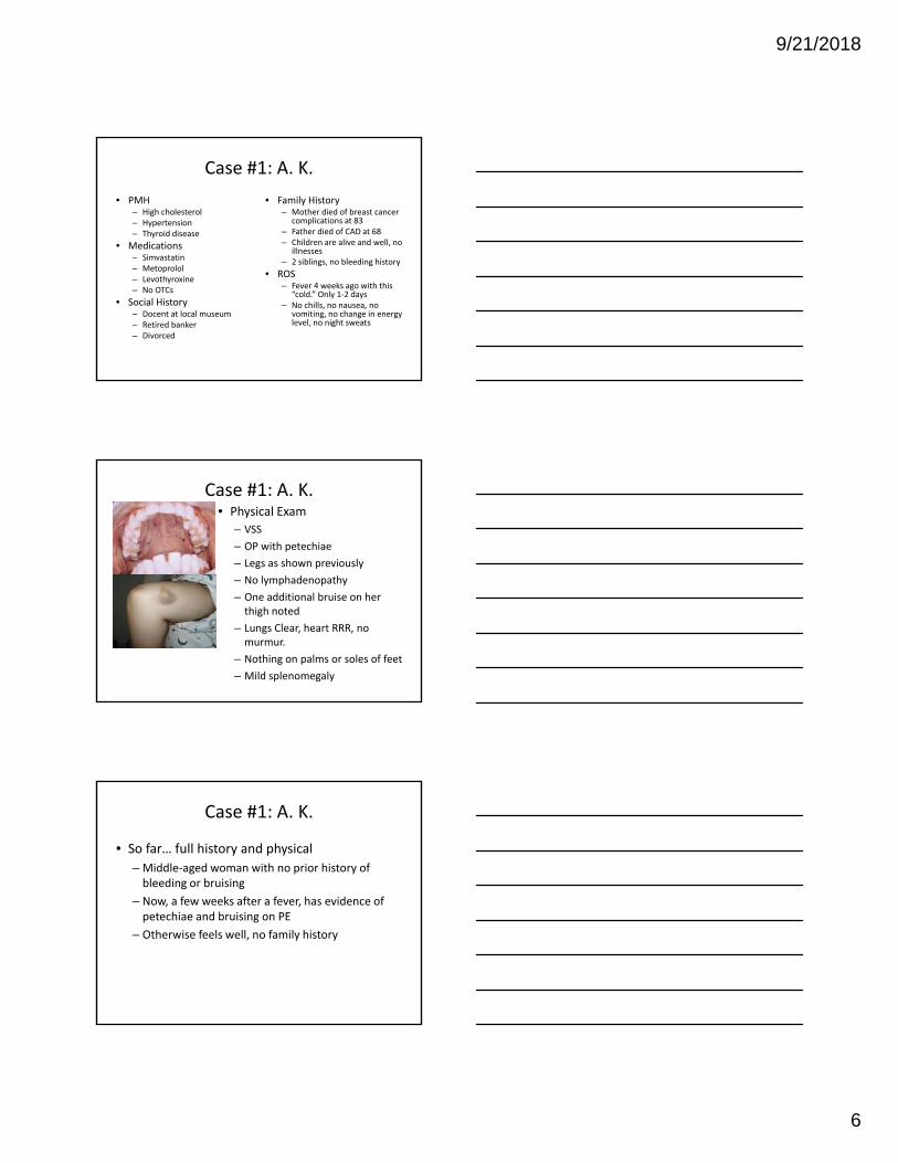
Case #1: A. K.
• Physical Exam
– VSS
– OP with petechiae
– Legs as shown previously
– No lymphadenopathy
– One additional bruise on her thigh noted
– Lungs Clear, heart RRR, no murmur.
– Nothing on palms or soles of feet
– Mild splenomegaly
Case #1: A. K.
• So far… full history and physical
– Middle‐aged woman with no prior history of bleeding or bruising
– Now, a few weeks after a fever, has evidence of petechiae and bruising on PE
– Otherwise feels well, no family history
Case #1: A. K.

9/21/2018
7
Case #1: Work Up
Test A.K. Normal
WBC 7.8 K/uL 4.0‐10.0 K/uL
HGN 12.8 gm/dL 12.0‐16.0 Gm/dL
MCV 89FL 85‐95 FL
PLT 8 K/uL 150‐400 K/uL
PT/INR 12 secINR=1.0
11‐13 sec
aPTT 28 sec 25‐32 sec
Bleeding Time 6.3 min 2‐8 min
Copyright ©2001 American Society of Hematology. Copyright restrictions may apply.
Lazarchick, J. ASH Image Bank 2001;2001:100177
Bleeding Disorders: Differential Diagnosis
Platelet Disorders Clotting Factor Abnormalities
Thrombocytopenia (Quantitative Platelet Disorders)
• Idiopathic Thrombocytopenic Purpura (ITP)
• Drug‐induced thrombocytopenia• Hypersplenism• Marrow infiltration by neoplasia• Viral infections (e.g., HIV, EBV, Rubella)• DIC• TTP‐HUS• Autoimmune condition (e.g. Lupus)• Gestational thrombocytopenia
Qualitative Platelet Disorders• Inherited
• Von Willebrand’s Disease• Glanzmann Thrombasthenia• Bernard‐Soulier Syndrome
• Acquired• Drugs• Chronic renal failure
Inherited• Factor VIII deficiency (Hemophilia A)• Factor IX deficiency (Hemophilia B)• Other Factor deficiencies (less
common)Acquired
• Vitamin K deficiency• Factor inhibitors • Failure of Synthetic Function of the
Liver• Drugs• DIC

9/21/2018
8
Copyright ©2001 American Society of Hematology. Copyright restrictions may apply.
Lazarchick, J. ASH Image Bank 2001;2001:100177
Figure 5. Bone marrow aspirate showing megakaryocytic hyperplasia with clustering of the megakaryocytes around a spicule
Case #1: ITP
• Middle aged woman with
– Petechiae, bruising
– New onset severe thrombocytopenia
– No additional complications in bone marrow
– Ruled out all other causes
• ITP Treatment
– Steroids
CASE 2

9/21/2018
9
Case #2: J.B.
CC; History
• Routine blood work in a 36yo in the burn unit
• “Something’s weird about the PT – INR is 5.”
• Now needs skin grafting for lower extremity burn. Also with inhalation injury.
Important points in HPI
• Characteristics– Mucosal vs. Deep Tissue
– Immediate vs Delayed
• Associated conditions– Epistaxis, hemoptysis, dark or
tarry stool?
– Skin – bruising, purpura, petechiae, telangiectasias?
– Trauma/Accident
– Childbirth
– Circumcision
• Social Exacerbations/Danger
Case #2: J.B.
• PMH
– Elevated cholesterol
– Apparently has had a blood transfusion in the past, after an automobile accident
• Medications
– None
• Social History
– Smoker X 20 years
• Family History
– Adopted
• PE
– VSS
– Intubated for airway protection
– 70% LE burns, wrapped
– No other bruising, petechia are visible
Case #2: J.B.
• So far… full history and physical
– Young man with elevated PT/INR
– Maybe lifelong
– ?Familial
– Not medication related
– May or may not have been the reason for the need for his blood transfusion

9/21/2018
10
Bleeding Disorder
Vascular
Platelet
QuantitativeQualitative
Clotting Factor
Deficiency
Inhibitor
Functional Defect
Case #2: Work Up
Test J.B. Normal
WBC 12.5 K/uL 4.0‐10.0 K/uL
HGN 12.1 gm/dL 12.0‐16.0 Gm/dL
MCV 98 FL 85‐95 FL
PLT 230 K/uL 150‐400 K/uL
PT/INR 28 secINR=3.1
10‐11.7 sec
aPTT 28 sec 25‐32 sec
Extrinsic(PT)
Pathway
Intrinsic(PTT)
Pathway
Contact Factors,
XIXII
VIIIX
TissueFactor
VII
X, V, phospholipids
Prothrombin (II) Thrombin
Fibrinogen (I)
Fibrin
CommonPathway

9/21/2018
11
Copyright ©2007 American Society of Hematology. Copyright restrictions may apply.
Crowther, M. A. et al. ASH‐SAP 2007;2007:361‐407
Prolonged PT
Warfarin
Liver Disease
Factor VII Deficiency
Prolonged PTT
Heparin
Factor VIII, IX, XI, XII Deficiency
Lupus anticoagulant
Prolonged PT and PTT
High‐dose Warfarin or Heparin
Liver Disease
DIC
Vitamin K deficiency
Prothrombin, fibrinogen, factor V, X or combined factor deficiency
Case #2: J.B.
• So far…
– Young man with an isolated elevated PT but a NORMAL aPTT
– Normal platelet count
• You check his Factor VII level – it’s 2%
• But does this discriminate between qualitative and quantitative abnormalities?

9/21/2018
12
How To Approach An Increased PT/ or PTT Test
There are two basic reasons for elevated PT or PTT:
(1) Deficiency of a clotting factor
(2) Inhibitor of a clotting factor
Key Point: mixing study is used to differentiate between (1) and (2)
Mixing Study:
A simple test to determine the cause of an elevated PT/ or APTT
aPTT
mix 1 part normal (NL) and 1 part patient (PT) plasma
PT
NLNL REPEAT aPTT
PROBLEM:
TEST: Mix Study
Mixing Study Interpretation
CorrectionCorrection
Deficiency of clotting Factor
Factor Assays
No CorrectionNo Correction
Inhibitor
LA Testing
n TEST RESULT

9/21/2018
13
Case #2: Work Up
Test J.B. Normal
Factor VII level 4% 50‐150%
PT/INR 28 secINR=3.1
10‐11.7 sec
Mixing Study Corrects the PT to 12 sec
Case #2: Factor VII Deficiency
• 95‐97% of all Inherited Coagulation defects– X‐linked inherited coagulation disorders:
• hemophilia A (factor VIII deficiency) • hemophilia B (factor IX deficiency)
– von Willebrand disease
• Remaining defects are VERY rare ‐‐ ranging from approximately 1 in 500,000 to 1 in 2 million
• Factor VII deficiency presents with a wide spectrum of clinical severity, correlating poorly with factor VII levels; some patients with undetectable levels are asymptomatic
Case #2: Treatment
• Considerations
– Fresh frozen plasma
– But there is a very short half‐life of factor VII (4 to 6 hours)
– Recombinant activated factor VII is available – but very, very costly

9/21/2018
14
CASE 3
Case #3: T.J.
CC; History
• “Heavy periods”
• 16 yo
• Periods are 5‐6 d in duration. Heavy tampon every couple of hours during d2‐3.
• “Have to get up during the night and “can’t make it through a movie without changing them.”
Important points in HPI
• Characteristics– Mucosal vs. Deep Tissue
– Immediate vs Delayed
• Associated conditions– Epistaxis, hemoptysis, dark or
tarry stool?
– Skin – bruising, purpura, petechiae, telangiectasias?
– Trauma/Accident
– Childbirth
– Circumcision
• Social Exacerbations/Danger
Case #3: T.J.
• Menorrhagia?• Definition: More than 80 ml of blood/cycle
• Characteristics• No joint bleeds
• Had to go back to dentist after excessive bleeding two days following an extraction
• Associated conditions• Also had two episodes of nose bleeding that required ER visits as a child. + Spontaneous. + packing, +cautery
• No blood transfusions.
• + gum bleeding
9/21/2018
15
Case #3: T.J.
• PMH– “They tell me I have
anemia and have to take iron.”
• Medications– Used to take a MVI– No OCPs
• Social History– Planning a career in
military– No tobacco, occasional
ETOH, + marijuana X 2– Denies DV
• Family History– Mother
• Blood transfusion after birth of second child
• + Menorrhagia hysterectomy
– Father• HTN, Chol
– Younger brother• + nose bleeds
– MGF• Died in Korean war
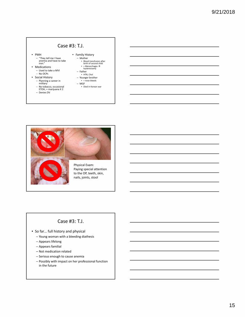
Physical Exam: Paying special attention to the OP, teeth, skin, nails, joints, stool
Case #3: T.J.
• So far… full history and physical
– Young woman with a bleeding diathesis
– Appears lifelong
– Appears familial
– Not medication related
– Serious enough to cause anemia
– Possibly with impact on her professional function in the future

9/21/2018
16
Bleeding Disorder
Vascular
Platelet
QuantitativeQualitative
Clotting Factor
Deficiency
Inhibitor
Functional Defect
Case #3: Laboratory Studies
Study What can we learn
CBC with differential
Platelet quantityIs there involvement of the other cell lines?What do the platelets look like?Is there evidence of a systemic disorder?
PT and aPTT Is there abnormalities in the coagulation system? Will pick up BOTH qualitative and quantitative defects, but won’t differentiateCan sometimes differentiate which clotting factor is the culprit
Bleeding Time Defect in platelet plug formation. Imperfect test. Stilldiscussed in the literature, but rarely used clinically. New tests have been developed to look at platelet function – PFA‐100
Case #3: Work Up
Test T.J. Normal
WBC 6.3 K/uL 4.0‐10.0 K/uL
HGN 10.8 gm/dL 12.0‐16.0 Gm/dL
MCV 72 FL 85‐95 FL
PLT 443 K/uL 150‐400 K/uL
PT/INR 12 secINR=1.0
11‐13 sec
aPTT 38 sec 25‐32 sec
Bleeding Time 7.4 min 2‐8 min

9/21/2018
17
Case #3: T.J.
• So far…
– Young woman with a lifelong, likely familial bleeding diathesis
– Microcytic anemia
– Normal platelet number, prolonged aPTT, normal PT, prolonged bleeding time
• What’s my differential?
– Common things are common
Bleeding Disorders: Differential Diagnosis
Platelet Disorders Clotting Factor Abnormalities
Thrombocytopenia (Quantitative Platelet Disorders)
• Idiopathic Thrombocytopenic Purpura (ITP)
• Drug‐induced thrombocytopenia• Hypersplenism• Marrow infiltration by neoplasia• Viral infections (e.g., HIV, EBV, Rubella)• DIC• TTP‐HUS• Autoimmune condition (e.g. Lupus)• Gestational thrombocytopenia
Qualitative Platelet Disorders• Inherited
• Von Willebrand’s Disease• Glanzmann Thrombasthenia• Bernard‐Soulier Syndrome
• Acquired• Drugs• Chronic renal failure
Inherited• Factor VIII deficiency (Hemophilia A)• Factor IX deficiency (Hemophilia B)• Other Factor deficiencies (less
common)Acquired
• Vitamin K deficiency• Factor inhibitors • Failure of Synthetic Function of the
Liver• Drugs• DIC
Case #3: T.J.
• Prolonged bleeding time, slightly prolonged PTT (normal platelet count, normal PT)
• Von Willebrand’s Disease

9/21/2018
18
Case #4: F.C.
• 68 yo F underwent resection of a pancreatic tumor
• Excessive bleeding from the drain at her surgical site required blood transfusions; surgeon is concerned about a bleeding disorder
• Has had prior surgeries without bleeding complications. No family history of bleeding disorders. No history of liver disease.
• Prior to surgery, platelet count, PT, PTT all normal
Case #4: F.C.
• White blood cell count 15.5 K/uL [4.0‐10.0 K/uL]
• Hemoglobin 8.7 g/dL [12.0‐16.0 gm/dL]
• Platelet count 90 K/uL [150‐400 K/uL]
• PTT 57 seconds [23‐31 seconds]
• PT 23 seconds [12‐14 seconds]
• Fibrinogen 100 mg/dL [150‐350 mg/dL]
• D‐dimer >20 µg/mL [<.5 µg/mL]
• AST 32 U/L [5‐35 U/L]
• ALT 33 U/L [7‐56 U/L]
• Alkaline phosphatase 57 U/L [20‐140 U/L]
• Total bilirubin normal
• Occasional schistocytes

9/21/2018
19
Case #4: DIC
Select Platelet Disorders Summary
Disorder Platelet count Bleeding Time Comments
ITP ↓ ↑ Anti‐GpIIB/IIIa antibodies splenic macrophage consumption of platelet‐antibody complex
Bernard‐SoulierSyndrome
‐‐‐‐ ↑ Defect in platelet plug formation. ↓GpIb defect in platelet‐to‐vWF adhesion
Glanzmann Thrombasthenia
‐‐‐‐ ↑ Defect in platelet plug formation. ↓GpIIb/IIIa defect in platelet‐to‐platelet aggregation
Mixed Platelet and Coagulation Disorders
Disorder Platelet Count
BleedingTime
PT PTT Comments
VonWillebrandDisease
‐‐‐‐ ↑ ‐‐‐‐ ↑ /nl Intrinsic pathway coagulation defect: ↓vWF↑PTT (vWF acts to carry/protect factor VIII)Defect in platelet plug forma on: ↓vWF defect in platelet‐to‐vWF adhesion
DIC ↓ ↑ ↑ ↑ Widespread activation of clottingdeficiency in clotting factors bleeding state.Findings: ↑fibrin degrada on products (D‐dimers), ↓fibrinogen
9/21/2018
20
Bleeding Disorders Summary
• Bleeding history is key!
• Use this history and physical exam to guide laboratory testing and evaluation
• CBC, PT, and PTT and peripheral blood smear are useful tests
• Any questions?
THANK YOU!


