
2
Objectives:
• Review the objectives on page 1 and 2
of the lecture handout
• Objectives marked with ‘*’ will not be tested over during student lab rotation
3
Historical Perspective: Urinalysis
• Physical examination of urine– Odor– Taste– Color– Clarity

4
Historical Perspective
• Chemical examination of urine– Limited reactions– Required large volumes of urine– Large volumes of reagent– Performed in test tubes– Time consuming and cumbersome– Clinical usefulness was not realized– Not routinely ordered
5
Historical Perspective
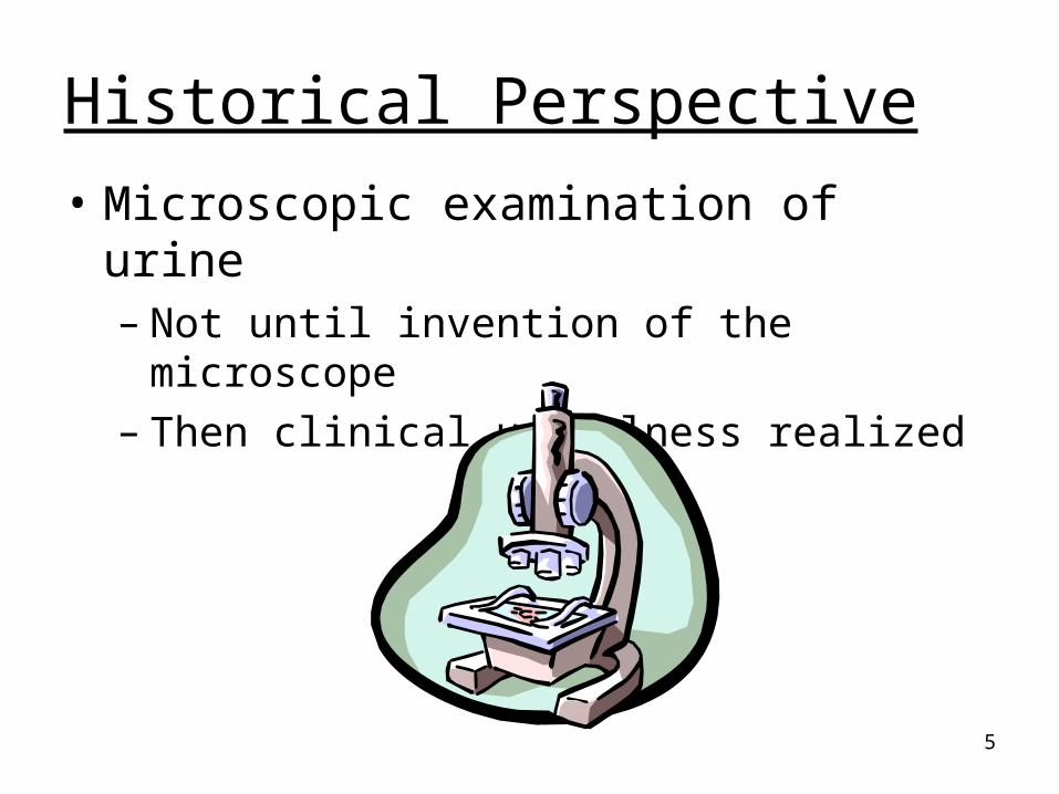
• Microscopic examination of urine– Not until invention of the microscope– Then clinical usefulness realized

6
Reagent Strip Testing
• Technology and necessity
• Chemical reactions ‘miniaturized’
• Required less urine
• Test results within minutes
• Easy to perform
• Increased test utilization
Brunzel, 2nd Ed, page 124

7
Reagent Strip Testing
• Ideal qualitative screening tool– Sensitive: Low concentration of substances
Negative result = normal
– Specific: Reacts with only one substance False negative and false
positive
– Cost effective: Relatively inexpensive tool that provides information about the health statusof the patient
8
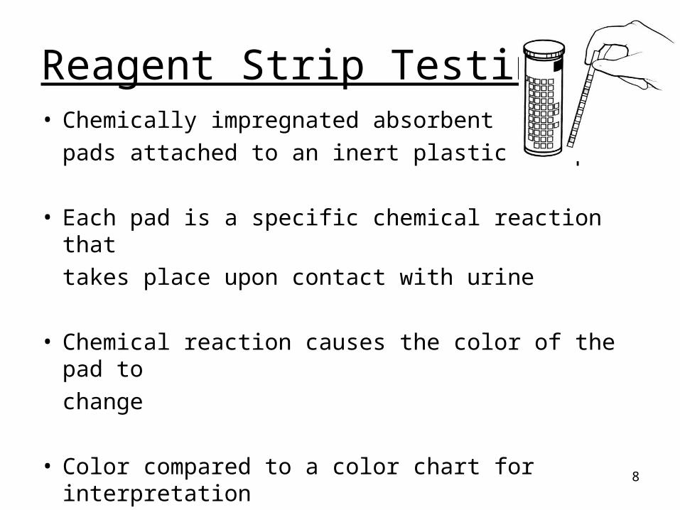
Reagent Strip Testing• Chemically impregnated absorbent
pads attached to an inert plastic strip
• Each pad is a specific chemical reaction that
takes place upon contact with urine
• Chemical reaction causes the color of the pad to
change
• Color compared to a color chart for interpretation
9
Reagent Strip Testing
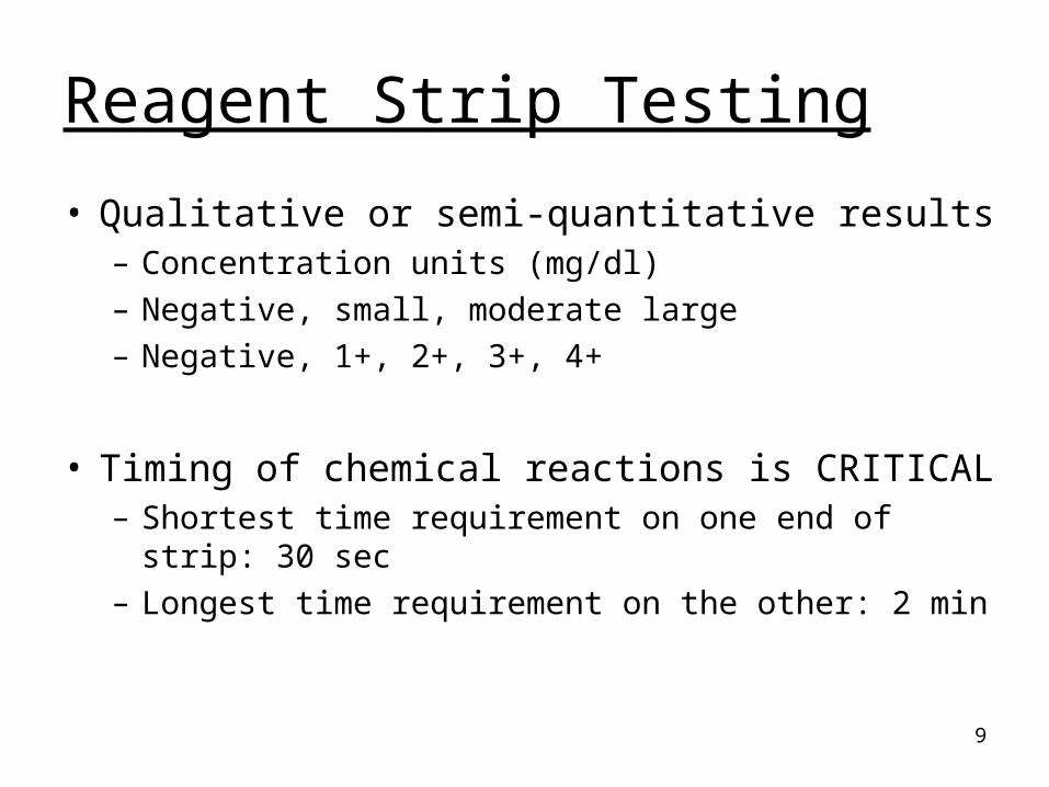
• Qualitative or semi-quantitative results– Concentration units (mg/dl)– Negative, small, moderate large– Negative, 1+, 2+, 3+, 4+
• Timing of chemical reactions is CRITICAL– Shortest time requirement on one end of strip: 30 sec– Longest time requirement on the other: 2 min

10
Reagent Strip Testing
• Principle of chemical reactions– False negative reactions– False positive reactions– Color interferences
• Alternative testing: used to confirm results that you may think are invalid due to– Interfering substance– Color interference (called color masking)
11
Care and Storage (pg 4)
Confirmatory Testing (pg 6)
Reading assignment:
Textbook, chapter 7
Page 124-130
12
Confirmatory Testing
• Alternative testing establishes the correctness or accuracy of another procedure
• Often used when urine is highly pigmented– Bilirubin reagent strip ictotest
13
Confirmatory Testing
• Characteristics:– Differ in sensitivity
• Ictotest vs Bilirubin reagent strip
– Differ in specificity• SSA vs Protein reagent strip• Clinitest vs Glucose reagent strip
– Differ in methodology/reaction
Ideallywant all 3
14
Differ in Specificity
• Clinitest reacts with all reducing
substances
• Glucose reagent strip reacts with only one reducing substance: glucose
15
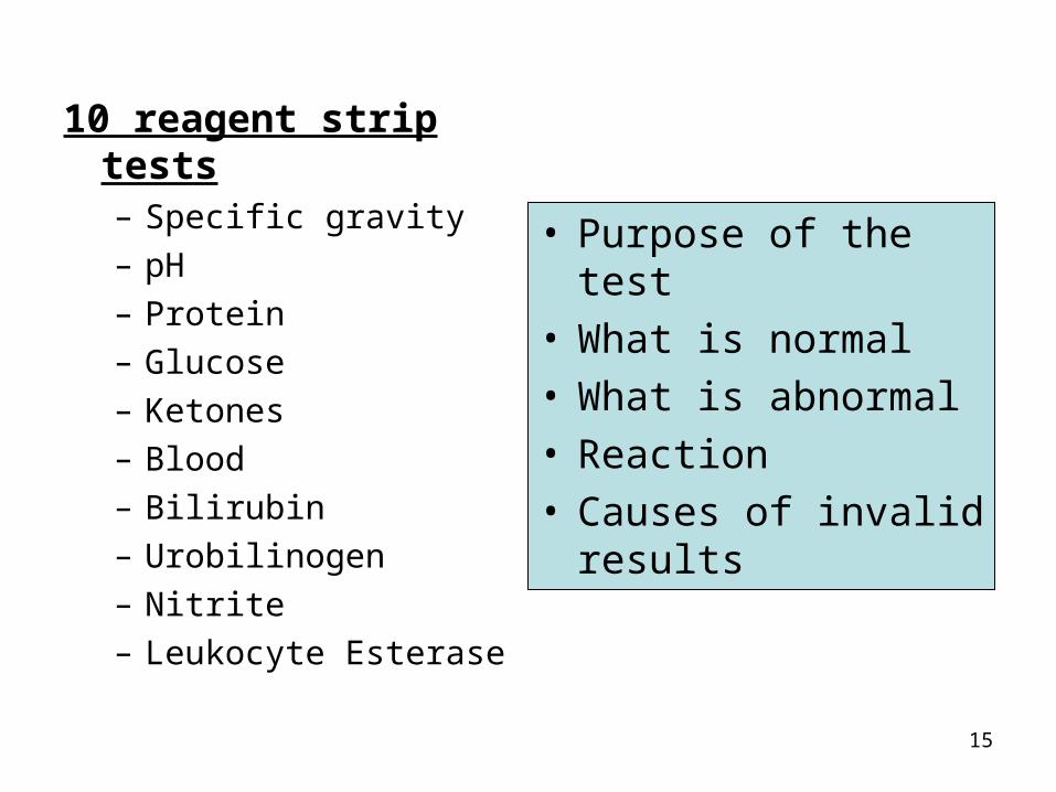
10 reagent strip tests– Specific gravity– pH– Protein– Glucose– Ketones– Blood– Bilirubin– Urobilinogen– Nitrite– Leukocyte Esterase
• Purpose of the test• What is normal• What is abnormal• Reaction• Causes of invalid
results
16
Specific Gravity: Purpose• Evaluates the concentrating and diluting
ability of the kidney– Density is related to the amount of
substances (solutes) in solution
– Increased density ~ increased solute in solution ~ hypertonic urine ~ concentrated urine
– Decreased density ~ decreased solute in solution ~ hypotonic urine ~ dilute urine
17
Specific Gravity: Normal
• Normal: 1.002 – 1.035
• Majority of urines: 1.010 – 1.025
• Physiologically impossible: 1.000>1.040
• Dependent upon hydration status
18
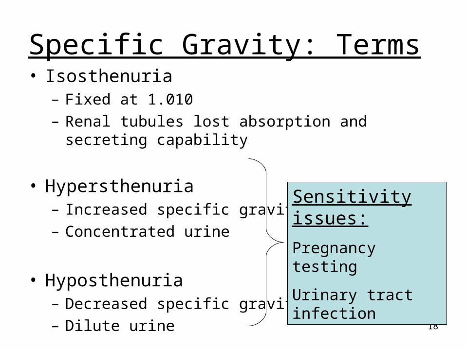
Specific Gravity: Terms• Isosthenuria
– Fixed at 1.010– Renal tubules lost absorption and secreting capability
• Hypersthenuria– Increased specific gravity– Concentrated urine
• Hyposthenuria– Decreased specific gravity– Dilute urine
Sensitivity issues:
Pregnancy testing
Urinary tract infection
19
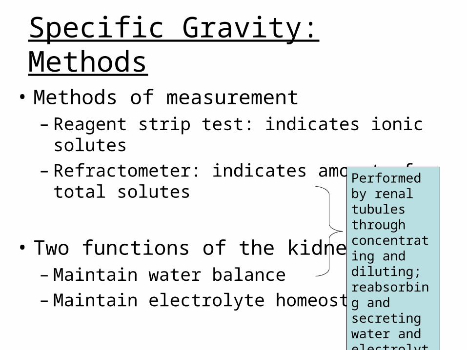
Specific Gravity: Methods
• Methods of measurement– Reagent strip test: indicates ionic solutes– Refractometer: indicates amount of total solutes
• Two functions of the kidney– Maintain water balance – Maintain electrolyte homeostasis
Performed by renal tubules through concentrating and diluting; reabsorbing and secreting water and electrolytes (ionic)
20
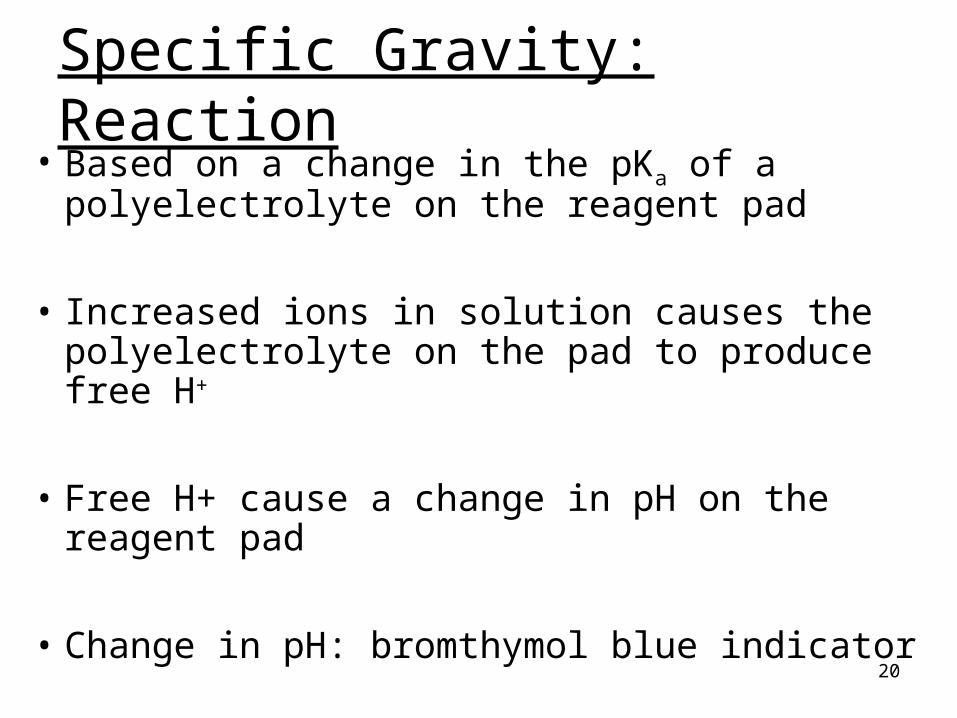
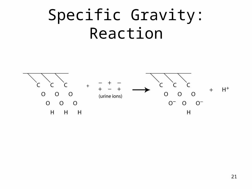
Specific Gravity: Reaction • Based on a change in the pKa of a
polyelectrolyte on the reagent pad
• Increased ions in solution causes the polyelectrolyte on the pad to produce free H+
• Free H+ cause a change in pH on the reagent pad
• Change in pH: bromthymol blue indicator
21
Specific Gravity: Reaction

22
Specific Gravity
• Sensitivity: 1.000
• Specificity: detects only ionic substances– Radiographic dye– Mannitol– Glucose
Does not interfere
23
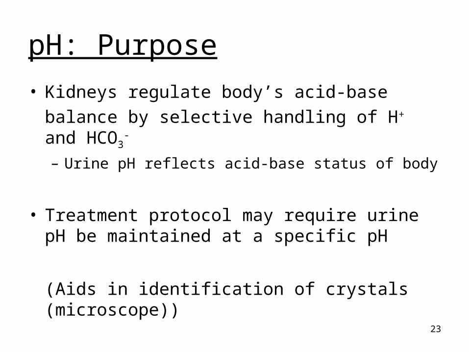
pH: Purpose
• Kidneys regulate body’s acid-base
balance by selective handling of H+ and HCO3-
– Urine pH reflects acid-base status of body
• Treatment protocol may require urine pH be maintained at a specific pH
(Aids in identification of crystals (microscope))

24
pH: Normal
• Normal: ranges from 4.5 – 8.0
• First morning void: acidic
• Physiologically impossible: <4.5
>8.01. Urine not handled properly
2. Old urine
3. Treatment induced

25
pH: Interpretation
• Made in conjunction with – Acid-base status– Renal function – Presence of infection in urinary tract– Diet: high protein, low protein– Medications– Age of urine sample
26
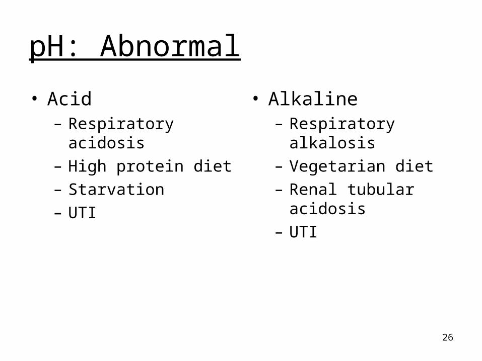
pH: Abnormal
• Acid– Respiratory acidosis– High protein diet– Starvation– UTI
• Alkaline– Respiratory alkalosis– Vegetarian diet– Renal tubular acidosis– UTI

27
pH: Reaction
• Double indicator system– Methyl red– Bromthymol blue
• Amount of free H+ influences acidity of urine and cause pH indicator to change color
Needed to measure the wide pH range: acid to alkaline

28
pH:
• Invalid test results due to:– Improper handling of urine sample
– Contamination of urine vessel prior to collection
– ‘Run-over’ phenomenon
29
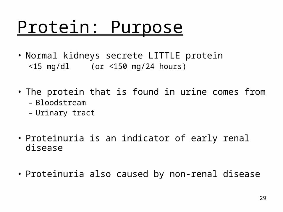
Protein: Purpose
• Normal kidneys secrete LITTLE protein<15 mg/dl (or <150 mg/24 hours)
• The protein that is found in urine comes from– Bloodstream– Urinary tract
• Proteinuria is an indicator of early renal disease
• Proteinuria also caused by non-renal disease

30
Renal Cause of Proteinuria: • Glomerular damage:
– Most serious cause of proteinuria– Most common cause of proteinuria– Glomerulonephritis– Nephrotic Syndrome
• Tubular dysfunction:– Reabsorption capability decreased– Toxin exposure, inherited disorder– Fancon’s syndrome: heavy metal poisoning

31
Classification of Proteinuria
• Functional
• Orthostatic (postural)
• Transient
• Pathologic– Pre-renal (overflow)– Renal: glomerular– Renal: tubular– Post-renal

32
Protein: Methods
• Reagent strip test
• SSA test
• Foam test
• Micro-albumin test

33
Protein: Reagent Strip
• The reagent pad is held at a constant pH of 3 by a buffer
• Proteins (anions) in solution cause anindicator dye to release H+ causing a colorchange
• ‘Protein error of indicators’

34
Protein: Reagent Strip
• Sensitivity: ~ 10-25 mg/dl
• Specificity: reacts with albumin– False positive: highly alkaline urine (pH > 8.0)– False negative:
Dilute urine
Presence of other proteins (Tamm-Horsfall, globulins,
myoglobin,
free light chains, hemoglobin)
35
Protein: SSA (Exton’s Test)
• Sulfosalicylic Acid (SSA) Precipitation Test
• Acid will precipitate proteins out of solution causing the solution to become cloudy
• Amount of cloudiness is related to the amount of protein present

36
Protein: SSA (Exton’s Test)
• Amount of cloudiness is evaluated, thus must use centrifuged urine
• Sensitivity: 5-10 mg/dl
• Specificity: detects all protein

37
Protein: SSA (Exton’s Test)
• False positive results: – Radiographic dyes– Turbid urine– Uncentrifuged urine
• False negative results:– Highly alkaline urine– Dilute urine
38
Protein: Foam test
• Shake aliquot of urine and observe color of resulting foam
• White foam: protein present

39
Protein: Micro-albumin test
• Measures very low concentration of albumin (better sensitivity than reagent strip test for albumin)
• Management of diabetic patient
• Methods vary: reagent strip test,
immunochemical reaction

40
Glucose: Purpose
• Healthy normal urine does not contain glucose
• Normally, glucose is filtered by the glomerulus and is reabsorbed back into the bloodstream through active transport mechanism
• Glucose in urine is pathologic

41
Glucose: Purpose
• Glucosuria
Glycosuria
• Caused by renal and non-renal disease– Pre-renal glycosuria: plasma glucose level
exceeds renal threshold (diabetes mellitus)
– Renal glycosuria: plasma glucose level below renal threshold, but tubules cannot reabsorb glucose back into bloodstream
Terms used interchangeably

42
Reducing Substances: Purpose
• Reducing Substances:– Glucose– Other sugars: galactosemia (inherited
metabolic disorder)
43
Glucose, Reducing Substances
• Normal: negative
• Abnormal: – Diabetes mellitus: glucose– Impaired renal tubular reabsorption: glucose
– Inborn error of metabolism: galactosemia
44
Methods
• Reagent strip: detects only glucose
• Copper Reduction: detects reducing substances
45
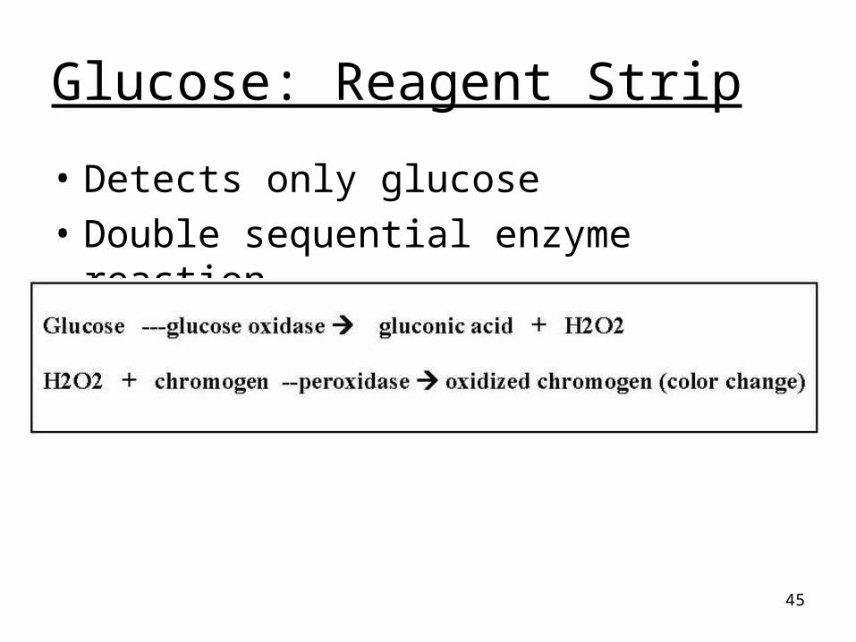
Glucose: Reagent Strip
• Detects only glucose
• Double sequential enzyme reaction

46
Glucose: Reagent Strip
• Sensitivity: ~ 30 mg/dl
• Specificity: – Reacts only with glucose– False positive:
• Strong oxidizing agents (bleach)• Peroxides
– False negative: • Ascorbic acid (reducing agent)• Improperly stored urine: glycolysis

47
Clinitest Reaction
• Copper Reduction Test:– Reducing substances are able to reduce
copper sulfate to cuprous oxide
– Pass-through phenomenon
– All children <2 years: metabolic disorder(galactosemia)
48
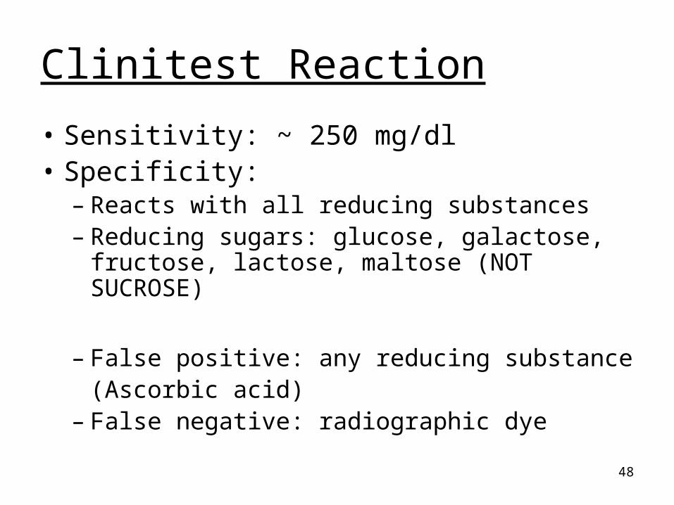
Clinitest Reaction
• Sensitivity: ~ 250 mg/dl• Specificity:
– Reacts with all reducing substances– Reducing sugars: glucose, galactose,
fructose, lactose, maltose (NOT SUCROSE)
– False positive: any reducing substance(Ascorbic acid)
– False negative: radiographic dye
49
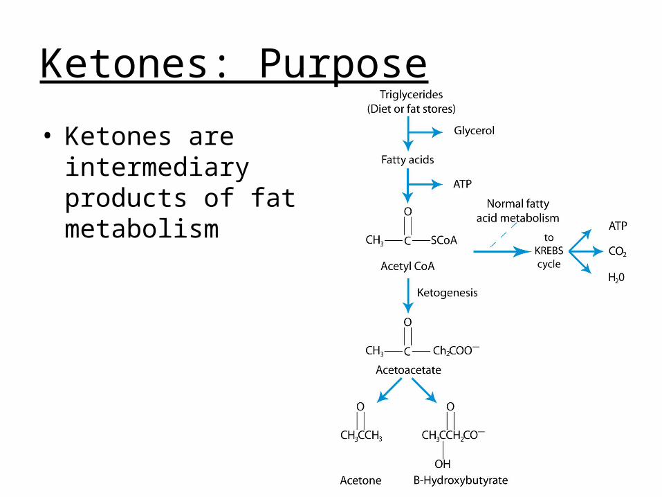
Ketones: Purpose
• Ketones are intermediary products of fat metabolism

50
Ketones
• Three ketone bodies– Acetone 2%– Acetoacetic acid 20%– Beta-hydroxybutyric acid 78%
• Characteristic ‘fruity breath’ ~ acetone

51
Ketones: Normal
• Normal: negative
• Abnormal:– Inability to utilize carbohydrates– Excessive loss of carbohydrates– Inadequate intake of carbohydrates

52
Ketones: Methods
• Reagent strip
• Acetest: tablet test

53
Ketones: Method
• Glycine: also measures acetone– Reagent strip: check package insert– Acetest tablets: contain glycine
54
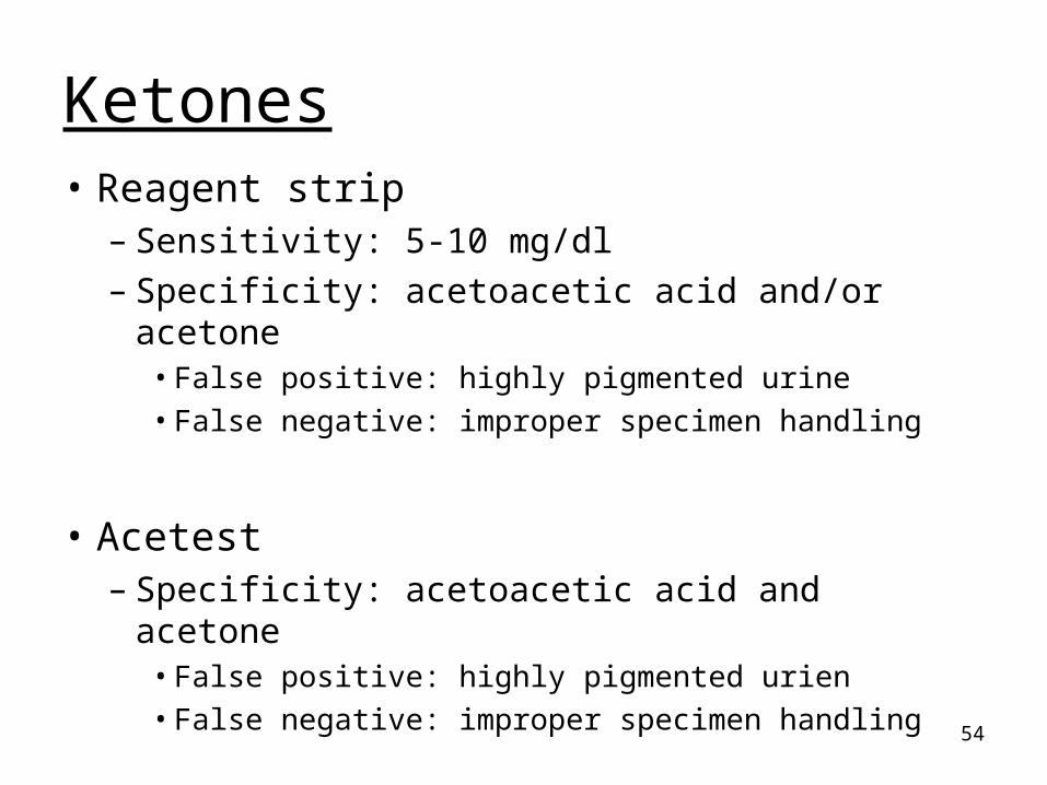
Ketones• Reagent strip
– Sensitivity: 5-10 mg/dl– Specificity: acetoacetic acid and/or acetone
• False positive: highly pigmented urine• False negative: improper specimen handling
• Acetest– Specificity: acetoacetic acid and acetone
• False positive: highly pigmented urien• False negative: improper specimen handling

55
Blood: Purpose
• Blood in urine indicates pathology
• Two forms found in urine– Intact RBC– Hemolyzed RBC
56
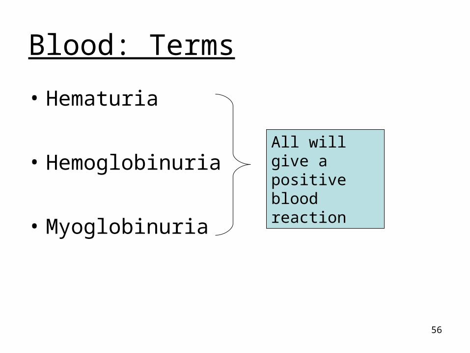
Blood: Terms
• Hematuria
• Hemoglobinuria
• Myoglobinuria
All will give a positive blood reaction
57
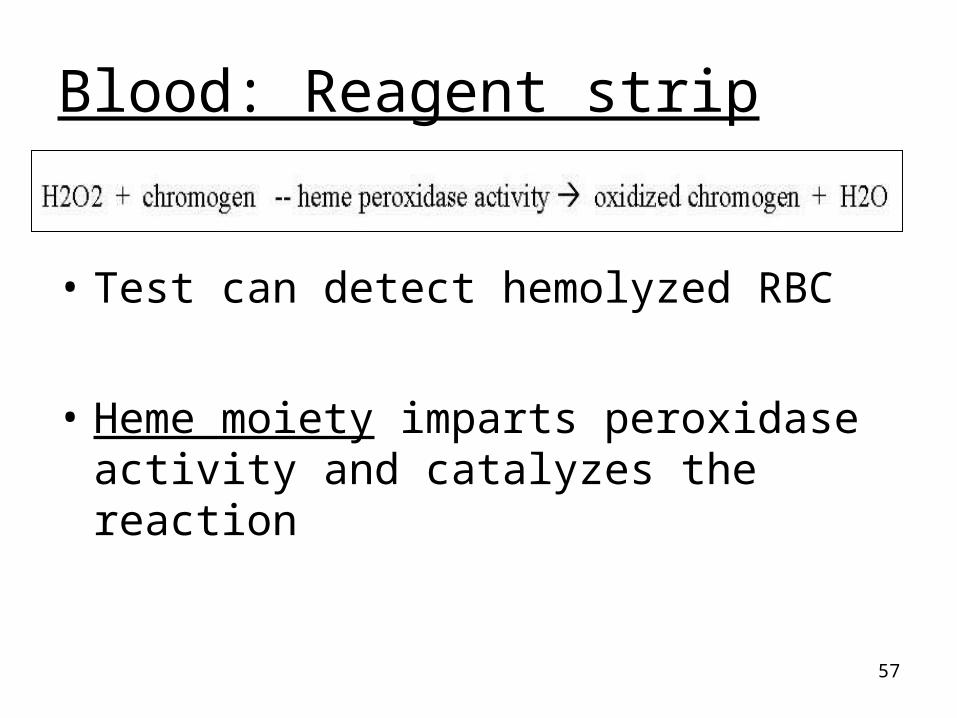
Blood: Reagent strip
• Test can detect hemolyzed RBC
• Heme moiety imparts peroxidase activity and catalyzes the reaction
58
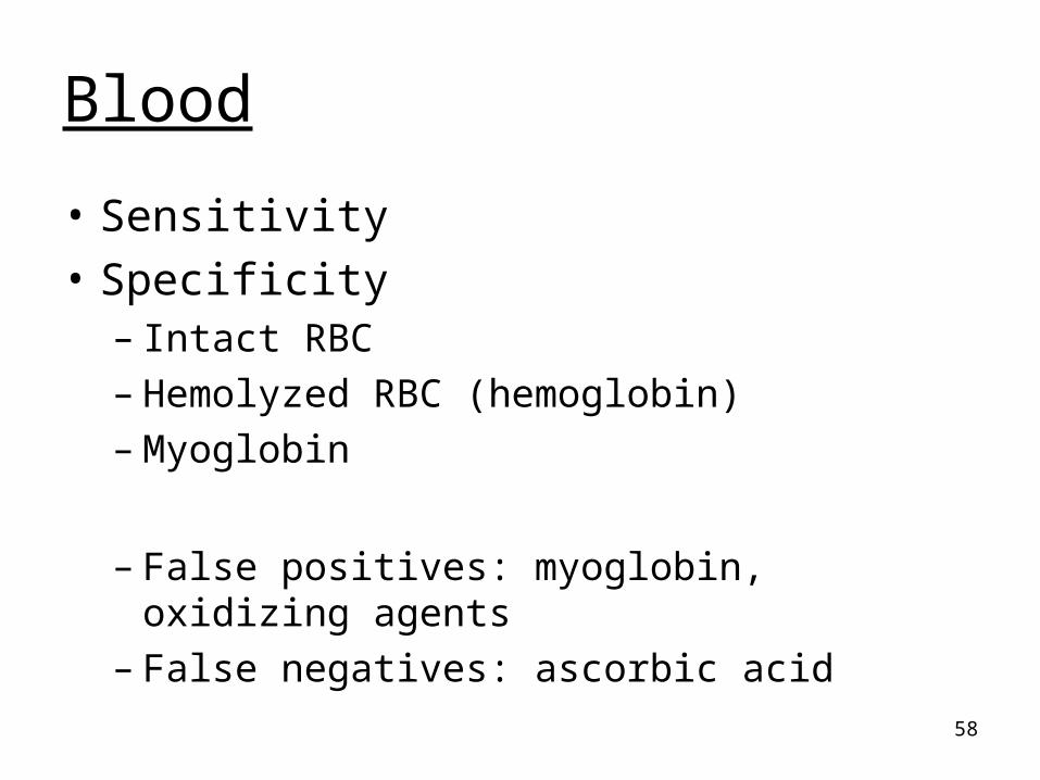
Blood
• Sensitivity
• Specificity– Intact RBC– Hemolyzed RBC (hemoglobin)– Myoglobin
– False positives: myoglobin, oxidizing agents– False negatives: ascorbic acid
59
Blood:
• Correlate reagent strip results– Microscopic findings– Color and clarity

60
Bilirubin and Urobilinogen
• Bilirubin in urine is always pathologic:
liver disease
• Urobilinogen in urine: normal to have a small amount:
0.2 – 1.0 mg/dl
61
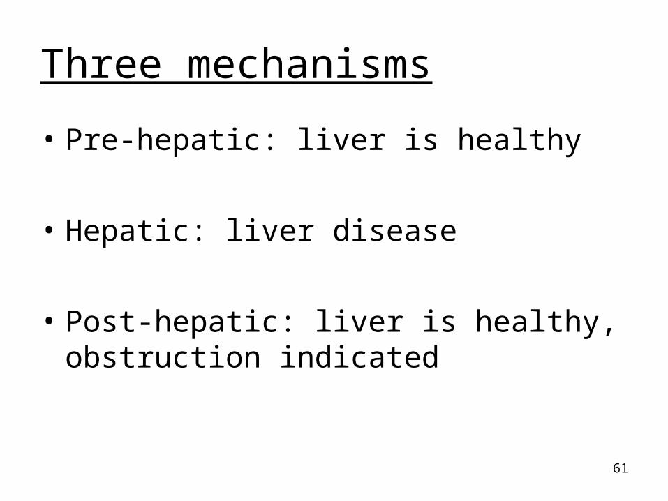
Three mechanisms
• Pre-hepatic: liver is healthy
• Hepatic: liver disease
• Post-hepatic: liver is healthy, obstruction indicated
62
Bilirubin: Methods
• Reagent strip
• Ictotest: tablet test
• Foam test

63
Bilirubin: Methods
• Reagent strip
• Ictotest: tablet test
• Same reaction
• Same specificity: conjugated bilirubin– False positive: urine color– False negative: low concentration, ascorbic
acid, improper specimen handling
64
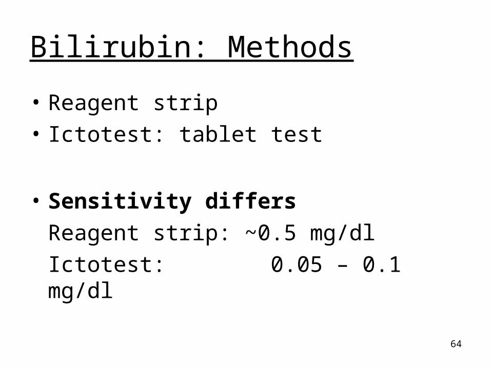
Bilirubin: Methods
• Reagent strip
• Ictotest: tablet test
• Sensitivity differs
Reagent strip: ~0.5 mg/dl
Ictotest: 0.05 – 0.1 mg/dl

65
Bilirubin: Methods
• Possible to have a negative reagent strip test and positive ictotest– Difference in sensitivity levels
• Always perform Ictotest when– Urine bilirubin test specifically ordered– Urine appearance is amber: even if bilirubin
reagent strip test is negative– Positive reagent strip test

66
Bilirubin: Foam Test
• Shake urine and observe resulting foam
• Yellow foam = bilirubin

67
Urobilinogen: Methods
• Reagent strip test– Two reactions dependent upon manufacturer
• Para-dimethylaminobenzaldehyde• Diazonium salt
– Cannot determine absence of UBG
• Watson-Schwartz assay

68
Urobilinogen: Methods• Para-dimethylaminobenzaldehyde
– Sensitivity: 0.2 mg/dl– Specificity:
• False positive: any ‘Ehrlich reactive compound’; color masking; urine at body temp
• False negative: improper specimen handling
• Diazonium salt– Sensitivity: 0.4 mg/dl– Specificity: reacts only with UBG
• False positive: color masking• False negative: improper specimen handling
69
Urobilinogen: Watson Schwartz
• Classic method used to differentiate
urobilinogen from porphobilinogen using a
differential extraction method
• Para-dimethylaminobenzaldehyde
70
Nitrite: Purpose
• Bacteria that contain a specific enzyme can reduce dietary nitrates to nitrites
• Rapid screening test for UTI
71
Nitrite: Normal
• Normal: negative
• Abnormal:– Cystitis: bladder– Pyelonephritis: kidney

72
Nitrite: Method• Reagent strip test
Nitrite + aromatic amine diazonium salt
Diazonium salt + aromatic compound pink color
• Sensitivity: 0.06-0.1 mg/dl nitrite
~ 10,000 organisms

73
Nitrite: Method• Reagent strip test
Specificity:– False positive: improper specimen handling; color
masking– False negative: bacteria cannot reduce nitrates
Bladder time not sufficient: need 4 hours
Low nitrite levels
Ascorbic acid
Antibiotic inhibition of bacteria
Further reduction of nitrites to nitrogen
74
Leukocyte Esterase: Purpose
• Increased WBC in urine is pathologic– Indicates inflammation, infection
• Neutrophils most common type of WBC found in urine
• Can detect intact WBC and lysed WBC
75
Leukocyte Esterase: Normal
• Normal: negative
• Abnormal:– Bacterial infection:
cystitis, pyelonephritis, urethritis– Non-bacterial infection: yeast, trichomonas

76
Leukocyte Esterase: Method
• Reagent strip:
Granules in cytoplasm of WBC contain an enzyme (esterase)
Ester –esterase aromatic compound
Aromatic compound + diazonium salt Purple colored complex

77
Leukocyte Esterase
• Sensitivity: 5-15 WBC/hpf
• Specificity:– False positive: vaginal contamination; color
masking– False negative: strong oxidizing agents
(bleach); lymphocytes (no granules)

78


![Electrophoresis Lecture Ricki PPT 10 [Compatibility Mode]](https://cdn.vdocuments.mx/doc/165x107/55cf96bd550346d0338d72bc/electrophoresis-lecture-ricki-ppt-10-compatibility-mode.jpg)


