1
Copyright © 2009 Pearson Education, Inc.
CAMPBELL
BIOLOGYReece • Urry • Cain • Wasserman • Minorsky • Jackson
© 2014 Pearson Education, Inc.
TENTH
EDITION
CAMPBELL
BIOLOGYReece • Urry • Cain • Wasserman • Minorsky • Jackson
© 2014 Pearson Education, Inc.
TENTH
EDITION
27Bacteria and Archaea
Lecture Presentation by
Dr Burns
NVC Biol 120
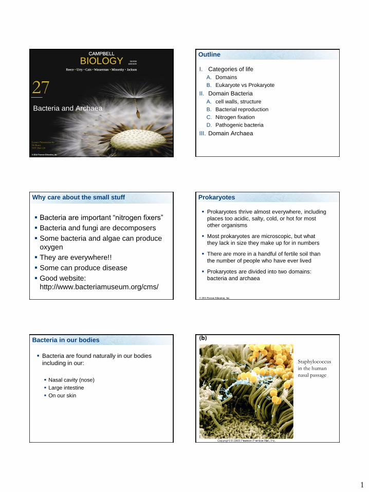
Outline
I. Categories of life
A. Domains
B. Eukaryote vs Prokaryote
II. Domain Bacteria
A. cell walls, structure
B. Bacterial reproduction
C. Nitrogen fixation
D. Pathogenic bacteria
III. Domain Archaea
Why care about the small stuff
Bacteria are important “nitrogen fixers”
Bacteria and fungi are decomposers
Some bacteria and algae can produce
oxygen
They are everywhere!!
Some can produce disease
Good website:
http://www.bacteriamuseum.org/cms/
Prokaryotes
Prokaryotes thrive almost everywhere, including
places too acidic, salty, cold, or hot for most
other organisms
Most prokaryotes are microscopic, but what
they lack in size they make up for in numbers
There are more in a handful of fertile soil than
the number of people who have ever lived
Prokaryotes are divided into two domains:
bacteria and archaea
© 2011 Pearson Education, Inc.
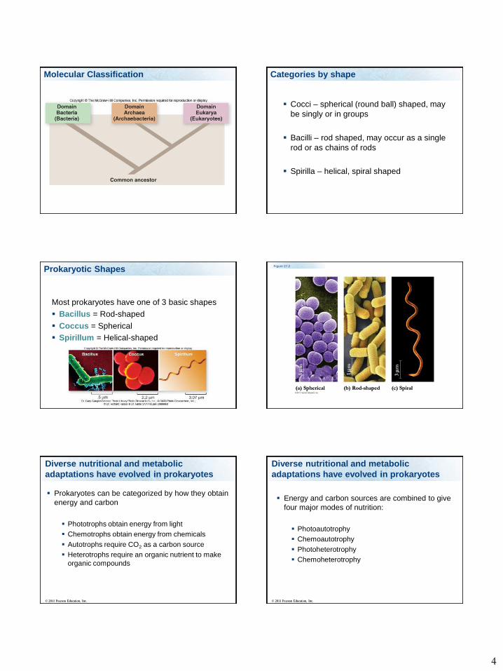
Bacteria in our bodies
Bacteria are found naturally in our bodies
including in our:
Nasal cavity (nose)
Large intestine
On our skin
Staphylococcus
in the human
nasal passage

2
Euprymna Scolopes have a symbiotic
relationship with luminescent bacteria.
Photo courtesy of Margaret McFall-Ngai.
Do prokaryotes have a nucleus
1. Yes
2. No
Yes N
o
50%50%
Are Domain Bacteria and Domain Archaea
prokaryotic or eukaryotic?
1. prokaryotic
2. eukaryotic
pro
kary
otic
euka
ryotic
50%50%
The First Cells
Microfossils are fossilized forms of microscopic
life
-Oldest are 3.5 billion years old
The First Cells
Stromatolites are mats of cyanobacterial cells
that trap mineral deposits
-Oldest are 2.7 billion years old
Prokaryotes
Domain Archaea – live in extreme
environments
Domain Bacteria (Eubacteria)
Microorganisms are any small organism – too
small to see with the naked eye include
Pathogens – cause disease
Most bacteria are not pathogens

3
Prokaryotic Characteristics
1. Unicellular
2. No membrane-bound nucleus, single
chromosome and histone-like proteins found
in nucleoid region
3. Lack membrane bound organelles
4. Contain ribosomes
Prokaryotic Characteristics
5. Many have flagella
6. Cell wall present in many species
7. Have plasma membrane
8. Reproduction by prokaryotic fission
9. Great metabolic diversity
Prokaryotes
No membrane bound organelles including:
nucleus, mitochondria, chloroplasts,
endoplasmic reticulum, golgi complex,
lysosomes.
They do contain small ribosomes, storage
granules, plasma membrane may be folded
Domain Bacteria
Prokarotes
Categorized by
1. Shape
2. Cell wall type
3. Where they get their energy and their
nutrients from.
4. Motility
Based on these molecular data, several
prokaryotic groupings have been proposed
Bergey’s Manual of Systematic Bacteriology
Contains about 7,000 bacterial and archaeal
species
The three-domain (Woese) system of phylogeny
is based on rRNA sequences
Molecular Classification
4
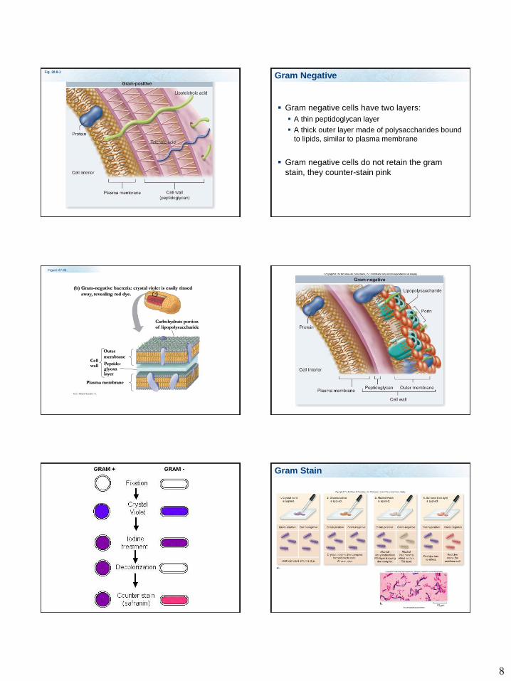
Molecular Classification Categories by shape
Cocci – spherical (round ball) shaped, may
be singly or in groups
Bacilli – rod shaped, may occur as a single
rod or as chains of rods
Spirilla – helical, spiral shaped
Most prokaryotes have one of 3 basic shapes
Bacillus = Rod-shaped
Coccus = Spherical
Spirillum = Helical-shaped
Prokaryotic ShapesFigure 27.2
(a) Spherical (b) Rod-shaped (c) Spiral
1
m
1
m
3
m
Diverse nutritional and metabolic
adaptations have evolved in prokaryotes
Prokaryotes can be categorized by how they obtain
energy and carbon
Phototrophs obtain energy from light
Chemotrophs obtain energy from chemicals
Autotrophs require CO2 as a carbon source
Heterotrophs require an organic nutrient to make
organic compounds
© 2011 Pearson Education, Inc.
Energy and carbon sources are combined to give
four major modes of nutrition:
Photoautotrophy
Chemoautotrophy
Photoheterotrophy
Chemoheterotrophy
© 2011 Pearson Education, Inc.
Diverse nutritional and metabolic
adaptations have evolved in prokaryotes

5
Table 27.1
Nitrogen Metabolism
Nitrogen is essential for the production of amino
acids and nucleic acids
Prokaryotes can metabolize nitrogen in a variety
of ways
In nitrogen fixation, some prokaryotes convert
atmospheric nitrogen (N2) to ammonia (NH3)
© 2011 Pearson Education, Inc.
Nitrogen Fixation
Some bacteria are able to take nitrogen gas and
fix it in the form that can be used by plants
Rhizobium fixes nitrogen for plants like
legumes.
Legumes and Rhizobium live in a mutualistic
relationship
Rhizobium provide usable nitrogen
Legumes provide sugar
Rhizobium live inside legume cells in nodules
Metabolic Cooperation
Cooperation between prokaryotes allows them
to use environmental resources they could not
use as individual cells
In the cyanobacterium Anabaena,
photosynthetic cells and nitrogen-fixing cells
called heterocysts (or heterocytes) exchange
metabolic products
© 2011 Pearson Education, Inc.
Figure 27.14
Photosynthetic
cells
Heterocyst
20 m

6
Photoautotroph - cyanobacteria
31
These organisms use the energy from the sun to fix
carbon into a sugar from CO2
1. Photoheterotrophs
2. Chemoheterotrophs
3. Photoautotrophs
4. Chemoautotrophs
Photo
hete
rotrophs
Chem
ohete
rotrophs
Photo
auto
troph
s
Chem
oauto
troph
s
25% 25%25%25%
These organisms use the energy from inorganic
compounds to fix carbon into a sugar from CO2
1. Photoheterotrophs
2. Chemoheterotrophs
3. Photoautotrophs
4. Chemoautotrophs
Photo
hete
rotrophs
Chem
ohete
rotrophs
Photo
auto
troph
s
Chem
oauto
troph
s
25% 25%25%25%
34
Cell-Surface Structures
An important feature of nearly all prokaryotic cells is
their cell wall, which maintains cell shape, protects
the cell, and prevents it from bursting in a hypotonic
environment
Eukaryote cell walls are made of cellulose or chitin
Bacterial cell walls contain peptidoglycan, a
network of sugar polymers cross-linked by
polypeptides
© 2011 Pearson Education, Inc.
Cell Wall
Prokaryotic cell plasma membranes are
usually covered with a cell wall. Keeps cell
from bursting in hypotonic solutions
Eubacteria cell walls contain peptidoglycan,
a combination of amino acids and sugars.

7
Cell Wall
Some species have a capsule surrounding the
cell wall. The capsule can provide protection
against immune system cells (phagocytes)
Some bacteria have pili – hair like structures
made of protein, help bacteria to adhere to
surfaces
Some pili are involved in transmitting DNA
between bacteria
Cell Walls
Archaea contain polysaccharides and
proteins but lack peptidoglycan
Scientists use the Gram stain to classify
bacteria by cell wall composition
Gram-positive bacteria have simpler walls
with a large amount of peptidoglycan
Gram-negative bacteria have less
peptidoglycan and an outer membrane that
can be toxic
© 2011 Pearson Education, Inc.
Cell Wall Types
There are two main cell wall types. The
differences have important implications in
treating the bacteria with antibiotics
Gram Positive
Gram Negative
40
The Bacterial Cell Wall Types
Gram positive
Gram positive cells have thick cell walls,
consisting mainly of peptidoglycan.
Gram positive cells absorb the violet “gram
stain”
Figure 27.3a
(a) Gram-positive bacteria: peptidoglycan traps crystal violet.
Peptido-
glycan
layer
Cell
wall
Plasma
membrane
8
Fig. 28.8-1
Gram Negative
Gram negative cells have two layers:
A thin peptidoglycan layer
A thick outer layer made of polysaccharides bound
to lipids, similar to plasma membrane
Gram negative cells do not retain the gram
stain, they counter-stain pink
Figure 27.3b
Outer
membrane
Peptido-glycanlayer
Plasma membrane
Cellwall
Carbohydrate portion
of lipopolysaccharide
(b) Gram-negative bacteria: crystal violet is easily rinsed
away, revealing red dye.
48
Gram Stain

9
49
External Layers
Capsule
A gelatinous layer found in some bacteria
Aids in attachment
Protects from the immune system
Motility
Most motile bacteria propel themselves by
flagella scattered about the surface or
concentrated at one or both ends
Flagella of bacteria, archaea, and eukaryotes
are composed of different proteins and likely
evolved independently
© 2011 Pearson Education, Inc.
© 2011 Pearson Education, Inc.
Video: Prokaryotic Flagella (Salmonella typhimurium)
Figure 27.6
Flagellum
Hook
Motor
Filament
RodPeptidoglycanlayer
Plasmamembrane
Cell wall
20 nm
53
Flagella
Flagella
Long, helical structures
Composed of the protein flagellin
Involved in locomotion
Motility
Most prokaryotes have a flagella
These flagella are not like eukaryotic flagella,
they are not composed of microtubules
Instead it has:
Basal body that acts like a motor
Hook
Filament
Protein is flagellin
10
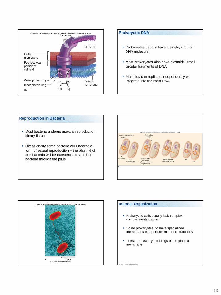
Prokaryotic DNA
Prokaryotes usually have a single, circular
DNA molecule.
Most prokaryotes also have plasmids, small
circular fragments of DNA.
Plasmids can replicate independently or
integrate into the main DNA
Reproduction in Bacteria
Most bacteria undergo asexual reproduction =
binary fission
Occasionally some bacteria will undergo a
form of sexual reproduction – the plasmid of
one bacteria will be transferred to another
bacteria through the pilus
Internal Organization
Prokaryotic cells usually lack complex compartmentalization
Some prokaryotes do have specialized membranes that perform metabolic functions
These are usually infoldings of the plasma membrane
© 2011 Pearson Education, Inc.
11
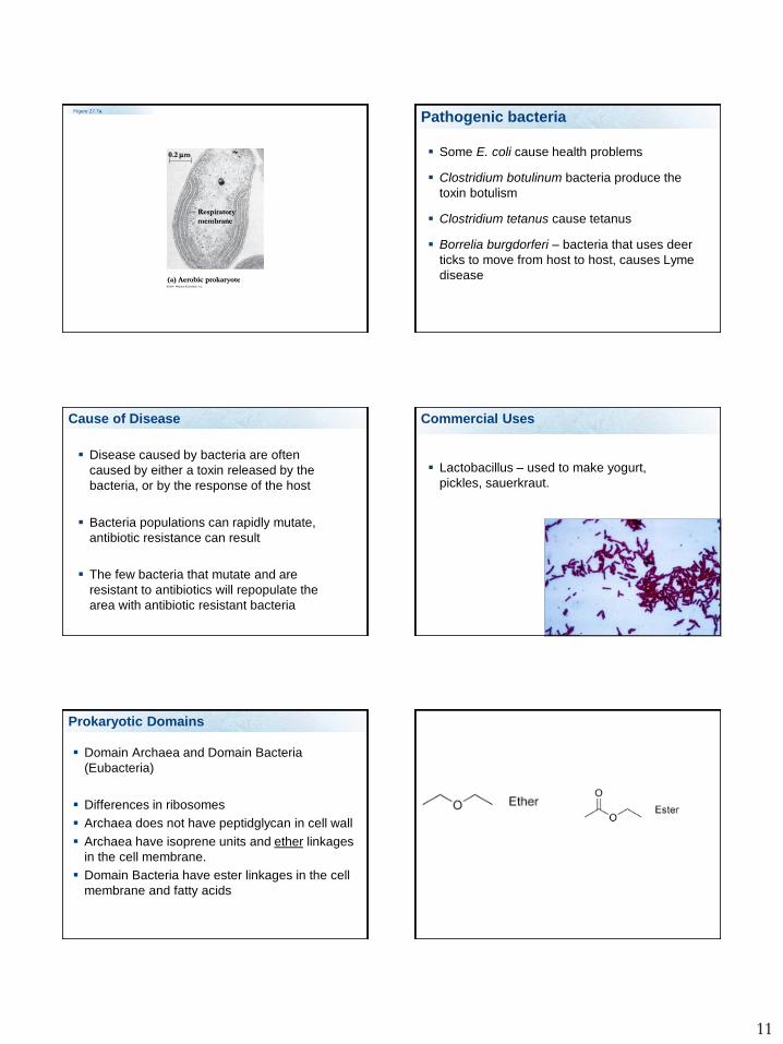
Figure 27.7a
(a) Aerobic prokaryote
Respiratory
membrane
0.2 m
Pathogenic bacteria
Some E. coli cause health problems
Clostridium botulinum bacteria produce the
toxin botulism
Clostridium tetanus cause tetanus
Borrelia burgdorferi – bacteria that uses deer
ticks to move from host to host, causes Lyme
disease
Cause of Disease
Disease caused by bacteria are often
caused by either a toxin released by the
bacteria, or by the response of the host
Bacteria populations can rapidly mutate,
antibiotic resistance can result
The few bacteria that mutate and are
resistant to antibiotics will repopulate the
area with antibiotic resistant bacteria
Commercial Uses
Lactobacillus – used to make yogurt,
pickles, sauerkraut.
Prokaryotic Domains
Domain Archaea and Domain Bacteria
(Eubacteria)
Differences in ribosomes
Archaea does not have peptidglycan in cell wall
Archaea have isoprene units and ether linkages
in the cell membrane.
Domain Bacteria have ester linkages in the cell
membrane and fatty acids

12
Plasma membrane Table 27.2
Domain Archaea
Prokaryotic – no nucleus
Extreme bacteria
Methanogens (methane makers)
Extreme halophiles (salt loving)
Extreme thermophiles (heat lovers)
Methanogens example
Discovered in 1983 contains methanococcus jannaschii
[©Stan Watson, Woods Hole Oceanographic Institute]
Pacific Ocean thermal vent
70
Halophile example
Dunaliella salina
Thermophile example
Yellowstone NP
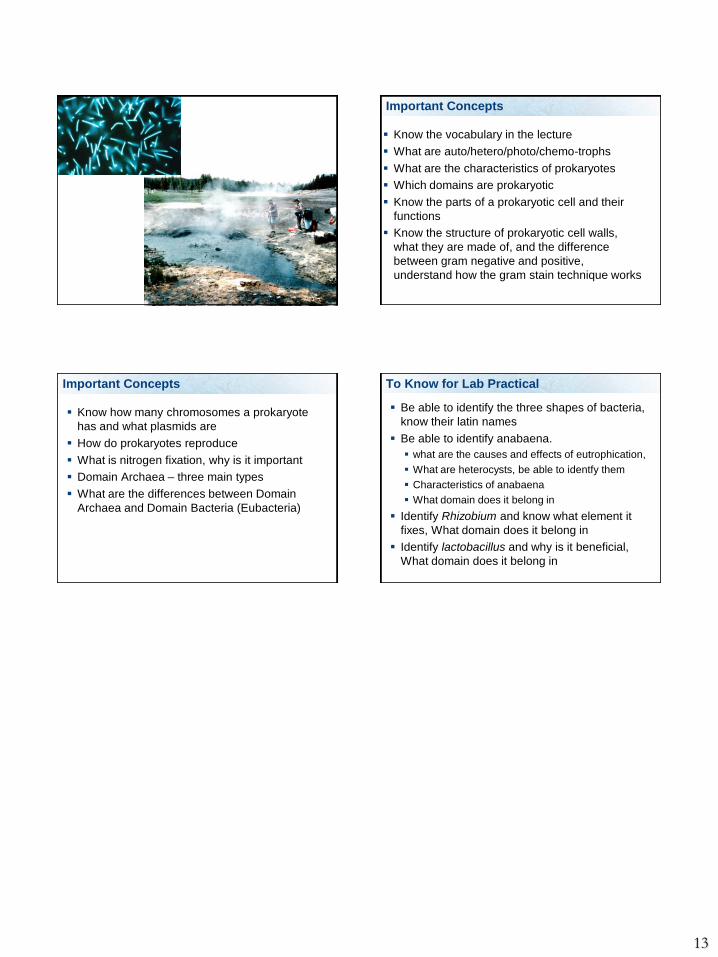
13
Important Concepts
Know the vocabulary in the lecture
What are auto/hetero/photo/chemo-trophs
What are the characteristics of prokaryotes
Which domains are prokaryotic
Know the parts of a prokaryotic cell and their
functions
Know the structure of prokaryotic cell walls,
what they are made of, and the difference
between gram negative and positive,
understand how the gram stain technique works
Important Concepts
Know how many chromosomes a prokaryote
has and what plasmids are
How do prokaryotes reproduce
What is nitrogen fixation, why is it important
Domain Archaea – three main types
What are the differences between Domain
Archaea and Domain Bacteria (Eubacteria)
To Know for Lab Practical
Be able to identify the three shapes of bacteria,
know their latin names
Be able to identify anabaena.
what are the causes and effects of eutrophication,
What are heterocysts, be able to identfy them
Characteristics of anabaena
What domain does it belong in
Identify Rhizobium and know what element it
fixes, What domain does it belong in
Identify lactobacillus and why is it beneficial,
What domain does it belong in


