
Biosynthesis of all-trans-Retinoic Acid and Regulation of Retinoids Homeostasis in
Primary Hippocampal Astrocytes and Neurons
By
Chao Wang
A dissertation submitted in partial satisfaction of the
requirements for the degree of
Doctor of Philosophy
in
Molecular and Biochemical Nutrition
in the
Graduate Division
of the
University of California, Berkeley
Committee in charge:
Professor Joseph L. Napoli, Chair
Professor Barry Shane
Professor Daniela Kaufer
Fall 2010

Biosynthesis of all-trans-Retinoic Acid and Regulation of Retinoids Homeostasis in
Primary Hippocampal Astrocytes and Neurons
Copyright 2010
By
Chao Wang

1
Abstract
Biosynthesis of all-trans-Retinoic Acid and Regulation of Retinoids Homeostasis in
Primary Hippocampal Astrocytes and Neurons
by
Chao Wang
Doctor of Philosophy in Molecular and Biochemical Nutrition
University of California, Berkeley
Professor Joseph L. Napoli, Chair
All-trans-retinoic acid stimulates neurogenesis, dendritic growth of hippocampal neurons
and higher cognitive functions, such as spatial learning and memory formation. Although
astrocyte-derived atRA has been considered a key factor in neurogenesis, little direct
evidence identifies hippocampus cell types and the enzymes that biosynthesize atRA. Nor
has any factor been reported to regulate atRA biosynthesis in adult CNS. Here we show
that primary rat astrocytes, but not neurons, biosynthesize atRA using multiple retinol
dehydrogenases (Rdh) of the short-chain dehydrogenase/reductase (SDR) gene family
and retinaldehyde dehydrogenases (Raldh). Astrocytes secrete atRA into their medium;
neurons sequester atRA. The first step, conversion of retinol into retinal, is rate-limiting
and the second step, conversion of retinal to atRA, is much more active and usually
affected in response to different stimulis. Both neurons and astrocytes can synthesize
retinyl esters and reduce retinal into retinol. siRNA knockdown indicates that Rdh10,
Rdh2 (mRdh1), and Raldh 1, 2 and 3 contribute to atRA production. Knockdown of the
Rdh Dhrs9 increased atRA synthesis ~40% by increasing Raldh1 expression.
Immunocytochemistry revealed cytosolic and nuclear expression of Raldh1 and cytosol
and perinuclear expression of Raldh2. atRA autoregulated its concentrations by inducing
retinyl ester synthesis via lecithin:retinol acyltransferase and stimulating its catabolism
via inducing Cyp26B1. Raldh1, Raldh2, Rdh2, Rdh10 and Dhrs9 increase their
expression as the elongation of in vitro culture time. Rdh1-/- and CrbpI-/- mice astrocytes
showed similar changes of retinoids metabolism except RE formation and partially
overlap genes compensation. Though shown a broad and strong expression pattern in
pure cultured astrocytes, Raldh1 expression dramatically dropped in astrocytes mixed
cultured with neurons or in hippocampus in vivo. In contrast, Raldh1 is widely expressed
in cultured neurons, with special intense signals on axons. CA1 neurons and mossy fibers
enriched Raldh1 expression pattern was confirmed as a postnatal development process by
immunohistochemistry. As a proinflammatory cytokine, TNFα oppositely down regulate
Raldh1 expression via JNK and MAPK pathway and up regulate Raldh3 expression
partially through P38 pathway, which resulted in different overall effect on atRA

2
biosynthesis in young and old astrocytes. These data show that adult hippocampus
astrocytes rely on multiple Rdh and Raldh to provide a paracrine source of atRA to
neurons, and atRA regulates its own biosynthesis in astrocytes directing flux of retinol.
Besides redundancy, different Rdh and Raldh may have unique function according to cell
types and/or subcellular locations. Cross talk between first and second step
dehydrogenation indicate a novel regulation mechanism that control the atRA
homeostasis in astrocytes.

i
Table of Contents
Abstract ......................................................................................................................................... 1
Table of Contents ............................................................................................................. i
List of Figures and Tables ............................................................................................... iii
Acknowledgement ..............................................................................................................v
Chapter I Literature Review
Part I General Review of Retinoids Metabolism ........................................................ 1
Vitamin A and retinoic acid .............................................................................................1
Structure and retinoid property ......................................................................................2
Retinoid uptake and processing .......................................................................................2
Cellular retinoid metabolism ..........................................................................................5
Cellular uptake and processing retinol ....................................................................... 5
ADHs ............................................................................................................................6
SDRs .............................................................................................................................7
Retinal dehydrogenases ................................................................................................9
Cellular retinoic acid binding proteins .......................................................................10
Retinoic acid degradation--cytochrome P450 enzymes .............................................10
RA receptors ...............................................................................................................11
RA target genes-possible autoregulation pathway .....................................................12
Part II Retinoic Acid Signaling and Function in Adult CNS ................................... 13
Overview ........................................................................................................................13
RA signaling components in adult CNS .................................................................... 13
Physiological function of RA in CNS- learning and synaptic plasticity .................... 14
RA and song learning .................................................................................................14
Physiological function of RA signaling in adult hippocampus .............................. 14
Pathological association of RA signaling on CNS diseases ....................................... 16
RA source in adult hippocampus ............................................................................... 18

ii
Part III Regulation of RA biosynthesis by hormones, cytokines or endotoxins ..... 19
Estrogen and RA signaling ......................................................................................... 19
LPS, PGE2, TNFα and RA signaling ......................................................................... 20
References ..................................................................................................................... 22
Chapter II Multiple Retinol and Retinal Dehydrogenases Catalyze
All-trans-Retinoic Acid Biosynthesis in Astrocytes
Introduction .................................................................................................................... 34
Materials and Methods ................................................................................................... 36
Results .............................................................................................................................. 39
Discussion ........................................................................................................................ 46
Figures and Legends ...................................................................................................... 50
References ...................................................................................................................... 69
Chapter III Localization in vitro and in vivo of RALDH1 Expression
Introduction .................................................................................................................... 74
Materials and Methods ................................................................................................... 76
Results .............................................................................................................................. 78
Discussion ........................................................................................................................ 81
Figures and Legends ...................................................................................................... 85
References ...................................................................................................................... 92
Chapter IV TNFα Regulates all-trans-RA Biosynthesis in Astrocytes through
Oppositely Changing RALDH1 and RALDH3 Expression
Introduction .................................................................................................................... 94
Materials and Methods ................................................................................................... 96
Results .............................................................................................................................. 97
Discussion ...................................................................................................................... 100
Figures and Legends .................................................................................................... 103
References .................................................................................................................... 108
Chapter V Summary and Future Directions
Part I Retinoids metabolism in cultured astrocytes and neurons ................................... 110
Part II Enzymes that participate in the retinoids homeostasis in CNS ........................ 113
Part III Future directions ................................................................................................ 115
Part IV Conclusion ........................................................................................................... 116
Figures and Legends .................................................................................................... 118

iii
References .................................................................................................................... 119
List of Figures and Tables
Figure II-1 Time course of atRA and RE biosynthesis from retinol by primary
astrocytes
50
Figure II-2 Effects of substrate concentrations on retinoid metabolism in
primary astrocytes
51
Figure II-3 Retinoid metabolism catalyzed by primary hippocampus neurons 52
Figure II-4 Co-cultured with neurons changed the atRA distribution
proportion in medium and cells
53
Figure II-5 Neurons sequester atRA secreted by astrocytes. 54
Figure II-6 Knockdown reveals Raldh that contribute to atRA biosynthesis 56
Figure II-7 Confocal images of Raldh1 and 2 expression 58
Figure II-8 Multiple Rdh contribute to atRA biosynthesis in astrocytes 60
Figure II-9 Dhrs9 functions as a retinol dehydrogenase in astrocytes 61
Figure II-10 atRA induces Cyp26B1 and Lrat mRNA in astrocytes 63
Figure II-11 Effect of culture duration on retinoid metabolism by primary
astrocytes
65
Figure II-12 Compensation effect of atRA biosynthesis in Rdh1-/- and
CrbpI-/- astrocytes
66
Figure II-13 Retinol homeostasis in astrocytes and neurons 67
Table II-1 Rate constants for retinol metabolism 68
Figure III-1 RALDH1 expression in primary cultured neuronal cells 85
Figure III-2 RALDH1 expression in adult rat hippocampus 86
Figure III-3 RALDH1 expression in astrocytes and oligodendrocytes in
hippocampus
87

iv
Figure III-4 RALDH1 is expressed in dendrites of CA1 but not CA3 neurons 88
Figure III-5 RADLH1 is co-localized with axons present in CA3 region 89
Figure III-6 RALDH1 is expressed on mossy fibers 90
Figure III-7 Developmental changes of RALDH1 expression in adult rat
hippocmapus
91
Figure IV-1 atRA production from retinol in 1 month old astrocytes remains
stable upon different stimulis.
103
Figure IV-2 TNFα affect retinal dehydrogenation in astrocytes through
oppositely changing RALDH1 and RALDH3 expression
104
Figure IV-3 TNFα induces similar change of RALDH1 and RALDH3
expression but different atRA production in 2weeks and 3months
old astrocytes
106
Figure IV-4 TNFα affect RALDH1 expression via JNK and MEK pathway;
RALDH3 expression partially via P38 pathway
107
Figure V-1 Retinoids homeostasis model in adult hippocampus 118

v
Acknowledgement
It’s my pleasure to acknowledge the contributions of many people to this work. I have
been blessed with generous help and support from them, to whom I would like to express
my deepest gratitude.
First and foremost, I would like to give my sincere gratitude to my PI, Dr. Joseph L.
Napoli, for giving me the opportunity to study this important and meaningful project, for
his keen wisdom, solid support and generous guidance during my past 6 years Ph.D.
research. I truly appreciate Dr. Barry Shane for serving on all my committees and for his
thoughtful concerns and suggestions on my projects. I am very grateful to have Dr.
Daniela Kaufer on my dissertation committee. I thank Dr. Kaufer for informative
suggestions on Raldh1 expression project and generous sharing of specific antibodies. I
very much appreciate Dr. Nancy Amy, Dr. Leonard F. Bjeldances, Dr. Robert O. Ryan and
Dr. John Ngai for serving on my qualify exam committee and providing important
guidance on this projects.
I would like to give my special thank my lab member Dr. Maureen Kane for her
altruistic help on measurement of retinoids and collaboration on the ethanol projects. The
training and guidance I received from Dr. Kane was the footstone for my whole project.
Her accurate and timely data analysis, patience to answer my questions and quickly
response to my requirements insure the progress of my project. I also want to thank Dr.
Na Chen for her patient guidance on primary cell culture and basic experimental
technologies; Dr. Charles Krois for assistance on animal handling and Dr. Kristin
Obrochta for help on immunohistochemistry. I also greatly appreciate all other colleges,
Sunny Wang, Nan Li, Alexandra Folias, Weiya Jiang, Min Zhang, Peirong Hu, James
Chithalen, Wentao Hao, Hua Tran, for sharing their knowledge and expertise, giving
suggestions and enjoying the fun of research.
I want to give special thanks to Dr. Xiao Shu and Dr. Aaron Li for being such wonderful
friends, not only giving me generous help on my research but also sharing all the joy and
despair, failure and success during my six years of life in Berkeley. I am deeply grateful
for the help and friendships from Roger Wong, Nan Wang, Su Yan, Yuyang Tian, Maryam
Ahmadian, Brie Fuqua, Seung-Min Lee and many others.
Last but not least, I would like express my deepest gratitude to my parents. Their
unconditional love and support have been my powerful and secure backing all the times
in my life.

1
Chapter I
Literature Review
Part I General Review of Retinoid Metabolism
Vitamin A and retinoic acid
Vitamin A, a fat-soluble vitamin, is essential for most forms of life, especially in
chordates. The vitamin has numerous functions information and maintenance of many
body tissues, for example, skin, bone, vasculature, and eye as well as the immune system
(Blomhoff and Blomhoff, 2006; Evans and Kaye, 1999; Kim, 2008). Vitamin A is
indispensable for normal vision, maintenance of epithelial cells, immune competence,
reproduction and embryo development and growth, and the maintenance and regulation
of both embryonic and adult central nervous system (Theodosiou et al., 2010). Vitamin A
is converted into an activated metabolite, all-trans-retinoic acid (atRA), through which it
performs its multiple effects on embryogenesis, cell proliferation, differentiation and
apoptosis (Petkovich, 2001). atRA is a rapid diffusing signaling molecule and exerts its
functions by controlling gene expression through activation of specific nuclear receptors
(Blomhoff and Blomhoff, 2006).
In humans, lower vitamin A intake than needed, impairs vision, featured by
xerophthalmia and even blindness in severe situations (Bendich and Langseth, 1989).
Deficiency of vitamin A (VAD) also increases susceptibility to infectious diseases,
because of impaired immune resistance (Kim, 2008). VAD is a serious problem in
developing countries. The WHO estimates that between 140 and 250 million preschool
children are at the risk of subclinical vitamin A deficiency, 3 million are clinically
vitamin A deficient and 1 million childhood deaths are associated with vitamin A
deficiency annually (Organization, 1995).
In contrast, vitamin A could function as a double-edged sword if uptake is excessive.
Excess dietary vitamin A, sometimes even marginally above the RDI (recommended
dietary intake) can result in damage to liver, skin, internal organs, the musculoskeletal
system, the central nervous system and embryo development (Blomhoff and Blomhoff,
2006; Collins and Mao, 1999). Hypervitaminosis A causes embryonic malformations,
reduced bone mineral density, and increased hip fractures (Melhus et al., 1998; Rothman
et al., 1995). Hypervitaminosis A results from large intake of dietary vitamin A, from
animal liver for example, or from drugs containing retinoids (synthetic or naturally
occurring compounds with vitamin A activity).
The function of vitamin A in the visual cycle was first revealed decades ago. 11-cis
-Retinal serves as the chromophore of visual pigment (Wald, 1968). Early research also
found that either hyper or hypo-vitaminosis A affect differentiation of epithelial cells and
atRA is a powerful signaling molecules in chordates (Wolbach and Howe, 1925). The

2
mechanism of atRA action was not resolved until retinoic acid receptors (RAR) were
discovered in 1987, which demonstrated that atRA affects gene expression through
nuclear transcription factors (Giguere et al., 1987). However, this is not the only
mechanism through which vitamin A exerts its biological activity. Additional mechanisms
of vitamin A actions have been identified, as detailed below.
Structure and retinoid property
Vitamin A is the term that designates any compound processing the biological activity
supported by retinol (after conversion into metabolites such as atRA). Some of synthetic
compounds, even though they don‘t have a retinol-like structure, have greater activity in
some assays of vitamin A function. So Sporn and Roberts proposed that ‗a retinoid should
be defined as a substance that can elicit specific biologic responses by binding to and
activating a specific receptor or set of receptors‘. Today, most researchers accept a
combination of definitions that the class of retinoids consists of RA analogs (with or
without biologic activity) and several compounds not structural close to RA that can exert
the biological activity of vitamin A.
All-trans-retinol is the parent retinoid compound containing a primary alcohol that can
be oxidized to other active retinoids. Retinyl palmitate is the predominant retinoid in
most animal tissues. However, other forms of fatty acid esters, such as retinyl oleate and
retinyl stearate also exist in natural materials. The all-trans configuration is the major
naturally occurring isomer, but cis configurations are also present in nature; for example,
11-cis-retinal is present in the retina of eye, 13-cis-retinoic acid is present in many tissues.
All retinoids, including retinol and atRA, are highly unstable in the presence of oxygen
and light. Exposure to oxidants or light will lead to degradation or isomerization. This
feature requires that any reagents or biological samples that containing retinoids must be
stored and handled under dim illumination; for example, yellow light causes the least
decay of retinoids (Gundersen and Blomhoff, 2001). In addition, glassware is required
when dealing with retinoids because plastic equipment can easily absorb retinoids, which
might result in shift of experiment results. Due to the instability of retinoids,
well-designed controls are especially required for each experiment.
All-trans-retinol is a fat soluble vitamin. There is increased water solubility from
retinol, retinal to atRA (60, 110, 210 nM) which means atRA can diffuse efficiently in
both aqueous phases, such as plasma and hydrophobic phases, such as membranes
(Blomhoff and Blomhoff, 2006). Most retinoids are bound with specific binding-proteins
during transportation between hydrophilic plasma or extra- and intracellular fluids.
Binding proteins also function in retinoid metabolism, such as the oxidation of retinol or
degradation of atRA (Napoli, 1997).
Retinoid uptake and processing
International units (IUs) are used for nutritional recommendations of vitamin A. The
term ‗retinol equivalents‘ (RE) is used to convert all kinds of performed retinol and
provitamin A carotenoids into a single unit. No animal species have ability to synthesize

3
vitamin A. All dietary vitamin A is from plant and microorganisms or from the tissues of
others animals that have gotten vitamin A in their diets. Carotenoids serve as provitamin
A compounds in plants and are cleaved to form retinoids in animals. Carotenoids are the
pigments responsible for the yellow, orange, red, or purple colors of many vegetables,
fruits and flowers. β-Carotene represents the most efficient precursor of vitamin A. One
β-carotene molecule can be cleaved into two retinal molecules in the small intestine and
further reduced into retinol. Alternatively, animals can obtain retinol by eating animal
tissues that have already converted carotenoids into retinoids. RE (retinyl esters),
particularly palmitate, represent the most abundant storage form, with liver serving as the
main, but not the only tissue for storage. The primary function of retinol and RE is
serving as the precursors of biologically activate retinoids. This process occurs in both
liver and non-hepatic tissues. The most established pathway of retinol metabolism
includes esterification of retinol into RE in liver or other tissues, reversible conversion of
retinol into retinal and then irreversible conversion of retinal into atRA. There are six
isoforms of biologically-active retinol derivatives, including all-trans, 9-cis, 13-cis,
9,13-cis, 11-cis and 11,13-di-cis, with all-trans being the predominant physiological form.
Some, but not all endogenous retinoids with direct biological activity, include: atRA,
11-cis-retinaldehyde, 3,4-didehydro-RA and 9-cis-RA.
In general, carotenoids are absorbed by enterocytes in the small intestine through
passive diffusion. After uptake, β-carotene is cleaved centrally to produce two molecules
of all-trans-retinaldehyde by the enzyme, β-carotenecyclooxygenase-I (BCOI). Dietary
RE is converted into retinol in the intestinal lumen before its uptake by triglyceride lipase.
Retinol is absorbed by enterocytes by a carrier-mediated process.
Retinol, either from β-carotene or RE, is bound to cellular retinol binding protein, type
II (CRBPII) for esterification in enterocytes. CRBPII is expressed primarily in the
absorptive cells in the intestine. CRBPII knock-out mice have reduced retinol uptake, but
when they are kept on a vitamin A-enriched diet, they develop and reproduce normally,
indicating that CRBPII functions in retinol absorption, especially during low amounts of
dietary vitamin A—the normal situation (E et al., 2002). It is generally believed that the
role of CRBPII is to solubilize retinol, protect retinol from degradation and more
importantly, deliver retinol to the esterification enzyme lecithin: retinol acetyltransferase
(LRAT) for conversion into RE. LRAT is widely expressed in many tissues and highly
expressed in the intestine, liver and the retinal-pigmented epithelial cells in eye (Herr and
Ong, 1992). It is the major enzyme that converts all-trans-retinol into RE. LRAT
knock-out mice develop normally with slight changes in the retina. Only trace levels of
RE are found in the liver, kidney and lung; indicating that this enzyme is important for
RE formation. However, there is an increase of RE storage level in adipose tissue in
LRAT-/- mice and during vitamin A deficiency, which are mobilized from adipose tissue
(O'Byrne et al., 2005). In LRAT-/- mice, retinol is absorbed as free retinol into cells, but
RE can be still found in circulation. This kind of retinol esterification is performed via an
acyl-CoA-dependent enzymatic process, which is catalyzed by acyl-CoA: retinol

4
acetyltransferase (ARAT) (Batten et al., 2004).
RE in enterocytes are secreted either into lymph as chylomicrons or into the portal
circulation as unesterified retinol. Chylomicrons consist of aggregates of triacylglycerol
and phospholipids packed together with carotenoids, RE, small amounts of retinol,
cholesterol esters and apolipoproteins. Chylomicrons in general circulation are reduced to
chylomicron remnants that contain high amounts of RE, which are either transported to
target tissues such as bone marrow, spleen, adipose tissue, skeletal muscle and kidney, or
cleared by the parenchymal cells of the liver.
In hepatocytes, RE is hydrolyzed. The released retinol binds with retinol-binding
protein (RBP), which is highly expressed in the hepatocyte endoplasmic reticulum (ER).
Retinol-RBP translocates from the ER to the Golgi complex, followed by secretion into
the plasma (Kanai et al., 1968). Besides secretion into the plasma, the majority of
unesterified retinol is transferred to stellate cells in the liver, where free retinol will be
re-esterified into RE and stored. In mammals, 50%-80% of total retinol is present in
hepatic stellate cells (Senoo, 2004). The stellate cells‘ ability of extensively storing or
mobilizing RE provides an adequate supply of vitamin A and a mechanism of ensuring
steady blood retinol level. Through RE storage and mobilization, the plasma retinol
concentration is maintained at a steady state of about 2 M in spite of fluctuation of daily
vitamin A intakes. CRBPI and LRAT are highly expressed in hepatic stellate cells which
are responsible for RE formation. Mice null in CRBPI or LRAT had impaired stellate cell
RE storage (Batten et al., 2004; Ghyselinck et al., 1999). Similar to CRBPII, one of the
functions of CRBPI is to deliver retinol to LRAT for esterification. CRBPs belong to the
greater family of fatty acid binding-proteins. CRBPI knock-out mice appear normal, but
have low storage of hepatic RE (Ghyselinck et al., 1999). Lipid droplets in the liver are
smaller and less abundant in CRBPI null mice. The plasma retinol concentration
remained the same as wild type mice, which suggests that CRBPI doesn‘t participate in
the release of RBP-retinol.
Besides hepatic stellate cells which can store around 80% of the total retinol and
retinyl esters (RE) in vertebrates, some extra hepatic tissues, for examples, cells in the
intestine, lung and kidney may accumulate RE in lipid droplets (Nagy et al., 1997). This
might result from these organs which have a high demand for retinol. For example, RE
storage in retinal pigment epithelial cells is a prerequisite for normal visual function.
atRA biosynthesis is also highly active in those tissues, which indicates that storage of
RE could provide a quick, localized source of precursors when the demand for RA
changes. CRBPs (I, II and III) plus LRAT and ARAT are considered to be important for
the esterification of retinol in these tissues using similar mechanism as in liver. However,
different tissues might utilize different combination of those binding proteins and
enzymes to esterify retinol.
RBP has been proved to be important but not indispensible for retinol transportation, as
long as the animal has a diet copious in vitamin A. Mice with RBP-/- have been
generated and show several phenotypes. Surprisingly, RBP-/- mice are viable and fertile

5
with impaired function of retina only when mice was fed with a diet low in vitamin A.
Adult RBP-/- mice fed with copious vitamin A diet are phenotypically normal (Quadro et
al., 1999; Quadro et al., 2003). Because RBP is the only known carrier for retinol in
circulation, the retinol level in RBP-/- mice is ten times lower than that in wild-type
littermates, while there is a significant increase of stored RE in liver. These data indicate
that RBP is required for the retinol mobilization and storage. These data also indicate that
RBP is not essential, but enhances the efficiency of uptake of retinol. The mechanism of
this RBP independent retinol transportation has not been well characterized. It might be
due to the simple passive diffusion of those unbound retinol in plasma.
When the RBP-retinol complex is transported in the circulation, it associates with
transthyretin (TTR) at 1:1 ratio, which reduces the glomerular filtration of retinol by
kidney. Mice null for TTR are viable and healthy without any development defects, as
long as they are fed diets copious in vitamin A. In Ttr-/- mice, the plasma level of retinol
is extremely low (almost zero) compared with wild-type littermates. However, retinol and
RE levels in liver, testes, kidney, spleen and eye in Ttr-/- mice are normal, indicating that
TTR is not involved in retinol uptake and storage in liver, and in retinoids delivery to
these target tissues (Gottesman et al., 2001).
Besides retinol and RE, many other retinoids, including all-trans-RA, 13-cis-RA,
13-cis-4-oxo-RA, all-trans-4-oxo-RA and all-trans-retinyl β-glucuronide are transported
in plasma. With exception of all-trans-retinoyl β-glucuronide, the other retinoids are
transported in plasma bound to albumin at concentration about 5-10 nM (Wyss and
Bucheli, 1997). The level of those retinoids fluctuates according to intake of vitamin A.
Two to four fold increases in these retinoids will occur after large doses of vitamin A
(Hartmann et al., 2005). Besides retinoids, carotenoids also can be transported in plasma
bound with lipoproteins.
Cellular retinoid metabolism
In some retinoid signaling, the active retinoids are synthesized in target cells. The
source of these active retinoids is RBP-bound retinol, which is taken up by the cells.
Besides that, cellular uptake of lipoproteins, such as chylomicrons containing retinol, RE
and carotenoids or the RE stored in the target cells or neighboring cells can contribute to
the biosynthesis of active retinoids. In some tissues, which have low demand for active
retinoids, circulation retinoids serve as a source of atRA.
Among all those active retinoids, atRA is the major active cellular retinoid metabolite
and has been widely studied and well characterized. The biosynthesis of atRA is a
two-step oxidation process. The rate limiting step of atRA biosynthesis is oxidation of
retinol to retinal. This two-step oxidation is catalyzed by specific enzymes and tightly
regulated. The major function of atRA is to act as a ligand of transcription factors.
Cellular uptake and processing of retinol
Holo-RBP delivers retinol to target cells. There is a specific cell surface receptor for

6
RBP present in target cells. This kind of receptor was first reported on retinal pigment
epithelium and intestinal epithelial cells in 1970s. Since then, accumulative data support
the presence of this receptor in many other tissues and cell types, such as placenta,
choroid plexus, testis and macrophages. This receptor was finally identified in 2007 as
stimulated by retinoic acid gene 6 (STRA6) (Kawaguchi et al., 2007). STRA6 is a widely
expressed multi-transmembrane protein. It mediates cellular retinol uptake and the
mutation of STRA6 cause a broad spectrum of malformations including anophthalmia,
congenital heart defects, and diaphragmatic hernia (Blaner, 2007, Pasutto et al., 2007).
STRA6 is broadly expressed in the murine embryo, but in the adult its expression
becomes more restricted. STRA6 mRNA is also up regulated in mammary gland tumors
and human colorectal tumors.
In target tissues, free retinol will associate with CRBP to form holo-CRBP for further
processing. CRBP encapsulates retinol and isolates this fat soluble molecule from the
aqueous environment by burying its hydroxyl function deep into the protein to stabilize
retinol and facilitate further processing of retinol, such as deliver retinol to enzymes for
oxidation or for esterification (Gottesman et al., 2001). In rat liver, the concentration of
retinol is about 5 μM and CRBP concentration is about 7 μM. This would result in the
‗free‘ retinol (non-CRBP-associated retinol) is about 0.25 nM, which is about 20,000 fold
lower than the holo-CRBP concentration. So the holo-CRBP is believed to be the most
potential substrate for atRA biosynthesis (Napoli, 1999).
ADHs
In some in vitro studies, both hepatic and extrahepatic alcohol dehydrogenases (ADH)
can catalyze oxidation of retinol into retinaldehyde. There are four classes of ADHs,
ADH1-ADH4. All vertebrate groups from bony fish to the human contain ADH1 and
ADH3. ADH4 is present in both rodent and human (Galter et al., 2003).
ADH1-/- mice exhibit a normal phenotype when maintained on a vitamin A sufficient
diet. ADH3 or ADH4 knock-out mice show some developmental defects only when fed
with VAD diet (Molotkov et al., 2002b; Pares et al., 2008). However, critical evidence
don‘t support the notion that ADHs function as the enzymes to catalyze retinol
dehydrogenation in vivo. Although ADH can oxidize retinol in vitro, even for ADH4,
which shows the highest retinol preference among all ADHs, it is still more than 10-fold
lower activity compared to ethanol as substrate. In addition, ADHs can‘t utilize
holo-CRBP as substrate for retinol oxidation, which is, as mentioned before, the
predominating form of retinol in cells. In cultured cells, expression of ADH didn‘t
catalyze retinol metabolism; reduced ADH expression didn‘t associate with a phenotype
of insufficient retinol activation (Pares et al., 2008). Careful inspection suggests that the
expression loci of ADH do not correlate with the putative sites of atRA biosynthesis
during embryogenesis, except for the widely expressed ADH3, which was not
characterized kinetically for retinol dehydrogenase activity. In ADH1, ADH3 and ADH4
knockout reports, no phenotypic or metabolic alterations are reported, which are
consistent with failing of retinol activation or genes changes that can compensate this
7
vitamin A activation interruption. No endogenous atRA level has been reported in those
knockout mice. Besides that, ADH1 was only proved to convert high dose of retinol (50
mg/kg) into atRA (Molotkov et al., 2002a). This dose of retinol provides ~100-fold more
than the recommended daily intake for a mouse and drive serum atRA about 2000-fold
over the steady-state value. It is believed that this high dose of retinol overwhelm
homeostatic mechanisms which can concentrate low level retinol in vivo to control the
atRA signaling sensitivity. ADH1 might be forced to participate into retinol metabolism
in this high concentration of retinol.
No genetic, metabolic or function-related data demonstrate that ADH can metabolize
physiological level of retinol in vivo or in cultured cells, which turn our focus on
SDR/RDHs, the in vivo retinol dehydrogenases.
SDRs
Besides ADHs which are cytosolic enzymes, retinoid-metabolizing activity can also be
found in the membrane fractions of many cells. The first two kinds of those
membrane-bound enzymes were rat retinol dehydrogenase 1-RoDH1 (now rename as
RDH2) purified from rat liver and RDH5 purified from bovine retinal pigment epithelium
(Chai et al., 1995; Suzuki et al., 1993). RDH2 can recognize holo-CRBP as substrate
whereas RDH5 can oxidize 11-cis-retinol to 11-cis-retinaldehyde indicating that it is
important for vision cycle and recognizes CRABP as substrate. The sequence identity of
these two enzymes is about 50% suggesting common evolutionary origins. After these
two short chain dehydrogenase/reductases (SDR) were cloned, numerous retinoid-active
SDRs were identified. To date, at least 17 different SDRs are identified in humans, rats
and mice, such as RDH1, RDH5, RDH11, RDH10, DHRS9, CRAD1-3 (Pares et al.,
2008).
Different from ADHs, microsome, which is the major location of SDRs, produce
retinaldehyde from retinol bound to CRBPI, the physiological form of intracellular retinol.
The microsomal rate of retinaldehyde production from holo-CRBPI exceeds the cytosolic
rate by 10-50 folds in different tissues, and microsomes harbor 80-90% of retinaldehyde
generating capacity (enzyme units) from holo-CRBPI. Two kinds of enzymes crosslink
with holo-CRBPI: RDH and LRAT. Pyridine nucleotide cofactors are required for this
crosslinking and apo-CRBPI cannot crosslink with RDH or LRAT (Napoli, 1999). Those
data promote the effort to clone rat RDH2 (mouse ortholog RDH1) and RDH5 and
following work has identified some SDRs with retinoid metabolizing activity in vitro.
Three of those SDRs have all-trans-retinol dehydrogenase potentials, including RDH1
(rat RDH2), RDH10 and DHRS9. Some of them have been discovered several times or in
multiple tissues, accounting for several different names.
There are at least 7 genes with high homology to RDH1 which cluster closely in mouse
chromosome 10D3. However, RDH1 is the only one with high activity for
all-trans-retinol. The others either lack the activity or have high activity with cis-retinoids.
Two enzymes in rat, originally named Rodh2 and Rodh1/3, are homologs of mouse
RDH1. Rat RDH2 and RDH2_rat have only 1 amino acid difference and might have

8
different promoters.
Rat RDH2/RDH2_rat (mouse RDH1), which exhibit widespread expression starting as
early as E7.5, serve as primary candidates for retinol oxidation enzymes because they
have the highest Vm/Km value of all SDR with all-trans-retinol. The expression pattern
of RDH2 also correlates with atRA activity regions. RDH1 mRNA expresses throughout
the embryo, especially enriched in the neural plate at E7.5 and in the neural tube, gut,
neural crest, and Rathke‘s pouch at E10.5. The mRNA of RDH1 is also expressed in the
developing eye, ventral regions, cartilage, liver and lung, but less intense in other tissues.
This expression pattern suggests that RDH1 participates in the precise atRA generation
during embryogenesis. However, RDH1 knockout mice didn‘t show any growth defects
on any level vitamin A diet, from marginal (0.6 IU to >30 IU) (Zhang et al., 2007). H&E
staining revealed no obvious differences in morphologies of multiple tissues in
RDH1-null mice compared to wild type mice. Further investigation showed that RDH1
inactivation lead to decrease liver Cyp26A1 mRNA and protein, a major atRA catabolic
enzyme, to spare retinoids in liver, indicating that RDH1 participate in the coordinate
modulation of atRA homeostasis.
Dhrs9 recognizes both CRBPI-bound and free retinol as substrate, and is strongly
expressed in epidermis in the atRA-dependent strata (Soref et al., 2001). Dhrs9 is
associated with intestinal development in zebra fish, and atRA biosynthesis in colon;
especially, Dhrs9 expression in colon cancer is impaired (Nadauld et al., 2004). During
estrus, Drhs9 co-expressed with CRBPI and CRABPII in uterine lining epithelium. Dhrs9
expression is regulated by estrogen in vivo (Li et al., 2004). However, Dhrs9 knockout
mouse is currently not available to precisely understand the contribution of this enzyme
to atRA biosynthesis.
RDH10 is associated with the development of renal progenitor cells and with
preimplantation mouse development (Romand et al., 2008; Wu et al., 2004). PPARγ can
induce atRA biosynthesis in dendritic cells through inducing RDH10. Recently, Sandell
et al. and Ashique et al. have stochastically produced mutants of RDH10 by ENU
mutagenesis, which generated T251C and A196V, respectively (Sandell et al., 2007;
Siegenthaler et al., 2009). Surprisingly, the mutant died at e13 and e17, respectively, due
to the cortex development defects which make RDH10 the only known RDH that is
indispensable for embryogenesis under normal vitamin A supply. RDH10 is currently the
only known NADP+-dependent retinol dehydrogenase and highly conserved among
different species, with 98.6% protein sequence identity between human and mouse (Wu
et al., 2002).
A retinaldehyde reductase (RRD), also known as human 2,4-dienoyl-CoA reductase,
was described in peroxisomes of liver (Lei et al., 2003).
In summary, many SDRs that can oxidize retinol into retinaldehyde have been
identified, but their tissue specific contributions to physiological retinol oxidation and
atRA biosynthesis remain to be clarified. Like ADHs, SDRs are also widely distributed in
metazoans and this SDR dependent retinol processing can be an evolutionary conserved

9
process.
Retinal dehydrogenases
The second step oxidation of atRA biosynthesis, following the reversible oxidation of
retinol into retinaldehyde by SDR, is an irreversible process that converts retinaldehyde
into atRA. This reaction is catalyzed by retinaldehyde dehydrogenases (RALDHs). There
are generally three RALDHs of the ALDH1A class in vertebrates, which are named as
ALDH1A1 or RALDH1, ALDH1A2 or RALDH2, and ALDH1A3 or RALDH3. Another
RALDH of the ALDH8 class called RALDH4 is mainly present in mouse liver
(Theodosiou et al., 2010).
Retinaldehyde dehydrogenase 1 is highly expressed in the dorsal retina of embryos and
in several adult epithelial tissues. The proposed function of RALDH1 is involved in
dorsoventral patterning of the eye and axonal path finding of retinal ganglion cells (Li et
al., 2000). However, only some minor effects were observed in the dorsal retina and the
axonal projection of Raldh1-/- mice, suggesting that this enzyme is not essential for RA
biosynthesis in most tissues (Duester et al., 2003). Overexpression of RALDH1 in
Xenopus embryos results in premature RA synthesis, which confirms that RALDH1
perform a retinal oxidation function in vivo (Duester et al., 2003). RALDH1 is also
reported to perform some function beyond as a retinal dehydrogenase, for example, a
thyroid binding protein (Yamauchi et al., 1999). This data indicate that expression of
Raldh1 is not always associated with retinal dehydrogenation.
RALDH2 is widely expressed in many embryonic and adult tissues. Overexpression of
RALDH2 in Xenopus embryos also results in high levels of atRA indicating the in vivo
RA synthesis function of RALDH2 (Haselbeck et al., 1999). RALDH2 can be first
detected at E7.5 during mouse embryogenesis, the same time RA can be detected. In
mouse embryos, RALDH2 is mainly present in mesenchymal tissues, such as lung bud
mesoderm, proximal limb bud, trunk mesoderm and heart until midgestation (Duester et
al., 2003). Raldh2-/- mice is embryonic lethal at E8.75 due to the failure development of
the heart. Besides that, Raldh2 null mice embryos have shortening of the anteroposterior
axis and defects on limb buds formation due to the absence of atRA, which is caused by
the reduced Hox expression. RALDH2 knockout mice also exhibit development defects
on hindbrain and neural crest. External administration of atRA can rescue the
RALDH2-/- phenotypes to a considerable extent (Niederreither et al., 1999; Niederreither
et al., 2002a; Niederreither et al., 2001; Niederreither et al., 2002b). All these data lead to
the conclusion that RALDH2 is essential and indispensible for development. However,
the importance of RALDH2 in adult atRA biosynthesis hasn‘t been fully investigated.
RALDH3 is mainly expressed in mouse and chicken retina, lens, and olfactory pit as
well as in the ureteric buds and surface ectoderm adjacent to the developing forebrain
(Duester et al., 2003). In vitro research proves that RALDH3 can oxidize retinal to atRA.
RALDH3 null mice died within 10 hr after birth because of the defects in nasal and
ocular development which cause respiratory distress (Dupe et al., 2003). This defect is
similar with the phenotypes observed in VAD mice or retinoid receptors null mice and

10
can be reversed by a simple maternal treatment with atRA.
RALDH4 is highly expressed in mouse liver and kidney and prefers
9-cis-retinaldehyde rather than all-trans-retinaldehyde as substrate, indicating that this
enzyme might play an important role in 9-cis-RA biosynthesis (Lin et al., 2003).
In summary, different organs and cells utilize different RALDHs to synthesize RA in
different life stage.
Cellular retinoic acid binding proteins
Two kinds of cellular RA binding proteins (CRABPI and CRABPII) bind newly
synthesized RA for either gene activation or RA degradation. CRABPs play an important
role in RA transportation, either entering into the nucleus to activate transcription
(autocrine) or delivered to nearby target cells (paracrine) (Napoli, 1996). CRABPI binds
all-trans-RA with higher affinity than CRABPII and both of them bind 9-cis-RA with
lower affinity than all-trans-RA. CRABPII is suggested to function as a RA signaling
facilitator which can quickly translocate to the nucleus upon binding with RA, where the
complex associates directly with retinoid receptors and facilitates the RA-receptor
interaction (Delva et al., 1999). In contrast, CRABPI is believed to transfer RA to the RA
catabolism enzymes like cytochrome P450s and assist the RA degradation process (Noy,
2000). Overexpression of CRABPI in F9 stem cell lines result in an increased rate of RA
degradation and lower sensitivity to RA compared to untransfected cells. Interestingly,
CRABPI and CRABPII double null mice are normal, as long as they are fed a diet
copious in vitamin A, and don‘t show any altered sensitivity to teratology after retinoid
administration (Lampron et al., 1995).
Retinoic acid degradation--cytochrome P450 enzymes
The atRA concentration is precisely controlled by the balance between atRA
biosynthesis and atRA degradation. Catabolism of atRA into more oxidized metabolites,
such as 4-hydroxy-RA, 18-hydroxy-RA and 4-oxo-RA, is catalyzed mainly by the
enzymes that belong to Cyp26 family. Three Cyp26 enzymes, CYP26A1, CYP26B1 and
CYP26C1, have been identified in both humans and rodents that function as the major
RA degradation enzymes (Thatcher and Isoherranen, 2009). Some other CYPs also have
been implicated in catabolizing atRA in vitro. However, whether these in vitro data can be
applied in vivo remained unclear (Theodosiou et al., 2010).
The first cytochrome P450 enzyme for RA catabolism CYP26A1 was cloned by White
et.al in zebrafish and shortly thereafter the human, mouse and rat homolog was cloned
(White et al., 1996). When transfected into COS cells, it can metabolize all-trans-RA into
more polar metabolites, such as 4-oxo-RA, 18-hydroxy-RA. CYP26A1 is highly
expressed in the liver, duodenum, colon, placenta and some region of the brain. An
RARE is found in the proximal upstream promoter region of CYP26A1 and the
transcripts are inducible by RA, indicating that CYP26A1 can sense the concentration of
RA and regulate the oxidation metabolism of RA accordingly. CYP26A1 null mice die

11
during mid to late gestation and display a prominent defect of spina bifida (Abu-Abed et
al., 2001). Generally speaking, CYP26A1 knockout mice show similar morphogenetic
defects as RA teratogenicity.
CYP26B1 was identified shortly after CYP26A1 and can also metabolize all-trans-RA
into polar metabolites including 4-oxo-RA, 4-hydroxy-RA and 18-hydroxy-RA.
CYP26B1 is mainly expressed in CNS, such as cerebellum and pons of the brain,
whereas CYP26A1 is expressed in low intensity in the brain. They have similar catabolic
activity, indicating that CYP26B1 might be the major catabolic enzyme that regulates
atRA in adult CNS (White et al., 2000). Besides brain, CYP26B1 mRNA is also
abundantly expressed in the placenta, ovary, testes and intestine, but not detectable in
liver. Overall, CYP26B1 expression is more widespread than CYP26A1. CYP26B1-/-
mice is also embryonic lethal as is CYP26A1-/-, but they show distinct development
defects. CYP26B1 null mice die shortly after birth, which is attributed to respiratory
failure and show greater defects in limb bud (Yashiro et al., 2004).
Most recently, Taimi et al. cloned the third enzyme CYP26C1 which can also convert
all-trans-RA to polar metabolites similar to CYP26A1 and B1 in transfected cells (Taimi
et al., 2004). However, CYP26C1 show higher activity with 9-cis-RA compared with
CYP26A1 and B1. CYP26C1 is mainly expressed during embryonic development.
CYP26C1-/- mice are viable and didn‘t show any defects in embryonic development, as
long as fed a diet copious in vitamin A.
Generally speaking, the non-overlapping expression pattern of three CYP26s indicates
individual roles for each CYP enzymes in the regulation of RA concentration both in
embryo development and in adulthood.
RA receptors
The effects of atRA are mediated through its binding to specific retinoid receptors,
which belong to the family of nuclear receptors (NR). There are three RARs (RARα,
RARβ and RARγ). Both all-trans-RA and 9-cis-RA bind to RAR. RXRs (RXRα, RXRβ
and RXRγ) show only 9-cis-RA binding activity (Chambon, 1996). RXR, originally
identified as an orphan receptor, serves as a heterodimeric partner to RAR, and many
other nuclear receptors, involved in regulating intermediary metabolism, such as thyroid
hormone receptor (TR), vitamin D receptor (VDR) or peroxisome proliferator-activated
receptor (PPAR) (Chambon, 1996). Transcriptional activation by RAR is determined by
the binding of RAR/RXR heterodimer to a specific DNA binding sequence called RAR
elements (RARE) (or RXR elements-RXRE) located within the promoters of target genes.
The binding recruits co-activators and co-repressors. The net effect may be either gene
repression, the release of gene repression, gene activation or gene transrepression (Wei,
2003). Knockout mice of three RARs showed obvious redundancy for each single RAR
in RA signaling. Mice null of RARα display some phenotypes of vitamin A deficiency
such as decreased viability, growth deficiency and male sterility (Mark et al., 2009).
RARβ-/- mice display a selective loss of striosomal compartmentalization in the rostral
12
striatum and locomotor defects, which are correlated with dopamine signaling and
suggest that RARβ might play an important role in brain function (Krezel et al., 1998;
Liao et al., 2008). RARγ is abundantly expressed in skin. Deletion of this gene shows
defects associated with vitamin A deficiency (Lohnes et al., 1993). The single RAR
knockout mice indicate some function redundancy among RARs. In contrast, the double
knockout mice exhibit more dramatic growth defects, which increase the mortality of
embryos or new pups (Lohnes et al., 1994; Mendelsohn et al., 1994). The RXRα and
RXRβ, but not RXRγ knockouts showed much lower viability than RARs null mice,
indicating that RXR is essential for retinoid signaling in vivo and each RXR has its own
function (Theodosiou et al., 2010).
Besides RAR and RXR, other nuclear receptors are also believed to participate in
retinoid signaling, and somehow compete with RAR/RXR heterodimer to regulate gene
expression. For example, atRA was previously reported to be a ligand for the PPARβ/δ
orphan receptor, which can induce differentiation and shows anti-apoptotic activities
through regulation of the PDK-1/Akt survival pathway. FABP5 is believed to act as the
binding protein that delivers RA to PPARβ/δ (Shaw et al., 2003; Tan et al., 2004). So one
model is the relative ratio of FABP5 and CRABPII determine which transcription factors
are selectively activated and result in the regulation of target genes (Schug et al., 2007).
RA target genes--possible autoregulation pathway
RA target genes, some of them are involved in RA homeostasis have been discovered,
including RARβ,CRBP, CRABP, CYP26, RALDH, laminin B1, Hox genes (Napoli,
1999). Some genes are found to be direct target of the classical RAR/RXR-dependent
RARE pathway, however, there are much more other genes have been proved as the
regulatory targets of RA but this regulatory effect is indirect through intermediate
transcription factors or non-classical association of receptors with other proteins
(Theodosiou et al., 2010).
Among those direct target genes, CRBP, LRAT, RALDH and CYP26 are the genes that
can be autoregulated by RA itself and then feedback regulate RA metabolism. In the
presence of RA, cells will increase CRBP and LRAT expression to convert more retinol
to RE, which will decrease the rates of atRA synthesis. In some examples, atRA can
decrease RALDH1 expression and increase CYP26 expression, which will result in the
similar effect as regulation of CRBP and LRAT—catabolize extra atRA and decrease
atRA synthesis (Napoli, 1999). In summary, atRA itself can regulate cellular retinol
uptake by inducing CRBP, enhances retinol esterification by inducing LRAT and limits
its own concentration by inducing CYP26.
Besides RAR/RXR dependent gene transcription signaling, a non-genomic signaling of
RA has been described recently. One example is the RA-dependent regulation of a type of
homeostatic synaptic plasticity in hippocampal neurons (Chen and Napoli, 2008). Chen et
al. showed that unliganded RARα serve as an RNA-binding protein associated with
mRNAs, such as glutamate receptor 1 (GluR1) to suppress translation. RA binding to

13
RARα releases RARα from the complex and relieves translational repression.
Part II Retinoic Acid Signaling and Function in Adult CNS
Overview
One of the first functions of atRA identified in the embryonic or early postnatal CNS
development, include patterning of anteroposterior axis of neural tube, neuronal cell
differentiation, and neurite outgrowth (Maden, 2002). The development of embryonic
hindbrain, retina, inner ear, olfactory system and spinal cord also is regulated by atRA
(Maden, 2002, 2006; Romand et al., 2006). Recently, cumulative evidence indicates the
importance of RA signaling in the adult CNS (Drager, 2006; Lane and Bailey, 2005). The
components of RA signaling, such as retinal dehydrogenase and binding proteins, are
present in the adult CNS, although the distribution pattern differs from that observed in
the embryo CNS. The presence of retinoid receptors in the adult CNS also indicates that
RA signaling might be important in specific areas of the adult brain, such as the
hippocampus, cortex, olfactory bulb and hypothalamus. Many neural genes are regulated
by atRA, directly or indirectly (Mey and McCaffery, 2004). RARE have been identified
in some of those genes. Disruption of signaling pathways indicates that RA signaling
participates in cognitive function of the brain, especially learning and memory formation
in the hippocampus. RA signaling is also implicated in the pathology of some
neurological diseases, such as Alzheimer‘s disease, schizophrenia, Huntington‘s disease
and Parkinson‘s disease.
RA signaling components in adult CNS
All-trans-RA biosynthesis is present in adult rabbit, mouse and rat brain. A comparable
or even relative higher rate of atRA biosynthesis than the rate in animal liver was
demonstrated in the cerebrum, cerebellum and meninges (Dev et al., 1993; Wagner et al.,
2002; Werner and Deluca, 2002).
RA signaling components have been discovered in the adult nervous system, including
synthetic and catabolic enzymes, binding proteins, retinoids receptors and RARE located
in many neuronal genes (Mey and McCaffery, 2004). RALDHs were found in the pia
mater, meninges, basal ganglia, hippocampus and auditory afferents in the adult brain. In
addition, RALDH1 was found throughout the blood vessels in the brain and RALDH2
was highly present in the meninges and perivascular cells of the olfactory bulb
(McCaffery and Drager, 1994; Thompson Haskell et al., 2002; Wagner et al., 2002). The
expression pattern of CRBPs and CRABPs in adult CNS is quite different from that in
embryos. In adult brain, CRBPI is the major CRBP and the distribution pattern parallels
that of the RALDHs with high expression in the meninges, hippocampus and the
olfactory bulb (Zetterstrom et al., 1999). CRABPI is mainly expressed in the
hippocampus and olfactory bulb, whereas CRABPII is restricted to cholinergic neurons in
the basal forebrain and nucleus accumbens and the pia mater (Zetterstrom et al., 1999).
The expression of those CRABPs does not entirely match RALDH expression. However,
the presence of RALDHs alone doesn‘t necessarily indicate the presence of atRA

14
synthesis. So the function of CRABPs in adult brain remains unclear and need to be
further investigated. For retinoids receptors, RARα and RARβ were detected at high
levels in adult CNS with low expression of RARγ. RARα protein is generally widely
distributed with particularly high expression in hippocampus and cortex. In contrast,
RARβ and RXRγ localizes in restricted areas, such as the caudate/putamen and nucleus
accumbens (Krezel et al., 1999). Besides retinoid receptors, RARE have been identified
in many neuronal genes, indicating that RA signaling is physiological active and
important in adult brain. Interestingly, RARE can bind not only RAR/RXR heterodimers,
but also some other transcription factor such as COUP-TF receptors which might indicate
a competitive regulation of gene expression (Pfahl et al., 1994).
Large numbers of neuronal specific genes are regulated by retinoids (Lane and Bailey,
2005). Those genes regulate different subgroups of neuronal events, including
transporters, metabolic enzymes, G-protein coupled receptors, ionotropic receptors, Ion
transport protein, cytoskeletal proteins and intracellular signaling molecules. Individual
neurotransmitter systems may be regulated by retinoids at different levels. Retinoids can
up regulate expression of glutamic acid decarboxylase (the enzyme involved in GABA
synthesis), the GABA transporter, and GABA receptor γ2 subunit. Among all those genes,
some, for example, oxytocin and neurogranin, have been confirmed as directly interacting
with RA via RAREs; some of them have been proved to be directly regulated by atRA,
but RARE haven‘t been proved present in their promoter regions. Many other genes are
indirect targets of retinoids—the regulation of gene expression might be a secondary
effect of retinoids (Lane and Bailey, 2005). However, all these data indicate that retinoids
are important for regulation of adult neuronal function.
Physiological function of RA in CNS- learning and synaptic plasticity
RA and song learning: Continued neurogenesis has been discovered in the high vocal
center of song birds, a region that can integrate auditory and motor activity and important
for male song birds to acquire their specie specific songs (Alvarez-Buylla and Kirn, 1997;
Denisenko-Nehrbass et al., 2000). RA signaling is believed to have an important function
in this process. Gene screen reveals that a mouse RALDH2 homolog is present in HVC,
which can utilize retinaldehyde to synthesize RA. This RA synthesis ability is present in
both young and adult song birds. But only the inhibition of RA synthesis in young song
birds by RALDH inhibitors can influence song production. These data indicate that RA
signaling is necessary at least for synaptic plasticity during song production acquisition.
Physiological function of RA signaling in adult hippocampus: Hippocampus is a
seahorse-shaped structure in the limbic lobe, a region of phylogenetically and
architectonically primitive cortex (McCaffery et al., 2006). The major function of
hippocampus is associated with the generation of intermediate, episodic, declarative and
spatial learning and memory and to consolidate these memories to permanent form.
Neural plasticity, the ability of the adult brain to actively remodel the pattern of neuronal

15
pathways, includes many neuronal events such as modulation of synaptic strength in the
form of long-term potentiation (LTP) and long-term depression (LTD), turnover of
synapses, neuritic reorganization and neurogenesis (Drager, 2006). RA signaling alters
many of these neural events from transcriptional or translational level to impact the
synaptic plasticity and finally affect the cognitive function of hippocampus (McCaffery et
al., 2006).
Components of RA signaling pathways are present in the adult hippocampus, including
RALDH2, which is restricted to the meninges, and CRBPI and CRABPI mRNA. In
addition, RARα and RXRα,β,γ mRNA was detected in the hippocampus and RARα is
expressed at a relative high level compared with other receptors (Zetterstrom et al., 1999;
Zetterstrom et al., 1994). Although these components were discovered in adult
hippocampus, the concentration of RA in hippocampus is uncertain due to methods
limitations. In our lab, we use LC/MS/MS to accurately measure atRA in discrete areas of
adult brain, including hippocampus. We found a relative high concentration of atRA in
the adult mice hippocampus, which is consistent with the hypothesis that hippocampus is
a RA signaling activating region in the adult brain (Kane et al., 2008).
RA signaling has been implicated in changes of LTP and LTD in adult hippocampus. In
RARβ-null or RARβ-RXRγ double knockout mice, LTP and LTD is impaired, and LTP is
totally abolished in CA1 region. Interestingly, only LTD is impaired in RXRγ knockout
mice, whereas LTP is normal, which indicates that RARβ is necessary for both LTP and
LTD, but RXRγ is only required for LTD (Chiang et al., 1998). This kind of impairment
of LTP and LTD might be a development deficit due to the lack of RA signaling receptor
during embryonic development. However, electron microscopy reveals that no
ultrastructural abnormalities were observed in these mutant mice. Presynaptic
neurotransmissions are also normal in the mutants. This evidence indicates that the RARβ
and/or RXRγ dependent RA signaling function during adulthood affect the synaptic
plasticity in hippocampus. However, RARβ is not detected in adult hippocampus, so this
effect must be through some indirect regulation of RARβ dependent signaling pathway,
such as neurogranin and neuromodulin which can regulate calcium availability and are
regulated by atRA (Chiang et al., 1998). Though RARα is the most abundant retinoid
receptor in the hippocampus, RARα null mice are early postnatal lethal. The importance
of RA signaling in synaptic plasticity is also supported by vitamin-A deficient mice and
aged mice (Etchamendy et al., 2003; Etchamendy et al., 2001; Misner et al., 2001). 12
weeks of VAD mice showed significantly reduced LTP and LTD, whereas LTD was
totally lost after 15 weeks. Both phenomena were improved after feeding a vitamin
A-sufficient diet. Similarly, in aged mice, the reduced mRNA level of RARβ, RXRβ
and/or γ concurred with a diminished level of LTP and LTD which can be reversed by
administration RA and be exacerbated by application of RAR antagonist.
LTP and LTD are generally considered to be physiologically associated with spatial
learning and memory formation. Retinoid receptors null mice, VAD mice/rats and aged
mice have all been used to study the effects of RA signaling on learning and memory

16
formation (Mey and McCaffery, 2004). Consistent with the effect on LTP and LTD, all
these animal models show correlative impairment of learning and memory formation.
RARβ or RARβ/RXRγ null mice, but not RXRγ null mice showed impaired learning and
memory formation in the Morris Water Maze test (Chiang et al., 1998). Rats fed with a
VAD diet for 12 weeks made significantly more errors than controls in the radial maze
spatial learning tasks, an effect that could be reversed by replenishing with a vitamin
A-sufficient diet for about 2 weeks (Misner et al., 2001). However, it is more difficult to
induce similar effects in mice. In one study, 28 weeks of VAD diet was needed for mice
to achieve obvious symptoms of vitamin A deficiency. Similar deficits in spatial learning
ability as observed in 12 weeks VAD rats were not observed until 39 weeks of feeding
mice the VAD diet, and this effect was not be reversed by dosing 150 μg/kg atRA for 10
days, which indicate that it is more difficult to induce a vitamin A deficient status in mice
and once it is induced, or has gone too far, the deleterious effect might be permanent
(Etchamendy et al., 2003). Aged mice provide another model to study the effects of RA
signaling on learning and memory. Old mice (21-23months) displayed a significant
impaired relational memory formation compared with young mice (4-5months), which is
consistent with reduced expression of RAR genes. This effect could be reversed by
administration of atRA for 10 days. Co-administration of an RAR antagonist blocked this
reversal (Etchamendy et al., 2001). Interestingly, another retinoid, 13-cis-RA also induces
the deficits in the performance of radial arm maze task, which is attributed to decreased
hippocampus neurogenesis (Crandall et al., 2004).
Taking all these data together, there is a clear link between RA signaling and synaptic
plasticity, as well as learning and memory formation. Absence of vitamin A or reduced
RA signaling by altering RAR, leads to reduce expression of neurogranin and other
important neuronal genes, which affects hippocampal synaptic plasticity and correlates
with impaired spatial learning. RA signaling may directly modify dendritic trees. Chen et
al have shown that atRA can quickly induce spine formation, which is associated with
new synapse formation, in cultured hippocampal neurons via a RARα-dependent
translational modification.
Pathological association of RA signaling on CNS diseases
In the corpus striatum, many RA signaling components were found, including CRABPI,
CRABPII and CRBPI. RARβ is the major RA receptor in the striatum and all three RXRs
were also present. Besides that, a high level of RALDH1 is present in dopaminergic
neurons of the substantia nigra (Krezel et al., 1998; Zetterstrom et al., 1999; Zetterstrom
et al., 1994). Two dopamine receptors, dopamine D2 receptor, which has an RARE, (the
knockout mice show a parkinsonian-like phenotype), and the D1 receptor rely on RA
regulation. The survivals of nigrostriatal dopaminergic neurons, whose lesions are
associated with Parkinson‘s disease, are also regulated by RA (Krezel et al., 1998).
RA signaling is also associated with motoneuron disease. In vitamin A depleted rats,
astrocytosis and a significant loss of motoneurons was found in the spinal cord ,which

17
indicates that RA signaling is not only important for embryonic neuron differentiation,
but also required for adult neuron survival (Maden, 2002). Interestingly, a small
population of spontaneous amyotrophic lateral sclerosis (ALS) patients showed
significant decrease of RARα and RALDH2 expression, which may contribute to neuron
loss in ALS (Maden and Hind, 2003).
13-cis-RA, which is the major component of Accutane (an oral treatment for severe
acne), may cause depression after chronic use. Recently, evidence pointed to
hippocampus as the major functional point for 13-cis-RA‘s deleterious effect in adult
brain (Crandall et al., 2004). A clinical dose (1 mg/kg/d) of 13-cis-RA administration
significantly suppresses hippocampal neurogenesis and severely disrupts hippocampus
dependent learning ability.
Indirect evidence indicates a link between RA signaling and schizophrenia. The
chromosomal location of loci of some RA signaling component genes are suggested to
link to schizophrenia. Abnormal RA signaling during development may increase the
susceptibility of schizophrenia (Goodman, 1998). Reelin, a secreted protein that may
promote synaptogenesis in GABAergic cells and is significantly down regulated in
schizophrenia, is controlled by RA through methylation of its promoters (Chen et al.,
2002).
The fact that RA can guide nervous system development raises the possibility that RA
can also act as a signal for nerve regeneration. Indirect evidence come from retinoids
receptors and binding proteins expression after nerve injury and explant cultures of dorsal
root ganglia, retina and spinal cord. A supportive effect of RA on axon growth and
neuronal survival is suggested due to increased secretion of some cytokines, such as IL-1,
IL-6 and TGFβ or transcriptional activation of the neurotrophin receptor genes, such as
Trk and GF-Rα1 through RARα. RA can also induce neuron and glia cells differentiation.
In cell culture experiments with embryonic tissues, RA increased axonal outgrowth from
the spinal cord, cerebellum and sympathetic ganglia. RA can also promote regeneration
of axons from differentiated nerve cells. These effects are usually synergistic with nerve
growth factor (NGF), BDNF and NT-3. RARβ2, but not other RARs, seems to be a
prerequisite for RA induced axonal growth, at least in spinal cord neurons. In mammalian
PNS, where axonal regeneration is available, the entire RA signaling components are
present, including CRBPI, CRABPI and II, enzymes for RA synthesis and CYP26A1,
which indicate the in vitro results discovered before might be physiologically important
for axon regeneration in vivo (For review, see Mey and McCaffery, 2004).
RA signaling has been implicated in the pathology of Alzheimer‘s disease (AD), a
disease characterized by the progressive memory impairment and deteriorating cognitive
ability. Formation of amyloid plaques, presence of neurofibrillary tangles and neuronal
loss is clinical phenotype of AD. A number of AD related genes are regulated by RA
signaling in vitro, including amyloid precursor protein, presenilins, and choline
acetyltransferase. Genetic analysis also reveals the association of chromosomes 10q23
and 12q13, the loci which include RBP4 and RARγ, with Alzheimer‘s disease. Recently,

18
the effects of RA signaling on amyloid plaque formation have been directly tested and
atRA might prevent the formation of amyloid plaque. atRA signaling has also been
demonstrated to affect the cholinergic neurotransmission which might also contribute to
the pathology of AD (For review, see Goodman, 2006; Lane and Bailey, 2005).
RA source in adult hippocampus
The relative high concentration of atRA detected in adult brain, especially in
hippocampus, as well as the evidence showing that RA signaling contributes to cognitive
function, raises questions about the sources of atRA. As mentioned before, the circulating
concentration of atRA is relatively low and may not support high demand for atRA in the
adult brain. Given that in other atRA signaling active organs, such as kidney and lung,
atRA is mainly from localized biosynthesis, as well as the presence of RA biosynthesis
components in adult CNS, our hypothesis is that a similar mechanism of atRA
biosynthesis is also present in adult brain, especially in RA signaling active regions such
as the hippocampus. Werner et al. and her colleagues showed that only 3H labeled retinol
but not 3H labeled atRA could be taken in by adult CNS, which provides direct evidence
to exclude the possibility that atRA in adult brain is from circulation, but supports the
hypothesis that localized biosynthesis of atRA might be present, because the substrate
can cross the blood-brain barrier (Werner and Deluca, 2002).
The primary goal of my project was to identify the cell types in the adult hippocampus
responsible for atRA biosynthesis, and to identify the enzymes active in atRA
biosynthesis in the hippocampus. Two major cell types are present in the adult
hippocampus, pyramidal neurons and glia cells. Pyramidal neurons are the acknowledged
functional unit for all cognitive roles in hippocampus, for example LTP and LTD. RA
signaling response elements, such as RAR has been detected in pyramidal neurons.
Pyramidal neurons can also respond to exogenous atRA and significantly increase the
spine numbers and spine lengths within 30 min treatment (Chen and Napoli, 2008). atRA
biosynthesis enzymes, such as RALDH1, are also found in pyramidal neurons. Pyramidal
neurons are candidates for atRA biosynthesis.
Glia cells are the other major cell type present in hippocampus. Generally speaking,
there are more glia cells than neurons in adult CNS. The ratio of glia cells: neurons is
10:1 in human and about 3:1 in rodents. The major function of glia cells is to provide
structural, nutritional and functional support to neurons (Farina et al., 2007). There are
different subtypes of glia cells. Astrocytes are the most abundant subtype that has been
well studied. Originally, astrocytes are only thought to provide metabolic support to
pyramidal neurons in adult hippocampus. However, more and more evidence has shown
that astrocytes are also implicated in the dynamic regulation of neurogenesis, synaptic
network formation, neuron electrical activity and specific neurological disease
(Nedergaard et al., 2003). To regulate and optimize the environment within which
neurons function, astrocytes maintain a tight control of ion concentration and pH
homeostasis, store glycogen and export lactate to neurons to provide metabolic support,
metabolize neurotransmitters including glutamate, and secret neurotrophic factors to

19
support neurons differentiation and synaptogenesis. In addition, astrocytes are also
involved in the induction and maintenance of blood-brain barrier, and participate in tissue
repair and nerve regeneration after CNS injury. Astrocytes can also regulate the immune
response in the CNS by secreting cytokines, such as TNFα, IL-1β (For review, seeFarina
et al., 2007; Markiewicz and Lukomska, 2006; Mori et al., 2005; Nedergaard et al., 2003;
Ransom et al., 2003).
Astrocytes are good candidates for RA biosynthesis. Astrocytes induce neurogenesis
from all kinds of neuronal stem cells, such as adult hippocampal stem cells, neonatal and
adult SVZ stem cells, NE-4C neuroectodermal cells and mouse ES cells in contacted or
non-contacted co-culture system (Nakayama et al., 2003; Song et al., 2002; Wuarin et al.,
1990). Glial condition medium also has similar effects, indicating that astrocytes can
secret intrinsic factors to induce neurogenesis. As atRA has long been recognized as an
autocoid that induces neuronal stem cell differentiation, the intrinsic factor could be atRA.
atRA showed a trophic effect on spinal neuron survival in the absence of astrocytes.
Retinol only has similar effects in the presence of astrocytes (Wuarin et al., 1990).
Metabolic labeling of retinol suggests that in the presence of astrocytes, retinol could be
oxidized into atRA. atRA biosynthesis components are also detected in some reports. For
example, Smith et al found that RALDH1 is co-localized with GFAP+ cells in olfactory
tissue (Asson-Batres and Smith, 2006). All these data indicate that astrocytes might be a
source for RA biosynthesis in adult hippocampus.
Astrocytes also occupy a strategic position, interposed between blood vessels and
neurons. This position facilitates astrocytes to intake precursors from circulation, process
them and finally provide the metabolites to neurons (Paemeleire, 2002). Astrocytes also
have some cytoarchitectural structures to perform this function. For example, astrocytes
project its processes toward blood vessels so called endfeet, which almost cover the
whole surface of blood vessels. On the other hand, astrocytes also project processes that
surround synapses of neurons. One example of how astrocytes utilize this
cytoarchitectural structure to support neurons‘ function is so called neuron-glia metabolic
coupling and plasticity. A glutamate-dependent astrocytes uptake and processing glucose
results in the release of lactate from astrocytes, which can be transported to neurons to
support the activity-dependent fuelling of the neuronal energy demands associated with
synaptic regulation (Magistretti, 2006).
Part III Regulation of RA biosynthesis by hormones, cytokines or endotoxins
We‘re trying to find out whether atRA biosynthesis can be regulated by some other
factors besides atRA itself. Those factors include the inflammatory cytokine TNFα, the
endotoxin lipopolysaccharide (LPS), prostaglandin E2 (PGE2), and the female sex
hormone estrogen.
Estrogen and RA signaling
It is believed that in the female reproductive system, there is interplay between RA
signaling and the ovarian hormone estrogen (Bucco et al., 1996; Li et al., 2004; Li and
20
Ong, 2003; Prins et al., 2002; Rexer and Ong, 2002). Vitamin A levels fluctuate in serum
and ovary, as do retinoid binding protein expression in the uterus during the human
menstrual cycle and the rat estrous cycle (Bucco et al., 1996). In estrogen treatment of
prepubertal rats uterus, increased RA synthesis is coincident with a down regulation of
RBP and CRBP mRNA and protein expression as well as an up regulation of CRABPII
mRNA and protein (Bucco et al., 1996). Further investigation proved that CRABPII is
directly induced by estrogen, but not atRA, in rat uterus (Li and Ong, 2003). In
ovariectomized rats, estrogen administration markedly increased uteri mRNA level of
Drhs9 and RALDH 2, which indicated that RA biosynthesis in rat uterus is directly
controlled by estrogen (Li et al., 2004). A similar effect was also found in adult rat
prostate where RARα, RARβ and atRA were markedly elevated in response to neonatal
estrogenic exposure (Prins et al., 2002). Thus, estrogen may be a regulator of RA
signaling.
For more than a decade, it has been known that estrogen influences synaptic plasticity
in hippocampus. Ovariectomy of female rats resulted in decreased dentritic spine density
and disruption of LTD on CA1 pyramidal neurons in the hippocampus, while systemic
application of estradiol to these rats caused an increase in the number of dendritic spines
and magnitude of LTD in this region (Gould et al., 1990). Along this line, the fluctuation
of spine density was observed during the estrus cycle in female rats. Chen et al also
demonstrated that atRA treatment induces dentritic filopodia growth and leads to spine
formation. Considering that estrogen is a key regulator of RA biosynthesis in the
reproductive system, it is possible that estrogen can also regulate RA biosynthesis in the
hippocampus and further affect synaptic plasticity.
LPS, PGE2, TNFα and RA signaling
LPS is the major component of the outer membrane of Gram-negative bacteria, acting
as a prototypical endotoxin and inducing a strong response from normal animal immune
systems by promoting the secretion of pro-inflammatory cytokines in many cell types,
especially in macrophages. atRA works synergistically with LPS to increase prostanoid
concentrations in rat, indicating that there might be cross talk between atRA and LPS‘s
signaling pathway (Devaux et al., 2001).
The pro-inflammatory cytokines induced by LPS include PGE2 and TNFα. PGE2
stimulates osteoblasts to release factors which stimulate bone resorption by osteoclasts.
PGE2 is also an important inflammatory cytokine that can induce immune responses such
as fever. atRA induces PGE2 synthesis in human neuroblastoma cells (Alique et al.,
2007).
TNFα is a pleiotropic cytokine implicated in the pathogenesis of neurological and
neurodegenerative disorders (Sriram and O'Callaghan, 2007). TNFα is a double-edge
sword for the normal function of adult brain. Under normal physiological conditions,
TNFα performs many different functions, including immune defense, cellular
homeostasis and protection against neurological insults. However, TNFα is also elicited

21
after some brain injury such as ischemia, infection such as HIV and in some
neurodegeneration such as Alzheimer‘s disease. The presence of TNFα in these
conditions is believed to be harmful and to delay recovery from injury or exacerbate the
disease (Sriram and O'Callaghan, 2007). Activation of different signal pathways by
TNFα, such as NF-κB, ERK1/2, P38 and JNK under different regulation might explain
how TNFα has such multifarious functions in the immune response in CNS (Wajant et al.,
2003). Astrocytes and microglia are the two major cell types in adult brain that can
synthesize and secret this cytokine. atRA is associated with the ability of astrocytes and
microglia to produce TNFα (Dheen et al., 2005). However, whether TNFα could affect
atRA production is unknown.

22
References
Abu-Abed, S., P. Dolle, et al. (2001). "The retinoic acid-metabolizing enzyme, CYP26A1,
is essential for normal hindbrain patterning, vertebral identity, and development of
posterior structures." Genes Dev 15(2): 226-240.
Akita, J., M. Takahashi, et al. (2002). "Neuronal differentiation of adult rat
hippocampus-derived neural stem cells transplanted into embryonic rat explanted
retinas with retinoic acid pretreatment." Brain Res 954(2): 286-293.
Alique, M., J. F. Herrero, et al. (2007). "All-trans retinoic acid induces COX-2 and
prostaglandin E2 synthesis in SH-SY5Y human neuroblastoma cells: involvement
of retinoic acid receptors and extracellular-regulated kinase 1/2." J
Neuroinflammation 4: 1.
Alliot, F., N. Delhaye-Bouchaud, et al. (1988). "Role of astroglial cell clones in the
survival and differentiation of cerebellar embryonic neurons." Brain Res Dev
Brain Res 44(2): 247-257.
Alvarez-Buylla, A. and J. R. Kirn (1997). "Birth, migration, incorporation, and death of
vocal control neurons in adult songbirds." J Neurobiol 33(5): 585-601.
Aoto, J., C. I. Nam, et al. (2008). "Synaptic signaling by all-trans retinoic acid in
homeostatic synaptic plasticity." Neuron 60(2): 308-320.
Asson-Batres, M. A. and W. B. Smith (2006). "Localization of retinaldehyde
dehydrogenases and retinoid binding proteins to sustentacular cells, glia,
Bowman's gland cells, and stroma: potential sites of retinoic acid synthesis in the
postnatal rat olfactory organ." J Comp Neurol 496(2): 149-171.
Banker, G. A. (1980). "Trophic interactions between astroglial cells and hippocampal
neurons in culture." Science 209(4458): 809-810.
Batten, M. L., Y. Imanishi, et al. (2004). "Lecithin-retinol acyltransferase is essential for
accumulation of all-trans-retinyl esters in the eye and in the liver." J Biol Chem
279(11): 10422-10432.
Bendich, A. and L. Langseth (1989). "Safety of vitamin A." Am J Clin Nutr 49(2):
358-371.
Blaner, W. S. (2007). "STRA6, a cell-surface receptor for retinol-binding protein: the plot
thickens." Cell Metab 5(3): 164-166.
Blomhoff, R. and H. K. Blomhoff (2006). "Overview of retinoid metabolism and
function." J Neurobiol 66(7): 606-630.
Brodeur, H., I. Gagnon, et al. (2003). "Cloning of monkey RALDH1 and characterization
of retinoid metabolism in monkey kidney proximal tubule cells." J Lipid Res
44(2): 303-313.
Bucco, R. A., W. L. Zheng, et al. (1996). "Regulation and localization of cellular
retinol-binding protein, retinol-binding protein, cellular retinoic acid-binding
protein (CRABP), and CRABP II in the uterus of the pseudopregnant rat."
Endocrinology 137(7): 3111-3122.

23
Chai, X., M. H. Boerman, et al. (1995). "Cloning of a cDNA for liver microsomal retinol
dehydrogenase. A tissue-specific, short-chain alcohol dehydrogenase." J Biol
Chem 270(8): 3900-3904.
Chambon, P. (1996). "A decade of molecular biology of retinoic acid receptors." FASEB
J 10(9): 940-954.
Chandrasekaran, V., Y. Zhai, et al. (2000). "Retinoic acid regulates the morphological
development of sympathetic neurons." J Neurobiol 42(4): 383-393.
Chen, N. and J. L. Napoli (2008). "All-trans-retinoic acid stimulates translation and
induces spine formation in hippocampal neurons through a membrane-associated
RARalpha." FASEB J 22(1): 236-245.
Chen, N., B. Onisko, et al. (2008). "The nuclear transcription factor RARalpha associates
with neuronal RNA granules and suppresses translation." J Biol Chem 283(30):
20841-20847.
Chen, Y., R. P. Sharma, et al. (2002). "On the epigenetic regulation of the human reelin
promoter." Nucleic Acids Res 30(13): 2930-2939.
Chetyrkin, S. V., O. V. Belyaeva, et al. (2001). "Characterization of a novel type of
human microsomal 3alpha -hydroxysteroid dehydrogenase: unique tissue
distribution and catalytic properties." J Biol Chem 276(25): 22278-22286.
Chiang, M. Y., D. Misner, et al. (1998). "An essential role for retinoid receptors RARbeta
and RXRgamma in long-term potentiation and depression." Neuron 21(6):
1353-1361.
Cocco, S., G. Diaz, et al. (2002). "Vitamin A deficiency produces spatial learning and
memory impairment in rats." Neuroscience 115(2): 475-482.
Collins, M. D. and G. E. Mao (1999). "Teratology of retinoids." Annu Rev Pharmacol
Toxicol 39: 399-430.
Connor, M. J. and N. Sidell (1997). "Retinoic acid synthesis in normal and Alzheimer
diseased brain and human neural cells." Mol Chem Neuropathol 30(3): 239-252.
Cooper, D. L., N. R. Isola, et al. (1993). "Members of the ALDH gene family are lens and
corneal crystallins." Adv Exp Med Biol 328: 169-179.
Cowherd, R. M., R. E. Lyle, et al. (1999). "Molecular regulation of adipocyte
differentiation." Semin Cell Dev Biol 10(1): 3-10.
Crandall, J., Y. Sakai, et al. (2004). "13-cis-retinoic acid suppresses hippocampal cell
division and hippocampal-dependent learning in mice." Proc Natl Acad Sci U S A
101(14): 5111-5116.
Delva, L., J. N. Bastie, et al. (1999). "Physical and functional interactions between
cellular retinoic acid binding protein II and the retinoic acid-dependent nuclear
complex." Mol Cell Biol 19(10): 7158-7167.
Denisenko-Nehrbass, N. I., E. Jarvis, et al. (2000). "Site-specific retinoic acid production
in the brain of adult songbirds." Neuron 27(2): 359-370.
Dev, S., A. J. Adler, et al. (1993). "Adult rabbit brain synthesizes retinoic acid." Brain
Res 632(1-2): 325-328.
24
Devaux, Y., C. Seguin, et al. (2001). "Retinoic acid and lipopolysaccharide act
synergistically to increase prostanoid concentrations in rats in vivo." J Nutr
131(10): 2628-2635.
Dheen, S. T., Y. Jun, et al. (2005). "Retinoic acid inhibits expression of TNF-alpha and
iNOS in activated rat microglia." Glia 50(1): 21-31.
Douville, J., R. Beaulieu, et al. (2009). "ALDH1 as a functional marker of cancer stem
and progenitor cells." Stem Cells Dev 18(1): 17-25.
Drager, U. C. (2006). "Retinoic acid signaling in the functioning brain." Sci STKE
2006(324): pe10.
Duester, G. (2000). "Families of retinoid dehydrogenases regulating vitamin A function:
production of visual pigment and retinoic acid." Eur J Biochem 267(14):
4315-4324.
Duester, G. (2001). "Genetic dissection of retinoid dehydrogenases." Chem Biol Interact
130-132(1-3): 469-480.
Duester, G., F. A. Mic, et al. (2003). "Cytosolic retinoid dehydrogenases govern
ubiquitous metabolism of retinol to retinaldehyde followed by tissue-specific
metabolism to retinoic acid." Chem Biol Interact 143-144: 201-210.
Dupe, V., N. Matt, et al. (2003). "A newborn lethal defect due to inactivation of
retinaldehyde dehydrogenase type 3 is prevented by maternal retinoic acid
treatment." Proc Natl Acad Sci U S A 100(24): 14036-14041.
E, X., L. Zhang, et al. (2002). "Increased neonatal mortality in mice lacking cellular
retinol-binding protein II." J Biol Chem 277(39): 36617-36623.
Edwards, R. B., A. J. Adler, et al. (1992). "Synthesis of retinoic acid from retinol by
cultured rabbit Muller cells." Exp Eye Res 54(4): 481-490.
Etchamendy, N., V. Enderlin, et al. (2003). "Vitamin A deficiency and relational memory
deficit in adult mice: relationships with changes in brain retinoid signalling."
Behav Brain Res 145(1-2): 37-49.
Etchamendy, N., V. Enderlin, et al. (2001). "Alleviation of a selective age-related
relational memory deficit in mice by pharmacologically induced normalization of
brain retinoid signaling." J Neurosci 21(16): 6423-6429.
Evans, T. R. and S. B. Kaye (1999). "Retinoids: present role and future potential." Br J
Cancer 80(1-2): 1-8.
Farina, C., F. Aloisi, et al. (2007). "Astrocytes are active players in cerebral innate
immunity." Trends Immunol 28(3): 138-145.
Galter, D., A. Carmine, et al. (2003). "Distribution of class I, III and IV alcohol
dehydrogenase mRNAs in the adult rat, mouse and human brain." Eur J Biochem
270(6): 1316-1326.
Ghyselinck, N. B. et al. (1999). "Cellular retinol-binding protein I is essential for vitamin
A homeostasis." EMBO J 18(18): 4903-4914.
Giguere, V., E. S. Ong, et al. (1987). "Identification of a receptor for the morphogen
retinoic acid." Nature 330(6149): 624-629.

25
Ginestier, C., M. H. Hur, et al. (2007). "ALDH1 is a marker of normal and malignant
human mammary stem cells and a predictor of poor clinical outcome." Cell Stem
Cell 1(5): 555-567.
Goodman, A. B. (1998). "Three independent lines of evidence suggest retinoids as causal
to schizophrenia." Proc Natl Acad Sci U S A 95(13): 7240-7244.
Goodman, A. B. (2006). "Retinoid receptors, transporters, and metabolizers as
therapeutic targets in late onset alzheimer disease." J Cell Physiol 209(3):
598-603.
Gottesman, M. E., L. Quadro, et al. (2001). "Studies of vitamin A metabolism in mouse
model systems." Bioessays 23(5): 409-419.
Gould, E., C. S. Woolley, et al. (1990). "Gonadal steroids regulate dendritic spine density
in hippocampal pyramidal cells in adulthood." J Neurosci 10(4): 1286-1291.
Grun, F., Y. Hirose, et al. (2000). "Aldehyde dehydrogenase 6, a cytosolic retinaldehyde
dehydrogenase prominently expressed in sensory neuroepithelia during
development." J Biol Chem 275(52): 41210-41218.
Gundersen, T. E. and R. Blomhoff (2001). "Qualitative and quantitative liquid
chromatographic determination of natural retinoids in biological samples." J
Chromatogr A 935(1-2): 13-43.
Halilagic, A., V. Ribes, et al. (2007). "Retinoids control anterior and dorsal properties in
the developing forebrain." Dev Biol 303(1): 362-375.
Hartmann, S., O. Brors, et al. (2005). "Exposure to retinyl esters, retinol, and retinoic
acids in non-pregnant women following increasing single and repeated oral doses
of vitamin A." Ann Nutr Metab 49(3): 155-164.
Haselbeck, R. J., I. Hoffmann, et al. (1999). "Distinct functions for Aldh1 and Raldh2 in
the control of ligand production for embryonic retinoid signaling pathways." Dev
Genet 25(4): 353-364.
Herr, F. M. and D. E. Ong (1992). "Differential interaction of lecithin-retinol
acyltransferase with cellular retinol binding proteins." Biochemistry 31(29):
6748-6755.
Jacobs, S., D. C. Lie, et al. (2006). "Retinoic acid is required early during adult
neurogenesis in the dentate gyrus." Proc Natl Acad Sci U S A 103(10):
3902-3907.
Jette, C., P. W. Peterson, et al. (2004). "The tumor suppressor adenomatous polyposis coli
and caudal related homeodomain protein regulate expression of retinol
dehydrogenase L." J Biol Chem 279(33): 34397-34405.
Jiang, F., Q. Qiu, et al. (2009). "Aldehyde dehydrogenase 1 is a tumor stem
cell-associated marker in lung cancer." Mol Cancer Res 7(3): 330-338.
Jones-Villeneuve, E. M., M. A. Rudnicki, et al. (1983). "Retinoic acid-induced neural
differentiation of embryonal carcinoma cells." Mol Cell Biol 3(12): 2271-2279.
Jurukovski, V., N. G. Markova, et al. (1999). "Cloning and characterization of retinol
dehydrogenase transcripts expressed in human epidermal keratinocytes." Mol

26
Genet Metab 67(1): 62-73.
Kanai, M., A. Raz, et al. (1968). "Retinol-binding protein: the transport protein for
vitamin A in human plasma." J Clin Invest 47(9): 2025-2044.
Kane, M. A., N. Chen, et al. (2005). "Quantification of endogenous retinoic acid in
limited biological samples by LC/MS/MS." Biochem J 388(Pt 1): 363-369.
Kane, M. A., A. E. Folias, et al. (2008). "Quantitative profiling of endogenous retinoic
acid in vivo and in vitro by tandem mass spectrometry." Anal Chem 80(5):
1702-1708.
Kane, M. A., A. E. Folias, et al. (2010). "Ethanol elevates physiological all-trans-retinoic
acid levels in select loci through altering retinoid metabolism in multiple loci: a
potential mechanism of ethanol toxicity." FASEB J 24(3): 823-832.
Kawaguchi, R., J. Yu, et al. (2007). "A membrane receptor for retinol binding protein
mediates cellular uptake of vitamin A." Science 315(5813): 820-825.
Kim, C. H. (2008). "Roles of retinoic acid in induction of immunity and immune
tolerance." Endocr Metab Immune Disord Drug Targets 8(4): 289-294.
Klann, R. C. and A. C. Marchok (1982). "Effects of retinoic acid on cell proliferation and
cell differentiation in a rat tracheal epithelial cell line." Cell Tissue Kinet 15(5):
473-482.
Kornyei, Z., E. Gocza, et al. (2007). "Astroglia-derived retinoic acid is a key factor in
glia-induced neurogenesis." FASEB J 21(10): 2496-2509.
Krezel, W., N. Ghyselinck, et al. (1998). "Impaired locomotion and dopamine signaling
in retinoid receptor mutant mice." Science 279(5352): 863-867.
Krezel, W., P. Kastner, et al. (1999). "Differential expression of retinoid receptors in the
adult mouse central nervous system." Neuroscience 89(4): 1291-1300.
Labrecque, J., P. V. Bhat, et al. (1993). "Purification and partial characterization of a rat
kidney aldehyde dehydrogenase that oxidizes retinal to retinoic acid." Biochem
Cell Biol 71(1-2): 85-89.
Laird, M. D., J. R. Vender, et al. (2008). "Opposing roles for reactive astrocytes following
traumatic brain injury." Neurosignals 16(2-3): 154-164.
Lampron, C., C. Rochette-Egly, et al. (1995). "Mice deficient in cellular retinoic acid
binding protein II (CRABPII) or in both CRABPI and CRABPII are essentially
normal." Development 121(2): 539-548.
Lane, M. A. and S. J. Bailey (2005). "Role of retinoid signalling in the adult brain." Prog
Neurobiol 75(4): 275-293.
Lei, Z., W. Chen, et al. (2003). "Reduction of all-trans-retinal in the mouse liver
peroxisome fraction by the short-chain dehydrogenase/reductase RRD: induction
by the PPAR alpha ligand clofibrate." Biochemistry 42(14): 4190-4196.
Li, H., E. Wagner, et al. (2000). "A retinoic acid synthesizing enzyme in ventral retina
and telencephalon of the embryonic mouse." Mech Dev 95(1-2): 283-289.
Li, X. H., B. Kakkad, et al. (2004). "Estrogen directly induces expression of retinoic acid
biosynthetic enzymes, compartmentalized between the epithelium and underlying
27
stromal cells in rat uterus." Endocrinology 145(10): 4756-4762.
Li, X. H. and D. E. Ong (2003). "Cellular retinoic acid-binding protein II gene expression
is directly induced by estrogen, but not retinoic acid, in rat uterus." J Biol Chem
278(37): 35819-35825.
Liao, W. L., H. C. Tsai, et al. (2008). "Modular patterning of structure and function of the
striatum by retinoid receptor signaling." Proc Natl Acad Sci U S A 105(18):
6765-6770.
Lin, M., M. Zhang, et al. (2003). "Mouse retinal dehydrogenase 4 (RALDH4), molecular
cloning, cellular expression, and activity in 9-cis-retinoic acid biosynthesis in
intact cells." J Biol Chem 278(11): 9856-9861.
Lohnes, D., P. Kastner, et al. (1993). "Function of retinoic acid receptor gamma in the
mouse." Cell 73(4): 643-658.
Lohnes, D., M. Mark, et al. (1994). "Function of the retinoic acid receptors (RARs)
during development (I). Craniofacial and skeletal abnormalities in RAR double
mutants." Development 120(10): 2723-2748.
Maden, M. (2002). "Retinoid signalling in the development of the central nervous
system." Nat Rev Neurosci 3(11): 843-853.
Maden, M. (2006). "Retinoids and spinal cord development." J Neurobiol 66(7): 726-738.
Maden, M. (2007). "Retinoic acid in the development, regeneration and maintenance of
the nervous system." Nat Rev Neurosci 8(10): 755-765.
Maden, M. and M. Hind (2003). "Retinoic acid, a regeneration-inducing molecule." Dev
Dyn 226(2): 237-244.
Magistretti, P. J. (2006). "Neuron-glia metabolic coupling and plasticity." J Exp Biol
209(Pt 12): 2304-2311.
Mark, M., N. B. Ghyselinck, et al. (2009). "Function of retinoic acid receptors during
embryonic development." Nucl Recept Signal 7: e002.
McCaffery, P. and U. C. Drager (1994). "High levels of a retinoic acid-generating
dehydrogenase in the meso-telencephalic dopamine system." Proc Natl Acad Sci
U S A 91(16): 7772-7776.
McCaffery, P., O. Koul, et al. (2004). "Ethanol increases retinoic acid production in
cerebellar astrocytes and in cerebellum." Brain Res Dev Brain Res 153(2):
233-241.
McCaffery, P., J. Zhang, et al. (2006). "Retinoic acid signaling and function in the adult
hippocampus." J Neurobiol 66(7): 780-791.
Melhus, H., K. Michaelsson, et al. (1998). "Excessive dietary intake of vitamin A is
associated with reduced bone mineral density and increased risk for hip fracture."
Ann Intern Med 129(10): 770-778.
Mendelsohn, C., D. Lohnes, et al. (1994). "Function of the retinoic acid receptors (RARs)
during development (II). Multiple abnormalities at various stages of
organogenesis in RAR double mutants." Development 120(10): 2749-2771.
Mey, J. and S. Hammelmann (2000). "OLN-93 oligodendrocytes synthesize

28
all-trans-retinoic acid in vitro." Cell Tissue Res 302(1): 49-58.
Mey, J. and P. McCaffery (2004). "Retinoic acid signaling in the nervous system of adult
vertebrates." Neuroscientist 10(5): 409-421.
Misner, D. L., S. Jacobs, et al. (2001). "Vitamin A deprivation results in reversible loss of
hippocampal long-term synaptic plasticity." Proc Natl Acad Sci U S A 98(20):
11714-11719.
Molotkov, A., L. Deltour, et al. (2002). "Distinct retinoid metabolic functions for alcohol
dehydrogenase genes Adh1 and Adh4 in protection against vitamin A toxicity or
deficiency revealed in double null mutant mice." J Biol Chem 277(16):
13804-13811.
Molotkov, A., X. Fan, et al. (2002). "Stimulation of retinoic acid production and growth
by ubiquitously expressed alcohol dehydrogenase Adh3." Proc Natl Acad Sci U S
A 99(8): 5337-5342.
Molotkov, A., X. Fan, et al. (2002). "Excessive vitamin A toxicity in mice genetically
deficient in either alcohol dehydrogenase Adh1 or Adh3." Eur J Biochem 269(10):
2607-2612.
Mori, T., A. Buffo, et al. (2005). "The novel roles of glial cells revisited: the contribution
of radial glia and astrocytes to neurogenesis." Curr Top Dev Biol 69: 67-99.
Moro, J. R., M. Iwata, et al. (2008). "Vitamin effects on the immune system: vitamins A
and D take centre stage." Nat Rev Immunol 8(9): 685-698.
Nadauld, L. D., I. T. Sandoval, et al. (2004). "Adenomatous polyposis coli control of
retinoic acid biosynthesis is critical for zebrafish intestinal development and
differentiation." J Biol Chem 279(49): 51581-51589.
Nagy, N. E., K. B. Holven, et al. (1997). "Storage of vitamin A in extrahepatic stellate
cells in normal rats." J Lipid Res 38(4): 645-658.
Nakayama, T., T. Momoki-Soga, et al. (2003). "Astrocyte-derived factors instruct
differentiation of embryonic stem cells into neurons." Neurosci Res 46(2):
241-249.
Napoli, J. L. (1996). "Retinoic acid biosynthesis and metabolism." FASEB J 10(9):
993-1001.
Napoli, J. L. (1999). "Interactions of retinoid binding proteins and enzymes in retinoid
metabolism." Biochim Biophys Acta 1440(2-3): 139-162.
Napoli, J. L. (2010). Vitamin A (Retinoids). Encyclopedia of Biological Chemistry,
Elseiver Ltd: in press.
Nedergaard, M., B. Ransom, et al. (2003). "New roles for astrocytes: redefining the
functional architecture of the brain." Trends Neurosci 26(10): 523-530.
Niederreither, K., V. Subbarayan, et al. (1999). "Embryonic retinoic acid synthesis is
essential for early mouse post-implantation development." Nat Genet 21(4):
444-448.
Niederreither, K., J. Vermot, et al. (2002). "Retinaldehyde dehydrogenase 2 (RALDH2)-
independent patterns of retinoic acid synthesis in the mouse embryo." Proc Natl

29
Acad Sci U S A 99(25): 16111-16116.
Niederreither, K., J. Vermot, et al. (2001). "Embryonic retinoic acid synthesis is essential
for heart morphogenesis in the mouse." Development 128(7): 1019-1031.
Niederreither, K., J. Vermot, et al. (2002). "Embryonic retinoic acid synthesis is required
for forelimb growth and anteroposterior patterning in the mouse." Development
129(15): 3563-3574.
Noy, N. (2000). "Retinoid-binding proteins: mediators of retinoid action." Biochem J 348
Pt 3: 481-495.
O'Byrne, S. M., N. Wongsiriroj, et al. (2005). "Retinoid absorption and storage is
impaired in mice lacking lecithin:retinol acyltransferase (LRAT)." J Biol Chem
280(42): 35647-35657.
Ong, D. E., P. N. MacDonald, et al. (1988). "Esterification of retinol in rat liver. Possible
participation by cellular retinol-binding protein and cellular retinol-binding
protein II." J Biol Chem 263(12): 5789-5796.
Organization, W. H. (1995). "Global prevalence of vitamin A deficiency." Geneva.
Paemeleire, K. (2002). "The cellular basis of neurovascular metabolic coupling." Acta
Neurol Belg 102(4): 153-157.
Pares, X., J. Farres, et al. (2008). "Medium- and short-chain dehydrogenase/reductase
gene and protein families : Medium-chain and short-chain
dehydrogenases/reductases in retinoid metabolism." Cell Mol Life Sci 65(24):
3936-3949.
Pasutto, F., H. Sticht, et al. (2007). "Mutations in STRA6 cause a broad spectrum of
malformations including anophthalmia, congenital heart defects, diaphragmatic
hernia, alveolar capillary dysplasia, lung hypoplasia, and mental retardation." Am
J Hum Genet 80(3): 550-560.
Penniston, K. L. and S. A. Tanumihardjo (2006). "The acute and chronic toxic effects of
vitamin A." Am J Clin Nutr 83(2): 191-201.
Pereira, F., E. Rosenmann, et al. (1991). "The 56 kDa androgen binding protein is an
aldehyde dehydrogenase." Biochem Biophys Res Commun 175(3): 831-838.
Petkovich, P. M. (2001). "Retinoic acid metabolism." J Am Acad Dermatol 45(5):
S136-142.
Pfahl, M., R. Apfel, et al. (1994). "Nuclear retinoid receptors and their mechanism of
action." Vitam Horm 49: 327-382.
Posch, K. C., R. D. Burns, et al. (1992). "Biosynthesis of all-trans-retinoic acid from
retinal. Recognition of retinal bound to cellular retinol binding protein (type I) as
substrate by a purified cytosolic dehydrogenase." J Biol Chem 267(27):
19676-19682.
Prins, G. S., W. Y. Chang, et al. (2002). "Retinoic acid receptors and retinoids are
up-regulated in the developing and adult rat prostate by neonatal estrogen
exposure." Endocrinology 143(9): 3628-3640.
Quadro, L., W. S. Blaner, et al. (1999). "Impaired retinal function and vitamin A

30
availability in mice lacking retinol-binding protein." EMBO J 18(17): 4633-4644.
Quadro, L., L. Hamberger, et al. (2003). "Understanding the physiological role of
retinol-binding protein in vitamin A metabolism using transgenic and knockout
mouse models." Mol Aspects Med 24(6): 421-430.
Ransom, B., T. Behar, et al. (2003). "New roles for astrocytes (stars at last)." Trends
Neurosci 26(10): 520-522.
Rexer, B. N. and D. E. Ong (2002). "A novel short-chain alcohol dehydrogenase from
rats with retinol dehydrogenase activity, cyclically expressed in uterine
epithelium." Biol Reprod 67(5): 1555-1564.
Rochette-Egly, C. and P. Germain (2009). "Dynamic and combinatorial control of gene
expression by nuclear retinoic acid receptors (RARs)." Nucl Recept Signal 7:
e005.
Rodnight, R., C. A. Goncalves, et al. (1997). "Control of the phosphorylation of the
astrocyte marker glial fibrillary acidic protein (GFAP) in the immature rat
hippocampus by glutamate and calcium ions: possible key factor in astrocytic
plasticity." Braz J Med Biol Res 30(3): 325-338.
Romand, R., P. Dolle, et al. (2006). "Retinoid signaling in inner ear development." J
Neurobiol 66(7): 687-704.
Romand, R., T. Kondo, et al. (2008). "Dynamic expression of the retinoic
acid-synthesizing enzyme retinol dehydrogenase 10 (rdh10) in the developing
mouse brain and sensory organs." J Comp Neurol 508(6): 879-892.
Ross, A. C. (2003). "Retinoid production and catabolism: role of diet in regulating retinol
esterification and retinoic Acid oxidation." J Nutr 133(1): 291S-296S.
Ross, A. C. and R. Zolfaghari (2004). "Regulation of hepatic retinol metabolism:
perspectives from studies on vitamin A status." J Nutr 134(1): 269S-275S.
Rothman, K. J., L. L. Moore, et al. (1995). "Teratogenicity of high vitamin A intake." N
Engl J Med 333(21): 1369-1373.
Sandell, L. L., B. W. Sanderson, et al. (2007). "RDH10 is essential for synthesis of
embryonic retinoic acid and is required for limb, craniofacial, and organ
development." Genes Dev 21(9): 1113-1124.
Schug, T. T., D. C. Berry, et al. (2007). "Opposing effects of retinoic acid on cell growth
result from alternate activation of two different nuclear receptors." Cell 129(4):
723-733.
Senoo, H. (2004). "Structure and function of hepatic stellate cells." Med Electron
Microsc 37(1): 3-15.
Shapiro, M. L. and H. Eichenbaum (1999). "Hippocampus as a memory map: synaptic
plasticity and memory encoding by hippocampal neurons." Hippocampus 9(4):
365-384.
Shaw, N., M. Elholm, et al. (2003). "Retinoic acid is a high affinity selective ligand for
the peroxisome proliferator-activated receptor beta/delta." J Biol Chem 278(43):
41589-41592.
31
Shearer, K. D., T. H. Goodman, et al. (2010). "Photoperiodic regulation of retinoic acid
signaling in the hypothalamus." J Neurochem 112(1): 246-257.
Shudo, K., H. Fukasawa, et al. (2009). "Towards retinoid therapy for Alzheimer's
disease." Curr Alzheimer Res 6(3): 302-311.
Siegenthaler, J. A., A. M. Ashique, et al. (2009). "Retinoic acid from the meninges
regulates cortical neuron generation." Cell 139(3): 597-609.
Silman, I. and J. L. Sussman (2005). "Acetylcholinesterase: 'classical' and 'non-classical'
functions and pharmacology." Curr Opin Pharmacol 5(3): 293-302.
Song, H., C. F. Stevens, et al. (2002). "Astroglia induce neurogenesis from adult neural
stem cells." Nature 417(6884): 39-44.
Soref, C. M., Y. P. Di, et al. (2001). "Characterization of a novel airway epithelial
cell-specific short chain alcohol dehydrogenase/reductase gene whose expression
is up-regulated by retinoids and is involved in the metabolism of retinol." J Biol
Chem 276(26): 24194-24202.
Sriram, K. and J. P. O'Callaghan (2007). "Divergent roles for tumor necrosis factor-alpha
in the brain." J Neuroimmune Pharmacol 2(2): 140-153.
Steven W. Levison, J. d. V., and James E. Goldman (2005). "Astrocyte development."
Developmental Neurobiology, 4th ed.
Suzuki, Y., S. Ishiguro, et al. (1993). "Identification and immunohistochemistry of retinol
dehydrogenase from bovine retinal pigment epithelium." Biochim Biophys Acta
1163(2): 201-208.
Taimi, M., C. Helvig, et al. (2004). "A novel human cytochrome P450, CYP26C1,
involved in metabolism of 9-cis and all-trans isomers of retinoic acid." J Biol
Chem 279(1): 77-85.
Takahashi, J., T. D. Palmer, et al. (1999). "Retinoic acid and neurotrophins collaborate to
regulate neurogenesis in adult-derived neural stem cell cultures." J Neurobiol
38(1): 65-81.
Tan, N. S., L. Michalik, et al. (2004). "Peroxisome proliferator-activated receptor-beta as
a target for wound healing drugs." Expert Opin Ther Targets 8(1): 39-48.
Thatcher, J. E. and N. Isoherranen (2009). "The role of CYP26 enzymes in retinoic acid
clearance." Expert Opin Drug Metab Toxicol 5(8): 875-886.
Theodosiou, M., V. Laudet, et al. (2010). "From carrot to clinic: an overview of the
retinoic acid signaling pathway." Cell Mol Life Sci 67(9): 1423-1445.
Thompson Haskell, G., T. M. Maynard, et al. (2002). "Retinoic acid signaling at sites of
plasticity in the mature central nervous system." J Comp Neurol 452(3): 228-241.
Toresson, H., A. Mata de Urquiza, et al. (1999). "Retinoids are produced by glia in the
lateral ganglionic eminence and regulate striatal neuron differentiation."
Development 126(6): 1317-1326.
Unsicker, K., H. Reichert-Preibsch, et al. (1987). "Astroglial and fibroblast growth
factors have neurotrophic functions for cultured peripheral and central nervous
system neurons." Proc Natl Acad Sci U S A 84(15): 5459-5463.

32
Vaccarino, F. M., D. M. Fagel, et al. (2007). "Astroglial cells in development,
regeneration, and repair." Neuroscientist 13(2): 173-185.
Wagner, E., T. Luo, et al. (2002). "Retinoic acid synthesis in the postnatal mouse brain
marks distinct developmental stages and functional systems." Cereb Cortex
12(12): 1244-1253.
Wajant, H., K. Pfizenmaier, et al. (2003). "Tumor necrosis factor signaling." Cell Death
Differ 10(1): 45-65.
Wald, G. (1968). "The molecular basis of visual excitation." Nature 219(5156): 800-807.
Wang, X., P. Penzes, et al. (1996). "Cloning of a cDNA encoding an aldehyde
dehydrogenase and its expression in Escherichia coli. Recognition of retinal as
substrate." J Biol Chem 271(27): 16288-16293.
Wei, L. N. (2003). "Retinoid receptors and their coregulators." Annu Rev Pharmacol
Toxicol 43: 47-72.
Werner, E. A. and H. F. Deluca (2002). "Retinoic acid is detected at relatively high levels
in the CNS of adult rats." Am J Physiol Endocrinol Metab 282(3): E672-678.
White, J. A., Y. D. Guo, et al. (1996). "Identification of the retinoic acid-inducible
all-trans-retinoic acid 4-hydroxylase." J Biol Chem 271(47): 29922-29927.
White, J. A., H. Ramshaw, et al. (2000). "Identification of the human cytochrome P450,
P450RAI-2, which is predominantly expressed in the adult cerebellum and is
responsible for all-trans-retinoic acid metabolism." Proc Natl Acad Sci U S A
97(12): 6403-6408.
Wolbach, S. B. and P. R. Howe (1925). "Tissue Changes Following Deprivation of
Fat-Soluble a Vitamin." J Exp Med 42(6): 753-777.
Wu, B. X., Y. Chen, et al. (2002). "Cloning and characterization of a novel all-trans
retinol short-chain dehydrogenase/reductase from the RPE." Invest Ophthalmol
Vis Sci 43(11): 3365-3372.
Wu, B. X., G. Moiseyev, et al. (2004). "Identification of RDH10, an All-trans Retinol
Dehydrogenase, in Retinal Muller Cells." Invest Ophthalmol Vis Sci 45(11):
3857-3862.
Wuarin, L., N. Sidell, et al. (1990). "Retinoids increase perinatal spinal cord neuronal
survival and astroglial differentiation." Int J Dev Neurosci 8(3): 317-326.
Wyss, R. and F. Bucheli (1997). "Determination of endogenous levels of 13-cis-retinoic
acid (isotretinoin), all-trans-retinoic acid (tretinoin) and their 4-oxo metabolites in
human and animal plasma by high-performance liquid chromatography with
automated column switching and ultraviolet detection." J Chromatogr B Biomed
Sci Appl 700(1-2): 31-47.
Yamauchi, K. and J. Nakajima (2002). "Effect of coenzymes and thyroid hormones on the
dual activities of Xenopus cytosolic thyroid-hormone-binding protein (xCTBP)
with aldehyde dehydrogenase activity." Eur J Biochem 269(8): 2257-2264.
Yamauchi, K., J. Nakajima, et al. (1999). "Xenopus cytosolic thyroid hormone-binding
protein (xCTBP) is aldehyde dehydrogenase catalyzing the formation of retinoic

33
acid." J Biol Chem 274(13): 8460-8469.
Yang, Y., W. Ge, et al. (2003). "Contribution of astrocytes to hippocampal long-term
potentiation through release of D-serine." Proc Natl Acad Sci U S A 100(25):
15194-15199.
Yashiro, K., X. Zhao, et al. (2004). "Regulation of retinoic acid distribution is required
for proximodistal patterning and outgrowth of the developing mouse limb." Dev
Cell 6(3): 411-422.
Yost, R. W., E. H. Harrison, et al. (1988). "Esterification by rat liver microsomes of
retinol bound to cellular retinol-binding protein." J Biol Chem 263(35):
18693-18701.
Zetterstrom, R. H., E. Lindqvist, et al. (1999). "Role of retinoids in the CNS: differential
expression of retinoid binding proteins and receptors and evidence for presence of
retinoic acid." Eur J Neurosci 11(2): 407-416.
Zetterstrom, R. H., A. Simon, et al. (1994). "Localization of cellular retinoid-binding
proteins suggests specific roles for retinoids in the adult central nervous system."
Neuroscience 62(3): 899-918.
Zhang, M., W. Chen, et al. (2001). "Molecular characterization of a mouse short chain
dehydrogenase/reductase active with all-trans-retinol in intact cells, mRDH1." J
Biol Chem 276(47): 44083-44090.
Zhang, M., P. Hu, et al. (2007). "Altered vitamin A homeostasis and increased size and
adiposity in the rdh1-null mouse." FASEB J 21(11): 2886-2896.
Zhao, D., P. McCaffery, et al. (1996). "Molecular identification of a major
retinoic-acid-synthesizing enzyme, a retinaldehyde-specific dehydrogenase." Eur
J Biochem 240(1): 15-22.
Ziouzenkova, O., G. Orasanu, et al. (2007). "Retinaldehyde represses adipogenesis and
diet-induced obesity." Nat Med 13(6): 695-702.
34
Chapter II
Multiple Retinol and Retinal Dehydrogenases Catalyze
All-trans-Retinoic Acid Biosynthesis in Astrocytes
Introduction
Vitamin A (retinol) metabolism produces the autacoid all-trans-retinoic acid (atRA),
which regulates multiple processes required for vertebrate reproduction, embryonic
development, immunity, growth and systems homeostasis (Maden, 2007; Moro et al.,
2008; Napoli, 1999; Penniston and Tanumihardjo, 2006; Ross and Zolfaghari, 2004).
atRA regulates proliferation, differentiation and apoptosis of many cell types, including
epithelial, preadipocytes, and neuronal stem cells (Cowherd et al., 1999; Klann and
Marchok, 1982; Wuarin et al., 1990). Molecular, cellular, and behavioral studies confirm
that central nervous system development and function rely on atRA (Cocco et al., 2002;
Etchamendy et al., 2003; Misner et al., 2001). atRA functions in the nervous system via
the nuclear hormone receptors RAR to regulate both transcription and translation (Chen
et al., 2008; Mark et al., 2009; Rochette-Egly and Germain, 2009). For example,
disrupting atRA signaling by knocking out RARβ severely compromises performance in
the Morris water maze test, commonly used to evaluate hippocampus-dependent spatial
learning in rodents (Chiang et al., 1998). Impairing atRA signaling impairs long-lasting,
activity-dependent changes in synaptic efficacy, including long-term potentiation and
long-term depression, viewed as potential cellular learning mechanisms (Shapiro and
Eichenbaum, 1999). atRA enhances hippocampus neuron function by stimulating
dendritic growth (Chen and Napoli, 2008). atRA also induces neurogenesis of adult
neural stem cells in culture and in vivo, and neuronal differentiation of embryonic
carcinoma cells (Akita et al., 2002; Jacobs et al., 2006; Jones-Villeneuve et al., 1983;
Takahashi et al., 1999).
A complex metabolic pathway, consisting of multiple steps and enzymes, controls atRA
homeostasis (Napoli, 2010). Depending on cell needs, all-trans-retinol undergoes storage
as RE, catalyzed primarily by lecithin: retinol acyltransferase (Lrat) (Ong et al., 1988;
Yost et al., 1988). Alternatively, dehydrogenation into all-trans-retinal, catalyzed by
retinol dehydrogenases (Rdh) that belong to the short-chain dehydrogenase/reductase
gene family, initiates atRA biosynthesis (Chai et al., 1995; Chetyrkin et al., 2001;
Jurukovski et al., 1999; Wu et al., 2002; Zhang et al., 2001). Dehydrogenation of
all-trans-retinal, catalyzed by retinal dehydrogenases (Raldh), which belong to the Aldh
gene family, produces atRA (Grun et al., 2000; Posch et al., 1992; Wang et al., 1996;
Zhao et al., 1996). Catabolism by members of the Cyp gene family balances biosynthesis
of atRA (Thatcher and Isoherranen, 2009). These steps function collectively to establish
35
the presence and amount of atRA at specific loci. The two dehydrogenation reactions, and
the catabolic reaction are each catalyzed by multiple isozymes. Rdh physiologically
active in generating atRA include Rdh2 (rRodh2, mRdh1), Rdh10, and Dhrs9. Knockout
or inadequate expression of each Rdh produces a phenotype associated with impaired
retinoid function, including enhanced adiposity (mRdh1), defects in head and body
development (Rdh10), or enhanced tumorigenesis (Dhrs9) (Jette et al., 2004;
Siegenthaler et al., 2009; Zhang et al., 2007). The three Raldh also have been associated
with generating atRA physiologically, through modifying adiposity (Raldh1) or
supporting embryonic development (Raldh2 and 3) (Dupe et al., 2003; Halilagic et al.,
2007; Niederreither et al., 1999; Ziouzenkova et al., 2007).
Astrocytes, the predominant glia cell type in the hippocampus, provide structural,
metabolic and functional support to neurons by secreting factors that induce neurogenesis
and formation of synaptic networks (Markiewicz and Lukomska, 2006). atRA has been
identified as one of the astrocyte-derived factors that instruct neural stem cell
differentiation (Kornyei et al., 2007; Nakayama et al., 2003; Song et al., 2002; Wuarin et
al., 1990). Consistent with supplying atRA to neurons, evidence has been generated
indicating that astrocytes biosynthesize atRA, including astrocytes from the rat spinal
cord, glial cells in the lateral ganglion eminence, Müller cells, and various brain areas
(Edwards et al., 1992; Kornyei et al., 2007; McCaffery et al., 2004; Toresson et al., 1999;
Wuarin et al., 1990). Expression of Raldh1 and Raldh2 has been detected in glial cells
and astrocytes (Asson-Batres and Smith, 2006; McCaffery et al., 2004). Expression of
Raldh also has been detected in human neural cells and rat superior cervical ganglion
neurons; the later also express Rdh. These data suggest that neurons also biosynthesize
atRA (Chandrasekaran et al., 2000; Connor and Sidell, 1997). Other research indicates
that the cortex meninges and hypothalamus tanycytes secrete atRA to diffuse throughout
nearby brain regions (Shearer et al., 2010; Siegenthaler et al., 2009). Given the
complexity of the brain, the specialized functions of brain regions, and the complexity of
retinoid homeostasis, conceivably atRA generation may occur in a region-specific
manner.
The goals of this research were to determine sources of atRA in the hippocampus, the
nature of the Rdh and Raldh that contribute to atRA generation, and to provide insight
into pathway regulation. We identify astrocytes as the primary, if not sole source, of
hippocampus atRA, and show that Rdh2, Dhrs9, Rdh10 and all three Raldh likely
contribute to astrocyte atRA biosynthesis. atRA autoregulates its homeostasis in
astrocytes by redirecting retinol metabolism to RE biosynthesis and inducing its
catabolism. We also report a novel cross talk between Dhrs9 and Raldh1, indicating a
new mechanism of regulating atRA production. These data reveal a complex path with
multiple functional isoenzymes for atRA biosynthesis in the hippocampus.

36
Materials and Methods
Cell culture—Primary cultures of astrocytes were prepared as described with some
modifications (Yang et al., 2003). Briefly, hippocampi without meninges from 2-day-old
Sprague-Dawley rats were dissected and resuspended in 2 ml Hanks‘ Balanced Salt
Solution containing 10 mM HEPES, pH 8.0 and 1,000 units/ml Penicillin-Streptomycin.
Hippocampi were dissociated by 0.05% trypsin and trituration through a series of Pasteur
pipettes with gradually reduced diameters. Astrocytes were plated in DMEM
supplemented with 10% FBS in 175 cm2 tissue culture flasks, and incubated in a 37C
incubator with 5% CO2. The medium was changed after 24 hr. Astrocytes formed a
confluent layer 14 days after plating. Flasks were sealed and shaken at 300 rpm for 6 hr
to separate oligodendrocytes and microglia cells. Astrocytes were trypsinized and
re-cultured in 6-well plates or 22 x 22 mm glass coverslips at a cell density of 2x10⁵
cells/well after an additional two weeks in culture.
To prepare the pure neuronal cultures, as described (Chen and Napoli, 2008),
hippocampal neurons were dissected and dissociated from E18 Sprague-Dawley rat
hippocampus in the same way as astrocytes, and plated in Neurobasal medium containing
2% B27 supplement (Invitrogen) on plates or glass cover slips pre-coated with
poly-D-Lysine(Sigma) at a cell density of 2x10⁵ cells/well in 24-well plates or 1x10
6
cells/well in 6-well plates. Half the medium was replenished each week. After 1 week
culture, 2 μM Ara-C (Sigma) was added to prevent glia proliferation. Co-cultured
neurons were prepared in the same manner as pure neuronal cultures except that neurons
were plated on a layer of 1 month primary astrocytes rather than directly on
poly-D-lysine-coated plates and no Ara C was added.
Purities of cultured astrocytes and neurons were confirmed by immunostaining with the
astrocyte marker glial fibrillary acidic protein (GFAP) or neuron dendrite marker
microtubule-associated protein 2 (MAP2).
Retinoid metabolism assays—Primary cultured astrocytes, neurons or co-cultured
astrocytes and neurons were incubated with the indicated concentrations of substrates all
dissolved in DMSO for the indicated times in DMEM containing 3% FBS. The medium
was aspirated and saved, cells were lysed on the plate with reporter lysis buffer
(Promega). atRA in the media and lysed cells, and RE and retinol in lysed cells were
extracted as described (Kane et al., 2008, 2010). atRA was quantified by LC/MS/MS
with atmospheric pressure chemical ionization. Retinol and RE were quantified by HPLC
(Kane et al., 2005). Retinoids were handled under yellow lights using only glass/stainless
steel containers, pipettes, and syringes. Background was assessed from control
incubations without cells. Results are averages of triplicates ± S.D, normalized to
pmol/million cells/4 hr or to control groups and shown as fold change.
RNA interference—The small interfering RNAs set (siRNA) of Raldh1 (#L-093355),
Raldh2 (#L-095884), Raldh3 (#L-098021), Rdh10 (#L-098615), Rdh2 (#L-095187),
Dhrs9 (#L-091764), Cyp26B1 (#L-093105) and non-targeting control siRNA

37
(#D-001810) were obtained from ON-TARGET plus SMART pool of Dharmacon
Research (Lafayette, CO). Each siRNA set was designed to target four different regions
of the specific gene. Astrocytes were plated in 6-well plates at a density of 2 x 10⁵/well 1
day before the transfection and transfected with 100 nM siRNA using DharmaFECT
siRNA transfection reagent I (Dharmacon) following manufacturer‘s protocol. 48 to 96 hr
post-transfection, the level of target gene knockdown was assayed by RT-PCR or western
blot. atRA was assayed 96 hr post-transfection.
RNA extraction, RT-PCR and quantitative RT-PCR analysis—Total RNA was extracted
from astrocytes with Trizol reagent (Invitrogen) following the manufacturer‘s protocol.
RNA concentration was determined by absorption at 260 nm; the ratio 260/280 nm was >
1.9. Semi-quantitative reverse transcription-polymerase chain reaction (RT-PCR) was
done using oligo (dT)12–18 as primer and superscript II reverse transcriptase (Invitrogen)
in a 20-μL reaction mixture. The cDNA was diluted and amplified with Taq polymerase
at following conditions: initial denaturing step at 93 °C for 3 min followed by 30-35
cycles of denaturation (93 °C, 30 s), annealing (60 °C, 30 s), and extension (72 °C,30 s),
then further extension at 72 °C for 10 min. Exon-specific primers were (forward and
reverse, respectively): Raldh1,
5‘-GAAGGGGACAAGGCAGATG-3‘,5‘-CCACACACCCCAATAGGTTC-3‘;
Raldh2,5‘-TTCTTCATTGAGCCTACCGTGTTC-3‘,5‘-TCTCTCCCATTTCCAGACAT
CTTG-3‘;Raldh3,5‘-CGATAAGCCCGATGTGGAC-3‘,5‘-CTGTGGATGATAGGAGAT
TGC-3‘;Rdh10,5‘-ACCTGTGATGTGGGGAAGAG-3‘,5‘-CAAGGTAAGGGCAAACC
AAA-3‘;Rdh2,5‘-GGACCAGACCAGCTCAGAAG-3‘,5‘-CAGGCTTTGGAGAAGTC
CAG-3‘;Dhrs9,5‘-ATGCTGCTTTGGGTGTTGGCCCTC-3‘,5‘-TCACACAGCTTGGG
GATTGGCCAG-3‘;Cyp26A1,5‘-TTCTGCAGATGAAGCGCAGG-3‘,5‘-TTTCGCTGC
TTGTGCGAGGA-3‘;Cyp26B1,5‘-CAGCTAGTGAGCACGGAGTG-3‘,5‘-CGGCAGA
GAGAAGACATTCTC-3‘;Lrat,5‘-AGGAGGCACAGGGAAGAAA-3‘,5‘-CACAAGCA
GAACGGGATG-3‘;β-actin,5‘-GGCATCCTGACCCTGAAGTAC-3‘,5‘-ACCCTCATAG
ATGGGCACAG-3‘.PCR products were electrophoresed in 1.5% agarose gels in the
presence of ethidium bromide, visualized by ultraviolet fluorescence, and recorded by a
digital camera connected to computer (ChemiImager 4400, Alpha Innotech). The cDNA
of rat β-actin was adopted as an intrinsic standard during RT-PCR.
Pre-designed real-time PCR primers for Raldh1 (Rn00755484_m1), Raldsh2
(Rn00588079_m1), Raldh3 (Rn00596232_m1), Rdh2 (Rn01505848_g1), Rdh10
(Rn00710727_m1), Dhrs9 (Rn00590763_m1) were obtained from Applied Biosystem
(Foster City, CA). The 20ul reaction mixture included 10ul TaqMan Universal PCR
master-mix, 9 μl diluted cDNA, 1 μl 20X TaqMan Gene Expression Assay Mix.
Real-time PCR was performed in 7500 real-time PCR system (Applied Biosystems). The
thermal cycling program consisted of 2 min at 50 °C, 10 min at 95 °C, followed by 40
cycles of 15 s at 95 °C, and 1 min at 60 °C. Reactions were quantified by selecting the
amplification cycle when the PCR product was first detected (threshold cycle, Ct). Each
reaction was performed in triplicate and the average Ct value was used in analysis. To

38
account for variability in total RNA input, expression was normalized to β-actin and
shown as (target gene/β-actin) x 105.
Western-blot analysis—Astrocytes were lysed with reporter lysis buffer following
manufacturer‘s protocol; protein content was normalized using protein assay kits
(Bio-Rad Laboratories). Protein was subjected to SDS–polyacrylamide gel
electrophoresis and transferred onto nitrocellulose membrane via standard protocol. The
odyssey western blotting system (LI-COR bioscience) was used for quantification. The
membrane was incubated in odyssey blocking buffer (1:1 dilution in PBS) for 1 hr and
then incubated with rabbit anti-Raldh1 antibody (1:200, abcam) overnight at 4 °C. After
incubation with IRDye infrared secondary antibodies (1:10,000 for IRDye800 goat
anti-rabbit; 1:15,000 for IRDye680 goat anti-mouse), the fluorescent signal was
visualized and density was measured by the Odyssey Infrared Imaging System. Results
are fold change compared with controls. Values are at least two independent reactions.
Construction of plasmid and transfection—To obtain a mammalian expression vector, the
full length open reading frame of rat Dhrs9 (NM_130819) was obtained by PCR
amplification from a rat brain cDNA library using forward primer
5‘-CGCGAATTCGCCACCATGCTGCTTTGGGTGTTGGCC-3‘, containing an EcoRI
site, and reverse primer 5‘- CCGGGATCCCACAGCTTGGGGATTGGCCAG-3‘,
containing a BamHI site. To obtain a FLAG-tagged Dhrs9 (Dhrs9-FLAG), the
gel-purified PCR product was ligated into pFLAG-CMV-5.1 (Sigma) vector, which
appends the FLAG tag to the C terminus of the protein. The sequence of the expression
vector was confirmed by sequencing. COS cell or primary cultured astrocytes were
transfected with Dhrs9-FLAG using Lipofectamine 2000 (Invitrogen) for 24 hr.
Expression of FLAG-tagged Dhrs9 was detected by immunofluorescent staining with anti
-FLAG antibody.
Immunofluorescent staining—Primary cultured neurons and astrocytes or transfected
COS cells and astrocytes grown on cover slips were fixed with 4% paraformaldehyde for
15 min, permeabilized with 0.2% Triton X-100 for 5 min and blocked with PBS
containing 10% goat serum or chicken serum for 1 hr at room temperature. Cells were
incubated with mouse anti-MAP2 (1:500, Millipore), mouse anti-GFAP (1:500,
Millipore), mouse monoclonal anti-FLAG M2 (1:500, Sigma), rabbit polyclonal
anti-Raldh1 (1:200, Abcam), goat polyclonal anti-Raldh2 (1:10, Santa Cruz) overnight at
4 °C, washed three times for 5-min each with PBST (1 x PBS + 0.25% Tween 20)
followed by incubation with goat anti-mouse Alexa 555, goat anti-rabbit Alexa 488,
donkey anti-mouse Alexa 555 or chicken anti-goat Alexa 488 secondary antibody
(Invitrogen) for 1 hr. After three times washing for 5 min each with PBST, coverslips
were mounted on slides by Vectashield mounting medium (with DAPI, Vector
Laboratories Inc. Burlingame, CA). Images were captured with a LSM 510 Meta UV/Vis
confocal microscope.
Statistical analysis—Statistical significance was assessed with two-tailed, unpaired
student‘s t-tests. Data are means ± S.D.

39
Results
Retinoids in adult rat hippocampus—We have reported concentrations of atRA in adult
mouse hippocampus using a sensitive liquid chromatography-tandem mass spectrometry
assay (Kane et al., 2005; Kane et al., 2008). Here we used the method to quantify
retinoids in adult rat hippocampus. The atRA concentration was ~3 ± 0.9 pmol/g tissue in
hippocampus of 2 month old male Sprague-Dawley rats. Retinol and RE concentrations
were ~56 ± 7.7 and ~539 ± 62 pmol/g tissue (n = 8).
Hippocampus astrocytes biosynthesize atRA—To test directly whether hippocampus
astrocytes metabolize retinol, we prepared primary cultures. Astrocyte purity was
assessed by assaying for expression of glial fibrillary acidic protein (GFAP), an astrocyte
marker (Rodnight et al., 1997). Immunostaining showed >98% of the cultured cells were
GFAP positive (Figure II-1A).
Rates of atRA and RE biosynthesis from retinol by primary astrocytes were determined
as a function of incubation time. The rates of total atRA produced, measured in medium
and cells, increased over 24 hr (Figure II-1B and C). At each time, astrocytes secreted
most atRA into the medium, resulting in a liner increase of atRA in the medium, with the
proportion remaining in the cells deceasing with incubation time. This linear increase of
atRA in the medium indicates a lack of feedback inhibition of synthesis and secretion. RE
increases were linear during the first 4 hr of incubation, but continued to increase over 24
hr (Figure II-1D). Four hr incubation was selected as the incubation time in the following
experiments to remain in the linear range of rate vs. time.
Rates of astrocytes converting retinol into atRA and RE depended on the retinol
concentration (Figure II-2A and B). Once again, astrocytes secreted the majority of the
synthesized atRA into their medium, with the proportion reaching ~96% with retinol
concentrations >1 μM. The substrate-rate curve of atRA biosynthesis (total atRA in cells
plus medium) had sigmoidal kinetics with an apparent K0.5 of ~2.1 μM and a Hill
coefficient of ~1.8. In contrast, RE synthesis was not saturated with retinol concentrations
as high as 8 μM, and no RE was detected in the medium. RE biosynthesis was much
faster than atRA biosynthesis, consistent with the much higher RE than atRA
concentrations in serum and tissues. The rate of retinal dehydrogenation into atRA was
not saturated at substrate concentrations up to 2 μM (Figure II-2C). In contrast, reduction
of retinal into retinol had an apparent K0.5 value of 0.3 μM (Figure II-2D). The rate of
retinal reduction into retinol exceeded the rate of dehydrogenation into atRA, at the lower
concentrations of retinal normally found in vivo ( 100 nM). This rate of reduction may
contribute to the sigmoidal kinetics observed during the production of atRA from retinol,
rather than reflecting a cooperative process. In other words, at the lower concentrations of
retinol the reduction of newly formed retinal would compete with the dehydrogenation of
retinal into atRA, reducing the overall rate of atRA biosynthesis. As the retinal
concentrations increases, however, the rate of dehydrogenation would exceed the rate of
reduction. Based on these data, we selected 2 μM retinol, 1 μM retinal, and 4 hr

40
incubation to monitor atRA biosynthesis in astrocytes, as reflecting the forward reactions
for both steps.
Retinoid metabolism by hippocampus neurons—Next, we tested capacity of neurons to
metabolize retinoids. About 95% of the cells in primary cultures of DIV14 neurons
expressed the dendritic marker MAP2 (Figure II-3A). Although atRA was detected in a
retinol concentration-dependent manner with the LC/MS assay, the amounts were linear
with substrate concentration, and the values were close to the limit of quantification of
the assay (Figure II-3B). Therefore, they are not as reliable as the data with astrocytes.
Neurons produced even less atRA from retinal, and atRA was at the limit of detection
(Figure II-3C). These data do not support the conclusion that neurons generate atRA. In
contrast, neurons showed a relatively robust rate of retinal reduction into retinol (Figure
II-3D). Retinal reduction by neurons was sigmoidal with an apparent K0.5 of 0.23 μM and
a Hill coefficient of 1.9. This likely reflects true reductase activity in neurons, because the
values are well within the capability of the HPLC assay used to quantify retinal, and are
too high relative to astrocytes to result solely from astrocyte contamination. Neurons
generated RE from retinol in a sigmoidal relationship with an apparent K0.5 of 5.9 μM and
a Hill coefficient of 1.4 (Figure II-3E). These data indicate that rat hippocampus neurons
have fairly robust retinal reductase and retinol acyltransferase activities, but low if any
retinol and retinal dehydrogenase activities.
Neurons sequester atRA secreted by astrocytes— Because hippocampus neurons do not
seem to generate atRA, or do so at a relatively low rate, secretion of atRA by astrocytes
might serve as a source of atRA for neurons. To test this hypothesis, we co-cultured
astrocytes and neurons. Neuron precursors were co-cultured on the top of 1 month old
astrocytes monolayer at an incremental ratio for 14 days (Figure II-4). If the cultured
neurons can uptake RA, there should be less RA accumulated in the co-culture medium
and more atRA accumulated in co-culture cells. As expected, when the ratio of neurons
increased, the atRA accumulated in the medium is significantly decreased compared with
the add up of total atRA accumulated by the same amount of neurons and astrocytes
which were cultured separately (Figure II-4A and 4D); in the meantime, a significant
increase of atRA accumulation, which was also correlated with the percentage of neurons,
was found in co-cultured system compare with the add up of separate atRA accumulation
in same amount of astrocytes and neurons(Figure II-4B and 4E). Besides 2 μM retinol we
usually used to assay the atRA biosynthesis, we also incubated the cells with a lower
concentration of retinol—0.5 μM which is more close to physiological concentration of
retinol. There is a similar change of atRA distribution proportion for low concentration of
substrate incubation which indicates that atRA transportation between neurons and
astrocytes could also happen under physiological condition. Notice that the absolutely
increased amount of RA in co-cultured cells (i.e. 0.32 pmol in 1:5 co-culture system with
0.5 μM retinol ) is less than the decreased amount of RA in the medium(4.55 pmol in 1:5
co-culture system with 0.5 μM retinol). This might reflect that neurons can not only take
up atRA but also utilize and degrade those RA within the incubation time.

41
Previous experiments show that both neurons and astrocytes have the ability to synthesize
RE from retinol and retain RE in the cells. Since there‘s no RE secretion, we hypothesize
that no RE was transported between neurons and astrocytes. In the co-culture system, no
matter what‘s the ratio of neurons, the RE produced in co-cultured cells is equal to the
summation of RE synthesized separately by same amount of neurons and astrocytes
(Figure II-4C and 4F). This result indicate that the RE formation is independent in
neurons and astrocytes and the co-culture doesn‘t change the individual RE biosynthesis.
However, since the astrocytes were cocultured with neurons for 2 weeks, we can‘t
exclude the possibility that the change of atRA distribution proportion is caused by
changing of astrocytes‘ atRA biosynthesis and secretion ability. We also don‘t know
whether neurons need direct cell-cell contact to sequester atRA secreted by astrocytes. To
test those possibilities, primary neurons were cultured on the surfaces of poly-D-lysine
coated plates and astrocytes were cultured on glass coverslips (Figure II-5A). A third
configuration involved placing the coverslips on the tops of neurons with the astrocytes
facing up. The coverslips prevented direct cell-cell contact between neurons and
astrocytes, and allowed easy separation of the neurons and astrocytes to quantify atRA
and RE after incubation with retinol. The coverslips covered about half the surface area
of the neurons, which ensured that at least half of the cultured neurons were exposed
directly to the medium.
Individual cultures revealed less atRA in neurons relative to astrocytes, and coculture
did not change the amount of atRA in astrocytes, but increased the amount of atRA in
neurons ~10-fold, relative to neurons cultured in the absence of astrocytes (Figure II-5B).
The amount of atRA in the medium did not change significantly, as expected given the
disparity between medium and cell atRA concentrations (Figure II-5C). We also
quantified RE and consistent with the intracellular retention of RE, independent culture
vs. co-culture made no difference in the intracellular concentrations of RE in neurons or
astrocytes (Figure II-5D). These results indicate that neurons can sequester atRA secreted
by astrocytes without requiring cell-cell contact.
Raldh contribute to atRA production in astrocytes—Raldh catalyze the second
dehydrogenation step in the pathway of atRA biosynthesis. Q-PCR revealed that
astrocytes express three Raldh (Raldh1, 2 and 3) (Figure II-6A). Expression varied
greatly, with ~500-fold higher expression of Raldh1 than Raldh2 or 3. To determine
which of these Raldh participate in astrocyte atRA biosynthesis, we used siRNA to
sequentially knockdown each mRNA. siRNA transfection reduced Raldh 1 and 3 mRNA
~80%, and Raldh2 mRNA ~60% after 3 days (Figure II-6B) and reduced Raldh1 protein
~90% after 4 days (Figure II-6C). Each Raldh knockdown occurred without affecting
β-actin expression (mRNA and protein), verifying the specificities of the siRNAs.
Astrocytes transfected with Raldh1 siRNA had a 64% reduction in atRA production from
retinol and a 77% reduction from retinal (Figure II-6D and 6E). Raldh2 knockdown also
caused significant reduction of atRA production: ~37% from retinol and ~44% reduction
from retinal. These results indicate that both Raldh1 and 2 contribute to atRA
42
biosynthesis in hippocampus astrocytes. Although the knockdown of Raldh3 mRNA was
as efficient as Raldh1, it caused only ~24% and ~13% reduction of atRA production from
retinol or retinal, respectively. Notably, no complementary up regulation of Raldh2 or 3
mRNA was observed during Raldh1 knock down (Figure II-6F); similar results were
observed when Raldh2 or 3 were knocked down (data not shown).
Although Raldh1 and 2 localize to the soluble fraction after differential centrifugation
of tissues or after heterologous expression in E. coli, each could occur in different
intracellular microenvironments (Brodeur et al., 2003; Labrecque et al., 1993; Wang et
al., 1996). To test this premise, primary astrocytes were immunostained with Raldh1 or 2
antibodies together with the astrocyte marker GFAP. Both Raldh1 and 2 expressed
throughout the cytoplasm (Figure II-7A and 7C). In addition, Raldh1 localized strongly to
the nuclei of astrocytes, whereas Raldh2 expressed more strongly in perinuclear areas
than throughout the cytoplasm and the nuclei (Figure II-7B and 7D). All GFAP-positive
cells expressed Raldh1 and 2, but GFAP-negative cells also expressed both, indicating
wider expression in glial cells.
Multiple Rdh contribute to atRA biosynthesis in astrocytes—Primary astrocytes express
Rdh10 and Dhrs9 much more robustly than Rdh2 (Figure II-8A). We knocked down each
using siRNA to evaluate their relative contributions to atRA production. siRNA
transfection eliminated ~50% of Rdh10 and >90% of Rdh2 and Dhrs9 mRNAs without
affecting β-actin (Figure II-8B). Knockdown of Rdh10 or Rdh2 caused ~25% and ~20%
reduction of atRA production from retinol, respectively (Figure II-8C). To exclude one
Rdh complementing for the loss of another by increased expression, we assayed the
impact of siRNA knockdown on the non-targeted Rdh by RT-PCR. Knockdown of Rdh10
or Rdh2 did not result in marked up regulation of the non-targeted paralog (Figure II-8D
and 8E). Knockdown of Dhrs9, surprisingly, caused ~40% increase of atRA production,
indicating its participation in retinoid metabolism in astrocytes—a result seeming
inconsistent with its purported function as a retinol dehydrogenase (Figure II-8C).
Dhrs9 functions as a retinol dehydrogenase—To confirm its function, Dhrs9 was
expressed in COS cells and over expressed in primary astrocytes. We confirmed
expression of FLAG-tagged Dhrs9 by immunostaining with an anti-FLAG antibody, and
tested the cells for ability to convert retinol into atRA (Figure II-9A). Expressing Dhrs9
in both COS cells and primary astrocytes increased atRA production from retinol,
confirming Dhrs9 function as a dehydrogenase. Next, we tested whether Dhrs9 impacted
the rate of atRA catabolism. Knockdown of Dhrs9 in astrocytes did not change the atRA
degradation rate (Figure II-9B). Consistent with this result, the mRNA of Cyp26A1 and
26B1, two of the main enzymes that catalyze atRA catabolism, did not decrease with
knockdown of Dhrs9 (Figure II-9C). Nor did the mRNA of Rdh10 and Rdh2 increase
after Dhrs9 knockdown. These data do not support a decrease in atRA catabolism or an
increase in expression of the enzymes in the first dehydrogenation step for the increase in
atRA production with knockdown of Dhrs9. To test whether Dhrs9 knockdown enhances
dehydrogenation or reduces reduction of retinal, atRA and retinol production from retinal
43
was measured in astrocytes transfected with Dhrs9 siRNA. Knockdown of Dhrs9 did not
change the rate of retinal reduction into retinol, confirming that Dhrs9 does not function
as a retinal reductase (Figure II-9D). atRA production from retinal, however, increased
with time after knockdown up to ~50% relative to the non-targeting siRNA transfected
cells, indicating an increase in retinal dehydrogenase activity (Figure II-9E). RT-PCR
revealed no increase of Raldh2 and 3 mRNA after Dhrs9 knockdown (Figure II-9C),
but an increase in Raldh1 mRNA (Figure II-9F, upper panel). Real-time PCR confirmed
the increase in Raldh1 mRNA with time after Dhrs9 knockdown (Figure II-9F, left lower
panel). An increase in protein accompanied the increase in Raldh1 mRNA (Figure II-9F,
right lower panel). To determine whether changes in retinal or atRA contribute to the
changes in Raldh1 expression, astrocytes were treated with either retinal or atRA, and
Raldh1 mRNA was measured by RT-PCR (Figure II-9G). Neither retinal nor atRA caused
changes in Raldh1 mRNA expression.
atRA autoregulates its homeostasis through Cyp26B1 and Lrat—To test the contributions
of redirecting retinol metabolism to RE biosynthesis and increasing catabolism on
maintaining atRA homeostasis, astrocytes were incubated with 1 μM atRA from 3 hr to 3
days. atRA induced Lrat and Cyp26B1 mRNA as early as 6 hr after treatment, but did not
affect Cyp26A1 mRNA (Figure II-10A). To test whether increases in RE formation
and/or decreases in atRA accompany these gene changes, atRA and RE biosynthesis from
retinol were measured in astrocytes exposed to graded doses of atRA for 72 hr (Figure
II-10B). As low as 0.1 μM atRA increased RE formation and decreased atRA
accumulation, with 0.3 to 0.5 μM producing maximum effects.
Because atRA induces Cyp26B1 in astrocytes, we tested whether Cyp26B1 serves as
the major catalyst of atRA degradation. Knockdown of Cyp26B1 mRNA by siRNA
resulted in a 5-fold increase in atRA recovery after incubating astrocytes with retinol
(Figure II-10C and 10D). To confirm this insight, the elimination t½ of atRA was
measured (Figure II-10E). atRA was relatively stable in medium without cells with a t½ of
~139 hr. Astrocytes transfected with the non-targeting construct catabolized half of the
atRA in ~9 hr. Cyp26B1 knockdown increased the t½ of atRA to ~17 hr, confirming a
major contribution of Cyp26B1 to atRA catabolism by astrocytes. These data indicate
that atRA autoregulates its homeostasis in astrocytes by re-directing retinol use from
atRA to RE formation, and by increasing the rate of atRA catabolism.
Rates of retinol metabolism change with time of astrocytes in primary culture—The
preceding work was done with primary astrocytes cultured for 1 month. To determine
whether length of time in culture alters ability of astrocytes to metabolize retinol, we
compared two additional times in culture to the 1 month cultures—2 weeks and 3 months
(Figure II-11A). Rates of atRA biosynthesis from retinol increased 4-fold after 1 month
compared to 2 weeks, and 19-fold after 3 months relative to 2 weeks in culture. Rates of
atRA biosynthesis from retinal increased 7-fold after 1 month compared with 2 weeks
and 11-fold after 3 months relative to 2 weeks in culture. RE production, in contrast,
decreased ~15% after 1 month of culture relative to 2 weeks, and ~34% after 3 months of
44
culture relative to 2 weeks. We used Q-PCR to determine gene expression changes that
might underlie the changes in metabolic rates(Figure II-11B). Raldh1 mRNA expression
underwent the largest changes with time, with a nearly 30-fold increase from 2 weeks to
1 month in culture. By 3 months, Raldh1 expression increased 47-fold relative to 2 weeks.
Increased Raldh1 protein also was observed by western blot (data not shown). Raldh2
mRNA increased 1000-fold from 2 weeks to 1 month, and 2.6-fold from 1month to 3
months, yet remained far less robustly expressed at all times than Raldh1. Raldh3 did not
change markedly with time in culture, and at 1 month was similar in expression to Raldh2,
as first observed in Figure II-6A. Rdh2 increased only ~30% from 2 weeks to 1 month,
and ~70% from 2 weeks to 3 months. Rdh10 mRNA doubled from 2 weeks to 3 months,
but did not change from 2 weeks to 1 month. Dhrs9 increased 57% from 2 weeks to 1
month and >5-fold from 2 weeks to 3 months. Cyp26A1 mRNA expression did not
change (data not shown), but Cyp26B1 mRNA increased with time in culture (Figure
II-11C). No change was detected of Lrat mRNA at 3 months.
Rdh1 or CrbpI knock out result in similar compensation of atRA biosynthesis and
transcriptional change in mice astrocytes--Rdh1 is the mouse homolog of rat Rdh2 and
the knockout mice is physiological normal with no significant defects during the
development on any level of vitamin A diet. However, Cyp26A1 has been proved to be
down regulated in liver to spare retinoids in Rdh1 null mice. CRBPI is the retinol binding
protein that facilitate retinol metabolism though the CRBPI null mice is completely
normal. To test the contribution of these two genes on retinoids metabolism in astrocytes,
1 month old primary cultured astrocytes from either Rdh1 null mice or CrbpI null mice
were incubated with retinol or retinal for 4hr to measure retinoids production (Figure
II-12A-12D). The expression of Rdh1 was barely detected in primary astrocytes by
RT-PCR (data now shown), however, Rdh1-/- astrocytes showed ~2 fold increase of atRA
production from retinol or retinal compared with astrocytes from wild-type mice. No
change of RE production from retinol or retinol reduction from retinal was observed in
Rdh1-/- astrocytes. These data indicate that Rdh1 knockout result in some
over-complementary effect in cultured astrocytes when treated with high concentration
substrates. To find out what genes are changed when Rdh1 is knocked out, we tested
expressions of Raldh1-3, Rdh10, Dhrs9 and Cyp26A1 and B1 by RT-PCR (Figure
II-12E). A significant up regulation of Raldh2 and Dhrs9 in Rdh1-/- astrocytes, as well as
a dramatic decrease of Cyp26A1, contributed to the increased ability of Rdh1-/-
astrocytes to synthesize and accumulate more atRA from retinol and retinal. Interestingly,
an increase expression of Cyp26B1 was detected in Rdh1-/- astrocytes, which is
consistent with feedback autoregulation of atRA in astrocytes.
A similar fold increase of atRA production from retinol was also detected in CrbpI-/-
astrocytes compared with Rdh1-/- astrocytes; whereas when incubated with retinal, ~4
fold increase of atRA biosynthesis was detected (Figure II-12A and 12B). 50% reduction
of RE formation in CrbpI-/- astrocytes is consistent with the previous report that CrbpI
facilitate the esterification of retinol by delivering substrate to Lrat (Figure II-12C). Same

45
as Rdh1-/- astrocytes, knockout of CrbpI doesn‘t affect the reduction of retinal, which
also indicates that the increase of atRA production from retinal must be due to the
complementary up regulation of Raldhs (Figure II-12D). Among three Raldhs, same as
Rdh1-/- astrocytes, Raldh2 is the one that is unregulated in CrbpI-/- astrocytes (Figure
II-12F). No change of Rdh10 or Dhrs9 was observed (data now shown). CrbpI expression
was confirmed by RT-PCR and no CrbpI mRNA was detected in null mice (data not
shown). We also measured the expression of CrbpII mRNA and a robust up regulation of
this retinol binding protein which is usually expressed relatively low in CNS was
detected, indicating a complementary up regulation of retinol binding proteins in
astrocytes. Consistent with atRA level, a similar down regulation of Cyp26A1 and up
regulation of Cyp26B1 was also observed in the CrbpI-/- mice astrocytes, indicating that
Rdh1-/- mice and CrbpI-/- might share a homologous regulation system to combat with
loss of one of the key components in retinoids metabolism. All these data demonstrated
that RDH1 and CRBPI participate into the retinoids homeostasis in adult astrocytes.

46
Discussion
Hippocampus neurons respond to atRA via RARα by quickly increasing dendrite
outgrowth through a novel mechanism of regulating translation to increase CamKII
kinase and GluR1 (Chen and Napoli, 2008; Chen et al., 2008). The source of atRA in the
hippocampus had not been established, whether from neurons, astrocytes or more
distance sources (e.g. meninges). Here we applied a sensitive LC/MS/MS assay to
specifically quantify atRA bio-generation in astrocytes and neurons. Our results do not
support major atRA biosynthesis by hippocampus neurons, suggesting a paracrine source.
Astrocytes seemed a likely source. Astrocytes are abundant in the adult CNS, accounting
for nearly half of the total cell content of brain (Laird et al., 2008). Astrocytes occupy a
strategic position, interposed between blood vessels and neurons. Astrocyte structures,
known as endfeet, contact blood vessel walls, enabling capture of retinol from circulation.
Astrocytes also extend processes that contact synapses, and regulate synaptic efficacy
through modulating uptake and release of neurotransmitters and neurotrophic factors.
This cytoarchitecture provides metabolic support for neurons by absorbing glucose from
circulation and exporting lactate for neuron use (Alliot et al., 1988; Banker, 1980;
Unsicker et al., 1987). Astrocytes or astrocyte-conditioned medium can induce neural fate
differentiation of ES cells or adult neural stem cells. atRA, and retinol in the presence of
astrocytes, induce neuronal differentiation, but retinol alone cannot induce differentiation
(Wuarin et al., 1990). Thus, our data considered with these previous reports, support the
hypothesis that hippocampus astrocytes absorb retinol from blood vessels and export
atRA to neurons to regulate synaptic plasticity. Nevertheless, our data do not exclude the
possibility that oligodendrocytes and microglia also synthesize atRA.
Our results show that Rdh2 (mRdh1), Rdh10 and Dhrs9 are all present and active in
hippocampus astrocytes. Rdh10 and Dhrs9 were expressed more intensely than Rdh2, yet
knockdown of Rdh2 decreased atRA biosynthesis, verifying a contribution. The result
with Dhrs9 knockdown also verifies a contribution to retinoid homeostasis, even though
its knockdown increased atRA biosynthesis by inducing Raldh1. The knockdowns verify
contributions for these Rdh, but do not necessarily reflect relative contribution.
Compensatory mechanisms may not be obvious, as illustrated by Dhrs9, or retinol
substrate may redirect to an unaffected Rdh. Moreover, these data do not exclude the
possibility that additional Rdh participate in retinol dehydrogenation. As for Raldh,
astrocytes expressed Raldh1 far more intensely then Raldh2 and 3, but all three catalyzed
atRA production in intact cells. These data show that multiple Rdh and Raldh are
expressed in and contribute to atRA biosynthesis in astrocytes.
These data stand in contrast to conclusions about sites of atRA biosynthesis in the CNS,
and ―the essential enzyme‖ based on localization of one enzyme in one step, as has been
attempted for some Raldh, without verifying catalytic activity by quantifying atRA. Such
an approach prompts several concerns. Absence of one Rdh or Raldh does not imply
absence of the others. On the other hand, the presence of a single enzyme does not define
47
a path: the enzyme may not have access to its substrate and/or it may not function
catalytically. Acetylcholine esterase, e.g. may have non-classical functions in which it
does not hydrolyze acetylcholine, but prompts neurite outgrowth and promotes adhesion
during synapse formation (Silman and Sussman, 2005). Members of the Aldh gene family,
including Raldh1, serve as corneal crystallins, which protect the eye from UV damage via
both catalytic and non-catalytic mechanisms (Cooper et al., 1993). Thus, it is important
to determine loci of multiple enzymes and to verify their function to generate realistic
models of atRA homeostasis.
Other than redundancy, other purposes may account for multiple catalytically active
Rdh and Raldh in the same cell type. Even though all GFAP positive astrocytes express
both Raldh1 and 2, and despite the fact that each isolates with the cytosolic faction upon
differential centrifugation, each isolates to distinct subcellular loci when assayed by
immunocytochemistry (30, 31). These loci include, but are not restricted to the cytoplasm.
The perinuclear locus of Raldh2 and the nuclear locus of Raldh1 imply generation of
distinct atRA pools. Distinct isozymes might also provide opportunity for differential
regulation. For example, proinflammatory cytokines induce astrocyte Raldh3, but not
Raldh1 or 2 (Wang and Napoli, unpublished data). These insights indicate that the
generation of atRA in the astrocyte is far from a simple process, and the complexity
involves both steps in generating atRA from retinol.
In contrast to differences in atRA biosynthesis, both neurons and astrocytes can
synthesize RE from retinol and reduce retinal into retinol. In astrocytes, conversion of
retinol into RE serves both to store retinol for future use and regulate the amount of
retinol shunted into atRA biosynthesis, whereas neurons may only sequester retinol as RE
to prevent atRA biosynthesis. Further insight into the flux of retinol in both neurons and
astrocytes can be deduced by comparing kinetic constants (Vm and K0.5 values) to the 60
nM retinol concentration in rat hippocampus (Table II-1). A substrate concentration much
lower than the apparent K0.5 with a relatively high Vm indicates an enzyme poised to
efficiently convert substrate into product and react to small increases in substrate
concentration with a disproportionate increase in rate. Such is the case for the retinoid
metabolizing enzymes in astrocytes and neurons. In astrocytes, the rate of RE
biosynthesis exceeds the overall rate of atRA biosynthesis (Figure II-13). The conversion
of retinal into atRA also exceeds the overall rate of retinol conversion into atRA by a
large margin, indicating retinal biosynthesis provides the rate limiting step of atRA
biosynthesis. The lower rate of retinal conversion into retinol vs. retinal conversion into
atRA confirms the latter as a fast step dependent on the retinal supply (retinol
dehydrogenation) and as a non-rate-limiting step. In the presence of atRA, Lrat induction
exacerbates this competition in favor of RE formation, while induction of catabolism
further decreases atRA concentrations. These studies support a model of atRA in
astrocytes controlling its steady-state concentrations by controlling its rate of catabolism
and the retinol concentration via Lrat, and neurons directing retinol into RE. This model
is consistent with previous conclusions that atRA induces Lrat and Cyp26 in liver (Ross,

48
2003; Ross and Zolfaghari, 2004).
Because Dhrs9 had been reported as a retinol dehydrogenase, we were surprised to
observe that its knockdown in astrocytes resulted in increased atRA production
(Chetyrkin et al., 2001; Rexer and Ong, 2002; Soref et al., 2001). We excluded the most
obvious possibility that Dhrs9 functioned as a reductase, and instead noted that Dhrs9
knockdown causes an increase in Raldh1 expression. This observation confirms function
for both Dhrs9 and Raldh1 in astrocyte atRA biosynthesis, and provides the first evidence
of communication between the two steps. The mechanism remains unresolved. Neither
atRA nor retinal changes atRA production by feedback inhibition, seemingly excluding
either as interfering directly with Raldh1 action. It is tempting to speculate that Dhrs9
mRNA may regulate Raldh1 expression. Future research will address this possibility. The
observation, nevertheless, provides novel insight into regulation of atRA homeostasis.
Neurons and astrocytes increase proliferation and then differentiation during the first
month of postnatal CNS development (Steven W. Levison, 2005; Vaccarino et al., 2007).
This coincides with the onset of increases in atRA biosynthetic enzymes in primary
astrocytes, likely to supply atRA to support rapid postnatal CNS development. The
quantitatively largest increase occurred in Raldh1 and seems to account for most, but not
all, of the increase in atRA biosynthesis, indicating a primary function for Raldh1 in the
adult nervous system.
The availability of Rdh1 null and CrbpI null mice provided another good model to
evaluate the contribution of them on retinoids homeostasis in astrocytes. Knockout of
Rdh1 or CrbpI both resulted in a similar change of atRA biosynthesis, which confirmed
the participation of Rdh1 and CrbpI in atRA biosynthesis in astrocytes. The increased
atRA production from both retinol and retinal elucidated that a complementary up
regulation of other retinoids homeostasis genes compensate loss of one of the first step
dehydrogenation enzymes or the binding proteins. This was confirmed by the gene
express pattern examination. However, this complementary effect should not be
considered as over compensation because i) the concentration of the substrate we used
was much higher than the physiological concentration. Given Raldh2 is an efficient
enzyme to synthesize atRA, the overdose of substrate could result in a robust
over-synthesized atRA in our in vitro model. ii) The continued increase of Raldhs and
Sdrs expression in prolonged cultured astrocytes and low Raldh1 expression in mixed
culture neurons and astrocytes (results in Chapter III) indicated that the presence of
neurons might feedback regulate atRA homeostasis in astrocytes and inhibit atRA
synthesis enzyme expression. In our pure in vitro astrocytes culture, lack of neurons
inhibition might result in over expression of either Raldh2 or Dhrs9, the genes have been
approved to contribute to atRA synthesis in astrocytes. The decrease of RE production in
CrbpI null astrocytes confirmed the function of this retinol binding protein in directing
retinol esterification. However, increased availability of retinol in CrbpI-/- astrocytes
didn‘t result in increase of atRA production from retinol compared with Rdh1-/-
astrocytes, indicating a tight control of atRA homeostasis at this concentration of

49
substrate in astrocytes. No change of retinal reduction indicated that Rdh1 doesn‘t
function as a retinal reductase and astrocytes don‘t up regulate atRA synthesis through
increasing the availability of retinal by decreasing retinal reduction.
Rdh1-/- and CrbpI-/- astrocytes shared some similar changes of retinoids metabolism
genes, including: i) increasing of Raldh2 and decreasing Cyp26A1, which result in net
increase of atRA accumulation; ii) increasing of Cyp26B1, which might prevent any over
accumulation of atRA. Besides these similar changes, Rdh1 knockout also specifically
increased Dhrs9 expression and CrbpI knockout specifically resulted in robust increased
expression of CrbpII, indicating that CrbpII, which is usually expressed in relatively low
level in CNS, function as a backup protein for any loss function of CrbpI. Some of these
genes changes are consistent with siRNA knockdown conclusions. For example, Dhrs9
function as one of the first step dehydrogenation enzyme and Cyp26B1 is the major
enzyme feedback regulated by excess atRA. Different from rat astrocytes, we detected a
relative high expression of Cyp26A1 in mice astrocytes and it seems like Cyp26A1 does
participate in the regulation of atRA homeostasis in mice. This might be due to species
specific function of Cyp26 enzymes. Both Rdh1 and CrbpI knockout astrocytes, which
means an impairment of the first step dehydrogenation, resulted in the increase of the
second step dehydrogenation. This phenomenon is consistent with increase of Raldh1
expression when Dhrs9 is knocked down in rat astrocytes though a different retinal
dehydrogenase, Raldh2, is up regulated in mice astrocytes. We don‘t know whether this
difference is gene specific (Rdh1 vs. Dhrs9) or species specific (mice vs. rat). However,
these data indicate that up regulation of retinal dehydrogenation through increasing
Raldhs expression to compensate decrease of retinol dehydrogenation might be a
common mechanism that astrocytes utilize to regulate atRA homeostasis.
In summary, our data support multiple Rdh and Raldh constituting a complex
metabolic system in astrocytes generating atRA to support neurons. This complexity may
reflect redundancy, except overlapping but distinct subcellular expression patterns of
Raldh1 and Raldh2 suggests more than redundancy, and perhaps generation of distinct
atRA pools. Cross talk between the first step and the second step dehydrogenation
provides another manner of regulation of atRA biosynthesis. Age related decreases in
relational memory and some neurodegenerative diseases have been related to lower atRA
levels in the brain (Etchamendy et al., 2001; Shudo et al., 2009). It is probable that
impaired expression or regulation of this synthetic and catabolic machinery contribute to
the atRA deficit.
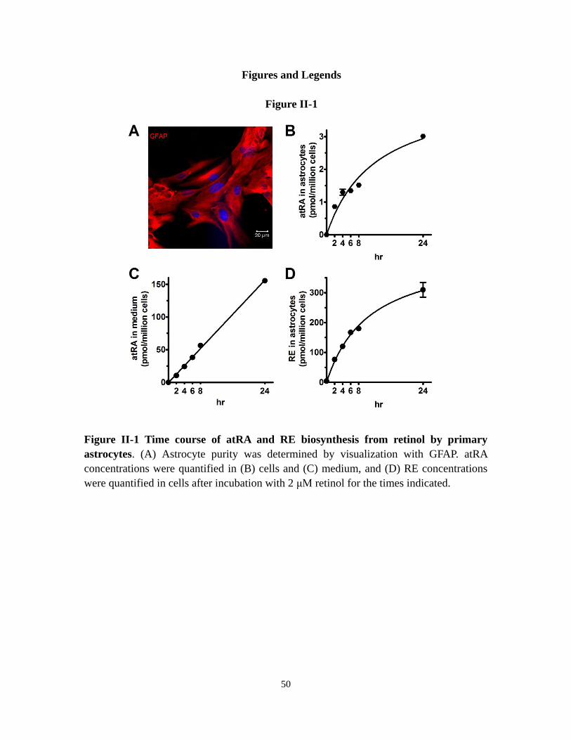
50
Figures and Legends
Figure II-1
Figure II-1 Time course of atRA and RE biosynthesis from retinol by primary
astrocytes. (A) Astrocyte purity was determined by visualization with GFAP. atRA
concentrations were quantified in (B) cells and (C) medium, and (D) RE concentrations
were quantified in cells after incubation with 2 μM retinol for the times indicated.
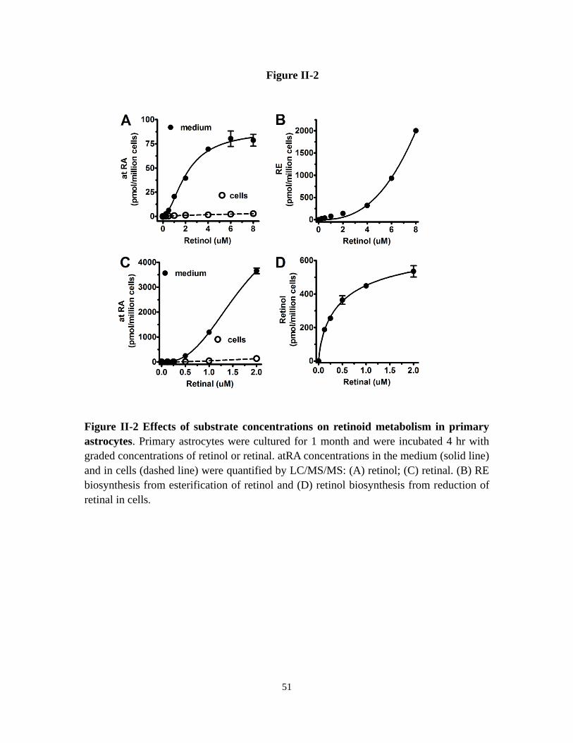
51
Figure II-2
Figure II-2 Effects of substrate concentrations on retinoid metabolism in primary
astrocytes. Primary astrocytes were cultured for 1 month and were incubated 4 hr with
graded concentrations of retinol or retinal. atRA concentrations in the medium (solid line)
and in cells (dashed line) were quantified by LC/MS/MS: (A) retinol; (C) retinal. (B) RE
biosynthesis from esterification of retinol and (D) retinol biosynthesis from reduction of
retinal in cells.

52
Figure II-3
Figure II-3 Retinoid metabolism catalyzed by primary hippocampus neurons. (A)
The purity of neurons (DIV14) was determined by MAP2 visualization. Primary cultured
neurons were incubated 4 hr with retinol or retinal. atRA accumulation in the medium
(solid line) and in cells (dashed line) from (B) retinol or (C) retinal was quantified by
LC/MS/MS. (D) Retinol biosynthesis from retinal and (E) RE production from retinol
was quantified by HPLC
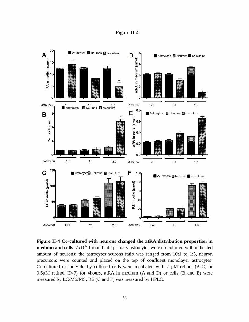
53
Figure II-4
Figure II-4 Co-cultured with neurons changed the atRA distribution proportion in
medium and cells. 2x10⁵ 1 month old primary astrocytes were co-cultured with indicated
amount of neurons: the astrocytes:neurons ratio was ranged from 10:1 to 1:5, neuron
precursors were counted and placed on the top of confluent monolayer astrocytes.
Co-cultured or individually cultured cells were incubated with 2 μM retinol (A-C) or
0.5μM retinol (D-F) for 4hours, atRA in medium (A and D) or cells (B and E) were
measured by LC/MS/MS, RE (C and F) was measured by HPLC.

54
Figure II-5
Figure II-5 Neurons sequester atRA secreted by astrocytes. (A) Astrocytes (2 x 10⁵)
were cultured on 22 x 22 mm glass coverslips and neurons (6 x 10⁵) were cultured
directly in plates. In cultures, coverslips were placed on the top of neurons with
astrocytes facing up to avoid direct contact between neurons and astrocytes. Cells were
incubated 4 hr with 2 μM retinol. atRA in the medium was quantified. Neurons and
astrocytes in cultures were separated and analyzed individually for atRA and RE in the
cells. (B) Intracellular atRA in astrocytes, neurons and cultures of astrocytes and neurons:

55
*P=0.0005, n = 3. (C) atRA in the medium of astrocyte cultures or cultures of astrocytes
and neurons. (D) Intracellular RE in astrocytes, neurons and cultures of astrocytes and
neurons

56
Figure II-6
Figure II-6 Knockdown reveals Raldh that contribute to atRA biosynthesis. (A)
Q-PCR analysis of Raldh mRNA expression in primary astrocyte cultures normalized to
β-actin mRNA. (B) Results of transfecting primary astrocytes with non-targeting siRNA
or with siRNA targeted to Raldh1, 2 or 3. mRNA was detected by RT-PCR 72 hr after

57
transfection. (C) Raldh1 was quantified by western blot 96 hr after transfection:
***P<0.001; (D) atRA biosynthesis from 2 μM retinol after 4 hr incubation. (E) atRA
biosynthesis from 1 μM retinal after 4hr incubation. (F) Seventy-two hr after Raldh1
siRNA transfection, Raldh2 and 3 mRNA were measured by RT-PCR. D and E data were
normalized to non-targeting siRNA transfected group: *P<0.01; n = 3. Four hr
incubations with substrates were done 96 hr after transfections. NT: Non-targeting.
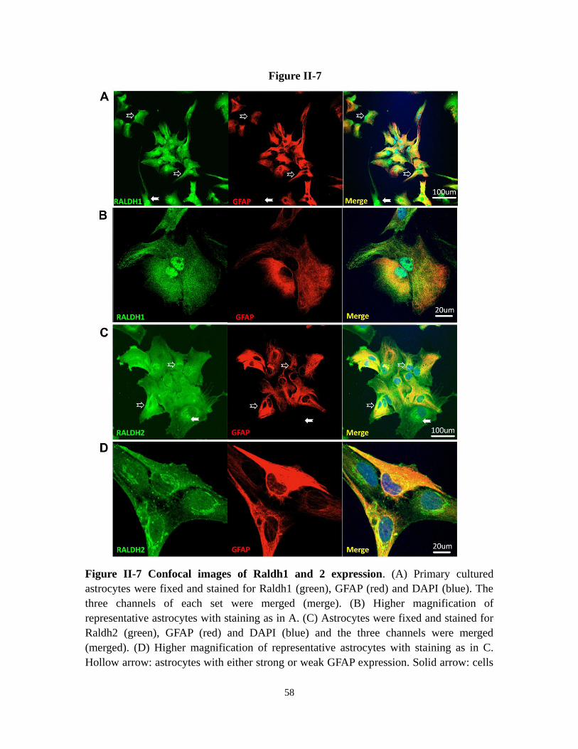
58
Figure II-7
Figure II-7 Confocal images of Raldh1 and 2 expression. (A) Primary cultured
astrocytes were fixed and stained for Raldh1 (green), GFAP (red) and DAPI (blue). The
three channels of each set were merged (merge). (B) Higher magnification of
representative astrocytes with staining as in A. (C) Astrocytes were fixed and stained for
Raldh2 (green), GFAP (red) and DAPI (blue) and the three channels were merged
(merged). (D) Higher magnification of representative astrocytes with staining as in C.
Hollow arrow: astrocytes with either strong or weak GFAP expression. Solid arrow: cells

59
without GFAP expression which expressed Raldh1 or 2. No signals were detected in
negative controls, which consisted of eliminating primary antibodies (data not shown).

60
Figure II-8
Figure II-8 Multiple Rdhs contribute to atRA biosynthesis in astrocytes. (A) Q-PCR
analysis of Rdh10, Rdh2 and Dhrs9 mRNA expression in astrocytes. (B) Effects of
transfecting astrocytes with non-targeting siRNA or siRNA homologous with Rdh10,
Rdh2 and Dhrs9. Rdh mRNA were detected by RT-PCR 72 hr after transfection. (C) Total
atRA production from 2 μM retinol in 4 hr was quantified 96 hr after transfection and was
normalized to the non-targeting cultures: *P<0.01, n = 4. (D) mRNA expression of Rdh2
and Dhrs9 was evaluated by RT-PCR 72 hr after Rdh10 siRNA transfection. (E) mRNA
expression of Rdh10 and Dhrs9 was evaluated by RT-PCR 72 hr after Rdh2 siRNA
transfection

61
Figure II-9
Figure II-9 Dhrs9 functions as a retinol dehydrogenase in astrocytes. (A) COS cells
(top) or primary astrocytes (bottom) were transfected with an empty vector or a vector
that expressed FLAG-tagged Dhrs9. Anti-FLAG antibody (red) and nuclei (DAPI blue)
images in cells transfected with the expression vector. Bar graphs show atRA production
from 2 μM retinol over 4 hr from cells transfected with empty vectors or
Dhrs9-expressing vectors: *P<0.005, n = 3. (B) Recovery of atRA was measured in
astrocytes transfected with non-targeting or Dhrs9 siRNA and incubated with 4 nM atRA

62
for 6 hr. (C) mRNA expression was measured by RT-PCR 72 hr after transfection with
Dhrs9 siRNA. (D) Retinol production was measured in astrocytes transfected with
non-targeting or Dhrs9 siRNA, incubated 4 hr with 1 μM retinal. (E) atRA production
from 1 μM retinal with time after transfection with non-targeting or Dhrs9 siRNA:
*P<0.0001, n = 3. (F) Dhrs9 and Raldh1 mRNAs were detected by RT-PCR (upper panel).
Raldh1 mRNA levels were quantified by Q-PCR (left lower panel). Raldh1 protein levels
were quantified by western blot (right lower panel) with time after transfection with
Dhrs9 siRNA: *P<0.05, n = 3. (G) Astrocytes were incubated with: (upper panel) 1 μM
retinal or (lower panel) 1 μM atRA. Radlh1 mRNA was measured by RT-PCR.

63
Figure II-10

64
Figure II-10 atRA induces Cyp26B1 and Lrat mRNA in astrocytes. (A) RT-PCR of
Cyp26A1, Cyp26B1 and Lrat mRNAs after incubating primary astrocytes with 1 μM
atRA. (B) Astrocytes were pre-incubated with graded concentrations of atRA for 72 hr.
atRA was removed, cells were incubated 4 hr with 2 μM retinol, atRA (solid line) and RE
(dashed line) production was quantified. (C) Astrocytes were transfected with
non-targeting or Cyp26B1 siRNA for 72 hr and Cyp26B1 mRNA was measured by
RT-PCR. (D) Astrocytes were transfected 96 hr with Cyp26B1 siRNA and then incubated
4 hr with 2 μM retinol. The amount of atRA produced was normalized to the amount
detected in cells transfected with non-targeting siRNA: *P<0.0001, n = 3. (E) Astrocytes
were transfected with non-targeting or Cyp26B1 siRNA (96 hr) and then incubated with 1
nM atRA. The natural log of the percent atRA remaining was plotted vs. incubation time:
filled circles, medium without cells; open circles, cells transfected with Cyp26B1 siRNA;
open diamonds, cells transfected with non-targeting siRNA.
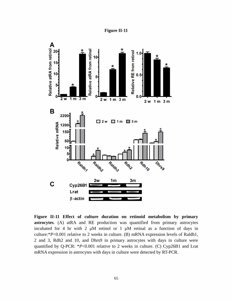
65
Figure II-11
Figure II-11 Effect of culture duration on retinoid metabolism by primary
astrocytes. (A) atRA and RE production was quantified from primary astrocytes
incubated for 4 hr with 2 μM retinol or 1 μM retinal as a function of days in
culture:*P<0.001 relative to 2 weeks in culture. (B) mRNA expression levels of Raldh1,
2 and 3, Rdh2 and 10, and Dhrs9 in primary astrocytes with days in culture were
quantified by Q-PCR: *P<0.001 relative to 2 weeks in culture. (C) Cyp26B1 and Lrat
mRNA expression in astrocytes with days in culture were detected by RT-PCR.

66
Figure II-12
Figure II-12 Compensation effect of atRA biosynthesis in Rdh1-/- and CrbpI-/-
astrocytes. Primary astrocytes from wild-type (WT) and Rdh1 knockout (Rdh1-/-) or
CrbpI (CrbpI -/-) knockout mice cultured for 1 month were incubated 4 hr with 2 μM
retinol or 1 μM retinal, atRA production from retinol (A) or retinal (B), RE production
from retinol (C) and retinol production from retinal (D) were quantified and normalized
to wild-type group. * P<0.0001, n=3. (E) Expressions of various mRNAs in wild-type or
Rdh1-/- mice astrocytes were measured by RT-PCR. (F) Expressions of various mRNAs
in wild-type or CrbpI-/- mice astrocytes were measured by RT-PCR.
WT
Rdh1 -/-
CrbpI -/-
0
1
2
3
*
*
Re
lati
ve
atR
A p
rod
uc
tio
n f
rom
RO
L
WT
Rdh1 -/-
CrbpI -/-
0
1
2
3
4
5
*
**
Re
lati
ve
atR
A p
rod
uc
tio
n f
rom
RA
L
WT
Rdh1 -/-
CrbpI -/-
0.0
0.5
1.0
1.5
*
Re
lati
ve
RE
pro
du
cti
on
fro
m R
OL
WT
Rdh1 -/-
CrbpI -/-
0.0
0.5
1.0
1.5
*
*R
ela
tiv
e R
OL
pro
du
cti
on
fro
m R
AL
Raldh2
Cyp26a1
Cyp26b1
Dhrs9
β- actin
Rdh1+/+ Rdh1-/-
Raldh2
Cyp26a1
Cyp26b1
Crbp II
β- actin
CrbpI +/+ CrbpI -/-
A B
C D
E F

67
Figure II-13
Figure II-13 Retinol homeostasis in astrocytes and neurons. The numbers next to each
step designate the rate of the step at 60 nM substrate relative to the other steps, calculated
from the data of Table 1. The dotted lines represent atRA control of Lrat and Cyp26B1
expression. NA, not applicable.
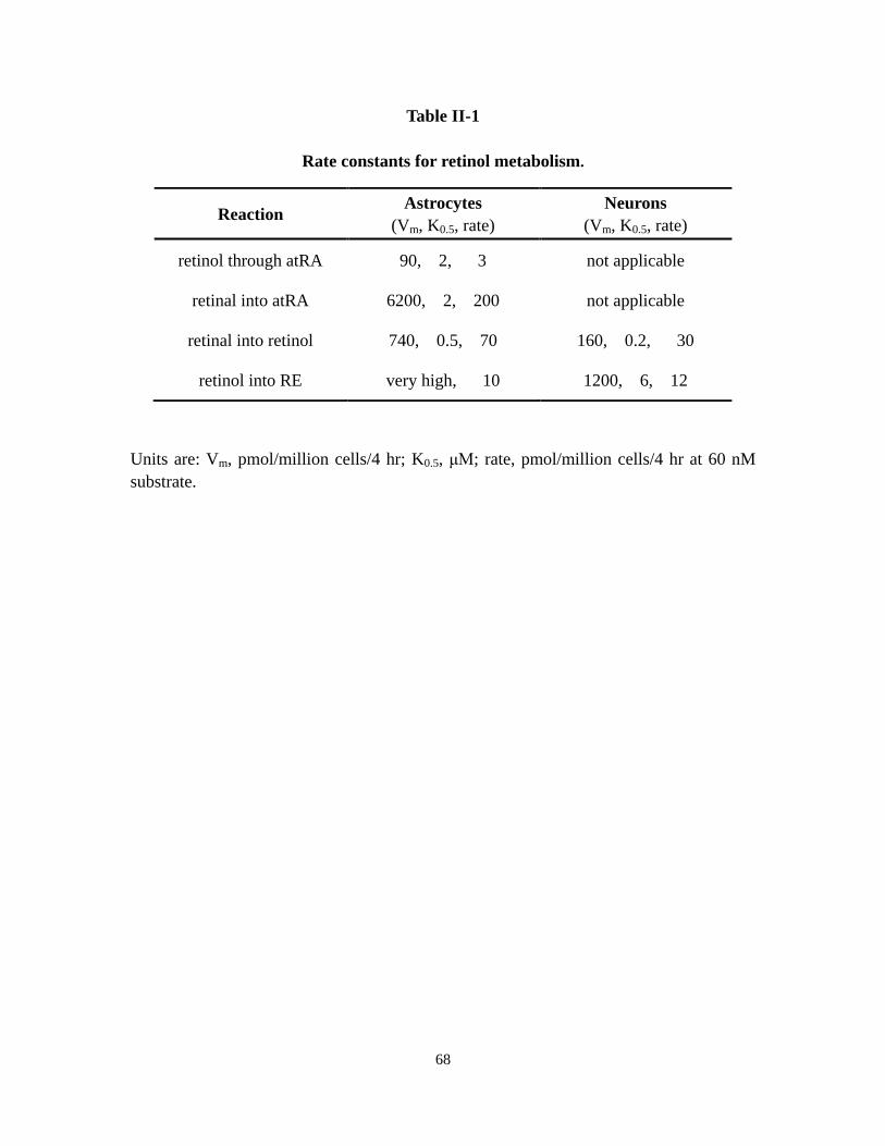
68
Table II-1
Rate constants for retinol metabolism.
Units are: Vm, pmol/million cells/4 hr; K0.5, μM; rate, pmol/million cells/4 hr at 60 nM
substrate.
Reaction Astrocytes
(Vm, K0.5, rate)
Neurons
(Vm, K0.5, rate)
retinol through atRA 90, 2, 3 not applicable
retinal into atRA 6200, 2, 200 not applicable
retinal into retinol 740, 0.5, 70 160, 0.2, 30
retinol into RE very high, 10 1200, 6, 12
69
References
Akita, J., M. Takahashi, et al. (2002). "Neuronal differentiation of adult rat
hippocampus-derived neural stem cells transplanted into embryonic rat explanted
retinas with retinoic acid pretreatment." Brain Res 954(2): 286-293.
Alliot, F., N. Delhaye-Bouchaud, et al. (1988). "Role of astroglial cell clones in the
survival and differentiation of cerebellar embryonic neurons." Brain Res Dev
Brain Res 44(2): 247-257.
Asson-Batres, M. A. and W. B. Smith (2006). "Localization of retinaldehyde
dehydrogenases and retinoid binding proteins to sustentacular cells, glia,
Bowman's gland cells, and stroma: potential sites of retinoic acid synthesis in the
postnatal rat olfactory organ." J Comp Neurol 496(2): 149-171.
Banker, G. A. (1980). "Trophic interactions between astroglial cells and hippocampal
neurons in culture." Science 209(4458): 809-810.
Brodeur, H., I. Gagnon, et al. (2003). "Cloning of monkey RALDH1 and characterization
of retinoid metabolism in monkey kidney proximal tubule cells." J Lipid Res
44(2): 303-313.
Chai, X., M. H. Boerman, et al. (1995). "Cloning of a cDNA for liver microsomal retinol
dehydrogenase. A tissue-specific, short-chain alcohol dehydrogenase." J Biol
Chem 270(8): 3900-3904.
Chandrasekaran, V., Y. Zhai, et al. (2000). "Retinoic acid regulates the morphological
development of sympathetic neurons." J Neurobiol 42(4): 383-393.
Chen, N. and J. L. Napoli (2008). "All-trans-retinoic acid stimulates translation and
induces spine formation in hippocampal neurons through a membrane-associated
RARalpha." FASEB J 22(1): 236-245.
Chen, N., B. Onisko, et al. (2008). "The nuclear transcription factor RARalpha associates
with neuronal RNA granules and suppresses translation." J Biol Chem 283(30):
20841-20847.
Chetyrkin, S. V., O. V. Belyaeva, et al. (2001). "Characterization of a novel type of
human microsomal 3alpha -hydroxysteroid dehydrogenase: unique tissue
distribution and catalytic properties." J Biol Chem 276(25): 22278-22286.
Chiang, M. Y., D. Misner, et al. (1998). "An essential role for retinoid receptors RARbeta
and RXRgamma in long-term potentiation and depression." Neuron 21(6):
1353-1361.
Cocco, S., G. Diaz, et al. (2002). "Vitamin A deficiency produces spatial learning and
memory impairment in rats." Neuroscience 115(2): 475-482.
Connor, M. J. and N. Sidell (1997). "Retinoic acid synthesis in normal and Alzheimer
diseased brain and human neural cells." Mol Chem Neuropathol 30(3): 239-252.
Cooper, D. L., N. R. Isola, et al. (1993). "Members of the ALDH gene family are lens and
corneal crystallins." Adv Exp Med Biol 328: 169-179.
Cowherd, R. M., R. E. Lyle, et al. (1999). "Molecular regulation of adipocyte

70
differentiation." Semin Cell Dev Biol 10(1): 3-10.
Dupe, V., N. Matt, et al. (2003). "A newborn lethal defect due to inactivation of
retinaldehyde dehydrogenase type 3 is prevented by maternal retinoic acid
treatment." Proc Natl Acad Sci U S A 100(24): 14036-14041.
Edwards, R. B., A. J. Adler, et al. (1992). "Synthesis of retinoic acid from retinol by
cultured rabbit Muller cells." Exp Eye Res 54(4): 481-490.
Etchamendy, N., V. Enderlin, et al. (2003). "Vitamin A deficiency and relational memory
deficit in adult mice: relationships with changes in brain retinoid signalling."
Behav Brain Res 145(1-2): 37-49.
Etchamendy, N., V. Enderlin, et al. (2001). "Alleviation of a selective age-related
relational memory deficit in mice by pharmacologically induced normalization of
brain retinoid signaling." J Neurosci 21(16): 6423-6429.
Grun, F., Y. Hirose, et al. (2000). "Aldehyde dehydrogenase 6, a cytosolic retinaldehyde
dehydrogenase prominently expressed in sensory neuroepithelia during
development." J Biol Chem 275(52): 41210-41218.
Halilagic, A., V. Ribes, et al. (2007). "Retinoids control anterior and dorsal properties in
the developing forebrain." Dev Biol 303(1): 362-375.
Jacobs, S., D. C. Lie, et al. (2006). "Retinoic acid is required early during adult
neurogenesis in the dentate gyrus." Proc Natl Acad Sci U S A 103(10):
3902-3907.
Jette, C., P. W. Peterson, et al. (2004). "The tumor suppressor adenomatous polyposis coli
and caudal related homeodomain protein regulate expression of retinol
dehydrogenase L." J Biol Chem 279(33): 34397-34405.
Jones-Villeneuve, E. M., M. A. Rudnicki, et al. (1983). "Retinoic acid-induced neural
differentiation of embryonal carcinoma cells." Mol Cell Biol 3(12): 2271-2279.
Jurukovski, V., N. G. Markova, et al. (1999). "Cloning and characterization of retinol
dehydrogenase transcripts expressed in human epidermal keratinocytes." Mol
Genet Metab 67(1): 62-73.
Kane, M. A., N. Chen, et al. (2005). "Quantification of endogenous retinoic acid in
limited biological samples by LC/MS/MS." Biochem J 388(Pt 1): 363-369.
Kane, M. A., A. E. Folias, et al. (2008). "Quantitative profiling of endogenous retinoic
acid in vivo and in vitro by tandem mass spectrometry." Anal Chem 80(5):
1702-1708.
Kane, M. A., A. E. Folias, et al. (2010). "Ethanol elevates physiological all-trans-retinoic
acid levels in select loci through altering retinoid metabolism in multiple loci: a
potential mechanism of ethanol toxicity." FASEB J 24(3): 823-832.
Klann, R. C. and A. C. Marchok (1982). "Effects of retinoic acid on cell proliferation and
cell differentiation in a rat tracheal epithelial cell line." Cell Tissue Kinet 15(5):
473-482.
Kornyei, Z., E. Gocza, et al. (2007). "Astroglia-derived retinoic acid is a key factor in
glia-induced neurogenesis." FASEB J 21(10): 2496-2509.
71
Labrecque, J., P. V. Bhat, et al. (1993). "Purification and partial characterization of a rat
kidney aldehyde dehydrogenase that oxidizes retinal to retinoic acid." Biochem
Cell Biol 71(1-2): 85-89.
Laird, M. D., J. R. Vender, et al. (2008). "Opposing roles for reactive astrocytes following
traumatic brain injury." Neurosignals 16(2-3): 154-164.
Maden, M. (2007). "Retinoic acid in the development, regeneration and maintenance of
the nervous system." Nat Rev Neurosci 8(10): 755-765.
Mark, M., N. B. Ghyselinck, et al. (2009). "Function of retinoic acid receptors during
embryonic development." Nucl Recept Signal 7: e002.
Markiewicz, I. and B. Lukomska (2006). "The role of astrocytes in the physiology and
pathology of the central nervous system." Acta Neurobiol Exp (Wars) 66(4):
343-358.
McCaffery, P., O. Koul, et al. (2004). "Ethanol increases retinoic acid production in
cerebellar astrocytes and in cerebellum." Brain Res Dev Brain Res 153(2):
233-241.
Misner, D. L., S. Jacobs, et al. (2001). "Vitamin A deprivation results in reversible loss of
hippocampal long-term synaptic plasticity." Proc Natl Acad Sci U S A 98(20):
11714-11719.
Moro, J. R., M. Iwata, et al. (2008). "Vitamin effects on the immune system: vitamins A
and D take centre stage." Nat Rev Immunol 8(9): 685-698.
Nakayama, T., T. Momoki-Soga, et al. (2003). "Astrocyte-derived factors instruct
differentiation of embryonic stem cells into neurons." Neurosci Res 46(2):
241-249.
Napoli, J. L. (1999). "Interactions of retinoid binding proteins and enzymes in retinoid
metabolism." Biochim Biophys Acta 1440(2-3): 139-162.
Napoli, J. L. (2010). Vitamin A (Retinoids). Encyclopedia of Biological Chemistry,
Elseiver Ltd: in press.
Niederreither, K., V. Subbarayan, et al. (1999). "Embryonic retinoic acid synthesis is
essential for early mouse post-implantation development." Nat Genet 21(4):
444-448.
Ong, D. E., P. N. MacDonald, et al. (1988). "Esterification of retinol in rat liver. Possible
participation by cellular retinol-binding protein and cellular retinol-binding
protein II." J Biol Chem 263(12): 5789-5796.
Penniston, K. L. and S. A. Tanumihardjo (2006). "The acute and chronic toxic effects of
vitamin A." Am J Clin Nutr 83(2): 191-201.
Posch, K. C., R. D. Burns, et al. (1992). "Biosynthesis of all-trans-retinoic acid from
retinal. Recognition of retinal bound to cellular retinol binding protein (type I) as
substrate by a purified cytosolic dehydrogenase." J Biol Chem 267(27):
19676-19682.
Rexer, B. N. and D. E. Ong (2002). "A novel short-chain alcohol dehydrogenase from
rats with retinol dehydrogenase activity, cyclically expressed in uterine

72
epithelium." Biol Reprod 67(5): 1555-1564.
Rochette-Egly, C. and P. Germain (2009). "Dynamic and combinatorial control of gene
expression by nuclear retinoic acid receptors (RARs)." Nucl Recept Signal 7:
e005.
Rodnight, R., C. A. Goncalves, et al. (1997). "Control of the phosphorylation of the
astrocyte marker glial fibrillary acidic protein (GFAP) in the immature rat
hippocampus by glutamate and calcium ions: possible key factor in astrocytic
plasticity." Braz J Med Biol Res 30(3): 325-338.
Ross, A. C. (2003). "Retinoid production and catabolism: role of diet in regulating retinol
esterification and retinoic Acid oxidation." J Nutr 133(1): 291S-296S.
Ross, A. C. and R. Zolfaghari (2004). "Regulation of hepatic retinol metabolism:
perspectives from studies on vitamin A status." J Nutr 134(1): 269S-275S.
Shapiro, M. L. and H. Eichenbaum (1999). "Hippocampus as a memory map: synaptic
plasticity and memory encoding by hippocampal neurons." Hippocampus 9(4):
365-384.
Shearer, K. D., T. H. Goodman, et al. (2010). "Photoperiodic regulation of retinoic acid
signaling in the hypothalamus." J Neurochem 112(1): 246-257.
Shudo, K., H. Fukasawa, et al. (2009). "Towards retinoid therapy for Alzheimer's
disease." Curr Alzheimer Res 6(3): 302-311.
Siegenthaler, J. A., A. M. Ashique, et al. (2009). "Retinoic acid from the meninges
regulates cortical neuron generation." Cell 139(3): 597-609.
Silman, I. and J. L. Sussman (2005). "Acetylcholinesterase: 'classical' and 'non-classical'
functions and pharmacology." Curr Opin Pharmacol 5(3): 293-302.
Song, H., C. F. Stevens, et al. (2002). "Astroglia induce neurogenesis from adult neural
stem cells." Nature 417(6884): 39-44.
Soref, C. M., Y. P. Di, et al. (2001). "Characterization of a novel airway epithelial
cell-specific short chain alcohol dehydrogenase/reductase gene whose expression
is up-regulated by retinoids and is involved in the metabolism of retinol." J Biol
Chem 276(26): 24194-24202.
Steven W. Levison, J. d. V., and James E. Goldman (2005). "Astrocyte development."
Developmental Neurobiology, 4th ed.
Takahashi, J., T. D. Palmer, et al. (1999). "Retinoic acid and neurotrophins collaborate to
regulate neurogenesis in adult-derived neural stem cell cultures." J Neurobiol
38(1): 65-81.
Thatcher, J. E. and N. Isoherranen (2009). "The role of CYP26 enzymes in retinoic acid
clearance." Expert Opin Drug Metab Toxicol 5(8): 875-886.
Toresson, H., A. Mata de Urquiza, et al. (1999). "Retinoids are produced by glia in the
lateral ganglionic eminence and regulate striatal neuron differentiation."
Development 126(6): 1317-1326.
Unsicker, K., H. Reichert-Preibsch, et al. (1987). "Astroglial and fibroblast growth
factors have neurotrophic functions for cultured peripheral and central nervous
73
system neurons." Proc Natl Acad Sci U S A 84(15): 5459-5463.
Vaccarino, F. M., D. M. Fagel, et al. (2007). "Astroglial cells in development,
regeneration, and repair." Neuroscientist 13(2): 173-185.
Wang, X., P. Penzes, et al. (1996). "Cloning of a cDNA encoding an aldehyde
dehydrogenase and its expression in Escherichia coli. Recognition of retinal as
substrate." J Biol Chem 271(27): 16288-16293.
Wu, B. X., Y. Chen, et al. (2002). "Cloning and characterization of a novel all-trans
retinol short-chain dehydrogenase/reductase from the RPE." Invest Ophthalmol
Vis Sci 43(11): 3365-3372.
Wuarin, L., N. Sidell, et al. (1990). "Retinoids increase perinatal spinal cord neuronal
survival and astroglial differentiation." Int J Dev Neurosci 8(3): 317-326.
Yang, Y., W. Ge, et al. (2003). "Contribution of astrocytes to hippocampal long-term
potentiation through release of D-serine." Proc Natl Acad Sci U S A 100(25):
15194-15199.
Yost, R. W., E. H. Harrison, et al. (1988). "Esterification by rat liver microsomes of
retinol bound to cellular retinol-binding protein." J Biol Chem 263(35):
18693-18701.
Zhang, M., W. Chen, et al. (2001). "Molecular characterization of a mouse short chain
dehydrogenase/reductase active with all-trans-retinol in intact cells, mRDH1." J
Biol Chem 276(47): 44083-44090.
Zhang, M., P. Hu, et al. (2007). "Altered vitamin A homeostasis and increased size and
adiposity in the rdh1-null mouse." FASEB J 21(11): 2886-2896.
Zhao, D., P. McCaffery, et al. (1996). "Molecular identification of a major
retinoic-acid-synthesizing enzyme, a retinaldehyde-specific dehydrogenase." Eur
J Biochem 240(1): 15-22.
Ziouzenkova, O., G. Orasanu, et al. (2007). "Retinaldehyde represses adipogenesis and
diet-induced obesity." Nat Med 13(6): 695-702.
Footnotes
The abbreviations used are: atRA, all-trans-retinoic acid; DIV, days in vitro; CNS, central
nervous system; Cyp26A1, Cytochrome P450 26A1; Cyp26B1, Cytochrome P450 26B1;
Dhrs9, dehydrogenase/reductase SDR family member 9; RAR, retinoic acid receptor;
GFAP, glial fibrillary acidic protein; HPLC, High-performance liquid chromatography;
LC/MS/MS, liquid chromatography/mass spectrometry; Lrat, lecithin:retinol
acyltransferase; MAP2, microtubule-associated protein 2; Raldh, retinaldehyde
dehydrogenase; RE, retinyl ester; Rdh, retinol dehydrogenase; SDR, short-chain
dehydrogenase/reductase; siRNA, small interfering RNA.

74
Chapter III
Localization in vitro and in vivo of RALDH1 Expression
Introduction
We have demonstrated that RALDH1 is a key component of atRA biosynthesis in adult
astrocytes, because knockdown of RALDH1 reduced atRA biosynthesis in astrocytes.
Astrocytes also up regulate RALDH1 expression to compensate for impairment of atRA
biosynthesis due to the loss of Dhrs9, indicating that RALDH1 is at least one of the
enzymes that the adult nervous system uses to control atRA homeostasis. An in vitro
culture time dependent increase of RALDH1 expression, which correlates with increased
atRA production in astrocytes also supports the conclusion that RALDH1 is a major
enzyme that contributes to atRA biosynthesis in astrocytes. Besides what we found, the
presence of RALDH1 has been reported in adult hippocampal neurons (Aoto et al., 2008)
and GFAP positive tissue in the adult olfactory bulb (Asson-Batres and Smith, 2006).
Immunostaining of RALDH1 in pure cultured astrocytes, showed wide expression in
cultured glia cells, not only in GFAP+ cells, but also in glia cells that do not express
GFAP. RALDH1 is expressed in both cytoplasm and nucleus of astrocytes, an uncommon
location that has not been reported before, to the best of our knowledge. As we know,
RALDH2 is a critical enzyme that provides atRA for embryo development, but in
cultured mature astrocytes, RALDH1 had the highest copy numbers of mRNA. We
hypothesize that there is a time-dependent increased expression of RALDH1 in
hippocampus, and this increment might correlate with the appearance and development of
astrocytes in adult hippocampus.
The anatomic structure of adult hippocampus is quite unique. Neurons in the
hippocampus are centralized in three areas, granule cells in the dentate gyrus, and
pyramidal neurons in CA1 and CA3 regions (Gross, 1993). The primary afferent to the
hippocampus is the perforant pathway (Witter et al., 2000). This pathway arises from the
entorhinal cortex and projects to the dentate gyrus. Granule cells in the dentate gyrus
project to mossy cells in the hilus of the dentate gyrus and the CA3 region of Ammon‘s
horn. The CA3 neurons in turn project to the CA1 by way of the Shaffer collateral. The
CA1 regions projects to the deep layers of the entorhinal cortex, thereby completing a
circuit. The axons that project from the dentate gyrus to the CA3 region are called mossy
fibers (Kobayashi, 2010). The pathway was so named by Ramon Cajal because the axons
display varicosities all along their lengths, giving them a ‗mossy‘ appearance. These
mossy fibers form multiple synapses with the elaborate dendritic spines of CA3
pyramidal cells in the stratum lucidum. A single mossy fiber projection may make as
many as 37 contacts with a single pyramidal cell. In rodents, the size of mossy fiber
75
projections show large interindividual variation, which show strong correlations with
spatial learning (McBain, 2008).
As shown in chapter II, no atRA production was found in cultured neurons, even
incubated with retinal. However, RALDH1 has been reported to be expressed in cultured
neurons (Aoto et al., 2008). Besides that, the in vivo RALDH1 location has never been
reported. Through immunostaining and immunohistochemistry, for the first time, we
elucidated the specific localization of RALDH1 in cultured astrocytes, neurons and in
adult hippocampus. Through co-immunostaining with other cell specific markers, we
demonstrated that RALDH1 expresses throughout the cultured neurons with particularly
high expression in axons; RALDH1 is also highly expressed in adult CA1 pyramidal
neurons and mossy fibers in the adult hippocampus. In contrast, RALDH1 is only highly
expressed in small portions of astrocytes, when in mixed culture with neurons, but has
low expression levels in the other astrocytes. Our data indicate that RALDH1 may not
function as a retinal dehydrogenase in neurons and its expression in astrocytes might be
regulated by neurons.

76
Materials and Methods
Cell culture--Mono neuronal precursors were obtained the same way as pure neurons
from the E18 Sprague-Dawley rat. To obtain mixed cultured glia-neurons, the precursors
were plated on poly-D-Lysine coated glass coverslips with Neurobasal medium
containing 2% B27 and 10% FBS at a density of 1x10⁵ cells/well in 24-well plates. Half
of the total medium was replenished every week. Mixed culture cells were fixed and
immunostained with RALDH1 antibody and cell marker after 2 weeks incubation in a
37C incubator with 5% CO2.
Diluted pure neurons were cultured to obtain a clearly individual neuron image. E18
rat mono neuronal precursors were plated at the density of 5x10³ cells/well in 24-well
plate on glass coverslips pre-coated with poly-D-lysine. Pure neurons were cultured as
described in Chapter II Materials and Methods.
RALDH1 construction of plasmid and transfection--To obtain a mammalian expression
vector, the full length open reading frame of mouse RALDH1 (NM_013467) was
obtained by PCR amplification from mouse brain cDNA library using the forward primer
5‘-ATGGATCCATGTCTTCGCCTGCACAAC-3‘, containing a BamHI site, and the
reverse primer 5‘- AAGAATTCTTAGGAGTTCTTCTGAGATATCTTC-3‘, containing a
EcoRI site. The gel-purified PCR product was cloned into pcDNA3.1+ vector. The proper
sequence of the expression vector was confirmed by sequencing. COS cells were
transfected with pcDNA3.1(+)-RALDH1 vector using Lipofectamine 2000 (Invitrogen).
After 24h transfection, cells were fixed for immunostaining of RALDH1.
Preparation of hippocampal slices—2 weeks to 1 month old Sprague-Dawley rats were
anesthetized with CO2 and decapitated. Brains were quickly excised and trimmed,
leaving a rectangular block of tissue containing the dorsal hippocampus. The tissue
blocks were immerged into liquid nitrogen slowly for 1 min until the tissue was
completely frozen. Frozen tissue blocks were embedded in O.C.T. compound (Tissue-Tek)
and glued to the stage of a cryostat-microtome. Six μM frozen hippocampal slices were
sectioned coronally and placed on the surfaces of glass slides. The slides were immerged
into 4% paraformaldehyde for half an hour before immunostaining procedures.
Immunocytochemistry and Immunohistochemistry--Immunocytochemistry was performed
in the same way as described in Chapter II materials and methods. Monoclonal Antibody
against pan-axonal neurofilament marker SMI-312 (1:500) was used to identify axons in
cultured neurons. For immunohistochemistry, fixed hippocampus slices were washed
three time for 5-min each in PBST (1xPBS+0.25% Tween 20), permeabilized with 0.2%
Triton X-100 for 15 min, and blocked with PBS containing 10% goat serum or donkey
serum overnight, depending on the origin of secondary antibody. Hippocampal slices
were then incubated with anti-NeuN (1:500, Millipore), rabbit polyclonal anti-RALDH1
(1:200 abcam), mouse anti MBP (1:200), rabbit anti MAP2 (1:1000), mouse anti Tau
(1:500), or goat polyclonal anti Synaptoporin (1:10) overnight at 4°C, washed three times
77
for 5-min each with PBST, followed by incubation with goat or donkey anti-mouse
Alexa 555, goat or donkey anti-rabbit Alexa 488, chicken anti-goat Alexa 488 secondary
antibody (Invitrogen) or rat cy3 red for 1 hr. After washing three times for 5 min each
with PBST, coverslips were mounted on slides with Vectashield mounting medium (with
DAPI, Vector Laboratories Inc. Burlingame, CA). Images were captured by a LSM 510
Meta UV/Vis confocal microscope.

78
Results
RALDH1 was highly expressed in pure neurons, but not astrocytes mixed cultured with
neurons—To test the specificity of the RALDH1 antibody, COS cells transfected with
rat-RALDH1-pcDNA3.1 expression vector or empty vector were immunostained with the
antibody (Figure III-1A). A strong cytoplasmic and weak nucleus expression pattern of
RALDH1 was found in transfected cells group but not in control cells.
In previous research, we found that RALDH1 was widely expressed in cultures of pure
astrocyte, not only in GFAP positive cells, but also in cells that did not express GFAP. In
pure glia cell cultures, astrocytes lost their in vivo stellate-like shape (Figure II-1A) and
the connection with pyramidal neurons during the differentiation. To address the question
whether the presence of neurons would affect RALDH1 expression, we mixed cultured
neurons and astrocytes. Immunostaining of GFAP showed that astrocytes in mixed
cultured neural cells maintained their star-like shape (Figure III-1B). RALDH1 was
unevenly expressed in these mixed cultured astrocytes (Figure III-1B, top panel). Most of
the astrocytes showed fairly low levels of RALDH1 expression (Figure III-1B, middle
panel), but a few (about 5%) showed strong expression of RALDH1 (Figure III-1B,
bottom panel). Surprisingly, we also found that RALDH1 was present in GFAP negative
cells in mixed cultures, especially highly present in some process-like structures. To test
whether those cells and processes with strong RALDH1 expression were neurons,
individual pyramidal neurons were immunostained with RALDH1 antibody together with
neuron markers (Figure III-1C). The neuronal markers included: NeuN—neuron nucleus
and cytoplasma marker, MAP2—neuron dendrite marker and SMI 312—neuron axon
marker. RALDH1 co-localized with all these cell markers, indicating that RALDH1 was
expressed throughout intact neurons, including soma, dendrites and axons. Compared
with other parts of neurons, the strongest signal of RALDH1 was detected on axons
(Figure III-1B, top panel).
RALDH1 is highly expressed in the CA1 and CA3 regions of adult rat hippocampus—To
elucidate the in vivo expression pattern of RALDH1 in adult rat hippocampus, 2 month
old adult male rat hippocampus slice were immunostained with RALDH1 and NeuN
antibody (Figure III-2A). Strong expression of RALDH1 was found in the CA1 neuron
cell body region. A closer examination of the CA1 region showed that RALDH1 was
expressed in CA1 soma of pyramidal neurons—colocalizing with the NeuN marker and
processes in the stratum radiatum layer (Figure III-2B, left panel). This expression pattern
correlated with the in vitro distribution of RALDH1 in pyramidal neurons. Besides the
strong RALDH1 expression in CA1 region, an even more intense RALDH1 signal was
detected in an area starting from the hilus of the dentate gyrus and extending along with
CA3 region. However, higher magnification showed that the strong RALDH1 signals in
CA3 region excluded the CA3 neuron soma (Figure III-2B, right panel), indicating that
CA3 neurons don‘t express RALDH1 in their cell bodies.
RALDH1 is not expressed in astrocytes and oligodendrocytes in adult hippocampus— We

79
next tested whether the structures highly expressing RALDH1 in the CA3 region
belonged to glia cells. Hippocampus slices double stained with RALDH1 and GFAP
clearly showed that i) a few astrocytes partially expressed RALDH1, which is consistent
with the in vitro mixed culture conclusions; ii) the intense RALDH1 signal in the CA3
region (for example, two selected rectangle areas) didn‘t co-localize with the GFAP
marker, which excluded the possibility that this structure belonged to astrocyte or its
processes (Figure III-3A). In summary, this in vivo RALDH1 expression pattern in
astrocytes didn‘t match what we observed in pure cultured astrocytes but matched more
closely with the mixed cultured results, indicating that the in vivo micro-environment will
affect RALDH1 expression. Myelin basic protein (MBP) is a protein important in the
process of myelination of nerves and is a major constituent of the myelin sheath of
oligodendrocytes (Montague et al., 2006). In mixed culture neural cells, RALDH1 is
weakly expressed in MBP positive oligodendrocytes (Figure III-3B, upper panel).
However, RALDH1 is not detected in vivo in MBP positive cells in the CA3 region
(Figure III-3B, lower panel). All these data suggest that the structure that expressed
RALDH1 was highly expressed in CA3 region was not belonged to glia cells in adult
hippocampus.
RALDH1 is localized in dendrites of CA1 neurons but not dendrites in the CA3
region—In cultured neurons, we found RALDH1 expressed in dendrites of pyramidal
neurons. We next tested whether RALDH1 was also expressed in dendrites of pyramidal
neurons in vivo. The co-localization of RALDH1 with MAP2 in the CA1 region indicated
that RALDH1 was expressed in the dendrites of CA1 neurons, consistent with in vitro
cultured neurons results (Figure III-4, left panel). However, high RALDH1 expression
structures in the CA3 region were not co-localized with MAP2, indicating that dendrites
in CA3 region do not express RALDH1 (Figure III-4, right panel). Given that RALDH1
is expressed in CA1 neuron soma, but not in CA3 neuron soma, primary cultured neurons
seem to mimic the RALDH1 expression pattern of CA1 neurons but not neurons in CA3
region.
RALDH1 is highly expressed in axons in CA3 region and co-localized with mossy
fibers—Tau proteins stabilize microtubules present in dendrites and are primarily in distal
parts of axons (Kempf et al., 1996). Co-immunostaining of both RALDH1 and Tau
further confirmed expression of RALDH1 in axons of cultured neurons (Figure III-5A).
Immunostaining of Tau proteins in the adult hippocampus exhibited a similar expression
pattern of RALDH1 in CA3 region (Figure III-5B). Higher magnification of the
RALDH1 expression structure in CA3 region showed perfect co-localization with Tau
signals, which indicated that the high RALDH1 expression structure in CA3 region are
the distal axons bundles (Figure III-5C).
Mossy fibers are the varicosities axons that projected in CA3 region from granule cells
in dentate gyrus and show similar structure as the Tau staining pattern. We then tested
whether those axon bundles that highly expressed RALDH1 in this area are mossy fibers.
Three selected areas in CA3 region all exhibited co-localization of RALDH1 and
80
synaptoporin, a mossy fiber specific marker (Figure III-6) (Sun et al., 2006). However,
these two proteins do not 100% overlap with each other. This result further confirmed the
mossy fibers-specific enriched RALDH1 expression in CA3 region. But RALDH1 might
have a different subcellular compartmentation with synaptoporin in mossy fibers.
Postnatal development of RALDH1 in adult hippocampus—Finally, we tested the
time-scale of RALDH1 expression in adult hippocampus to see whether its expression,
especially in mossy fibers, is a transitional or developmental change. RALDH1 was
barely detected in the hippocampus of P2 rats (Figure III-7). Weak expression of
RALDH1 was detected in one week old rat hippocampus, mainly in the CA3 region. The
expression level of RALDH1 gradually increased in the following weeks and reached a
steady level after 3 weeks of postnatal development (Figure III-7, left panel). A close
examination clearly showed the incremental expression of RALDH1 in CA3 region
(Figure III-7, right panel). In the same time, RALDH1 also began to appear in CA1
regions, including neurons soma and dendrites (data not shown). All these data indicate
that RALDH1 expression changes with postnatal development.

81
Discussion
Distinct expression levels and distribution patterns of RALDH1 in vitro and in vivo
in astrocytes
RALDH1 is a major enzyme responsible for atRA biosynthesis in astrocytes, as shown
by knockdown experiments in primary cultures. The distribution pattern and expression
level of RALDH1 in astrocytes serves as an indicator of atRA biosynthesis. However, one
of the disadvantages of using the primary cell culture model is the different
developmental environment for these cells. The media used to culture primary astrocytes
and pyramidal neurons only contains basic necessary nutrients and growth factors that
maintain cell survival and stimulate proliferation. However, more and more evidence
indicates that the development of complex nervous systems requires many integrated
signals that function in a spatial and temporal specific manner (Chen and Tonegawa,
1997). For example, formation of synaptic plasticity relies on soluble factors secreted by
astrocytes and contact with oligodendrocytes (Markiewicz and Lukomska, 2006).
Absence of these essential factors during the ex vivo culture of neural cells may cause
those cells to undergo unconventional changes. Our pure cultured astrocytes lost the
star-like processes morphology. In the mixed culture system, astrocytes showed
morphology close to the in vivo morphology, which may be due to the presence of
pyramidal neurons, which partially compensate for loss of micro-environmental factors
required for astrocyte development.
RALDH1 showed an expression pattern in pure astrocyte cultures of particularly high
expression level in the nucleus. However, RALDH1 is only highly expressed in a small
portion of astrocytes in the mixed culture system, leaving most of the astrocytes with a
relative low level of RALDH1 expression. This uneven expression pattern is even more
obvious in hippocampus slices, with only a few astrocytes showing low level of
RALDH1, whereas the others don‘t show detectable level of RALDH1. The differences
in RALDH1 expression might indicate that astrocytes have ability to change atRA
biosynthesis by changing the level of RALDH1 during different circumstances. The
presence of mature neurons might have a negative effect on RALDH1 expression in
nearby astrocytes. In pure cultured astrocytes, lacking this inhibitory effect from neurons,
a culture-time dependent increase of RALDH1 expression was seen (Chapter II). The
presence of neurons in mixed culture systems or in vivo restricts RALDH1 expression to
a relative low and stable level perhaps to regulate the biosynthesis of atRA. This is
necessary for maintaining the normal function of neurons, because astrocytes have ability
to efficiently synthesize and secret atRA into the environment and atRA can stimulate
spine growth of nearby neurons. The high level of RALDH1 might result in excessive
atRA secretion, which may damage synaptic plasticity or affect neurogenesis. The
inhibitory mechanism, either due to soluble factors secreted by neurons, or
neuron-astrocyte contact, need to be further investigated.
Although most astrocytes showed relative low levels of RALDH1 expression, a small

82
portion of mixed cultured astrocytes exhibited strong RALDH1 expression all throughout
the cell body; immunohistochemistry of hippocampus slices also revealed scattered
astrocytes, which had detectable RALDH1 expression, indicating that those astrocytes
reserve or regain the high atRA biosynthesis ability. The signals and regulatory
mechanisms that control RALDH1 expression in this small number of astrocytes are
unclear. Figuring out the age of these astrocytes or the developmental stage of the
neurons contacted with these astrocytes may provide a possible explanation, because
either the young astrocytes or the developmental neurons might need more atRA for
differentiation or formation of synaptic plasticity.
Function of RALDH1 in neurons
Our previous results didn‘t support the conclusion that primary cultured neurons can
synthesize atRA. Even incubated with retinal, no obvious atRA production can be
detected in cells or in medium. However, a relatively strong signal of RALDH1 was
detected in cultured neurons, which challenges this conclusion.
The high level of RALDH1 was detected all throughout cultured neurons, from soma
to dendrites, with particularly strong signal in axons. Similar results have been reported
by other groups (Aoto et al., 2008). There are several possibilities that could explain the
conflict between high expression of RALDH1 and barely detectable atRA after retinal
incubation in pyramidal neurons.
First, if RALDH1 in neurons functions as a retinal dehydrogenase, the low production
of atRA could be due to the low accessibility of RALDH1 to substrate or a fast and
efficient degradation of synthesized atRA. However, the latter situation is less likely
because in the astrocytes-neurons co-culture experiment, we detected significantly
accumulation of atRA in neurons. To exclude this possibility, CYP26 enzymes
inhibitors could be used to block atRA degradation. Retinol or retinal are small
hydrophobic molecules and can easily diffuse across the cell membrane to reach target
enzymes in cell culture models. This property is observed in many cells types such as
astrocytes, COS cells and CHO cells. However, neurons are a specific cell type in CNS
and tightly control the uptake of substances from the environment. Whether the
pyramidal neurons in hippocampus have a specific mechanism to load retinol or retinal,
or this loading needs specific assistance from other glia cells, for example, astrocytes,
needs to be further investigated. Moreover, the presence of one ‗atRA synthesis essential
enzyme‘ is not sufficient for atRA production. Other components for atRA biosynthesis
need to be detected to evaluate the presence of atRA biosynthesis in neurons.
Secondly, besides function as a retinal dehydrogenase, RALDH1 was also reported as a
binding protein. Pereira et al. reported the 56 kD androgen binding protein in genital skin
fibroblasts is an aldehyde dehydrogenase (Pereira et al., 1991). A cytosolic
thyroid-hormone-binding protein (xCTBP), predominantly responsible for the major
binding activity of T3 in the cytosol of Xenopus liver, is identical to aldehyde
dehydrogenase class 1 (ALDH1) (Yamauchi et al., 1999). Whether RALDH1 in neurons
also function as a hormone binding protein is unknown and this possibility should be

83
considered as a potential function in neurons.
It is also possible that RALDH1 shows both aldehyde dehydrogenase activity and
hormone binding activity. There might be other factors in neurons, for example, NAD+ or
NADPH, that could modulate balance between enzyme activity and binding activity
(Yamauchi and Nakajima, 2002).
In vivo RALDH1 expression in adult hippocampus
In vivo RALDH1 expression in CA1 neurons is highly consistent with the in vitro
culture neurons, with particular high expression in neuron soma and dendrites. The high
expression of RALDH1 in axons in vitro is barely seen in vivo because axons are too thin
to be clearly observed in the hippocampus slice. Neurons in the cortex also show similar
RALDH1 expression pattern with particular strong expression on dendrites (data not
shown). These data indicate that this wide RALDH1 expression pattern is generally
present in neurons in vivo. However, not all the neurons in hippocampus showed
homogeneous RALDH1 expression pattern.
Oligodendrocytes are the major cell type responsible for myelination of axons in the
CNS (Bradl and Lassmann, 2010). Relative low expression of RALDH1 in
oligodendrocytes in vitro and in vivo suggests that atRA signaling might not be active in
this cell type in adult hippocampus. However, we could not exclude the presence of other
types of RADLH in oligodendrocytes. Actually, atRA biosynthesis was observed in the
OLN-93 cell line, a permanent cell line derived from spontaneously transformed cells in
rat brain glial cultures and resembling primary oligodendrocytes in their antigenic
properties (Mey and Hammelmann, 2000).
Granule cells are the major neurons in the dentate gyrus and also undergo adult
neurogenesis. A very weak RALDH1 signal was detected in these cells‘ bodies. However,
the processes near the granule cell layer, with axons like structures, showed strong
RALDH1 expression pattern. This area belongs to the subgranular zone, one of two areas
in which neurogenesis persisted in the adult hippocampus. The presence of RALDH1 in
this region might be associated with active atRA signaling that contributes to survival and
differentiation of newly formed neurons.
CA3 neuron is the important participant of the perforant pathway, receiving input from
mossy fibers and exporting signals to CA1 neurons. However, RALDH1 expression in
CA3 neurons is totally different from CA1 region. No RALDH1 signals were detected in
CA3 neurons. However, the strongest RALDH1 signals were detected in the mossy fibers
which encompass the CA3 neurons bodies and dendrites. The perfect co-localization of
RADLH1 with Tau proteins is consistent with in vitro observation of high RALDH1
signal on axons. In adult, synaptoporin is found exclusively in the mossy fiber terminals
present in the hilar region of the dentate gyrus and the regio inferior of the cornu
ammonis (Sun et al., 2006). Co-localization of RALDH1 with synaptoporin further
confirmed its unique mossy fiber enriched expression pattern. The expression of
RALDH1 on mossy fiber is a typical postnatal developmental change, with obvious
RALDH1 expression from 1-month-old rat. The function of this highly expressed

84
RALDH1 in mossy fiber is unknown. If it functions as a retinal dehydrogenase, it might
indicate a highly active atRA signaling present in mossy fibers, which may contribute to
the dynamic synaptic plasticity changes in this area.
Besides function as retinal dehydrogenase and binding proteins, RALDH1 is also
reported as stem cell marker, especially tumor stem cell (Douville et al., 2009; Ginestier
et al., 2007; Jiang et al., 2009). So the presence of RALDH1 in adult rat hippocampus
might have a broad potential functions rather than atRA biosynthesis. The cell type and
subcellular location specific expression pattern of RALDH1 further imply the possible
complex functions of this protein. Glia specific or neurons specific knock out RALDH1
would be a good model to study the exact function of RALDH1 in different area of adult
CNS.
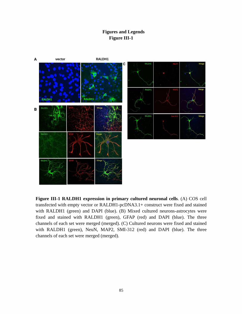
85
Figures and Legends
Figure III-1
Figure III-1 RALDH1 expression in primary cultured neuronal cells. (A) COS cell
transfected with empty vector or RALDH1-pcDNA3.1+ construct were fixed and stained
with RALDH1 (green) and DAPI (blue). (B) Mixed cultured neurons-astrocytes were
fixed and stained with RALDH1 (green), GFAP (red) and DAPI (blue). The three
channels of each set were merged (merged). (C) Cultured neurons were fixed and stained
with RALDH1 (green), NeuN, MAP2, SMI-312 (red) and DAPI (blue). The three
channels of each set were merged (merged).

86
Figure III-2
Figure III-2 RALDH1 expression in adult rat hippocampus. (A) 2 month old adult
Sprague-Dawley rat hippocampus slice were fixed and stained with RALDH1 (green),
NeuN (red) and DAPI (blue). (B) Higher magnification of representative CA1 and CA3
region of hippocampus with same stain as in A. upper panel, 25x; lower panel, 100x.

87
Figure III-3
Figure III-3 RALDH1 expression in astrocytes and oligodendrocytes in
hippocampus. (A) upper panel, two representative regions of adult rat hippocampus
were stained with RALDH1 (green), GFAP (red) and DAPI (blue). Lower panel, higher
magnification of selected regions in upper panel with same staining. (B) upper panel,
mixed cultured neuron-glia were fixed and stained with RALDH1 (green) and MBP (red).
The three channels of each set were merged (merged). Lower panel left, CA3 region of
adult rat hippocampus were stained with RALDH1(green), MBP(red) and DAPI (blue);
lower panel right, higher magnification of selected region with same staining.
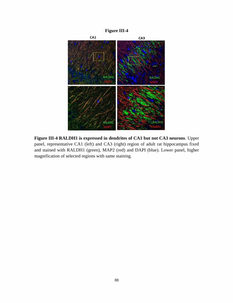
88
Figure III-4
Figure III-4 RALDH1 is expressed in dendrites of CA1 but not CA3 neurons. Upper
panel, representative CA1 (left) and CA3 (right) region of adult rat hippocampus fixed
and stained with RALDH1 (green), MAP2 (red) and DAPI (blue). Lower panel, higher
magnification of selected regions with same staining.

89
Figure III-5
Figure III-5 RADLH1 is co-localized with axons present in CA3 region. (A) Cultured
individual neuron was fixed and stained with RALDH1 (green), Tau (red) and DAPI
(blue). (B) Representative CA3 region of adult hippocampus were fixed and stained with
Tau (green), RALDH1 (red) and DAPI (blue). (C) Higher magnification of selected
regions in B with same staining. The three channels were merged (merged).

90
Figure III-6
Figure III-6 RALDH1 is expressed on mossy fibers. Three representative areas of CA3
region of adult hippocampus were fixed and stained with synaptoporin (green), RALDH1
(red) and DAPI.

91
Figure III-7
Figure III-7 Developmental changes of RALDH1 expression in adult rat
hippocampus. Left panel, hippocampus from P2, P7, P14 and P21 Sprague-Dawley rats
were fixed and stained with RALDH1 (green), NeuN (red) and DAPI (blue). Right panel,
higher magnification of selected regions in left panel with same staining.

92
References
Aoto, J., C. I. Nam, et al. (2008). "Synaptic signaling by all-trans retinoic acid in
homeostatic synaptic plasticity." Neuron 60(2): 308-320.
Asson-Batres, M. A. and W. B. Smith (2006). "Localization of retinaldehyde
dehydrogenases and retinoid binding proteins to sustentacular cells, glia,
Bowman's gland cells, and stroma: potential sites of retinoic acid synthesis in the
postnatal rat olfactory organ." J Comp Neurol 496(2): 149-171.
Bradl, M. and H. Lassmann (2010). "Oligodendrocytes: biology and pathology." Acta
Neuropathol 119(1): 37-53.
Chen, C. and S. Tonegawa (1997). "Molecular genetic analysis of synaptic plasticity,
activity-dependent neural development, learning, and memory in the mammalian
brain." Annu Rev Neurosci 20: 157-184.
Douville, J., R. Beaulieu, et al. (2009). "ALDH1 as a functional marker of cancer stem
and progenitor cells." Stem Cells Dev 18(1): 17-25.
Ginestier, C., M. H. Hur, et al. (2007). "ALDH1 is a marker of normal and malignant
human mammary stem cells and a predictor of poor clinical outcome." Cell Stem
Cell 1(5): 555-567.
Gross, C. G. (1993). "Hippocampus minor and man's place in nature: a case study in the
social construction of neuroanatomy." Hippocampus 3(4): 403-415.
Jiang, F., Q. Qiu, et al. (2009). "Aldehyde dehydrogenase 1 is a tumor stem
cell-associated marker in lung cancer." Mol Cancer Res 7(3): 330-338.
Kempf, M., A. Clement, et al. (1996). "Tau binds to the distal axon early in development
of polarity in a microtubule- and microfilament-dependent manner." J Neurosci
16(18): 5583-5592.
Kobayashi, K. (2010). "Hippocampal mossy fiber synaptic transmission and its
modulation." Vitam Horm 82: 65-85.
Markiewicz, I. and B. Lukomska (2006). "The role of astrocytes in the physiology and
pathology of the central nervous system." Acta Neurobiol Exp (Wars) 66(4):
343-358.
McBain, C. J. (2008). "Differential mechanisms of transmission and plasticity at mossy
fiber synapses." Prog Brain Res 169: 225-240.
Mey, J. and S. Hammelmann (2000). "OLN-93 oligodendrocytes synthesize
all-trans-retinoic acid in vitro." Cell Tissue Res 302(1): 49-58.
Montague, P., A. S. McCallion, et al. (2006). "Myelin-associated oligodendrocytic basic
protein: a family of abundant CNS myelin proteins in search of a function." Dev
Neurosci 28(6): 479-487.
Pereira, F., E. Rosenmann, et al. (1991). "The 56 kDa androgen binding protein is an
aldehyde dehydrogenase." Biochem Biophys Res Commun 175(3): 831-838.
Sun, T., H. S. Xiao, et al. (2006). "Differential expression of synaptoporin and
synaptophysin in primary sensory neurons and up-regulation of synaptoporin after
93
peripheral nerve injury." Neuroscience 141(3): 1233-1245.
Witter, M. P., P. A. Naber, et al. (2000). "Cortico-hippocampal communication by way of
parallel parahippocampal-subicular pathways." Hippocampus 10(4): 398-410.
Yamauchi, K. and J. Nakajima (2002). "Effect of coenzymes and thyroid hormones on the
dual activities of Xenopus cytosolic thyroid-hormone-binding protein (xCTBP)
with aldehyde dehydrogenase activity." Eur J Biochem 269(8): 2257-2264.
Yamauchi, K., J. Nakajima, et al. (1999). "Xenopus cytosolic thyroid hormone-binding
protein (xCTBP) is aldehyde dehydrogenase catalyzing the formation of retinoic
acid." J Biol Chem 274(13): 8460-8469.

94
Chapter IV
TNFα Regulates all-trans-RA Biosynthesis in Astrocytes Through
Oppositely Changing RALDH1 and RALDH3 Expression
Introduction
Vitamin A deficiency can cause profound reduction of resistance to infection, which
indicates that retinoids play an important role in the maintenance and regulation of
normal immune system function (Kim, 2008). Overall, RA signaling appears to establish
a Th2-type T cell non-inflammatory environment (Pino-Lagos et al., 2010). atRA
regulates NO production, enhances IL-1 production and inhibits TNFα production in
monocyte/macrophages (Dheen et al., 2005; Kim, 2008; Wang et al., 2007). RA can also
regulate migration of dendritic cells (DC) to present antigen to T cells. Not only gut
dendritic cells, but also splenic DCs synthesize RA to regulate intestine immunity
(Darmanin et al., 2007; Geissmann et al., 2003). Vitamin A also improves antitumor
immunity through mechanisms such as induction of cell differentiation and enhancement
of migration to lymph nodes (Malkovsky et al., 1983; Mirza et al., 2006). RA synthesis in
the immune system is regulated by a wide spectrum of inflammatory mediators, such as
IL-4, TLR receptors agonist and atRA acts as an important modulator of immune system
(Yokota et al., 2009).
CNS immunity is important for the trauma recovery and for age related
neurodegenerative diseases, such as Alzheimer‘s disease (Hohlfeld et al., 2007).
Microglia activation in response to an insult—secretion of a series of inflammatory
cytokines, determines the magnitude of neuroinflammatory response (Yang et al., 2010).
Glia cells, mainly microglia and astrocytes, become ‗activated‘ or ‗reactivated‘ in
response to various insults and secret proinflammatory cytokines, chemokines, and
trophic factors to activate neural immunity (Popovich and Longbrake, 2008). One of the
important proinflammatory cytokines secreted in the brain immune response is TNFα
(Sriram and O'Callaghan, 2007). TNFα is synthesized as a 26-kDa membrane-bound
polypeptide precursor that is cleaved by proteolysis to a 12-kDa subunit. TNFα exerts its
effects in autocrine and/or paracrine fashions (Wajant et al., 2003). The biological actions
of TNFα are mediated through two distinct cell surface receptors, TNFR1 (p55) and
TNFR2 (p75). TNFα exhibits both neurotoxic and neuroprotective roles in the adult CNS,
especially during the recovery after CNS injury (Perry et al., 2002).
TNFα contributes to the normal function of the hippocampus. Following transit global
ischemia, TNFα was selectively induced in the striatum and hippocampus, but not in
other brain areas (Buttini et al., 1996; Meistrell et al., 1997). Furthermore, expression of

95
TNFα after ischemic brain injury first appeared in neurons and then in astrocytes. TNFα
also plays a neuroprotective role against excitotoxic injury in the hippocampus, and
mediates neuroprotection in response to acute nitric oxide excitotoxicity (Cheng et al.,
1994; Turrin and Rivest, 2006). A lack of highly expressed TNFα is associated with
significant chemically induced damage to hippocampus (Little et al., 2002). Mice null of
both TNFα receptors are more susceptible to hippocampal excitotoxic and ischemic
injury (Bruce et al., 1996).
Astrocytes synthesize and secret TNFα, whereas TNFα can stimulate proliferation of
astrocytes, which is called reactive astrogliosis, a characteristic response of astrocytes to
inflammation and trauma of adult CNS (Balasingam et al., 1994). TNFα receptor 2 was
quickly and highly induced in astrocytes upon TNFα treatment, which was considered as
the potential mechanism that TNFα used to stimulate proliferation of astrocytes.
Because astrocytes can synthesize atRA through multiple retinol dehydrogenases and
retinal dehydrogenases, and TNFα is an active regulator of astrocyte activity in the CNS,
we tested whether there was any connection between these two important modulators.
TNFα performs its regulatory effect through two kinds of receptors TNF R1 and TNF R2
and activates various signaling pathways, such as NF-κB, c-JNK inducing MAP kinase
cascade or p38-MAP kinase cascade (Wajant et al., 2003). Our data show that TNFα can
regulate retinal dehydrogenation through oppositely changing RALDH1 and RALDH3
expression. Signaling pathways associated with RALDH1 and RALDH3 regulation by
TNFα is different. We also found that TNFα‘s regulation on changing atRA biosynthesis
is more effective in young astrocytes.
96
Materials and Methods
Cell culture and treatment--Primary astrocytes were cultured as described in Chapter II. 1
month old primary astrocytes were trypsinized and reseeded in 6-well plates at the
concentration of 2x10⁵/well 24 hr before the treatment. Medium were changed the day
before the experiments. All of the stimulis, including estrogen (sigma), LPS (sigma),
PGE2 (sigma) and TNFα (Invitrogen) were dissolved in water to make the stock solution
according to the product manuscript and stored properly. Upon treatment, the stock
solution were diluted to desired concentration in the medium and applied on the cultured
astrocytes. After 12 hr to 48 hr treatment, new medium containing either retinol or retinal
were replaced and incubated for additional 4 hr, retinoids production were assayed and
measured as described before.
RT-PCR, Q-PCR and western blot --Same as described in Chapter II, materials and
methods
Inhibition of TNFα signal pathways--The inhibitors for TNFα signal pathways were
purchased from Enzo life sciences, Inc., including NF-κB pathway inhibitors IKK-NBD,
MAP kinase kinase inhibitor PD98059, P38 inhibitor SB203580 and JNK inhibitor
SP600125. All the inhibitors were dissolved in proper solvent as a stock solution
according to the product manuscript. Astrocytes medium were changed the day before the
experiment. A final concentration of 20 μM IKK-NBD were pretreated astrocytes for 3 hr,
and a final concentration of 20 μM PD98059, SB203580 and SP600125 were pretreated
astrocytes for 1 hr, then 50 ng/ml TNFα were added to the medium for additional 24 hr
incubation. Q-PCR of RALDH1 and RLADH3 expression and atRA production from
retinol and retinal were detected as described before.

97
Results
Estrogen, LPS, PGE2 and TNFα don’t affect atRA production from retinol in 1 month old
astrocytes—To test the effect of different stimuli on atRA production in astrocytes, atRA
production from retinol in 1 month old astrocytes were assayed after incremental time
incubation with LPS, PGE2 or TNFα. Up to 24 hr incubation of 1 μM PGE2, 1μg/ml LPS
or 50 ng/ml TNFα did not change atRA production from retinol (Figure IV-1A). Dose
response of these stimuli was also tested. 10 μM estrogen had no effect on atRA
production, but 50 μM and 100 μM estrogen slightly decreased atRA biosynthesis from
retinol after 3 days treatment (Figure IV-1B). However, this high concentration of
estrogen also cause significant cell death, which indicated that such high concentration
estrogen might be toxic to astrocytes (data not shown). Up to 10 μg/ml LPS and up to 100
ng/ml TNFα had no effect on atRA synthesis from retinol (Figure IV-1C and 1D) in 1
month old astrocytes. All these data suggest that atRA homeostasis is fairly stable in 1
month old astrocytes and resistant to these extracellular stimuli.
TNFα treatment impaired atRA production from retinal via inversely regulation of
RALDH1 and RALDH3 expression in 1 month old astrocytes--Although TNFα didn‘t
affect atRA biosynthesis from retinol, retinal dehydrogenation activity of astrocytes was
significantly reduced after TNFα treatment (Figure IV-2A and 2B). 1 month old
astrocytes pre-incubated with 100 ng/ml TNFα for 24 hr synthesized significantly less
atRA from retinal compared with control group and the reduction was more robust after
48 hr pre-incubation, which indicated that a translational change or activity impairment
of retinal dehydrogenase occurred upon TNFα treatment (Figure IV-2A). Dose response
of TNFα treatment on astrocytes revealed that as low as 10 ng/ml TNFα treatment could
result in the similar reduction of retinal dehydrogenation activity as 100 ng/ml TNFα
treatment (Figure IV-2B). RALDH1 and RALDH2 play key roles in atRA production
from retinal in astrocytes. To test whether reduction of retinal dehydrogenation after
TNFα treatment is due to the change of RALDH activity, the expression level of
RALDHs were measured either after different times of incubation or under different
concentrations of TNFα (Figure IV-2C and 2D). A significant TNFα incubation time
dependent and dose dependent decrease of RALDH1 expression was found. The time
dependent reduction of RALDH1 is consistent with the timeline of decrease retinal
dehydrogenation upon TNFα treatment. Surprisingly, TNFα incubation also resulted in a
robust increase of RALDH3 expression in the same time. Quantification analysis
revealed that decrease of RALDH1 expression and increase of RALDH3 expression
depended on the TNFα concentration (FigureIV-2E and 2F). TNFα induced decrease of
RALDH1 protein was confirmed by western-blot (Figure IV-2G). The opposite change of
RALDH1 and RALDH3 expression resulted in a net decrease of atRA production from
retinal, which indicated that increase of RALDH3 activity could not compensate the loss
of RALDH1 activity in 1 month old astrocytes. These results were also consistent with
previous conclusion that RALDH1 is the major retinal dehydrogenase that contributes to

98
atRA biosynthesis.
TNFα treatment result in different atRA production in 2 weeks and 3 months
astrocytes--There was a significant increase of RALDH1 expression along with extended
culture time in primary astrocytes. However, RALDH3 expression didn‘t change during
that period (Chapter II). To test whether TNFα can induce similar gene expression
changes in young age astrocytes, we compared RALDH1, RALDH3 expression and atRA
production from retinol or retinal in 2 weeks and 3 months old astrocytes. Same as 1
month old astrocyte, 100 ng/ml TNFα up to 48 hr treatment didn‘t change atRA
production from retinol (Figure IV-3A) but caused a time dependent decrease of atRA
production from retinal (Figure IV-3B). Opposite expression of RALDH1 and RALDH3
were also observed in 3 months old astrocytes, confirmed by RT-PCR and Q-PCR
analysis (Figure IV-3D). TNFα treatment induced similar expression change of RALDH1
and RALDH3 in 2 weeks old astrocytes as 1 month or 3 months old astrocytes (Figure
IV-3C). No other genes changes were observed in 2 weeks old astrocytes (data not
shown). However, a significant increase of atRA production from retinol and retinal were
observed in 2 weeks old astrocytes (Figure IV-3A and 3B). Increase of atRA production
from retinol and retinal started as early as 12 hr TNFα treatment, consistent with the
RALDH1 and RALDH3 mRNA change (Figure IV-3C and 3D). No additional increase
atRA accumulation were detected as elongation of incubation time with TNFα.
Interestingly, atRA synthesis from retinol increased around 3 fold while incubation with
retinal only increased ~1.5 fold. Q-PCR analysis revealed the relative amount of
RALDH1 and RALDH3 expression upon TNFα treatment in 2 weeks or 3 months
astrocytes (Figure IV-3C and 3D lower panel). In the absence of TNFα, consistent with
previous results, ~500 fold increase of RALDH1 expression were detected in 3 months
old astrocytes while RALDH3 didn‘t change. RALDH1 is always the predominant
RALDH according to their relative amount in 2 weeks and 3 months astrocytes. However,
in 2 weeks old astrocytes, upon TNFα induction, RALDH3, instead of RALDH1, become
the primary RALDH with highest expression level (~100 fold higher than RALDH1).The
increase amount of RALDH3 mRNA is much higher than the decrease amount of
RALDH1 mRNA. In contrast, in 3 months old astrocytes, though down regulation of
RALDH1 upon TNFα treatment , the predominant RALDH is still RALDH1, with ~5
fold higher expression level than the increased RALDH3. The decrease amount of
RALDH1 is much higher than the increase amount of RALDH3 mRNA. Besides
RALDH1 and RALDH3, we also observed an increased CRBPI expression in both 2
weeks and 3 months astrocytes (Figure IV-3C and 3D, upper panel). All these data
indicate that the different change of atRA production upon TNFα treatment in 2 weeks
and 3 months astrocytes might be due to the different absolute amount change of
RALDH1 and RALDH3.
TNFα regulate RLADH1 and RALDH3 expression through different signal
pathways--The opposite regulation of RALDH1 and RALDH3 expression in astrocytes
by TNFα raised the question that how the same stimulation can cause inverse effect. As
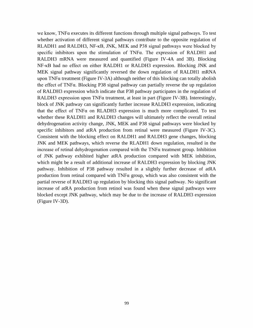
99
we know, TNFα executes its different functions through multiple signal pathways. To test
whether activation of different signal pathways contribute to the opposite regulation of
RLADH1 and RALDH3, NF-κB, JNK, MEK and P38 signal pathways were blocked by
specific inhibitors upon the stimulation of TNFα. The expression of RALDH1 and
RALDH3 mRNA were measured and quantified (Figure IV-4A and 3B). Blocking
NF-κB had no effect on either RALDH1 or RALDH3 expression. Blocking JNK and
MEK signal pathway significantly reversed the down regulation of RALDH1 mRNA
upon TNFα treatment (Figure IV-3A) although neither of this blocking can totally abolish
the effect of TNFα. Blocking P38 signal pathway can partially reverse the up regulation
of RALDH3 expression which indicate that P38 pathway participates in the regulation of
RALDH3 expression upon TNFα treatment, at least in part (Figure IV-3B). Interestingly,
block of JNK pathway can significantly further increase RALDH3 expression, indicating
that the effect of TNFα on RLADH3 expression is much more complicated. To test
whether these RALDH1 and RALDH3 changes will ultimately reflect the overall retinal
dehydrogenation activity change, JNK, MEK and P38 signal pathways were blocked by
specific inhibitors and atRA production from retinal were measured (Figure IV-3C).
Consistent with the blocking effect on RALDH1 and RALDH3 gene changes, blocking
JNK and MEK pathways, which reverse the RLADH1 down regulation, resulted in the
increase of retinal dehydrogenation compared with the TNFα treatment group. Inhibition
of JNK pathway exhibited higher atRA production compared with MEK inhibition,
which might be a result of additional increase of RALDH3 expression by blocking JNK
pathway. Inhibition of P38 pathway resulted in a slightly further decrease of atRA
production from retinal compared with TNFα group, which was also consistent with the
partial reverse of RALDH3 up regulation by blocking this signal pathway. No significant
increase of atRA production from retinol was found when these signal pathways were
blocked except JNK pathway, which may be due to the increase of RALDH3 expression
(Figure IV-3D).

100
Discussion
Retinoic acid is a powerful inducer for cell precursor differentiation. Chen‘s research
also proved that high concentration of RA can stimulate the outgrowth of neurite broadly
and quickly (Chen and Napoli, 2008). The concentration of atRA must be tightly
regulated in adult hippocampus to maintain the normal function of mature neurons and
induce the differentiation of neuron precursors. In previous chapters, we already
demonstrated that astrocytes can synthesize atRA in hippocampus but this ability is
tightly regulated by either the expression of multiple retinol and retinal dehydrogenases
or by feedback regulation of RA degradation and RE formation through atRA itself.
However, we haven‘t found any other factors beyond the atRA itself which participate
into the regulation of atRA homeostasis in adult CNS.
In reproductive system, Ong and his colleges have demonstrated that estrogen, a
primary female sex hormone, can regulate RALDH2 and CRBP expression to control
atRA production in uterus. The RA production ability is fluctuated along with the change
of estrogen level in female (Li et al., 2004; Li and Ong, 2003). Since hippocampus is also
able to synthesize and secret estrogen, we hypothesize that the estrogen in the
hippocampus can regulate atRA production (Kretz et al., 2004). The other important
system that might interact with retinoids signaling is the immune system. Lots of
evidence showed that retinoids, especially atRA is an important inducer and regulator of
the function of immune system, such as, secretion of different inflammatory cytokines
like IL-6 and IFNγ (Pino-Lagos et al., 2010). PGE2, LPS and TNFα also play important
functions in regulating the immune response by inducing the expression of these
cytokines (Sriram and O'Callaghan, 2007; Zolfaghari et al., 2007). In central nerve
system, astrocytes and microglia are two major cell types that respond to environmental
insult and regulate the immune response. Since we proved that astrocytes could
synthesize and secret atRA, we are wondering whether these factors that can induce
inflammatory cytokines up regulation can also alert atRA synthesis to exhibit their
functions.
Our data revealed that none of these factors, including estrogen, LPS, PGE2 and TNFα
can affect atRA synthesis from retinol in 1 month old astrocytes. However, TNFα
significantly reduced atRA production from retinal in the same age astrocytes which
indicated that TNFα could affect retinal dehydrogenation activity in astrocytes. Decrease
of RALDH1 upon TNFα treatment can contribute to the decrease of atRA synthesis from
retinal. Surprisingly, we also detected a robust increase of RALDH3 expression which
should have reversal effect on atRA production from retinal. The up regulation of
RALDH3 reached the maximum level earlier than decrease expression of RALDH1—
there was no further increased expression of RALDH3 from 12h to 48h whereas a
continuous decrease of RALDH1 expression was found upon TNFα treatment. So the
response of RALDH3 to TNFα may be much earlier and more sensitive than RALDH1
because as low as 10 ng/ml TNFα can induce dramatic increase of RALDH3 but not on

101
RALDH. This quick and sensitive response of RALDH3 upon TNFα treatment indicates
that RALDH3 might be the enzyme that responds to TNFα in the early inflammation
period to regulate atRA production. The longer response time and higher response dose of
RALDH1 expression upon TNFα treatment indicate that change of RALDH1 might be a
secondary effect of TNFα treatment. Whether increase of RALDH3 is a precondition for
down regulation of RALDH1 is unclear and need to be further investigated upon the
deletion of RALDH3 in astrocytes.
The net effect of increase of RALDH3 and decrease of RALDH1 in 1 month old
astrocytes is to cause a decrease of atRA production from retinal after at least 24 hr
treatment. However, this reverse change of two enzymes which have similar catalytic
activity sounds biologically inefficient. Considering that RALDH1 is only highly
expressed in pure cultured astrocytes but not in mixed culture or in hippocampus in vivo,
the response we found in these one month old astrocytes might not reflect the real
situation in vivo. We utilized much younger—2weeks old astrocytes, which exhibit fair
low RALDH1 level, to investigate whether the quick increase of RALDH3 has any
significant biological effect upon TNFα treatment. Different from 1months and 3months
old astrocytes, these 2weeks old, low RALDH1 expression astrocytes showed a dramatic
response to TNFα treatment and upon this stimuli, atRA production was significantly
increased from both retinol and retinal, indicating that in astrocytes with low expression
of RALDH1, TNFα can up regulate atRA biosynthesis through increase RALDH3
expression. In other words, in low RALDH1 expression astrocytes, the increase amount
of RALDH3 expression upon TNFα stimulation can overcome the decrease of RALDH1
expression, result in a net increase of atRA production. Vice versa, in high RALDH1
expression astrocytes, such as pure astrocytes older than 1 month, the increase of
RALDH3 cannot compensate the decrease of RALDH1 which result in a net decrease of
atRA production from retinal.
The 2 weeks old astrocytes not only mimic the low RALDH1 expression astrocytes‘
response to TNFα treatment, but may also reflect the response of new formed astrocytes
to elevated TNFα level in the regeneration after CNS trauma. In this pathological
condition, increased TNFα level is usually one of the representative phenomenon
indicating the activation of immune response in the injury area (Sriram and O'Callaghan,
2007). Proliferation and activation of newly formed astrocytes is also present after CNS
trauma (Balasingam et al., 1994). According to our data, elevated TNFα might quickly
stimulate the increase expression of RALDH3, which cause increase secretion of atRA
from astrocytes and these atRA is known to play an important role in brain trauma
recovery (Maden and Hind, 2003; Mey et al., 2005; Schrage et al., 2006; Yee and
Rawson, 2000). The decrease of RALDH1 expression upon TNFα treatment might be a
secondary effect responding to increase of atRA production. However, inhibition of
TNFα signaling pathway revealed that this change can be directly abolished by blocking
JNK and MEK signaling pathway, indicating that TNFα can directly regulate RALDH1
expression. The biological meaning of opposite down regulation RALDH1 remained
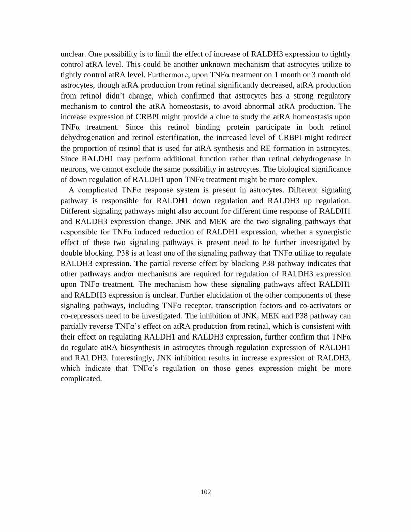
102
unclear. One possibility is to limit the effect of increase of RALDH3 expression to tightly
control atRA level. This could be another unknown mechanism that astrocytes utilize to
tightly control atRA level. Furthermore, upon TNFα treatment on 1 month or 3 month old
astrocytes, though atRA production from retinal significantly decreased, atRA production
from retinol didn‘t change, which confirmed that astrocytes has a strong regulatory
mechanism to control the atRA homeostasis, to avoid abnormal atRA production. The
increase expression of CRBPI might provide a clue to study the atRA homeostasis upon
TNFα treatment. Since this retinol binding protein participate in both retinol
dehydrogenation and retinol esterification, the increased level of CRBPI might redirect
the proportion of retinol that is used for atRA synthesis and RE formation in astrocytes.
Since RALDH1 may perform additional function rather than retinal dehydrogenase in
neurons, we cannot exclude the same possibility in astrocytes. The biological significance
of down regulation of RALDH1 upon TNFα treatment might be more complex.
A complicated TNFα response system is present in astrocytes. Different signaling
pathway is responsible for RALDH1 down regulation and RALDH3 up regulation.
Different signaling pathways might also account for different time response of RALDH1
and RALDH3 expression change. JNK and MEK are the two signaling pathways that
responsible for TNFα induced reduction of RALDH1 expression, whether a synergistic
effect of these two signaling pathways is present need to be further investigated by
double blocking. P38 is at least one of the signaling pathway that TNFα utilize to regulate
RALDH3 expression. The partial reverse effect by blocking P38 pathway indicates that
other pathways and/or mechanisms are required for regulation of RALDH3 expression
upon TNFα treatment. The mechanism how these signaling pathways affect RALDH1
and RALDH3 expression is unclear. Further elucidation of the other components of these
signaling pathways, including TNFα receptor, transcription factors and co-activators or
co-repressors need to be investigated. The inhibition of JNK, MEK and P38 pathway can
partially reverse TNFα‘s effect on atRA production from retinal, which is consistent with
their effect on regulating RALDH1 and RALDH3 expression, further confirm that TNFα
do regulate atRA biosynthesis in astrocytes through regulation expression of RALDH1
and RALDH3. Interestingly, JNK inhibition results in increase expression of RALDH3,
which indicate that TNFα‘s regulation on those genes expression might be more
complicated.

103
Figures and Legends
Figure IV-1
Figure IV-1 atRA production from retinol in 1 month old astrocytes remains stable
upon different stimulis. (A) 1 month old primary astrocytes were treated with 1 μM
PGE2, 1 μg/ml LPS and 50 ng/ml TNFα for the time indicated, atRA production from 2
μM ROL for 4 hr incubation were quantified and normalized to control group.
(B)Primary astrocytes cultured for 1 month were incubated for 3 days with graded
concentrations of estrogen, atRA production were quantified and normalized to
non-treated group. (C)Primary astrocytes cultured for 1 month were incubated for 12 hr
with graded concentrations of LPS, atRA production from 2 μM retinol 4 hr incubation
were quantified and normalized to non-treated group. (D)Primary astrocytes cultured for
1 month were incubated for 24 hr with graded concentrations of TNFα, atRA production
0 5 10 15 20 250.6
0.8
1.0
1.2
1.4 1uM PGE
1ug/ml LPS
50ng/ml TNF
hr
Re
lati
ve
RA
pro
du
cti
on
fro
m R
OL
0 10 50 1000.0
0.5
1.0
Estrogen (nM)
rela
tiv
e R
A p
rod
uc
tio
n f
rom
RO
L
0 0.1 1 100.0
0.5
1.0
LPS (ug/ml)
Re
lati
ve
RA
pro
du
cti
on
fro
m R
OL
0 10 50 1000.0
0.5
1.0
TNFα (ng/ml)
Re
lati
ve
RA
pro
du
cti
on
fro
m R
OL
A
B C
D

104
from 2 μM retinol 4 hr incubation were quantified and normalized to non-treated group
Figure IV-2
Figure IV-2 TNFα affect retinal dehydrogenation in astrocytes through oppositely
changing RALDH1 and RALDH3 expression. 1 month astrocytes were incubated with
100 ng/ml TNFα for the time indicated, atRA production from 1 μM retinal for 4 hr
incubation were quantified and normalized to untreated group (A); mRNA of RALDH1
and RALDH3 expression were measured by RT-PCR (C). Primary astrocytes cultured for
1 month were incubated for 24 hr with graded concentrations of TNFα, atRA production
0 10 50 1000.0
0.5
1.0
*
TNF α (ng/ml)
**
Rela
tiv
e a
tRA
pro
du
cti
on
fro
m R
AL
0.6
0.8
1.0
1.2
*
*
0 12 24 48
hr
100ng/ml TNFα
Rela
tiv
e a
tRA
pro
du
cti
on
fro
m R
AL
0 10 50 1000
1000
2000
3000
*
*
TNF α (ng/ml)
Rela
tiv
e R
ald
h1 m
RN
A
0 10 50 1000
20
40
60
80
**
TNF α (ng/ml)
*
Rela
tiv
e R
ald
h3 m
RN
ARaldh1
Raldh3
β-actin
0 12 24 48
TNFα hr
Raldh1
Raldh3
β-actin
0 10 50 100
TNFα (ng/ml)
Raldh1
β-actin
0 10 50 100
TNFα (ng/ml)
A B
DC
FE
G

105
incubated with 1 μM retinal were quantified and normalized to untreated group (B);
mRNA of Raldh1 and Raldh3 expression were measured by RT-PCR (D) and quantified
by Q-PCR (E-F); Protein level of RALDH1 were measured by western-blot(G)
106
Figure IV-3
Figure IV-3 TNFα induces similar change of RALDH1 and RALDH3 expression but
different atRA production in 2weeks and 3months old astrocytes. A and B) 2 weeks
or 3 months astrocytes were incubated with 100 ng/ml TNFα for 12 hr, 24 hr and 48 hr,
atRA production from 2 μM retinol or 1 μM retinal 4 hr incubation were measured. C and
D) RALDH1, RALDH3 and CRBPI mRNA were detected by RT-PCR (upper panel),
RALDH1 and RALDH3 mRNA were quantified by Q-PCR (lower panel) from 2 weeks
old (C) or 3 months (D) astrocytes which received same TNFα treatment as A and B.
*P<0.01, n=3
2week 3month0
20
40
60
80
100
120con 12h
100ng/ml TNFα2um ROL 4h
* **
24h 48hR
A(p
mo
l/m
illo
n c
ells)
2week 3month0
20
40
60
400
500
600
700con 12h
100ng/ml TNFα1um RAL 4h
** *
24h
**
48h
RA
(pm
ol/m
illo
n c
ells)
RALDH1 RALDH30
50
100
150
200
0h 12h 24h 48h
* **
*
*
*
rela
tiv
e m
RN
A
RALDH1 RALDH30
1000
2000
3000
0h 12h 24h 48h
***
* **
rela
tiv
e m
RN
A
A B
C D
107
Figure IV-4
Figure IV-4 TNFα affect RALDH1 expression via JNK and MEK pathway;
RALDH3 expression partially via P38 pathway. 1 month old primary astrocytes were
incubated with 50 ng/ml TNFα for 24 hr after pre-treatment with either 20 μM NBD
(NF-κB inhibitor) for 3 hr, or 20 μM SP600125 (JNK inhibitor), PD98059 (MEK
inhibitor), SB253058 (P38 inhibitor) for 1 hr, separately. (A) mRNA of RALDH1 were
detected by RT-PCR (upper panel) and quantified by Q-PCR (lower panel) .(B) mRNA of
Raldh3 were detected by RT-PCR (upper panel) and quantified by Q-PCR (lower panel).
(C-D) Primary astrocytes which were pretreated with same inhibitors as indicated in A
were incubated with 50 ng/ml TNFα for 48 hr, atRA production from 1 μM retinal (C) or
2 μM retinol (D) were quantified and normalized to untreated group. a,b,c,d indicate
statistically different groups; columns labeled with different letters are significant
different with each other.
Raldh1
β-actin
-TNFαNBDSP600125PD98059SB253085
----
+
-
-
--
+
-+--
+
--+-
+
+
-
--
+
---+
Raldh3
β-actin
-TNFαNBDSP600125PD98059SB253085
----
+
-
-
--
+
-+--
+
--+-
+
+
-
--
+
---+
contr
olTN
F
NBD+T
NF
SP60
0125
+TNF
PD98
059+
TNF
SB20
3580
+TNF
0
500
1000
Re
lati
ve
Ra
ldh
1 m
RN
A
a
b b b
C C
contr
olTN
F
NBD+T
NF
SP60
0125
+TNF
PD98
059+
TNF
SB20
3580
+TNF
0
50
100
150
Re
lati
ve
Ra
ldh
3 m
RN
A
a
b
b
d
C
b
0.0
0.5
1.0
1.5
Re
lati
ve
atR
A p
rod
uc
tio
nfr
om
RA
L
a
bb d
a
c
-TNFαNBDSP600125PD98059SB253085
----
+
-
-
--
+
-+--
+
--+-
+
+
-
--
+
---+
-TNFαNBDSP600125PD98059SB253085
----
+
-
-
--
+
-+--
+
--+-
+
+
-
--
+
---+
-TNFαNBDSP600125PD98059SB253085
----
+
-
-
--
+
-+--
+
--+-
+
+
-
--
+
---+
-TNFαNBDSP600125PD98059SB253085
----
+
-
-
--
+
-+--
+
--+-
+
+
-
--
+
---+
-TNFαNBDSP600125PD98059SB253085
----
+
-
-
--
+
-+--
+
--+-
+
+
-
--
+
---+
-TNFαNBDSP600125PD98059SB253085
----
+
-
-
--
+
-+--
+
--+-
+
+
-
--
+
---+
-TNFαNBDSP600125PD98059SB253085
----
+
-
-
--
+
-+--
+
--+-
+
+
-
--
+
---+
-TNFαNBDSP600125PD98059SB253085
----
+
-
-
--
+
-+--
+
--+-
+
+
-
--
+
---+
0.0
0.5
1.0
1.5
2.0R
ela
tiv
e a
tRA
pro
du
cti
on
fro
m R
OL
a
b
a
a
c
a
-TNFαNBDSP600125PD98059SB253085
----
+
-
-
--
+
-+--
+
--+-
+
+
-
--
+
---+
-TNFαNBDSP600125PD98059SB253085
----
+
-
-
--
+
-+--
+
--+-
+
+
-
--
+
---+
-TNFαNBDSP600125PD98059SB253085
----
+
-
-
--
+
-+--
+
--+-
+
+
-
--
+
---+
-TNFαNBDSP600125PD98059SB253085
----
+
-
-
--
+
-+--
+
--+-
+
+
-
--
+
---+
-TNFαNBDSP600125PD98059SB253085
----
+
-
-
--
+
-+--
+
--+-
+
+
-
--
+
---+
-TNFαNBDSP600125PD98059SB253085
----
+
-
-
--
+
-+--
+
--+-
+
+
-
--
+
---+
-TNFαNBDSP600125PD98059SB253085
----
+
-
-
--
+
-+--
+
--+-
+
+
-
--
+
---+
-TNFαNBDSP600125PD98059SB253085
----
+
-
-
--
+
-+--
+
--+-
+
+
-
--
+
---+
BA
C D

108
References
Balasingam, V., T. Tejada-Berges, et al. (1994). "Reactive astrogliosis in the neonatal
mouse brain and its modulation by cytokines." J Neurosci 14(2): 846-856.
Bruce, A. J., W. Boling, et al. (1996). "Altered neuronal and microglial responses to
excitotoxic and ischemic brain injury in mice lacking TNF receptors." Nat Med
2(7): 788-794.
Buttini, M., K. Appel, et al. (1996). "Expression of tumor necrosis factor alpha after focal
cerebral ischaemia in the rat." Neuroscience 71(1): 1-16.
Chen, N. and J. L. Napoli (2008). "All-trans-retinoic acid stimulates translation and
induces spine formation in hippocampal neurons through a membrane-associated
RARalpha." FASEB J 22(1): 236-245.
Cheng, B., S. Christakos, et al. (1994). "Tumor necrosis factors protect neurons against
metabolic-excitotoxic insults and promote maintenance of calcium homeostasis."
Neuron 12(1): 139-153.
Darmanin, S., J. Chen, et al. (2007). "All-trans retinoic acid enhances murine dendritic
cell migration to draining lymph nodes via the balance of matrix
metalloproteinases and their inhibitors." J Immunol 179(7): 4616-4625.
Dheen, S. T., Y. Jun, et al. (2005). "Retinoic acid inhibits expression of TNF-alpha and
iNOS in activated rat microglia." Glia 50(1): 21-31.
Geissmann, F., P. Revy, et al. (2003). "Retinoids regulate survival and antigen
presentation by immature dendritic cells." J Exp Med 198(4): 623-634.
Hohlfeld, R., M. Kerschensteiner, et al. (2007). "Dual role of inflammation in CNS
disease." Neurology 68(22 Suppl 3): S58-63; discussion S91-56.
Kim, C. H. (2008). "Roles of retinoic acid in induction of immunity and immune
tolerance." Endocr Metab Immune Disord Drug Targets 8(4): 289-294.
Kretz, O., L. Fester, et al. (2004). "Hippocampal synapses depend on hippocampal
estrogen synthesis." J Neurosci 24(26): 5913-5921.
Li, X. H., B. Kakkad, et al. (2004). "Estrogen directly induces expression of retinoic acid
biosynthetic enzymes, compartmentalized between the epithelium and underlying
stromal cells in rat uterus." Endocrinology 145(10): 4756-4762.
Li, X. H. and D. E. Ong (2003). "Cellular retinoic acid-binding protein II gene expression
is directly induced by estrogen, but not retinoic acid, in rat uterus." J Biol Chem
278(37): 35819-35825.
Little, A. R., S. A. Benkovic, et al. (2002). "Chemically induced neuronal damage and
gliosis: enhanced expression of the proinflammatory chemokine, monocyte
chemoattractant protein (MCP)-1, without a corresponding increase in
proinflammatory cytokines(1)." Neuroscience 115(1): 307-320.
Maden, M. and M. Hind (2003). "Retinoic acid, a regeneration-inducing molecule." Dev
Dyn 226(2): 237-244.
109
Malkovsky, M., C. Dore, et al. (1983). "Enhancement of specific antitumor immunity in
mice fed a diet enriched in vitamin A acetate." Proc Natl Acad Sci U S A 80(20):
6322-6326.
Meistrell, M. E., 3rd, G. I. Botchkina, et al. (1997). "Tumor necrosis factor is a brain
damaging cytokine in cerebral ischemia." Shock 8(5): 341-348.
Mey, J., J. M. D, et al. (2005). "Retinoic acid synthesis by a population of NG2-positive
cells in the injured spinal cord." Eur J Neurosci 21(6): 1555-1568.
Mirza, N., M. Fishman, et al. (2006). "All-trans-retinoic acid improves differentiation of
myeloid cells and immune response in cancer patients." Cancer Res 66(18):
9299-9307.
Perry, S. W., S. Dewhurst, et al. (2002). "Tumor necrosis factor-alpha in normal and
diseased brain: Conflicting effects via intraneuronal receptor crosstalk?" J
Neurovirol 8(6): 611-624.
Pino-Lagos, K., Y. Guo, et al. (2010). "Retinoic acid: A key player in immunity."
Biofactors.
Popovich, P. G. and E. E. Longbrake (2008). "Can the immune system be harnessed to
repair the CNS?" Nat Rev Neurosci 9(6): 481-493.
Schrage, K., G. Koopmans, et al. (2006). "Macrophages and neurons are targets of
retinoic acid signaling after spinal cord contusion injury." Eur J Neurosci 23(2):
285-295.
Sriram, K. and J. P. O'Callaghan (2007). "Divergent roles for tumor necrosis factor-alpha
in the brain." J Neuroimmune Pharmacol 2(2): 140-153.
Turrin, N. P. and S. Rivest (2006). "Tumor necrosis factor alpha but not interleukin 1
beta mediates neuroprotection in response to acute nitric oxide excitotoxicity." J
Neurosci 26(1): 143-151.
Wajant, H., K. Pfizenmaier, et al. (2003). "Tumor necrosis factor signaling." Cell Death
Differ 10(1): 45-65.
Wang, X., C. Allen, et al. (2007). "Retinoic acid enhances the production of IL-10 while
reducing the synthesis of IL-12 and TNF-alpha from LPS-stimulated
monocytes/macrophages." J Clin Immunol 27(2): 193-200.
Yang, I., S. J. Han, et al. (2010). "The role of microglia in central nervous system
immunity and glioma immunology." J Clin Neurosci 17(1): 6-10.
Yee, K. K. and N. E. Rawson (2000). "Retinoic acid enhances the rate of olfactory
recovery after olfactory nerve transection." Brain Res Dev Brain Res 124(1-2):
129-132.
Yokota, A., H. Takeuchi, et al. (2009). "GM-CSF and IL-4 synergistically trigger
dendritic cells to acquire retinoic acid-producing capacity." Int Immunol 21(4):
361-377.
Zolfaghari, R., C. J. Cifelli, et al. (2007). "Lipopolysaccharide opposes the induction of
CYP26A1 and CYP26B1 gene expression by retinoic acid in the rat liver in vivo."
Am J Physiol Gastrointest Liver Physiol 292(4): G1029-1036.
110
Chapter V
Summary and Future Directions
This dissertation established a retinoid homeostasis model in primary cultured
astrocytes and neurons, revealed specific RALDH1 expression pattern in vitro and in vivo,
and elucidated effects and mechanisms of TNFα on atRA biosynthesis in astrocytes. To
our knowledge, we are the first group to demonstrate that primary cultured astrocytes, but
not neurons, from hippocampus origin can synthesize atRA through multiple retinol
dehydrogenases and retinal dehydrogenases by an accurate LC/MS/MS technology. Our
work also systematically and comprehensively elucidates retinoids metabolism kinetics
(both time-course and dose-curve) and atRA feedback regulation on its own homeostasis.
Besides that, several novel phenomenon, maybe neural cells specific, were observed in
our research, including transportation of atRA from astrocytes to pyramidal neurons,
different subcellular locations of Raldh1 and Raldh2, up regulation of Raldh to
compensate for impairment of retinol dehydrogenation, culture neurons and mossy fiber
enriched Raldh1 expression pattern and opposite regulation of Raldh1 and Raldh3
expression upon TNFα treatment in astrocytes. All these data indicate that retinoid
metabolism in adult CNS, especially hippocampal neurons and astrocytes, not only
follow the known homeostasis model that has been found in other type of cells, but also
exhibit some undiscovered, maybe hippocampus specific, characteristics which may
imply CNS specific regulation of retinoids homeostasis model.
Part I Retinoids metabolism in cultured astrocytes and neurons
Determining the source of atRA in the rodent hippocampus was the initial purpose of
this research. The strength of our specific LC/MS/MS technology enables us to accurately
measure low level of retinoids isomers which is present in small volume of biological
samples, such as different loci of adult CNS. Around 3 pmol/g atRA in adult rat
hippocampus, plus the presence of high concentration of retinol and RE, suggest that
biosynthesis of atRA may be the way that hippocampus acquire the atRA for the normal
physiological function, which has been proved by RARβ null mice, VAD mice or aged
mice losing spatial learning and memory formation ability. We also considered the
possibility of obtaining atRA from circulation. However, the presence of only retinol
binding protein, but not CRABP, on blood-brain barrier and radiolabeled retinoids
chasing research minimize this possibility (MacDonald et al., 1990; Pardridge et al., 1985;
Werner and Deluca, 2002). The meninges is believed to serve as the major source of atRA
for early embryonic brain development and differentiation (Romand et al., 2008; Zhang
et al., 2003). Highly expressed Raldh2 in meninges and the comprehensive study of

111
Foxc1 mutant mice which lost meninges in forebrain indicate that atRA from meninges
during embryonic development is indispensible for neural precursor cells differentiation
(Siegenthaler et al., 2009). However, whether meninges is also an important source for
atRA in adult brain remained unclear. One possibility is that during embryonic
development, most precursors haven‘t fully developed and lack the ability to synthesize
atRA. Meninges then act as a temporal source to provide atRA to nearby neural
precursors to induce differentiation. In adult brain, the presence of functional glia cells
which gain the ability to synthesize atRA and the increased volume of the brain make it
unnecessary and impossible for the meninges to continue to provide atRA to the neurons
or other cell types which need atRA to perform normal function. Loss contact with
neurons also make meninges an unlike source for atRA in adult CNS. However, the
remaining high expression level of Raldh2 in adult meninges still indicate that it might
still play an important function in adult brain, and further investigation need to addressed
on this issue (McCaffery et al., 2006).
Since we exclude other possibilities, the most likely source of high atRA we detected
in adult hippocampus is from in situ biosynthesis. The presence of retinoids metabolism
enzymes and binding proteins and retinoic acid receptor in adult hippocampus support
this hypothesis. However, as we know, the adult brain, including hippocampus, is highly
heterogeneously. Neurons and glia cells are the two major cell types that fully
differentiate with different structures and functions. In glia cells, astrocytes account for
the majority of total cells and support the normal function of neurons. The objectives of
our project are to figure out i) how retinoids are metabolized in these two cell types and
which cell can synthesize atRA; ii) what enzymes, and to what extent, participate in
retinoids metabolism in these cells; iii) is there any factors that can affect retinoids
metabolism in these cells.
Previous research has provided some hints on retinoids metabolism in neurons and
astrocytes (See Chapter I). Research in our lab indicates that neurons can quickly respond
to atRA stimulation and incorporate RARα into neurons to suppress translation. Raldh1
was also found to be present in neurons. No direct evidence indicate that neurons can
synthesize atRA. In contrast, lots of indirect evidence indicate that astrocytes can
synthesize atRA, including coculture astrocytes with RAR reporter cell resulting in
positive signal (Kornyei et al., 2007); converting retinol to active form to induce neuron
survival only in the presence of astrocytes. However, neither of these evidence directly
proved atRA biosynthesis in astrocytes. We utilize the advantages of our LC/MS/MS
technology, which has been demonstrated to specifically and sensitively detected all
different isoforms of RA, to accurately detect the presence of atRA production from
astrocytes incubated with either retinol or retinal, and measure the kinetics parameters of
different retinoids metabolism reactions. According to these data, we conclude that: i)
atRA biosynthesis is only present in cultured astrocytes; ii) RE formation and retinal
reduction occur in both astrocytes and neurons, with similar rates under physiological
concentrations of substrate. We also showed cultured neurons can actively take up atRA

112
from astrocytes without the assistance of cell-cell contact. Besides that, atRA can
suppress its own biosynthesis through both up regulation of atRA degradation and RE
formation to redirect the flow of retinol in astrocytes. Neurons can also respond to atRA
to increase RE formation .
According to these data and the architecture of neurons and astrocytes position, we
established a general model how neurons and astrocytes utilize retinol as the substrate
and maintain a controlled level of atRA (Figure V-1). In this model, the substrate of atRA
biosynthesis is the RBP binding retinol which is circulating in the capillary all over the
adult brain. The endfeet of astrocytes covering the whole capillary, which is part of the
blood-brain barrier, make sure that only astrocytes get access to the retinol. Stra6, the
only known cell membrane receptor for RBP, has been detected in astrocytes (data now
shown). The uptake retinol will bind with CrbpI, whose presence was confirmed by
RT-PCR detection of messenger RNA. Two known fates of CRBP-retinol (holo-CRBP)
are present in astrocytes: esterification and dehydrogenation. The majority of retinol is
converted to RE—the storage form of retinoids, for further utilization. Esterification may
also serve as mechanism that astrocytes get rid of excess retinol. However, receptor relied
uptake of retinol should control the amount of substrate flow into the astrocytes and RE
formation may mainly serve as a backup mechanism in case any vitamin A deficiency.
The dramatic increase rate of RE formation under high concentration retinol indicates
that astrocyte has a broad capacity to handle large amount of retinol. atRA is synthesized
in astrocytes under strict regulation. RE formation is one of the major ways to control the
amount of retinol that can be oxidized. The high retinal reduction activity, atRA secretion
ability and atRA feedback autoregulation work together to tightly control the
concentration of atRA inside of astrocytes. The atRA remained in astrocytes may regulate
the some specific gene expression or function of astrocytes. The composition of whole
signaling pathway, such as receptors and binding proteins need to be further elucidated in
astrocytes.
Secretion of synthesized atRA from astrocytes and quickly induced spine growth of
hippocampal pyramidal neurons by atRA indicate that neurons might be able to take up
the atRA secreted by astrocytes for utilization. Our co-culture data support this
hypothesis and prove that cell-cell contact is not necessary for this atRA uptake. We don‘t
know whether the in vivo situation is same as what we observed in primary culture cells.
If it is the case, we speculate that atRA are secreted into the micro-environment around
the nearby neurons by astrocytes in hippocampus, and then taken up by neurons under a
specific, highly regulated mechanism. We don‘t know whether neurons can feedback
regulate the amount of atRA synthesized or secreted by astrocytes, and we also cannot
exclude the possibility that atRA is transported through direct neuron-astrocyte contact.
The in vivo transportation atRA need to be revealed by an accurate chasing method in
which atRA can be clearly labeled and visualized.
atRA can increase its own degradation and RE formation in astrocytes and neurons
indicate that the common feedback regulation mechanism is also present in CNS cells.

113
We don‘t know whether esterification of retinol is also present in neurons in vivo, or the
RE formation we observed in neurons is just an adaption of in vitro culture. However, it
at least indicate that neurons can gain the ability to protect themselves from toxic retinol
by converting them to RE. How atRA is utilized and metabolized in neurons remains
unclear. Knockout and knockdown of retinoids metabolizing enzymes and binding
proteins, such as CYP26 enzymes and CRABPs, need to be studied to elucidate the whole
atRA metabolism model inside of neurons.
Part II Enzymes that participate in the retinoids homeostasis in CNS
Previous atRA biosynthesis research usually focused on only one specific
dehydrogenase, for example Raldh2 during embryonic development. However, our
research confirmed that at least three retinol dehydrogenases (Rdh2, Rdh10 and Dhrs9)
and three retinal dehydrogenases (Raldh1, 2 and 3) contribute to atRA biosynthesis. We
didn‘t exclude other possible isoenzymes‘ participation in this process. The contribution
of each isoenzyme is different. However, to what extend this result can be applied to in
vivo status is unknown. We used one month old astrocyte to evaluate the contribution of
each enzyme. However, the culture time dependent increased expression of some, but not
all of the enzymes indicate that in vitro culture condition definitely affect the expression
level of these dehydrogenases. It‘s inappropriate to estimate the in vivo contribution of
any isoenzyme just according to the in vitro knockdown data.
The knockdown results indicate different contributions of each isoenzyme in cultured
astrocytes. Knockdown Raldh1 showed the most dramatic effect on decrease of atRA
biosynthesis, which is consistent with its highest expression level. However, according to
in vitro and in vivo immunostaining detection of its protein level, we found a fair low
expression of Raldh1 in astrocytes in the presence of neurons, either in co-cultured
system or in hippocampus slice. Besides that, Raldh1 is also detected in some unexpected
location, including the nuclear of astrocytes, all over the cultured neurons and enriched
expression on mossy fibers. All these results indicate that Raldh1 in adult hippocampus,
not only function as a retinal dehydrogenase, but also have some other specific functions
which need to be further addressed. Culture time dependent increase of Raldh1
expression and TNFα induced long term decrease expression of Raldh1 indicate that this
is a gene that can respond to environmental stimulation for a long term regulation of
retinoids homeostasis.
As an indispensable retinal dehydrogenase during embryonic CNS development, the
presence of Raldh2 is usually considered as the powerful evidence to imply the presence
of atRA biosynthesis. Our data also support this notion, with significant reduction of
retinal dehydrogenation when Raldh2 was knocked down; even its messenger RNA level
is about 500-1000 fold lower than Raldh1. Besides that, culture time dependent increased
Raldh2 expression also confirms its participation in atRA biosynthesis in astrocytes. The
perinuclear enriched expression pattern also suggests Raldh2 might contribute to
subcellular specific synthesis of atRA.

114
Knockdown of Raldh3 didn‘t result in as much change of atRA biosynthesis as its
isoenzymes. Raldh3 also didn‘t increase its expression level as Raldh1 and Raldh2 when
cultured long time in vitro. However, increased Raldh3 expression was observed when
stimulated by TNFα for less than 12 hr and result in increase of atRA biosynthesis.
According to these data, change of Raldh3 expression in astrocytes may serve as a quick
response to environmental change, such as different immune cytokines level, to regulate
atRA level.
The other possible function of Raldh is to act as a regulator to compensate for any
changes that might perturb atRA levels. According to our data, increase in Raldh1 mRNA
was observed when Dhrs9 was knocked down in cultured astrocytes; Raldh2 was
unregulated in Rdh1 and CrbpI knockout mice astrocytes. Dhrs9, mice Rdh1 (homolog of
rat Rdh2) and CrbpI are the enzymes or binding protein that participate in the first step
retinol oxidation. It seems like astrocytes will up regulate retinal oxidation to compensate
the disturbing of the retinol dehydrogenation. To our knowledge, this is a novel regulation
mechanism on atRA homeostasis which has never been reported before. The remaining
questions are: i) whether this is an astrocyte, or CNS specific regulation, or this
phenomenon is present in other tissues; ii) whether this is an in vitro culture dependent
phenomenon or up regulation of Raldh can also be detected in vivo; iii) The selection of
up regulation of Raldh1 or Raldh2 is species (rat vs. mouse) specific or linked to the
specific first step oxidation components (Dhrs9, Rdh1 and CrbpI); iv) the molecular and
cellular mechanism for up regulation of retinal dehydrogenases. As we know, no up
regulation of any Raldhs was detected in Rdh1 knockout mice liver. Our research also
excludes the possibility that retinal or atRA can regulate Raldh expression. To solve this
puzzle, new hypothesis need to be raised and tested.
Both Rdh2 and Rdh10 knockdown result in slightly decrease of atRA production from
retinol, and Dhrs9 knockdown result in increase of atRA production which is due to the
increase expression of Raldh1. The different response on Rdhs knockdown indicates that
they might have different impact on atRA biosynthesis. We didn‘t detect the subcellular
location of these Rdhs due to the lack of specific antibodies. Our results also can‘t
exclude the possibility that other retinol dehydrogenases may also contribute to the first
step oxidation. However, our data at least prove that more than one single retinol
dehydrogenase contribute to retinol dehydrogenation.
In our research, we only test the enzymes which function as a retinol dehydrogenase.
However, because astrocytes showed strong ability to reduce retinal, which is believed as
an important regulatory method to control atRA biosynthesis, we speculate that a balance
equalization system is present in astrocytes to control the first step oxidation. This system
is the bottleneck to limit the atRA synthesis by controlling how much retinal can be
utilized for oxidation. This balance system contains multiple retinol dehydrogenases,
retinal reductases and retinol/retinal binding proteins. Dhrs9‘s function in this system
needs to be confirmed by knocking it down in Raldh1 null astrocyte. Our research
demonstrated that Dhrs9 doesn‘t function as retinal reductase. Up regulation of CrbpII in

115
CrbpI knockout astrocytes indicate that both binding proteins are present and have
redundant function in astrocytes. Rdh2 and Rdh10 may also reflect the redundancy of
retinol oxidation in astrocytes. Future investigation should focus on other possible
members of this balance system and their contribution, especially the possible oxidase
and reductase and their preferred substrate concentration.
We proved that the major degradation enzyme for atRA degradation is Cyp26B1,
which can be induced by atRA itself, consistent with previous report. Cyp26B1, but not
Cyp26A1 or C1, has also been reported widely expressed in adult CNS. Consistent with
that, low level of Cyp26A1 was detected in cultured astrocytes and it cannot be regulated
by atRA itself. Increase Cyp26B1 expression was also observed in astrocytes which
exhibited spontaneously increased atRA biosynthesis, such as long time in vitro cultured
astrocytes or Rdh1 and CrbpI knockout astrocytes. Up regulation of Cyp26B1 in these
cells doesn‘t require exogenous atRA stimulation. If the regulation of Cyp26B1 does
require atRA, the subcellular localized atRA biosynthesis, for example, the specific
subcellular expression of Raldh1 and Raldh2, might provide a regional high
concentration atRA which can induce Cyp26B1 expression. As expected, Lrat was also
up regulated by atRA in astrocytes, which is consistent with increased RE formation.
These atRA induced gene changes establish the autoregulation of retinoids homeostasis in
astrocytes, which is also present in other system though the participants vary. This
variation can also be found in neurons. Our data demonstrate that cultured neurons can
also respond to atRA stimulation and increase RE formation. However, the only changed
component of retinoids signaling is CrbpI in neurons. The mechanism that causes this
different response is unclear. atRA delivery, recruiting co-activator or co-repressor, atRA
receptor expression pattern could be the directions for further investigation.
Part III Future Directions
Raldh1 is the enzyme broadly studied in our research. However, a lot of unexplained
phenomenon makes the follow up research on Raldh1‘s function in adult CNS a
fascinating work. The first unsolved problem is the different expression level of Raldh1
in different astrocytes. Culture time dependent robust increased Raldh1 expression makes
it the predominant retinal dehydrogenase in pure cultured astrocytes, which is confirmed
by siRNA knockdown. However, fairly low expression of this enzyme in mixed cultured
astrocytes with neurons and even lower and rarer expression in vivo make us doubt, to
what extent this enzyme contribute to atRA biosynthesis in adult hippocampus. Whether
the dramatic increase of Raldh1 expression in pure astrocytes is due to the presence of
inducer in environment or the lack the inhibition signals from neurons needs to be
addressed. If neurons do inhibit Raldh1 expression in astrocytes, figuring out this
suppression factors will be the major task which can provide the insight on how neurons
can regulate atRA biosynthesis. The studies focused on the regulation of Raldh1
expression will also provide some clues on the mechanism of how Dhrs9 knockdown can
up regulate Raldh1. The TNFα research indicates that JNK and MAPK signaling pathway
116
might participate in the regulation of Raldh1 expression. Further research focused on
these two signaling pathways might help to resolve this problem.
The second unsolved problem is the function other than retinal dehydrogenase of
Raldh1. Raldh1 was found highly expressed in cultured astrocytes nucleus, an unusual
location that has never been reported before. To further confirm this nucleus specific
expression pattern, nucleus protein need to be separated and Raldh1 protein level need to
be detected by western blot. Raldh1 was also detected in neurons—throughout cultured
neurons and specifically in CA1 neurons and mossy fibers. No evidence showed that
Raldh1 in neurons can dehydrogenate retinal though exogenous retinal can be taken up
by cultured neurons. The functions of Raldh1 in astrocytes nucleus and neurons remained
unclear. Raldh1 knockout mouse is a good model to study the specific function of Raldh1.
Cultured astrocytes and neurons from Raldh1 knockout mice will help us to further
evaluate its contribution to atRA biosynthesis. Any unusual phenomenon in either
cultured neural cells or histology of hippocampus will provide hints for generate new
hypothesis and direct followup research.
Raldh2 and Raldh3 are less studied compared with Raldh1. Similar questions are also
need to be addressed. For example, the mechanism of Raldh2 up regulation in Rdh1 and
CrbpI null astrocytes and TNFα induced P38 signaling dependent Raldh3 expression.
Because both Raldh2 and Raldh3 knockout mice are lethal, conditional CNS specific
Raldh2 and Raldh3 knockout mice might be an appropriate model to study their functions
in adult hippocampus. The elucidation of in vivo expression pattern of these two enzymes,
especially under different stress like VAD or inflammation, may help to understand the
specific functions of these multiple retinal dehydrogenases in adult CNS.
There are also some other projects that are related to retinoids homeostasis model in
adult hippocampus. One major project is to elucidate atRA metabolism in neurons. The
kinetics of atRA degradation in neurons, the enzymes that participate in atRA metabolism,
RE formation and retinal reduction, the mechanism of atRA induced up regulation of
CrbpI expression need to be addressed. The other project is to elucidate the functions and
contributions of binding proteins in adult CNS retinoids metabolism. One interesting
question is that since atRA has to bind with binding proteins all the time, which retinoids
binding proteins are essential for atRA secretion from astrocytes and transportation to
neurons. Besides binding proteins, are there any other components, such as RAR or
Raldh, also involved in atRA secretion and transportation?
Part IV Conclusion
Our research opens the gate towards elucidation of retinoids metabolism in adult
hippocampus. Consistent with previous report, our data directly demonstrate astrocytes,
but not neurons, can synthesize and secret atRA, which can be taken up by neurons,
through multiple retinol dehydrogenases and retinal dehydrogenases. TNFα may act as a
potential regulator to change atRA synthesis through affecting the retinal
dehydrogenation. The communication between the first and second step dehydrogenation
indicate a novel manner to regulate atRA homeostasis. These data indicate that glia cells

117
may serve as the major source of atRA in adult hippocampus. Since atRA play an
important role in adult brain and is associated with many pathological and age related
impairment of brain function, the elucidation of atRA biosynthesis and metabolism in
adult hippocampus would greatly help us to find out potential targets to manipulate atRA
level, which may contribute to the amelioration and treatment of retinoids related human
CNS diseases.
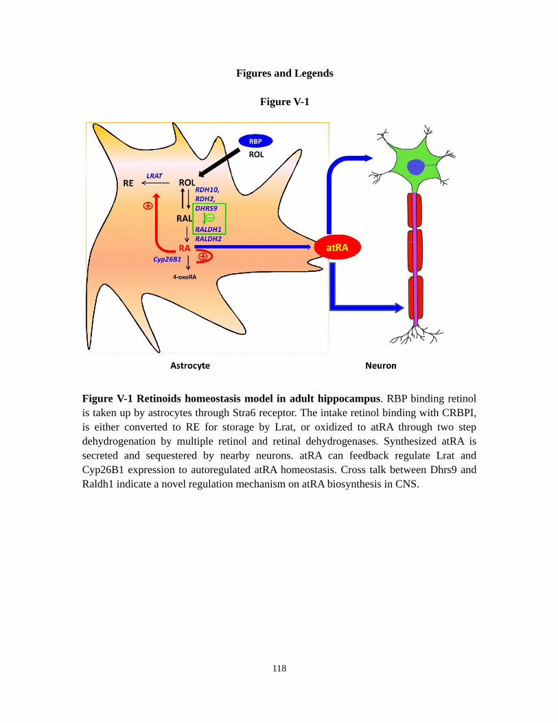
118
Figures and Legends
Figure V-1
Figure V-1 Retinoids homeostasis model in adult hippocampus. RBP binding retinol
is taken up by astrocytes through Stra6 receptor. The intake retinol binding with CRBPI,
is either converted to RE for storage by Lrat, or oxidized to atRA through two step
dehydrogenation by multiple retinol and retinal dehydrogenases. Synthesized atRA is
secreted and sequestered by nearby neurons. atRA can feedback regulate Lrat and
Cyp26B1 expression to autoregulated atRA homeostasis. Cross talk between Dhrs9 and
Raldh1 indicate a novel regulation mechanism on atRA biosynthesis in CNS.

119
References
Abu-Abed, S., Dolle, P., Metzger, D., Beckett, B., Chambon, P., and Petkovich, M.
(2001). The retinoic acid-metabolizing enzyme, CYP26A1, is essential for normal
hindbrain patterning, vertebral identity, and development of posterior structures. Genes
Dev 15, 226-240.
Akita, J., Takahashi, M., Hojo, M., Nishida, A., Haruta, M., and Honda, Y. (2002).
Neuronal differentiation of adult rat hippocampus-derived neural stem cells transplanted
into embryonic rat explanted retinas with retinoic acid pretreatment. Brain Res 954,
286-293.
Alique, M., Herrero, J.F., and Lucio-Cazana, F.J. (2007). All-trans retinoic acid induces
COX-2 and prostaglandin E2 synthesis in SH-SY5Y human neuroblastoma cells:
involvement of retinoic acid receptors and extracellular-regulated kinase 1/2. J
Neuroinflammation 4, 1.
Alliot, F., Delhaye-Bouchaud, N., Geffard, M., and Pessac, B. (1988). Role of astroglial
cell clones in the survival and differentiation of cerebellar embryonic neurons. Brain Res
Dev Brain Res 44, 247-257.
Alvarez-Buylla, A., and Kirn, J.R. (1997). Birth, migration, incorporation, and death of
vocal control neurons in adult songbirds. J Neurobiol 33, 585-601.
Aoto, J., Nam, C.I., Poon, M.M., Ting, P., and Chen, L. (2008). Synaptic signaling by
all-trans retinoic acid in homeostatic synaptic plasticity. Neuron 60, 308-320.
Asson-Batres, M.A., and Smith, W.B. (2006). Localization of retinaldehyde
dehydrogenases and retinoid binding proteins to sustentacular cells, glia, Bowman's gland
cells, and stroma: potential sites of retinoic acid synthesis in the postnatal rat olfactory
organ. J Comp Neurol 496, 149-171.
Balasingam, V., Tejada-Berges, T., Wright, E., Bouckova, R., and Yong, V.W. (1994).
Reactive astrogliosis in the neonatal mouse brain and its modulation by cytokines. J
Neurosci 14, 846-856.
Banker, G.A. (1980). Trophic interactions between astroglial cells and hippocampal
neurons in culture. Science 209, 809-810.
Batten, M.L., Imanishi, Y., Maeda, T., Tu, D.C., Moise, A.R., Bronson, D., Possin, D.,
Van Gelder, R.N., Baehr, W., and Palczewski, K. (2004). Lecithin-retinol acyltransferase
is essential for accumulation of all-trans-retinyl esters in the eye and in the liver. J Biol
Chem 279, 10422-10432.
Bendich, A., and Langseth, L. (1989). Safety of vitamin A. Am J Clin Nutr 49, 358-371.
Blaner, W.S. (2007). STRA6, a cell-surface receptor for retinol-binding protein: the plot
thickens. Cell Metab 5, 164-166.
Blomhoff, R., and Blomhoff, H.K. (2006). Overview of retinoid metabolism and function.
J Neurobiol 66, 606-630.
Bradl, M., and Lassmann, H. (2010). Oligodendrocytes: biology and pathology. Acta
Neuropathol 119, 37-53.
Brodeur, H., Gagnon, I., Mader, S., and Bhat, P.V. (2003). Cloning of monkey RALDH1
120
and characterization of retinoid metabolism in monkey kidney proximal tubule cells. J
Lipid Res 44, 303-313.
Bruce, A.J., Boling, W., Kindy, M.S., Peschon, J., Kraemer, P.J., Carpenter, M.K.,
Holtsberg, F.W., and Mattson, M.P. (1996). Altered neuronal and microglial responses to
excitotoxic and ischemic brain injury in mice lacking TNF receptors. Nat Med 2,
788-794.
Bucco, R.A., Zheng, W.L., Wardlaw, S.A., Davis, J.T., Sierra-Rivera, E., Osteen, K.G.,
Melner, M.H., Kakkad, B.P., and Ong, D.E. (1996). Regulation and localization of
cellular retinol-binding protein, retinol-binding protein, cellular retinoic acid-binding
protein (CRABP), and CRABP II in the uterus of the pseudopregnant rat. Endocrinology
137, 3111-3122.
Buttini, M., Appel, K., Sauter, A., Gebicke-Haerter, P.J., and Boddeke, H.W. (1996).
Expression of tumor necrosis factor alpha after focal cerebral ischaemia in the rat.
Neuroscience 71, 1-16.
Chai, X., Boerman, M.H., Zhai, Y., and Napoli, J.L. (1995). Cloning of a cDNA for liver
microsomal retinol dehydrogenase. A tissue-specific, short-chain alcohol dehydrogenase.
J Biol Chem 270, 3900-3904.
Chambon, P. (1996). A decade of molecular biology of retinoic acid receptors. FASEB J
10, 940-954.
Chandrasekaran, V., Zhai, Y., Wagner, M., Kaplan, P.L., Napoli, J.L., and Higgins, D.
(2000). Retinoic acid regulates the morphological development of sympathetic neurons. J
Neurobiol 42, 383-393.
Chen, C., and Tonegawa, S. (1997). Molecular genetic analysis of synaptic plasticity,
activity-dependent neural development, learning, and memory in the mammalian brain.
Annu Rev Neurosci 20, 157-184.
Chen, N., and Napoli, J.L. (2008). All-trans-retinoic acid stimulates translation and
induces spine formation in hippocampal neurons through a membrane-associated
RARalpha. FASEB J 22, 236-245.
Chen, N., Onisko, B., and Napoli, J.L. (2008). The nuclear transcription factor RARalpha
associates with neuronal RNA granules and suppresses translation. J Biol Chem 283,
20841-20847.
Chen, Y., Sharma, R.P., Costa, R.H., Costa, E., and Grayson, D.R. (2002). On the
epigenetic regulation of the human reelin promoter. Nucleic Acids Res 30, 2930-2939.
Cheng, B., Christakos, S., and Mattson, M.P. (1994). Tumor necrosis factors protect
neurons against metabolic-excitotoxic insults and promote maintenance of calcium
homeostasis. Neuron 12, 139-153.
Chetyrkin, S.V., Belyaeva, O.V., Gough, W.H., and Kedishvili, N.Y. (2001).
Characterization of a novel type of human microsomal 3alpha -hydroxysteroid
dehydrogenase: unique tissue distribution and catalytic properties. J Biol Chem 276,
22278-22286.
Chiang, M.Y., Misner, D., Kempermann, G., Schikorski, T., Giguere, V., Sucov, H.M.,

121
Gage, F.H., Stevens, C.F., and Evans, R.M. (1998). An essential role for retinoid
receptors RARbeta and RXRgamma in long-term potentiation and depression. Neuron 21,
1353-1361.
Cocco, S., Diaz, G., Stancampiano, R., Diana, A., Carta, M., Curreli, R., Sarais, L., and
Fadda, F. (2002). Vitamin A deficiency produces spatial learning and memory impairment
in rats. Neuroscience 115, 475-482.
Collins, M.D., and Mao, G.E. (1999). Teratology of retinoids. Annu Rev Pharmacol
Toxicol 39, 399-430.
Connor, M.J., and Sidell, N. (1997). Retinoic acid synthesis in normal and Alzheimer
diseased brain and human neural cells. Mol Chem Neuropathol 30, 239-252.
Cooper, D.L., Isola, N.R., Stevenson, K., and Baptist, E.W. (1993). Members of the
ALDH gene family are lens and corneal crystallins. Adv Exp Med Biol 328, 169-179.
Cowherd, R.M., Lyle, R.E., and McGehee, R.E., Jr. (1999). Molecular regulation of
adipocyte differentiation. Semin Cell Dev Biol 10, 3-10.
Crandall, J., Sakai, Y., Zhang, J., Koul, O., Mineur, Y., Crusio, W.E., and McCaffery, P.
(2004). 13-cis-retinoic acid suppresses hippocampal cell division and
hippocampal-dependent learning in mice. Proc Natl Acad Sci U S A 101, 5111-5116.
Darmanin, S., Chen, J., Zhao, S., Cui, H., Shirkoohi, R., Kubo, N., Kuge, Y., Tamaki, N.,
Nakagawa, K., Hamada, J., et al. (2007). All-trans retinoic acid enhances murine
dendritic cell migration to draining lymph nodes via the balance of matrix
metalloproteinases and their inhibitors. J Immunol 179, 4616-4625.
Delva, L., Bastie, J.N., Rochette-Egly, C., Kraiba, R., Balitrand, N., Despouy, G.,
Chambon, P., and Chomienne, C. (1999). Physical and functional interactions between
cellular retinoic acid binding protein II and the retinoic acid-dependent nuclear complex.
Mol Cell Biol 19, 7158-7167.
Denisenko-Nehrbass, N.I., Jarvis, E., Scharff, C., Nottebohm, F., and Mello, C.V. (2000).
Site-specific retinoic acid production in the brain of adult songbirds. Neuron 27, 359-370.
Dev, S., Adler, A.J., and Edwards, R.B. (1993). Adult rabbit brain synthesizes retinoic
acid. Brain Res 632, 325-328.
Devaux, Y., Seguin, C., Grosjean, S., de Talance, N., Schwartz, M., Burlet, A., Zannad, F.,
Meistelman, C., Mertes, P.M., and Ungureanu-Longrois, D. (2001). Retinoic acid and
lipopolysaccharide act synergistically to increase prostanoid concentrations in rats in vivo.
J Nutr 131, 2628-2635.
Dheen, S.T., Jun, Y., Yan, Z., Tay, S.S., and Ling, E.A. (2005). Retinoic acid inhibits
expression of TNF-alpha and iNOS in activated rat microglia. Glia 50, 21-31.
Douville, J., Beaulieu, R., and Balicki, D. (2009). ALDH1 as a functional marker of
cancer stem and progenitor cells. Stem Cells Dev 18, 17-25.
Drager, U.C. (2006). Retinoic acid signaling in the functioning brain. Sci STKE 2006,
pe10.
Duester, G., Mic, F.A., and Molotkov, A. (2003). Cytosolic retinoid dehydrogenases
govern ubiquitous metabolism of retinol to retinaldehyde followed by tissue-specific

122
metabolism to retinoic acid. Chem Biol Interact 143-144, 201-210.
Dupe, V., Matt, N., Garnier, J.M., Chambon, P., Mark, M., and Ghyselinck, N.B. (2003).
A newborn lethal defect due to inactivation of retinaldehyde dehydrogenase type 3 is
prevented by maternal retinoic acid treatment. Proc Natl Acad Sci U S A 100,
14036-14041.
E, X., Zhang, L., Lu, J., Tso, P., Blaner, W.S., Levin, M.S., and Li, E. (2002). Increased
neonatal mortality in mice lacking cellular retinol-binding protein II. J Biol Chem 277,
36617-36623.
Edwards, R.B., Adler, A.J., Dev, S., and Claycomb, R.C. (1992). Synthesis of retinoic
acid from retinol by cultured rabbit Muller cells. Exp Eye Res 54, 481-490.
Etchamendy, N., Enderlin, V., Marighetto, A., Pallet, V., Higueret, P., and Jaffard, R.
(2003). Vitamin A deficiency and relational memory deficit in adult mice: relationships
with changes in brain retinoid signalling. Behav Brain Res 145, 37-49.
Etchamendy, N., Enderlin, V., Marighetto, A., Vouimba, R.M., Pallet, V., Jaffard, R., and
Higueret, P. (2001). Alleviation of a selective age-related relational memory deficit in
mice by pharmacologically induced normalization of brain retinoid signaling. J Neurosci
21, 6423-6429.
Evans, T.R., and Kaye, S.B. (1999). Retinoids: present role and future potential. Br J
Cancer 80, 1-8.
Farina, C., Aloisi, F., and Meinl, E. (2007). Astrocytes are active players in cerebral
innate immunity. Trends Immunol 28, 138-145.
Galter, D., Carmine, A., Buervenich, S., Duester, G., and Olson, L. (2003). Distribution
of class I, III and IV alcohol dehydrogenase mRNAs in the adult rat, mouse and human
brain. Eur J Biochem 270, 1316-1326.
Geissmann, F., Revy, P., Brousse, N., Lepelletier, Y., Folli, C., Durandy, A., Chambon, P.,
and Dy, M. (2003). Retinoids regulate survival and antigen presentation by immature
dendritic cells. J Exp Med 198, 623-634.
Ghyselinck, N.B., Bavik, C., Sapin, V., Mark, M., Bonnier, D., Hindelang, C., Dierich, A.,
Nilsson, C.B., Hakansson, H., Sauvant, P., et al. (1999). Cellular retinol-binding protein I
is essential for vitamin A homeostasis. EMBO J 18, 4903-4914.
Giguere, V., Ong, E.S., Segui, P., and Evans, R.M. (1987). Identification of a receptor for
the morphogen retinoic acid. Nature 330, 624-629.
Ginestier, C., Hur, M.H., Charafe-Jauffret, E., Monville, F., Dutcher, J., Brown, M.,
Jacquemier, J., Viens, P., Kleer, C.G., Liu, S., et al. (2007). ALDH1 is a marker of normal
and malignant human mammary stem cells and a predictor of poor clinical outcome. Cell
Stem Cell 1, 555-567.
Goodman, A.B. (1998). Three independent lines of evidence suggest retinoids as causal
to schizophrenia. Proc Natl Acad Sci U S A 95, 7240-7244.
Goodman, A.B. (2006). Retinoid receptors, transporters, and metabolizers as therapeutic
targets in late onset alzheimer disease. J Cell Physiol 209, 598-603.
Gottesman, M.E., Quadro, L., and Blaner, W.S. (2001). Studies of vitamin A metabolism
123
in mouse model systems. Bioessays 23, 409-419.
Gould, E., Woolley, C.S., Frankfurt, M., and McEwen, B.S. (1990). Gonadal steroids
regulate dendritic spine density in hippocampal pyramidal cells in adulthood. J Neurosci
10, 1286-1291.
Gross, C.G. (1993). Hippocampus minor and man's place in nature: a case study in the
social construction of neuroanatomy. Hippocampus 3, 403-415.
Grun, F., Hirose, Y., Kawauchi, S., Ogura, T., and Umesono, K. (2000). Aldehyde
dehydrogenase 6, a cytosolic retinaldehyde dehydrogenase prominently expressed in
sensory neuroepithelia during development. J Biol Chem 275, 41210-41218.
Gundersen, T.E., and Blomhoff, R. (2001). Qualitative and quantitative liquid
chromatographic determination of natural retinoids in biological samples. J Chromatogr
A 935, 13-43.
Halilagic, A., Ribes, V., Ghyselinck, N.B., Zile, M.H., Dolle, P., and Studer, M. (2007).
Retinoids control anterior and dorsal properties in the developing forebrain. Dev Biol 303,
362-375.
Hartmann, S., Brors, O., Bock, J., Blomhoff, R., Bausch, J., Wiegand, U.W., Hartmann,
D., and Hornig, D.H. (2005). Exposure to retinyl esters, retinol, and retinoic acids in
non-pregnant women following increasing single and repeated oral doses of vitamin A.
Ann Nutr Metab 49, 155-164.
Haselbeck, R.J., Hoffmann, I., and Duester, G. (1999). Distinct functions for Aldh1 and
Raldh2 in the control of ligand production for embryonic retinoid signaling pathways.
Dev Genet 25, 353-364.
Herr, F.M., and Ong, D.E. (1992). Differential interaction of lecithin-retinol
acyltransferase with cellular retinol binding proteins. Biochemistry 31, 6748-6755.
Hohlfeld, R., Kerschensteiner, M., and Meinl, E. (2007). Dual role of inflammation in
CNS disease. Neurology 68, S58-63; discussion S91-56.
Jacobs, S., Lie, D.C., DeCicco, K.L., Shi, Y., DeLuca, L.M., Gage, F.H., and Evans, R.M.
(2006). Retinoic acid is required early during adult neurogenesis in the dentate gyrus.
Proc Natl Acad Sci U S A 103, 3902-3907.
Jette, C., Peterson, P.W., Sandoval, I.T., Manos, E.J., Hadley, E., Ireland, C.M., and Jones,
D.A. (2004). The tumor suppressor adenomatous polyposis coli and caudal related
homeodomain protein regulate expression of retinol dehydrogenase L. J Biol Chem 279,
34397-34405.
Jiang, F., Qiu, Q., Khanna, A., Todd, N.W., Deepak, J., Xing, L., Wang, H., Liu, Z., Su, Y.,
Stass, S.A., et al. (2009). Aldehyde dehydrogenase 1 is a tumor stem cell-associated
marker in lung cancer. Mol Cancer Res 7, 330-338.
Jones-Villeneuve, E.M., Rudnicki, M.A., Harris, J.F., and McBurney, M.W. (1983).
Retinoic acid-induced neural differentiation of embryonal carcinoma cells. Mol Cell Biol
3, 2271-2279.
Jurukovski, V., Markova, N.G., Karaman-Jurukovska, N., Randolph, R.K., Su, J., Napoli,
J.L., and Simon, M. (1999). Cloning and characterization of retinol dehydrogenase

124
transcripts expressed in human epidermal keratinocytes. Mol Genet Metab 67, 62-73.
Kanai, M., Raz, A., and Goodman, D.S. (1968). Retinol-binding protein: the transport
protein for vitamin A in human plasma. J Clin Invest 47, 2025-2044.
Kane, M.A., Chen, N., Sparks, S., and Napoli, J.L. (2005). Quantification of endogenous
retinoic acid in limited biological samples by LC/MS/MS. Biochem J 388, 363-369.
Kane, M.A., Folias, A.E., Wang, C., and Napoli, J.L. (2008). Quantitative profiling of
endogenous retinoic acid in vivo and in vitro by tandem mass spectrometry. Anal Chem
80, 1702-1708.
Kane, M.A., Folias, A.E., Wang, C., and Napoli, J.L. (2010). Ethanol elevates
physiological all-trans-retinoic acid levels in select loci through altering retinoid
metabolism in multiple loci: a potential mechanism of ethanol toxicity. FASEB J 24,
823-832.
Kawaguchi, R., Yu, J., Honda, J., Hu, J., Whitelegge, J., Ping, P., Wiita, P., Bok, D., and
Sun, H. (2007). A membrane receptor for retinol binding protein mediates cellular uptake
of vitamin A. Science 315, 820-825.
Kempf, M., Clement, A., Faissner, A., Lee, G., and Brandt, R. (1996). Tau binds to the
distal axon early in development of polarity in a microtubule- and
microfilament-dependent manner. J Neurosci 16, 5583-5592.
Kim, C.H. (2008). Roles of retinoic acid in induction of immunity and immune tolerance.
Endocr Metab Immune Disord Drug Targets 8, 289-294.
Klann, R.C., and Marchok, A.C. (1982). Effects of retinoic acid on cell proliferation and
cell differentiation in a rat tracheal epithelial cell line. Cell Tissue Kinet 15, 473-482.
Kobayashi, K. (2010). Hippocampal mossy fiber synaptic transmission and its
modulation. Vitam Horm 82, 65-85.
Kornyei, Z., Gocza, E., Ruhl, R., Orsolits, B., Voros, E., Szabo, B., Vagovits, B., and
Madarasz, E. (2007). Astroglia-derived retinoic acid is a key factor in glia-induced
neurogenesis. FASEB J 21, 2496-2509.
Kretz, O., Fester, L., Wehrenberg, U., Zhou, L., Brauckmann, S., Zhao, S., Prange-Kiel, J.,
Naumann, T., Jarry, H., Frotscher, M., et al. (2004). Hippocampal synapses depend on
hippocampal estrogen synthesis. J Neurosci 24, 5913-5921.
Krezel, W., Ghyselinck, N., Samad, T.A., Dupe, V., Kastner, P., Borrelli, E., and
Chambon, P. (1998). Impaired locomotion and dopamine signaling in retinoid receptor
mutant mice. Science 279, 863-867.
Krezel, W., Kastner, P., and Chambon, P. (1999). Differential expression of retinoid
receptors in the adult mouse central nervous system. Neuroscience 89, 1291-1300.
Labrecque, J., Bhat, P.V., and Lacroix, A. (1993). Purification and partial characterization
of a rat kidney aldehyde dehydrogenase that oxidizes retinal to retinoic acid. Biochem
Cell Biol 71, 85-89.
Laird, M.D., Vender, J.R., and Dhandapani, K.M. (2008). Opposing roles for reactive
astrocytes following traumatic brain injury. Neurosignals 16, 154-164.
Lampron, C., Rochette-Egly, C., Gorry, P., Dolle, P., Mark, M., Lufkin, T., LeMeur, M.,

125
and Chambon, P. (1995). Mice deficient in cellular retinoic acid binding protein II
(CRABPII) or in both CRABPI and CRABPII are essentially normal. Development 121,
539-548.
Lane, M.A., and Bailey, S.J. (2005). Role of retinoid signalling in the adult brain. Prog
Neurobiol 75, 275-293.
Lei, Z., Chen, W., Zhang, M., and Napoli, J.L. (2003). Reduction of all-trans-retinal in
the mouse liver peroxisome fraction by the short-chain dehydrogenase/reductase RRD:
induction by the PPAR alpha ligand clofibrate. Biochemistry 42, 4190-4196.
Li, H., Wagner, E., McCaffery, P., Smith, D., Andreadis, A., and Drager, U.C. (2000). A
retinoic acid synthesizing enzyme in ventral retina and telencephalon of the embryonic
mouse. Mech Dev 95, 283-289.
Li, X.H., Kakkad, B., and Ong, D.E. (2004). Estrogen directly induces expression of
retinoic acid biosynthetic enzymes, compartmentalized between the epithelium and
underlying stromal cells in rat uterus. Endocrinology 145, 4756-4762.
Li, X.H., and Ong, D.E. (2003). Cellular retinoic acid-binding protein II gene expression
is directly induced by estrogen, but not retinoic acid, in rat uterus. J Biol Chem 278,
35819-35825.
Liao, W.L., Tsai, H.C., Wang, H.F., Chang, J., Lu, K.M., Wu, H.L., Lee, Y.C., Tsai, T.F.,
Takahashi, H., Wagner, M., et al. (2008). Modular patterning of structure and function of
the striatum by retinoid receptor signaling. Proc Natl Acad Sci U S A 105, 6765-6770.
Lin, M., Zhang, M., Abraham, M., Smith, S.M., and Napoli, J.L. (2003). Mouse retinal
dehydrogenase 4 (RALDH4), molecular cloning, cellular expression, and activity in
9-cis-retinoic acid biosynthesis in intact cells. J Biol Chem 278, 9856-9861.
Little, A.R., Benkovic, S.A., Miller, D.B., and O'Callaghan, J.P. (2002). Chemically
induced neuronal damage and gliosis: enhanced expression of the proinflammatory
chemokine, monocyte chemoattractant protein (MCP)-1, without a corresponding
increase in proinflammatory cytokines(1). Neuroscience 115, 307-320.
Lohnes, D., Kastner, P., Dierich, A., Mark, M., LeMeur, M., and Chambon, P. (1993).
Function of retinoic acid receptor gamma in the mouse. Cell 73, 643-658.
Lohnes, D., Mark, M., Mendelsohn, C., Dolle, P., Dierich, A., Gorry, P., Gansmuller, A.,
and Chambon, P. (1994). Function of the retinoic acid receptors (RARs) during
development (I). Craniofacial and skeletal abnormalities in RAR double mutants.
Development 120, 2723-2748.
MacDonald, P.N., Bok, D., and Ong, D.E. (1990). Localization of cellular retinol-binding
protein and retinol-binding protein in cells comprising the blood-brain barrier of rat and
human. Proc Natl Acad Sci U S A 87, 4265-4269.
Maden, M. (2002). Retinoid signalling in the development of the central nervous system.
Nat Rev Neurosci 3, 843-853.
Maden, M. (2006). Retinoids and spinal cord development. J Neurobiol 66, 726-738.
Maden, M. (2007). Retinoic acid in the development, regeneration and maintenance of
the nervous system. Nat Rev Neurosci 8, 755-765.

126
Maden, M., and Hind, M. (2003). Retinoic acid, a regeneration-inducing molecule. Dev
Dyn 226, 237-244.
Magistretti, P.J. (2006). Neuron-glia metabolic coupling and plasticity. J Exp Biol 209,
2304-2311.
Malkovsky, M., Dore, C., Hunt, R., Palmer, L., Chandler, P., and Medawar, P.B. (1983).
Enhancement of specific antitumor immunity in mice fed a diet enriched in vitamin A
acetate. Proc Natl Acad Sci U S A 80, 6322-6326.
Mark, M., Ghyselinck, N.B., and Chambon, P. (2009). Function of retinoic acid receptors
during embryonic development. Nucl Recept Signal 7, e002.
Markiewicz, I., and Lukomska, B. (2006). The role of astrocytes in the physiology and
pathology of the central nervous system. Acta Neurobiol Exp (Wars) 66, 343-358.
McBain, C.J. (2008). Differential mechanisms of transmission and plasticity at mossy
fiber synapses. Prog Brain Res 169, 225-240.
McCaffery, P., and Drager, U.C. (1994). High levels of a retinoic acid-generating
dehydrogenase in the meso-telencephalic dopamine system. Proc Natl Acad Sci U S A 91,
7772-7776.
McCaffery, P., Koul, O., Smith, D., Napoli, J.L., Chen, N., and Ullman, M.D. (2004).
Ethanol increases retinoic acid production in cerebellar astrocytes and in cerebellum.
Brain Res Dev Brain Res 153, 233-241.
McCaffery, P., Zhang, J., and Crandall, J.E. (2006). Retinoic acid signaling and function
in the adult hippocampus. J Neurobiol 66, 780-791.
Meistrell, M.E., 3rd, Botchkina, G.I., Wang, H., Di Santo, E., Cockroft, K.M., Bloom, O.,
Vishnubhakat, J.M., Ghezzi, P., and Tracey, K.J. (1997). Tumor necrosis factor is a brain
damaging cytokine in cerebral ischemia. Shock 8, 341-348.
Melhus, H., Michaelsson, K., Kindmark, A., Bergstrom, R., Holmberg, L., Mallmin, H.,
Wolk, A., and Ljunghall, S. (1998). Excessive dietary intake of vitamin A is associated
with reduced bone mineral density and increased risk for hip fracture. Ann Intern Med
129, 770-778.
Mendelsohn, C., Lohnes, D., Decimo, D., Lufkin, T., LeMeur, M., Chambon, P., and
Mark, M. (1994). Function of the retinoic acid receptors (RARs) during development (II).
Multiple abnormalities at various stages of organogenesis in RAR double mutants.
Development 120, 2749-2771.
Mey, J., D, J.M., Brook, G., Liu, R.H., Zhang, Y.P., Koopmans, G., and McCaffery, P.
(2005). Retinoic acid synthesis by a population of NG2-positive cells in the injured spinal
cord. Eur J Neurosci 21, 1555-1568.
Mey, J., and Hammelmann, S. (2000). OLN-93 oligodendrocytes synthesize
all-trans-retinoic acid in vitro. Cell Tissue Res 302, 49-58.
Mey, J., and McCaffery, P. (2004). Retinoic acid signaling in the nervous system of adult
vertebrates. Neuroscientist 10, 409-421.
Mirza, N., Fishman, M., Fricke, I., Dunn, M., Neuger, A.M., Frost, T.J., Lush, R.M.,
Antonia, S., and Gabrilovich, D.I. (2006). All-trans-retinoic acid improves differentiation
127
of myeloid cells and immune response in cancer patients. Cancer Res 66, 9299-9307.
Misner, D.L., Jacobs, S., Shimizu, Y., de Urquiza, A.M., Solomin, L., Perlmann, T., De
Luca, L.M., Stevens, C.F., and Evans, R.M. (2001). Vitamin A deprivation results in
reversible loss of hippocampal long-term synaptic plasticity. Proc Natl Acad Sci U S A 98,
11714-11719.
Molotkov, A., Deltour, L., Foglio, M.H., Cuenca, A.E., and Duester, G. (2002a). Distinct
retinoid metabolic functions for alcohol dehydrogenase genes Adh1 and Adh4 in
protection against vitamin A toxicity or deficiency revealed in double null mutant mice. J
Biol Chem 277, 13804-13811.
Molotkov, A., Fan, X., Deltour, L., Foglio, M.H., Martras, S., Farres, J., Pares, X., and
Duester, G. (2002b). Stimulation of retinoic acid production and growth by ubiquitously
expressed alcohol dehydrogenase Adh3. Proc Natl Acad Sci U S A 99, 5337-5342.
Montague, P., McCallion, A.S., Davies, R.W., and Griffiths, I.R. (2006).
Myelin-associated oligodendrocytic basic protein: a family of abundant CNS myelin
proteins in search of a function. Dev Neurosci 28, 479-487.
Mori, T., Buffo, A., and Gotz, M. (2005). The novel roles of glial cells revisited: the
contribution of radial glia and astrocytes to neurogenesis. Curr Top Dev Biol 69, 67-99.
Moro, J.R., Iwata, M., and von Andriano, U.H. (2008). Vitamin effects on the immune
system: vitamins A and D take centre stage. Nat Rev Immunol 8, 685-698.
Nadauld, L.D., Sandoval, I.T., Chidester, S., Yost, H.J., and Jones, D.A. (2004).
Adenomatous polyposis coli control of retinoic acid biosynthesis is critical for zebrafish
intestinal development and differentiation. J Biol Chem 279, 51581-51589.
Nagy, N.E., Holven, K.B., Roos, N., Senoo, H., Kojima, N., Norum, K.R., and Blomhoff,
R. (1997). Storage of vitamin A in extrahepatic stellate cells in normal rats. J Lipid Res
38, 645-658.
Nakayama, T., Momoki-Soga, T., and Inoue, N. (2003). Astrocyte-derived factors instruct
differentiation of embryonic stem cells into neurons. Neurosci Res 46, 241-249.
Napoli, J.L. (1996). Retinoic acid biosynthesis and metabolism. FASEB J 10, 993-1001.
Napoli, J.L. (1997). Retinoid binding-proteins redirect retinoid metabolism: biosynthesis
and metabolism of retinoic acid. Semin Cell Dev Biol 8, 403-415.
Napoli, J.L. (1999). Interactions of retinoid binding proteins and enzymes in retinoid
metabolism. Biochim Biophys Acta 1440, 139-162.
Napoli, J.L. (2010). Vitamin A (Retinoids). In Encyclopedia of Biological Chemistry
(Elseiver Ltd), p. in press.
Nedergaard, M., Ransom, B., and Goldman, S.A. (2003). New roles for astrocytes:
redefining the functional architecture of the brain. Trends Neurosci 26, 523-530.
Niederreither, K., Subbarayan, V., Dolle, P., and Chambon, P. (1999). Embryonic retinoic
acid synthesis is essential for early mouse post-implantation development. Nat Genet 21,
444-448.
Niederreither, K., Vermot, J., Fraulob, V., Chambon, P., and Dolle, P. (2002a).
Retinaldehyde dehydrogenase 2 (RALDH2)- independent patterns of retinoic acid

128
synthesis in the mouse embryo. Proc Natl Acad Sci U S A 99, 16111-16116.
Niederreither, K., Vermot, J., Messaddeq, N., Schuhbaur, B., Chambon, P., and Dolle, P.
(2001). Embryonic retinoic acid synthesis is essential for heart morphogenesis in the
mouse. Development 128, 1019-1031.
Niederreither, K., Vermot, J., Schuhbaur, B., Chambon, P., and Dolle, P. (2002b).
Embryonic retinoic acid synthesis is required for forelimb growth and anteroposterior
patterning in the mouse. Development 129, 3563-3574.
Noy, N. (2000). Retinoid-binding proteins: mediators of retinoid action. Biochem J 348
Pt 3, 481-495.
O'Byrne, S.M., Wongsiriroj, N., Libien, J., Vogel, S., Goldberg, I.J., Baehr, W.,
Palczewski, K., and Blaner, W.S. (2005). Retinoid absorption and storage is impaired in
mice lacking lecithin:retinol acyltransferase (LRAT). J Biol Chem 280, 35647-35657.
Ong, D.E., MacDonald, P.N., and Gubitosi, A.M. (1988). Esterification of retinol in rat
liver. Possible participation by cellular retinol-binding protein and cellular retinol-binding
protein II. J Biol Chem 263, 5789-5796.
Organization, W.H. (1995). Global prevalence of vitamin A deficiency. Geneva.
Paemeleire, K. (2002). The cellular basis of neurovascular metabolic coupling. Acta
Neurol Belg 102, 153-157.
Pardridge, W.M., Sakiyama, R., and Coty, W.A. (1985). Restricted transport of vitamin D
and A derivatives through the rat blood-brain barrier. J Neurochem 44, 1138-1141.
Pares, X., Farres, J., Kedishvili, N., and Duester, G. (2008). Medium- and short-chain
dehydrogenase/reductase gene and protein families : Medium-chain and short-chain
dehydrogenases/reductases in retinoid metabolism. Cell Mol Life Sci 65, 3936-3949.
Pasutto, F., Sticht, H., Hammersen, G., Gillessen-Kaesbach, G., Fitzpatrick, D.R.,
Nurnberg, G., Brasch, F., Schirmer-Zimmermann, H., Tolmie, J.L., Chitayat, D., et al.
(2007). Mutations in STRA6 cause a broad spectrum of malformations including
anophthalmia, congenital heart defects, diaphragmatic hernia, alveolar capillary dysplasia,
lung hypoplasia, and mental retardation. Am J Hum Genet 80, 550-560.
Penniston, K.L., and Tanumihardjo, S.A. (2006). The acute and chronic toxic effects of
vitamin A. Am J Clin Nutr 83, 191-201.
Pereira, F., Rosenmann, E., Nylen, E., Kaufman, M., Pinsky, L., and Wrogemann, K.
(1991). The 56 kDa androgen binding protein is an aldehyde dehydrogenase. Biochem
Biophys Res Commun 175, 831-838.
Perry, S.W., Dewhurst, S., Bellizzi, M.J., and Gelbard, H.A. (2002). Tumor necrosis
factor-alpha in normal and diseased brain: Conflicting effects via intraneuronal receptor
crosstalk? J Neurovirol 8, 611-624.
Petkovich, P.M. (2001). Retinoic acid metabolism. J Am Acad Dermatol 45, S136-142.
Pfahl, M., Apfel, R., Bendik, I., Fanjul, A., Graupner, G., Lee, M.O., La-Vista, N., Lu,
X.P., Piedrafita, J., Ortiz, M.A., et al. (1994). Nuclear retinoid receptors and their
mechanism of action. Vitam Horm 49, 327-382.
Pino-Lagos, K., Guo, Y., and Noelle, R.J. (2010). Retinoic acid: A key player in immunity.

129
Biofactors.
Popovich, P.G., and Longbrake, E.E. (2008). Can the immune system be harnessed to
repair the CNS? Nat Rev Neurosci 9, 481-493.
Posch, K.C., Burns, R.D., and Napoli, J.L. (1992). Biosynthesis of all-trans-retinoic acid
from retinal. Recognition of retinal bound to cellular retinol binding protein (type I) as
substrate by a purified cytosolic dehydrogenase. J Biol Chem 267, 19676-19682.
Prins, G.S., Chang, W.Y., Wang, Y., and van Breemen, R.B. (2002). Retinoic acid
receptors and retinoids are up-regulated in the developing and adult rat prostate by
neonatal estrogen exposure. Endocrinology 143, 3628-3640.
Quadro, L., Blaner, W.S., Salchow, D.J., Vogel, S., Piantedosi, R., Gouras, P., Freeman, S.,
Cosma, M.P., Colantuoni, V., and Gottesman, M.E. (1999). Impaired retinal function and
vitamin A availability in mice lacking retinol-binding protein. EMBO J 18, 4633-4644.
Quadro, L., Hamberger, L., Colantuoni, V., Gottesman, M.E., and Blaner, W.S. (2003).
Understanding the physiological role of retinol-binding protein in vitamin A metabolism
using transgenic and knockout mouse models. Mol Aspects Med 24, 421-430.
Ransom, B., Behar, T., and Nedergaard, M. (2003). New roles for astrocytes (stars at last).
Trends Neurosci 26, 520-522.
Rexer, B.N., and Ong, D.E. (2002). A novel short-chain alcohol dehydrogenase from rats
with retinol dehydrogenase activity, cyclically expressed in uterine epithelium. Biol
Reprod 67, 1555-1564.
Rochette-Egly, C., and Germain, P. (2009). Dynamic and combinatorial control of gene
expression by nuclear retinoic acid receptors (RARs). Nucl Recept Signal 7, e005.
Rodnight, R., Goncalves, C.A., Wofchuk, S.T., and Leal, R. (1997). Control of the
phosphorylation of the astrocyte marker glial fibrillary acidic protein (GFAP) in the
immature rat hippocampus by glutamate and calcium ions: possible key factor in
astrocytic plasticity. Braz J Med Biol Res 30, 325-338.
Romand, R., Dolle, P., and Hashino, E. (2006). Retinoid signaling in inner ear
development. J Neurobiol 66, 687-704.
Romand, R., Kondo, T., Cammas, L., Hashino, E., and Dolle, P. (2008). Dynamic
expression of the retinoic acid-synthesizing enzyme retinol dehydrogenase 10 (rdh10) in
the developing mouse brain and sensory organs. J Comp Neurol 508, 879-892.
Ross, A.C. (2003). Retinoid production and catabolism: role of diet in regulating retinol
esterification and retinoic Acid oxidation. J Nutr 133, 291S-296S.
Ross, A.C., and Zolfaghari, R. (2004). Regulation of hepatic retinol metabolism:
perspectives from studies on vitamin A status. J Nutr 134, 269S-275S.
Rothman, K.J., Moore, L.L., Singer, M.R., Nguyen, U.S., Mannino, S., and Milunsky, A.
(1995). Teratogenicity of high vitamin A intake. N Engl J Med 333, 1369-1373.
Sandell, L.L., Sanderson, B.W., Moiseyev, G., Johnson, T., Mushegian, A., Young, K.,
Rey, J.P., Ma, J.X., Staehling-Hampton, K., and Trainor, P.A. (2007). RDH10 is essential
for synthesis of embryonic retinoic acid and is required for limb, craniofacial, and organ
development. Genes Dev 21, 1113-1124.

130
Schrage, K., Koopmans, G., Joosten, E.A., and Mey, J. (2006). Macrophages and neurons
are targets of retinoic acid signaling after spinal cord contusion injury. Eur J Neurosci 23,
285-295.
Schug, T.T., Berry, D.C., Shaw, N.S., Travis, S.N., and Noy, N. (2007). Opposing effects
of retinoic acid on cell growth result from alternate activation of two different nuclear
receptors. Cell 129, 723-733.
Senoo, H. (2004). Structure and function of hepatic stellate cells. Med Electron Microsc
37, 3-15.
Shapiro, M.L., and Eichenbaum, H. (1999). Hippocampus as a memory map: synaptic
plasticity and memory encoding by hippocampal neurons. Hippocampus 9, 365-384.
Shaw, N., Elholm, M., and Noy, N. (2003). Retinoic acid is a high affinity selective
ligand for the peroxisome proliferator-activated receptor beta/delta. J Biol Chem 278,
41589-41592.
Shearer, K.D., Goodman, T.H., Ross, A.W., Reilly, L., Morgan, P.J., and McCaffery, P.J.
(2010). Photoperiodic regulation of retinoic acid signaling in the hypothalamus. J
Neurochem 112, 246-257.
Shudo, K., Fukasawa, H., Nakagomi, M., and Yamagata, N. (2009). Towards retinoid
therapy for Alzheimer's disease. Curr Alzheimer Res 6, 302-311.
Siegenthaler, J.A., Ashique, A.M., Zarbalis, K., Patterson, K.P., Hecht, J.H., Kane, M.A.,
Folias, A.E., Choe, Y., May, S.R., Kume, T., et al. (2009). Retinoic acid from the
meninges regulates cortical neuron generation. Cell 139, 597-609.
Silman, I., and Sussman, J.L. (2005). Acetylcholinesterase: 'classical' and 'non-classical'
functions and pharmacology. Curr Opin Pharmacol 5, 293-302.
Song, H., Stevens, C.F., and Gage, F.H. (2002). Astroglia induce neurogenesis from adult
neural stem cells. Nature 417, 39-44.
Soref, C.M., Di, Y.P., Hayden, L., Zhao, Y.H., Satre, M.A., and Wu, R. (2001).
Characterization of a novel airway epithelial cell-specific short chain alcohol
dehydrogenase/reductase gene whose expression is up-regulated by retinoids and is
involved in the metabolism of retinol. J Biol Chem 276, 24194-24202.
Sriram, K., and O'Callaghan, J.P. (2007). Divergent roles for tumor necrosis factor-alpha
in the brain. J Neuroimmune Pharmacol 2, 140-153.
Steven W. Levison, J.d.V., and James E. Goldman (2005). Astrocyte development.
Developmental Neurobiology, 4th ed.
Sun, T., Xiao, H.S., Zhou, P.B., Lu, Y.J., Bao, L., and Zhang, X. (2006). Differential
expression of synaptoporin and synaptophysin in primary sensory neurons and
up-regulation of synaptoporin after peripheral nerve injury. Neuroscience 141,
1233-1245.
Suzuki, Y., Ishiguro, S., and Tamai, M. (1993). Identification and immunohistochemistry
of retinol dehydrogenase from bovine retinal pigment epithelium. Biochim Biophys Acta
1163, 201-208.
Taimi, M., Helvig, C., Wisniewski, J., Ramshaw, H., White, J., Amad, M., Korczak, B.,

131
and Petkovich, M. (2004). A novel human cytochrome P450, CYP26C1, involved in
metabolism of 9-cis and all-trans isomers of retinoic acid. J Biol Chem 279, 77-85.
Takahashi, J., Palmer, T.D., and Gage, F.H. (1999). Retinoic acid and neurotrophins
collaborate to regulate neurogenesis in adult-derived neural stem cell cultures. J
Neurobiol 38, 65-81.
Tan, N.S., Michalik, L., Desvergne, B., and Wahli, W. (2004). Peroxisome
proliferator-activated receptor-beta as a target for wound healing drugs. Expert Opin Ther
Targets 8, 39-48.
Thatcher, J.E., and Isoherranen, N. (2009). The role of CYP26 enzymes in retinoic acid
clearance. Expert Opin Drug Metab Toxicol 5, 875-886.
Theodosiou, M., Laudet, V., and Schubert, M. (2010). From carrot to clinic: an overview
of the retinoic acid signaling pathway. Cell Mol Life Sci 67, 1423-1445.
Thompson Haskell, G., Maynard, T.M., Shatzmiller, R.A., and Lamantia, A.S. (2002).
Retinoic acid signaling at sites of plasticity in the mature central nervous system. J Comp
Neurol 452, 228-241.
Toresson, H., Mata de Urquiza, A., Fagerstrom, C., Perlmann, T., and Campbell, K.
(1999). Retinoids are produced by glia in the lateral ganglionic eminence and regulate
striatal neuron differentiation. Development 126, 1317-1326.
Turrin, N.P., and Rivest, S. (2006). Tumor necrosis factor alpha but not interleukin 1 beta
mediates neuroprotection in response to acute nitric oxide excitotoxicity. J Neurosci 26,
143-151.
Unsicker, K., Reichert-Preibsch, H., Schmidt, R., Pettmann, B., Labourdette, G., and
Sensenbrenner, M. (1987). Astroglial and fibroblast growth factors have neurotrophic
functions for cultured peripheral and central nervous system neurons. Proc Natl Acad Sci
U S A 84, 5459-5463.
Vaccarino, F.M., Fagel, D.M., Ganat, Y., Maragnoli, M.E., Ment, L.R., Ohkubo, Y.,
Schwartz, M.L., Silbereis, J., and Smith, K.M. (2007). Astroglial cells in development,
regeneration, and repair. Neuroscientist 13, 173-185.
Wagner, E., Luo, T., and Drager, U.C. (2002). Retinoic acid synthesis in the postnatal
mouse brain marks distinct developmental stages and functional systems. Cereb Cortex
12, 1244-1253.
Wajant, H., Pfizenmaier, K., and Scheurich, P. (2003). Tumor necrosis factor signaling.
Cell Death Differ 10, 45-65.
Wald, G. (1968). The molecular basis of visual excitation. Nature 219, 800-807.
Wang, X., Allen, C., and Ballow, M. (2007). Retinoic acid enhances the production of
IL-10 while reducing the synthesis of IL-12 and TNF-alpha from LPS-stimulated
monocytes/macrophages. J Clin Immunol 27, 193-200.
Wang, X., Penzes, P., and Napoli, J.L. (1996). Cloning of a cDNA encoding an aldehyde
dehydrogenase and its expression in Escherichia coli. Recognition of retinal as substrate.
J Biol Chem 271, 16288-16293.
Wei, L.N. (2003). Retinoid receptors and their coregulators. Annu Rev Pharmacol Toxicol

132
43, 47-72.
Werner, E.A., and Deluca, H.F. (2002). Retinoic acid is detected at relatively high levels
in the CNS of adult rats. Am J Physiol Endocrinol Metab 282, E672-678.
White, J.A., Guo, Y.D., Baetz, K., Beckett-Jones, B., Bonasoro, J., Hsu, K.E., Dilworth,
F.J., Jones, G., and Petkovich, M. (1996). Identification of the retinoic acid-inducible
all-trans-retinoic acid 4-hydroxylase. J Biol Chem 271, 29922-29927.
White, J.A., Ramshaw, H., Taimi, M., Stangle, W., Zhang, A., Everingham, S., Creighton,
S., Tam, S.P., Jones, G., and Petkovich, M. (2000). Identification of the human
cytochrome P450, P450RAI-2, which is predominantly expressed in the adult cerebellum
and is responsible for all-trans-retinoic acid metabolism. Proc Natl Acad Sci U S A 97,
6403-6408.
Witter, M.P., Naber, P.A., van Haeften, T., Machielsen, W.C., Rombouts, S.A., Barkhof,
F., Scheltens, P., and Lopes da Silva, F.H. (2000). Cortico-hippocampal communication
by way of parallel parahippocampal-subicular pathways. Hippocampus 10, 398-410.
Wolbach, S.B., and Howe, P.R. (1925). Tissue Changes Following Deprivation of
Fat-Soluble a Vitamin. J Exp Med 42, 753-777.
Wu, B.X., Chen, Y., Fan, J., Rohrer, B., Crouch, R.K., and Ma, J.X. (2002). Cloning and
characterization of a novel all-trans retinol short-chain dehydrogenase/reductase from the
RPE. Invest Ophthalmol Vis Sci 43, 3365-3372.
Wu, B.X., Moiseyev, G., Chen, Y., Rohrer, B., Crouch, R.K., and Ma, J.X. (2004).
Identification of RDH10, an All-trans Retinol Dehydrogenase, in Retinal Muller Cells.
Invest Ophthalmol Vis Sci 45, 3857-3862.
Wuarin, L., Sidell, N., and de Vellis, J. (1990). Retinoids increase perinatal spinal cord
neuronal survival and astroglial differentiation. Int J Dev Neurosci 8, 317-326.
Wyss, R., and Bucheli, F. (1997). Determination of endogenous levels of 13-cis-retinoic
acid (isotretinoin), all-trans-retinoic acid (tretinoin) and their 4-oxo metabolites in human
and animal plasma by high-performance liquid chromatography with automated column
switching and ultraviolet detection. J Chromatogr B Biomed Sci Appl 700, 31-47.
Yamauchi, K., and Nakajima, J. (2002). Effect of coenzymes and thyroid hormones on
the dual activities of Xenopus cytosolic thyroid-hormone-binding protein (xCTBP) with
aldehyde dehydrogenase activity. Eur J Biochem 269, 2257-2264.
Yamauchi, K., Nakajima, J., Hayashi, H., Horiuchi, R., and Tata, J.R. (1999). Xenopus
cytosolic thyroid hormone-binding protein (xCTBP) is aldehyde dehydrogenase
catalyzing the formation of retinoic acid. J Biol Chem 274, 8460-8469.
Yang, I., Han, S.J., Kaur, G., Crane, C., and Parsa, A.T. (2010). The role of microglia in
central nervous system immunity and glioma immunology. J Clin Neurosci 17, 6-10.
Yang, Y., Ge, W., Chen, Y., Zhang, Z., Shen, W., Wu, C., Poo, M., and Duan, S. (2003).
Contribution of astrocytes to hippocampal long-term potentiation through release of
D-serine. Proc Natl Acad Sci U S A 100, 15194-15199.
Yashiro, K., Zhao, X., Uehara, M., Yamashita, K., Nishijima, M., Nishino, J., Saijoh, Y.,
Sakai, Y., and Hamada, H. (2004). Regulation of retinoic acid distribution is required for

133
proximodistal patterning and outgrowth of the developing mouse limb. Dev Cell 6,
411-422.
Yee, K.K., and Rawson, N.E. (2000). Retinoic acid enhances the rate of olfactory
recovery after olfactory nerve transection. Brain Res Dev Brain Res 124, 129-132.
Yokota, A., Takeuchi, H., Maeda, N., Ohoka, Y., Kato, C., Song, S.Y., and Iwata, M.
(2009). GM-CSF and IL-4 synergistically trigger dendritic cells to acquire retinoic
acid-producing capacity. Int Immunol 21, 361-377.
Yost, R.W., Harrison, E.H., and Ross, A.C. (1988). Esterification by rat liver microsomes
of retinol bound to cellular retinol-binding protein. J Biol Chem 263, 18693-18701.
Zetterstrom, R.H., Lindqvist, E., Mata de Urquiza, A., Tomac, A., Eriksson, U., Perlmann,
T., and Olson, L. (1999). Role of retinoids in the CNS: differential expression of retinoid
binding proteins and receptors and evidence for presence of retinoic acid. Eur J Neurosci
11, 407-416.
Zetterstrom, R.H., Simon, A., Giacobini, M.M., Eriksson, U., and Olson, L. (1994).
Localization of cellular retinoid-binding proteins suggests specific roles for retinoids in
the adult central nervous system. Neuroscience 62, 899-918.
Zhang, J., Smith, D., Yamamoto, M., Ma, L., and McCaffery, P. (2003). The meninges is
a source of retinoic acid for the late-developing hindbrain. J Neurosci 23, 7610-7620.
Zhang, M., Chen, W., Smith, S.M., and Napoli, J.L. (2001). Molecular characterization of
a mouse short chain dehydrogenase/reductase active with all-trans-retinol in intact cells,
mRDH1. J Biol Chem 276, 44083-44090.
Zhang, M., Hu, P., Krois, C.R., Kane, M.A., and Napoli, J.L. (2007). Altered vitamin A
homeostasis and increased size and adiposity in the rdh1-null mouse. FASEB J 21,
2886-2896.
Zhao, D., McCaffery, P., Ivins, K.J., Neve, R.L., Hogan, P., Chin, W.W., and Drager, U.C.
(1996). Molecular identification of a major retinoic-acid-synthesizing enzyme, a
retinaldehyde-specific dehydrogenase. Eur J Biochem 240, 15-22.
Ziouzenkova, O., Orasanu, G., Sharlach, M., Akiyama, T.E., Berger, J.P., Viereck, J.,
Hamilton, J.A., Tang, G., Dolnikowski, G.G., Vogel, S., et al. (2007). Retinaldehyde
represses adipogenesis and diet-induced obesity. Nat Med 13, 695-702.
Zolfaghari, R., Cifelli, C.J., Lieu, S.O., Chen, Q., Li, N.Q., and Ross, A.C. (2007).
Lipopolysaccharide opposes the induction of CYP26A1 and CYP26B1 gene expression
by retinoic acid in the rat liver in vivo. Am J Physiol Gastrointest Liver Physiol 292,
G1029-1036.


