
Adequacy of bacteria as
supplementary food source
for Daphnia magna
Maria da Conceição Paiva MarinhoMestrado em Ecologia e AmbienteDepartamento de Biologia
Ano 2016/2017
Orientador Sara Cristina Ferreira Marques Antunes, Professora Auxiliar Convidadade Ciências da Universidade do Porto
Coorientador Olga Maria Oliveira da Silva Lage, Professora Auxiliar, Faculdade de Ciênciasda Universidade do Porto
Adequacy of bacteria as supplementary food source for Daphnia magna
Adequacy of bacteria as
supplementary food source
Daphnia magna
Maria da Conceição Paiva Marinho
Mestrado em Ecologia e Ambiente
Sara Cristina Ferreira Marques Antunes, Professora Auxiliar Convidada, Faculdade
Olga Maria Oliveira da Silva Lage, Professora Auxiliar, Faculdade de Ciências
FCUP i
Adequacy of bacteria as supplementary food source for Daphnia magna

FCUP ii
Adequacy of bacteria as supplementary food source for Daphnia magna
Todas as correções determinadas
pelo júri, e só essas, foram efetuadas. O Presidente do Júri,
Porto, ______/______/_________
FCUP iii
Adequacy of bacteria as supplementary food source for Daphnia magna
Dissertação submetida à Faculdade de Ciências da Universidade do Porto, para a obtenção do grau de Mestre em Ecologia e Ambiente, da responsabilidade do Departamento de Biologia.
A presente tese foi desenvolvida sob a orientação científica da Doutora Sara Cristina Ferreira Marques Antunes, Professora Auxiliar Convidada do Departamento de Biologia da FCUP; e coorientação científica da Doutora Olga Maria Oliveira da Silva Lage, Professora Auxiliar do Departamento de Biologia da Faculdade de Ciências da Universidade do Porto

FCUP iv
Adequacy of bacteria as supplementary food source for Daphnia magna
Agradecimentos
À minha orientadora científica Prof. Doutora Sara Antunes e Coorientadora Prof.
Doutora Olga Lage, agradeço todo o apoio científico fundamental para o
desenvolvimento deste trabalho. Agradeço os conhecimentos transmitidos, a
amabilidade com que me receberam, o apoio, a paciência e a motivação dados
durante todo este percurso.
Aos colegas do laboratório 1.14 e 1.35 agradeço pelo bom ambiente de trabalho.
Agradeço a todos os Professores que me acompanharam ao longo do mestrado, pela
sua disponibilidade e pela transmissão de conhecimentos.
À minha mãe e ao meu pai (in memorian), por todo o amor e carinho que me deram e
a quem devo tudo o que sou e quem sou. A minha eterna gratidão.
Ao meu marido Nelo, pelo o amor, companheirismo, compreensão e encorajamento.
Obrigada por fazeres parte da minha vida e por caminhares ao meu lado.
À minha filha Francisca, agradeço pela compreensão e paciência. És a minha maior
motivação.
Aos meus sogros, agradeço todo o apoio dado.
Por último, agradeço a todas as pessoas que ao longo do meu Mestrado em Ecologia
e Ambiente me ajudaram, directa ou indirectamente, a cumprir os meus objectivos e a
realizar mais esta etapa da minha formação académica, o meu muito obrigada.

FCUP v
Adequacy of bacteria as supplementary food source for Daphnia magna
Resumo
Daphnia é um microcrustáceo de água doce que integra o zooplâncton. Diferentes
espécies são cultivadas em laboratório e utilizadas como organismos modelo em
várias áreas de estudo. A alimentação deste organismo é baseada em algas, contudo,
várias abordagens têm sido desenvolvidas para inferir uma dieta que melhore o seu
desempenho em laboratório. Neste sentido, a qualidade nutricional da dieta fornecida
às culturas de Daphnia pode influenciar o seu crescimento, a reprodução e a
sobrevivência. Nos ecossistemas aquáticos, as bactérias fazem parte do seston e
podem contribuir significativamente para a dieta do zooplâncton, nomeadamente de
Daphnia. As bactérias apresentam elevados níveis de fósforo, suportando as altas
necessidades deste composto por Daphnia. Além do fósforo, compostos bioquímicos
como ácidos gordos e esteróis foram identificados como nutrientes essenciais na dieta
deste organismo. O objetivo deste estudo foi avaliar o potencial de diferentes bactérias
(a Actinobacteria Arthrobacter e os Planctomycetes Gemmata obscuriglobus e
Rhodopirellula rubra) como fonte de alimento alternativa ou suplementar para Daphnia
magna. Para tal, foram realizados ensaios de longa duração (21 dias) desenvolvidos
de acordo com protocolos padronizados que avaliam os parâmetros da história de
vida, com a alimentação por bactérias provenientes de diferentes fases de
crescimento, (exponencial e estacionário). Os ensaios em que D. magna apenas foi
alimentada com bactérias mostraram a ineficiência destas como única fonte alimentar.
Contudo, quando usadas como suplemento à microalga Raphidocelis subcapitata
(alimento padrão em cultura laboratorial) verificou-se que, para as concentrações mais
elevadas, a idade à primeira reprodução diminuiu, a produção de neonatos foi
significativamente mais elevada assim como o crescimento somático dos organismos
e a taxa de incremento populacional. Foi ainda comprovada, visualmente, a
capacidade de absorção e metabolização das bactérias por D. magna uma vez que a
cor rosa típica das três bactérias foi incorporada pelos organismos
expostos/alimentados, inclusivamente esta situação foi registada nos ovos. Os
planctomycetes permitiram melhores resultados face aos da actinobacteria
Arthrobacter, mas G. obscuriglobus que possui esteróis não induziu um melhor
desempenho de D. magna comparativamente ao outro planctomycete testado (R.
rubra). O uso da sonicação para separar as células bacterianas em agregados antes
de serem fornecidas como alimento mostrou ser uma técnica eficaz. Este estudo
permitiu comprovar que a adição de um suplemento alimentar bacteriano ao alimento

FCUP vi
Adequacy of bacteria as supplementary food source for Daphnia magna
padrão fornecido a D. magna permite obter um melhor desenvolvimento e
desempenho de D. magna em culturas laboratoriais.
Palavras-chave: Daphnia magna, história de vida, Planctomycetes, Actinobacteria,
dieta, recursos alimentares, Raphidocelis subcapitata.
FCUP vii
Adequacy of bacteria as supplementary food source for Daphnia magna
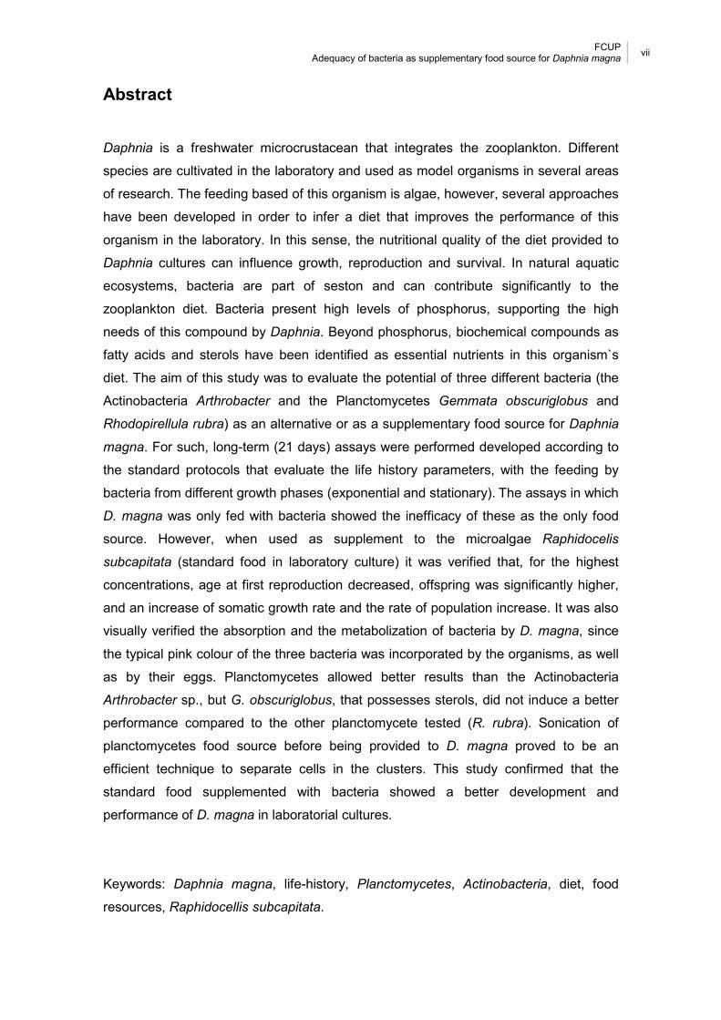
Abstract
Daphnia is a freshwater microcrustacean that integrates the zooplankton. Different
species are cultivated in the laboratory and used as model organisms in several areas
of research. The feeding based of this organism is algae, however, several approaches
have been developed in order to infer a diet that improves the performance of this
organism in the laboratory. In this sense, the nutritional quality of the diet provided to
Daphnia cultures can influence growth, reproduction and survival. In natural aquatic
ecosystems, bacteria are part of seston and can contribute significantly to the
zooplankton diet. Bacteria present high levels of phosphorus, supporting the high
needs of this compound by Daphnia. Beyond phosphorus, biochemical compounds as
fatty acids and sterols have been identified as essential nutrients in this organism`s
diet. The aim of this study was to evaluate the potential of three different bacteria (the
Actinobacteria Arthrobacter and the Planctomycetes Gemmata obscuriglobus and
Rhodopirellula rubra) as an alternative or as a supplementary food source for Daphnia
magna. For such, long-term (21 days) assays were performed developed according to
the standard protocols that evaluate the life history parameters, with the feeding by
bacteria from different growth phases (exponential and stationary). The assays in which
D. magna was only fed with bacteria showed the inefficacy of these as the only food
source. However, when used as supplement to the microalgae Raphidocelis
subcapitata (standard food in laboratory culture) it was verified that, for the highest
concentrations, age at first reproduction decreased, offspring was significantly higher,
and an increase of somatic growth rate and the rate of population increase. It was also
visually verified the absorption and the metabolization of bacteria by D. magna, since
the typical pink colour of the three bacteria was incorporated by the organisms, as well
as by their eggs. Planctomycetes allowed better results than the Actinobacteria
Arthrobacter sp., but G. obscuriglobus, that possesses sterols, did not induce a better
performance compared to the other planctomycete tested (R. rubra). Sonication of
planctomycetes food source before being provided to D. magna proved to be an
efficient technique to separate cells in the clusters. This study confirmed that the
standard food supplemented with bacteria showed a better development and
performance of D. magna in laboratorial cultures.
Keywords: Daphnia magna, life-history, Planctomycetes, Actinobacteria, diet, food
resources, Raphidocellis subcapitata.
FCUP viii
Adequacy of bacteria as supplementary food source for Daphnia magna
Table of contents
Agradecimentos ........................................................................................................... iv
Resumo ........................................................................................................................ v
Abstract ....................................................................................................................... vii
Table of contents ........................................................................................................ viii
Table index .................................................................................................................. ix
Figure index .................................................................................................................. x
CHAPTER 1. Introduction ............................................................................................. 1
CHAPTER 2. Material and Methods .............................................................................. 5
2.1 Daphnia magna cultures ...................................................................................... 5
2.2 Arthrobacter, Gemmata obscuriglobus and Rhodopirellula rubra cultures ........... 5
2.3 Chronic assays .................................................................................................... 6
2.4 Statistical analysis ............................................................................................... 7
CHAPTER 3. Results .................................................................................................... 9
CHAPTER 4. Discussion ............................................................................................ 14
CHAPTER 5. Conclusions .......................................................................................... 18
References ................................................................................................................. 19
FCUP ix
Adequacy of bacteria as supplementary food source for Daphnia magna
Table index
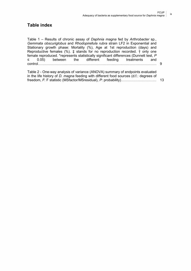
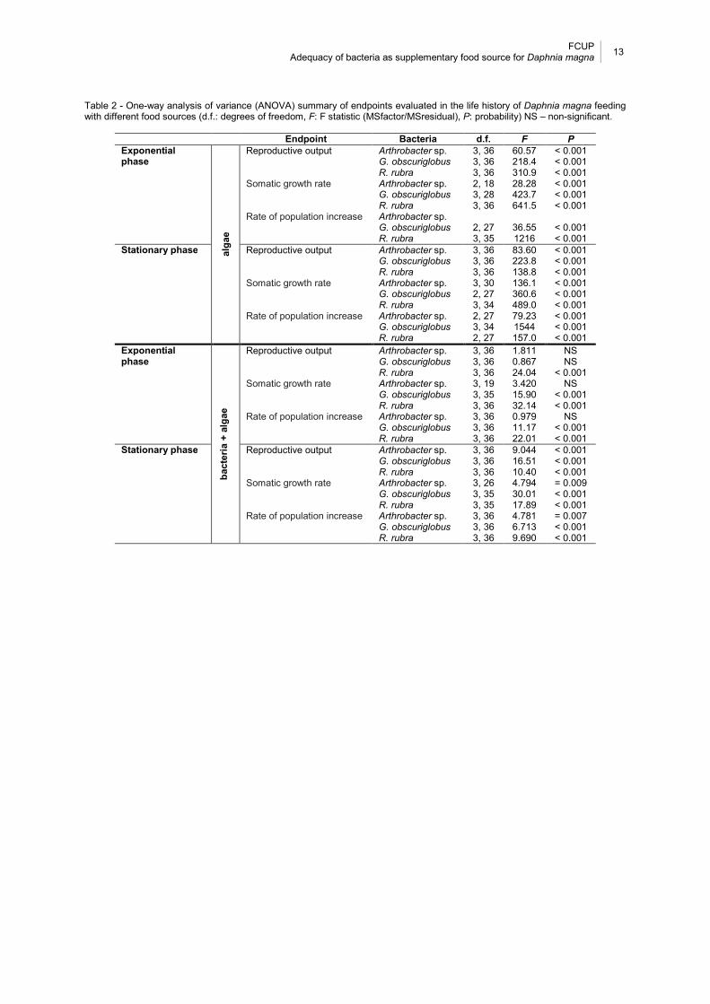
Table 1 – Results of chronic assay of Daphnia magna fed by Arthrobacter sp., Gemmata obscuriglobus and Rhodopirellula rubra strain LF2 in Exponential and Stationary growth phase: Mortality (%), Age at 1st reproduction (days) and Reproductive females (%). ‡ stands for no reproduction recorded. † only one female reproduced. *represents statistically significant differences (Dunnett test, P ≤ 0.05) between the different feeding treatments and control………………………………………………………..……………………………… 9 Table 2 - One-way analysis of variance (ANOVA) summary of endpoints evaluated in the life history of D. magna feeding with different food sources (d.f.: degrees of freedom, F: F statistic (MSfactor/MSresidual), P: probability)………………………… 13

FCUP x
Adequacy of bacteria as supplementary food source for Daphnia magna
Figure index
Fig. 1 – Daphnia magna. Body length is measured from the top of the head to the base of tail spine. .................................................................................................................. 7
Fig. 2 - Daphnia magna female after 21 days fed with different food treatments evidencing the pink or green coloration due to the bacteria or alga respective food sources. Under each image, the body length (mm) of the organism is presented. Control: D. magna fed with R. subcapitata; Only bacteria: D. magna fed with G.
obscuriglobus, R. rubra and Arthrobacter sp. with different concentrations 25 μL, 250 μL and 2500 μL; Bacteria + alga: D. magna fed with G. obscuriglobus, R. rubra and Arthrobacter sp. with different concentrations 25 μL, 250 μL and 2500 μL + R.
Subcapitata. ................................................................................................................ 10
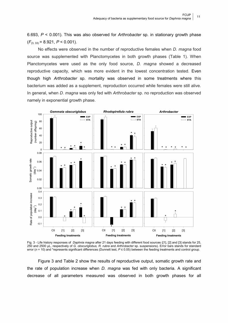
Fig. 3 - Life history responses of Daphnia magna after 21 days feeding with different food sources ([1], [2] and [3] stands for 25, 250 and 2500 µL, respectively of G.
obscuriglobus, R. rubra and Arthrobacter sp. suspensions). Error bars stands for standard error (n = 10) and *represents significant differences (Dunnett test, P ≤ 0.05) between the feeding treatments and control group. .................................................... 11
Fig. 4 - Life history responses of Daphnia magna after 21 days feeding with different food sources ([1]+A, [2]+A and [3]+A stands for 25, 250 and 2500 µL respectively of G.
obscuriglobus, R. rubra and Arthrobacter sp. suspensions plus R. subcapitata ratio). Error bars stands for standard error (n = 10) and *represents significant differences (Dunnett test, P ≤ 0.05) between the feeding treatments and control group. ............... 12
FCUP 1
Adequacy of bacteria as supplementary food source for Daphnia magna
CHAPTER 1. Introduction
In the last century, several organisms denominated “model organisms” were used as
important tools for research allowing the increase of our knowledge in numerous biological
processes (Levy and Currie, 2015). Among the several model organisms, the cladoceran of
the genus Daphnia is one of the principal standard organisms in aquatic ecotoxicology.
Furthermore, this freshwater microcrustacean plays an important role in aquatic food webs
as a primary consumer and is a vital food source for many higher trophic levels including fish
species (Hebert, 1978; Tessier et al., 2000; Forró et al., 2008; Antunes et al., 2016). As main
component of freshwater zooplankton (Hebert, 1978), the role of this organism is well
documented in the literature and Daphnia is widely used in research areas such as ecology,
ecotoxicology, ecophysiology, evolution, genomics, roles in host-parasite, predator-prey
interactions and phenotypic plasticity (Ebert, 2005, 2011; Lampert, 2006; Stollewerk, 2010;
Colbourne et al., 2011; Seda and Petrusek, 2011; Harris et al., 2012). Short life cycle, small
size, high fecundity, parthenogenetic reproduction and easy maintenance in laboratory
cultures are characteristics that support the significant advantages of the water flea Daphnia
magna as a model and standard organism (Lambert, 2006; Seda and Petrusek, 2011;
Antunes et al., 2016).
Generally, Daphnia species are cultivated and grown in laboratory for use as model
organisms in research studies. The nutritional quality and quantity of diet provided to water
fleas are important variables that not only influence their growth but also, reproduction and
survival (Gulati and DeMott, 1997; Bukovinszky et al., 2012; Sarpe et al., 2014). On the other
hand, the diet in Daphnia`s cultures may compromise its performance and the results
gathered in laboratory assays (Lanno et al., 1989; Antunes et al., 2004; Ieromina et al.,
2014). Uni-algal cultures, namely the green microalgae Raphidocelis subcapitata or Chlorella
vulgaris, are currently used as the only food source for this organism under laboratory
conditions (Antunes et al., 2004; Becker and Boersma, 2005; Choi et al., 2016). However,
alternative or supplementary food sources should be considered in order to reduce
dependence on a single food source (Bouchnak and Steinberg, 2010; Martin-Creuzburg et
al., 2011; Buratini and Aragão, 2012; Antunes et al., 2016).
The search of the adequate food source for Daphnia culture and the way food
influence the growth and reproduction of this organism has been the aim of several studies
(Weers and Gulati, 1997; von Elert, 2002; Becker and Boersma, 2005; Brett et al., 2006;
Freese and Martin-Creuzburg, 2013). Different feeding regimes are used in order to assess
their contribution to the characteristics of life history of daphnids. For Daphnia cyanobacteria
FCUP 2
Adequacy of bacteria as supplementary food source for Daphnia magna
have lower nutritional value than green algae (Arnold, 1971), and these lower nutritional
value than diatoms (Choi et al., 2014). Infante and Abella (1985) showed that Daphnia size
and embryo production decreased with increase density of Oscillatoria, which may be related
to its filamentous nature and/or to chemical inhibition. The cyanobacteria, Aphanizomenon
gracile, Synechococcus elongatus, and Microcystis aeruginosa showed to promote negative
effects on survival, growth, and food uptake of D. pulicaria, due to size and morphological
restrictions as well as nutritional inadequacy and toxicity effects (Lampert, 1981). More
successful reproduction rates were obtained when D. magna was fed with the diatom
Stephanodiscus hantzschii than with the green alga Chlorella vulgaris (Choi et al., 2016).
These result correlated with a higher proportion of long-chain poly unsaturated fatty acids
(PUFAs) in S. hantzschii (Choi et al., 2016). In addition, D. magna assimilated better the
carbon and nitrogen originated from S. hantzschii. Taub and Dollar (1968) concluded that
Chlorella pyrenoidosa and Chlamydomonas reinhardii are inadequate food sources for
normal longevity and reproduction in D. pulex, reduced ovulation and an incomplete
development of embryos was recorded. The performance of D. longispina fed with
Scendesmus quadricola, Oscillatoria ocutasama, horse manure and yeast was studied by
Mona et al. (2014). Survival, longevity and swimming activity were not affected by the
different foods sources, however, the best performance of D. longispina was achieved with
S. quadricola followed by the yeast, O. ocutasama and horse manure. Normally, the growth
of Daphnia is determined by food availability and quality (Acharya et al., 2004), where
phosphorus (P), and certain PUFAs are essential nutritional substances for freshwater
zooplankton (Gulati and DeMott, 1997). Urabe and Sterner (2001) found that D. obtusa fed
with Scenedesmus acutus on low N content or low P content showed slow growth, reduced
eggs viability and reduced survival, mainly before maturation. The fatty acid composition of
Rhodomonas and Cryptomonas are different from the Scenedesmus and Chlamydomonas,
they contained high percentages of long-chained PUFAs, being considered the best food for
Daphnia (Ahlgren et al., 1990).
As non-selective filter feeders, cladocerans do not discriminate between foods with
regard to their nutritional quality (DeMott, 1986) and ingest small suspended organisms such
as algae, bacteria, ciliates, and flagellates as well as detritus (Hebert, 1978; Hessen, 1992;
Tessier et al., 2000). In aquatic ecosystems, particulate organic matter in suspension may
contain a high proportion of bacteria, being able to maintain Daphnia population by itself
(Picard and Lair, 2000; Freese and Martin-Creuzburg, 2013). Furthermore, when algal
biomass is low or when Daphnia reaches a high population density, bacteria can be an
important portion of the feeding of these organisms (Kankaala, 1988; Pace, 1990). As a key

FCUP 3
Adequacy of bacteria as supplementary food source for Daphnia magna
species in many freshwater ecosystems, it efficiently consumes heterotrophic bacteria
(Brendelberger, 1991) and this grazing is able to affect the biomass, shape the structure and
species composition of the bacterial community (Jurgens et al., 1999; Degans et al., 2002).
Heterotrophic bacteria are important components of aquatic food webs being responsible for
the carbon and energy production and transfer in the pelagic aquatic organisms (Azam et al.,
1983; Weisse and MacIsaac, 2000). Relatively to algae, bacteria present a higher
phosphorus to carbon (P:C) ratio (Vadstein, 2000), allowing to sustain the high P Daphnia
needs (Hessen and Andersen, 1990; DeMott et al., 1998). Among the various food quality
parameters, besides P others elements like C and nitrogen (N) (Urabe et al., 1997; DeMott et
al., 1998; Darchambeau et al., 2003) and organic nutrients such as PUFAs or sterols were
identified as essential elements in daphnids diet (Mueller-Navarra, 1995; von Elert et al.,
2003; Becker and Boersma, 2005; Martin-Creuzburg et al., 2005). PUFAs and sterols are
lipids already documented to be essential in somatic growth and reproduction rates (Mueller-
Navarra et al., 2000; von Elert et al., 2003). On the other hand, bacteria can be a viable
source of carbon and elemental nutrients (Vadstein 2000; Biddanda et al., 2001), but, in
general they are devoid of PUFAs and sterols (Volkman, 2003; Martin-Creuzburg et al.,
2011). Furthermore, several studies have shown that these organisms are poor food quality
for daphnids (Martin-Creuzburg et al., 2011; Taipale et al., 2012; Wenzel et al., 2012). Other
studies already demonstrated that Daphnia performance is limited by the absence in sterols
and PUFAs on cyanobacterial diet (Martin-Creuzburg et al., 2005, 2008).
Arthrobacter is usually considered a genus of soil bacteria (Hagedorn and Holt,
1975). However, representative strains have been isolated from several environments such
as air, food, water, sewage, activated sludge, oil brine, plants, biofilms, cyanobacterial mats,
sediment and inclusively from human clinical samples (Funke et al., 1996; Crocker et al.,
2000; Irlinger et al., 2005; Chang et al., 2007; Kim et al., 2008; Mages et al., 2008;
Sutthiwong et al., 2014). Species that are members of this genus are catalase-positive,
aerobic and produce little or no acid from glucose (Chang et al., 2007; Sutthiwong et al.,
2014). Phylogenetically, Arthrobacter sp. is Actinobacteria which are Gram-positive bacteria
with high GC content (Eschbach et al., 2003). The main distinctive feature of this genus is
the life cycle in which the shape of cells is typically rods during exponential growth, being
replaced by cocci in the stationary phase (Sutthiwong et al., 2014; Busse, 2016). In natural
environments they play an important role in the transformation of a wide variety of organic
matter (Crocker et al., 2000), including aromatic hydrocarbons, that they use as the only
sources of carbon or nitrogen (Stevenson, 1967; Hagedorn and Holt, 1975).

FCUP 4
Adequacy of bacteria as supplementary food source for Daphnia magna
Planctomycetes is a peculiar phylum of the domain Bacteria characterized by
members with unique morphological, genetic, metabolic and physiological identity. Their
unusual complex cell plan, common budding reproduction, endocytosis capacity, eukaryote
homologs of membrane-coating proteins, ability to synthesize sterols (Gemmata
obscuriglobus; Pearson et al., 2003), absence of tubulin like protein FtZ (Fuerst et al., 2013;
Sagulenko et al., 2014; Pinos et al., 2016) and crateriform pits on the cell surface (Lage et
al., 2013), have led to a growing interest in these organisms over the last decades. Some of
these features are commonly found in eukaryotes (Reynaud and Devos, 2011; Fuerst and
Sagulenko, 2012). This phylum along with Verrucomicrobia, Chlamydiae, Lentisphaerae,
Poribacteria, OP3 and WWE2 form the PVC superphylum (Wagner and Horn, 2006; Gupta et
al., 2012; Lagkouvardos et al., 2014; Pinos et al., 2016). They are a ubiquitous group of
bacteria, found in a great variety of ecosystems including aquatic and terrestrial ones
(Winkelmann et al., 2010; Andrew et al., 2012), and in association with several organisms
(Fuerst et al., 1997; Webster and Bourne, 2007; Lage and Bondoso, 2011; Izumi et al., 2013;
Lage, 2013). Planctomycetes exhibit great metabolic diversity and play an important role in
the global environmental cycles (Pinos et al., 2016 and references therein).
In laboratory, there are some variables that influence D. magna development. The
diet provided is one of these variables and has been shown to be a determining factor in D.
magna performance. Recent studies suggested that bacteria could be considered as
supplementary and/or nutritional food for the cladocerans D. magna and D. longispina
(Antunes et al., 2016). Although D. magna fed with the planctomycete Rhodopirellila rubra
plus algae Raphidocelis subcapitata showed favorable growth performance on the highest
concentration tested, concern was raised regarding the bacteria clusters size that could
impair absorption. As a follow up, this study aims to evaluate the potential of other bacteria:
the Actinobacteria Arthrobacter, the planctomycete Rhodopirellula rubra strain LF2 and the
sterol producing planctomycete Gemmata obscuriglobus as an additional and/or nutritional
food sources for D. magna. Furthermore, the effect of sonication to separate bacteria in
clusters before feeding D. magna was also tested. For such, long-term (21 days) assays will
be performed, developed according to standard protocols, and evaluated the life history
parameters after feeding from different bacterial growth phases (exponential and stationary).
FCUP 5
Adequacy of bacteria as supplementary food source for Daphnia magna
CHAPTER 2. Material and Methods
2.1 Daphnia magna cultures
Cultures of D. magna (clone A, sensu Baird et al., 1989a), was grown under controlled
laboratory conditions for several generations as pure parthenogenic cultures. Monoclonal
cultures of D. magna were reared in laboratory at temperature (20±2 ºC), photoperiod
(16hL:8hD) and maintained in ASTM synthetic hard water medium (ASTM, 1980). D. magna
cultures were supplemented with a standard organic additive (suspension extracted from the
brown algae Ascophyllum nodosum) (Baird et al., 1989b), which provides essential
micronutrients to Daphnia. The cultures were fed with the microalgae Raphidocelis
subcapitata (formerly known as Selenastrum subcapitata and Pseudokirchneriella
subcapitata) with a ratio of 3.0 x 10⁵ cells.mL-1.day-1. The microalgae was kept in cultures in
Woods Hole MBL medium (Stein, 1973), under controlled conditions of temperature (20±2
ºC) and photoperiod 16hL:8hD (~ 6000 lux), being harvested in the exponential growth phase
(5 – 7 days) (Environment Canada, 1992; OECD, 2006). Algal cell concentrations were
calculated based on the correlation of absorbance measured at λ=440 nm and cell density
previously determined. Daphnia magna cultures were fed three times a week when the
culture medium was renewed. Neonates born between the 3rd to 5th broods were used for
renewing the culture or for initiating assays.
2.2 Arthrobacter sp., Gemmata obscuriglobus and Rhodopirellula rubra
cultures
The procedures for culturing the three bacteria were similar. Specific media were used to
grown the different bacteria: G. obscuriglobus was cultured in nutrient agar (NA) and nutrient
broth (NB), Arthrobacter sp. was grown in M14 medium (Lage and Bondoso, 2011), and R.
rubra was cultured in the medium M13 (Lage and Bondoso, 2011). All bacteria were grown at
26 ºC, first in solid culture medium and then upscaled in liquid media to a volume of 600 mL
in a continuous stirring of 200 rpm. After 3 or 7 days of growth for exponential and stationary
phases, respectively, the cells were collected by centrifugation at 4000 rpm. Cell pellets were
resuspended in distilled water and the optical density at λ=600 nm adjusted to 0.2 arbitrary
units (AU). The all cell suspensions were stored at -20 ºC for posterior use in the feeding
assays (see below chronic assays). Due to the clustering of the cells, 50 mL planctomycetes
FCUP 6
Adequacy of bacteria as supplementary food source for Daphnia magna
cell suspensions were defrost and sonicate for 30 s, in a Misonix Microson Ultrasonic Cell
Disruptor XL set to intensity 10 watts. Next, the cell suspensions were adjusted to 0.2
arbitrary units (AU) at λ=600 nm before provided as food to the D. magna assays.
2.3 Chronic assays
To evaluate the potential of Arthrobacter sp., G. obscuriglobus and R. rubra to be used as
additional or as nutritional food source for D. magna in laboratory cultures, two distinct
conditions of each bacteria were tested: 1) bacteria from exponential growth phase; and 2)
bacteria from stationary growth phase. Feeding assays were adapted from chronic standard
protocols for life-history parameters evaluation (ASTM, 1997; OECD, 2012). D. magna in the
feeding assays were kept under the same temperature, photoperiod regimes and medium
renewed with a standard organic additive as described for rearing cultures. In each assay, 7
food conditions were tested: 1) a negative control fed with R. subcapitata with a ratio of
3.0×10⁵ cells mL-1 day-1; 2) three different concentrations of bacteria (25, 250 and 2500 μL of
cells suspension stands for 1/1000, 10/1000 100/1000, v/v), herein designated [1], [2] and
[3], and 3) the same bacteria concentrations described above (25, 250 and 2500 μL of cells
suspension) adding a standard food ratio of R. subcapitata (3.0×10⁵ cells mL-1 day-1), herein
designated by [1]+A, [2]+A, and [3]+A.
Ten individualized D. magna replicates were used in each treatment. All assays started
with neonates born between the 3th and 5th brood, and an aged with less than 24 h. Assays
were conducted for 21 days in glass vessels in a final volume of 50 mL of ASTM synthetic
hard water medium. Organisms were fed with alga every day (bacteria were given only when
the culture medium was changed, every two days), the medium was renewed every two
days, and the assays were monitored daily for mortality and reproductive state. Neonates
born during the assay were counted and discarded. For each assay, several endpoints were
quantified: mortality (%), age at first reproduction (days), somatic growth rate (day-1),
reproductive females (%), reproductive output and rate of population increase (r, day-1).
For somatic growth rate calculation, D. magna body length was measured, from the top
of the head to the base of the caudal spine (Fig. 1). An average of initial body length was
measured at the beginning of the assay in a sub-sample of 20 neonates from the same
brooding of the organism used in the assay. At the end of the test (21 days) all the survivor
organisms were measured in a binocular stereoscope. The somatic growth rate was
calculated, according to the following expression:

FCUP 7
Adequacy of bacteria as supplementary food source for Daphnia magna
growth rate =ln���� – ln��
∆
where lf is the body length (mm) of the organism after 21 days of the assay, li is the average
body length (mm) of a subsample (n=20) of neonates coming from the same batch of
neonates that initiated the test, and ∆t is the duration of the test (in days).
Survival and fecundity related data were used to compute for the estimation the per
capita intrinsic rate of population increase (r), which was iterated from the Euler–Lotka
equation:
� = � ���������
���
where r is the intrinsic rate of population increase (day-1), x is the age class (in days), lx is the
probability of surviving at age x, and mx is the fecundity at age x. Standard errors for r were
estimated using the jack-knifing technique described by Meyer et al. (1986).
Fig. 1 – Daphnia magna. Body length is measured from the top of the head to the base of tail spine.
2.4 Statistical analysis
One-way Analysis of Variance (ANOVA) was used to analyze the endpoints measured in the
long-term (21 days) assays (age at 1st reproduction, somatic growth rate, reproductive output

FCUP 8
Adequacy of bacteria as supplementary food source for Daphnia magna
and rate of population increase). This analysis allows to determine statistically differences
between the food treatments. If ANOVA was significant a Dunnett test was conducted to
assess statistical differences between the different food treatments and the control. For all
analyses, the level of significance (α) used was 0.05.

FCUP 9
Adequacy of bacteria as supplementary food source for Daphnia magna
CHAPTER 3. Results
By visual assessment, D. magna fed with R. subcapitata (control), has no additional body
coloration besides green/brown color in the gut due to the alga and the organic additive.
However, when D. magna was fed with any of the bacteria it exhibits a pink coloration due to
bacterial pigments that increased with bacterial concentrations. This pink coloration was
even clearly evident in the eggs in the marsupial camera (Fig. 2).
Table 1 shows the results of mortality, age at first reproduction and percentage of
reproductive female in the different assays. When compared to Planctomycetes, higher
levels of mortality were observed when D. magna was fed with Arthrobacter sp. in both
growth phase assays. Higher mortality was also observed in G. obscuriglobus in the highest
concentration tested for both growth phases when D. magna was fed with only bacteria.
However, when this bacterial concentration was added with alga no mortality occurred.
Table 1 – Results of 21 days feeding assays of Daphnia magna fed by Arthrobacter sp., Gemmata obscuriglobus and Rhodopirellula rubra strain LF2 in Exponential and Stationary growth phase: Mortality (%), Age at 1st reproduction (days) and Reproductive females (%). ‡ stands for no reproduction recorded. † only one female reproduced. *represents statistically significant differences (Dunnett test, P ≤ 0.05) between the different feeding treatments and control.
Exponential phase Stationary phase
Treatments
Mortality Age at 1st
reproduction Reproductive
females Mortality
Age at 1st reproduction
Reproductive females
G.
obscuriglo
bus Ctl 0 10.1±0.10 100 0 10.2±0.13 100
[1] 0 >21days‡ 0 0 >16days 10 [2] 10 14.1±0.35 100 0 12.4±0.34 100 [3] 70 12.3±0.25 40 100 13days† 10 [1]+A 0 9.7±0.15 100 0 10.0±0.00 100 [2]+A 0 9.6±0.16* 100 10 9.9±0.11 90 [3]+A 10 9.0±0.00* 100 0 9.8±0.20 100
R.
rubra
Ctl [1] [2] [3] [1]+A [2]+A [3]+A
0 0 0 0 0 0 0
10.9±0.10 ≥ 21days 13.8±0.66 13.7±0.42 10.8±0.25 9.8±0.25*
10.0±0.26*
100 10
100 100 100 100 100
0 0
10 10 0
10 0
11.1±0.31 >21days‡ 13.6±0.48 13.0±0.47 10.8±0.20 10.1±0.31* 9.6±0.22*
100 0
90 90 100 90 100
Art
hro
bacte
r
Ctl [1] [2] [3] [1]+A [2]+A [3]+A
10 30 30 100 70 30 40
10.8±0.13 >21days‡ >21days‡
--- 10.8±0.13 11.0±0.41 10.5±0.38
100 0 0 0
100 90 80
10 20 0
20 30 40 10
10.6±0.16 ≥21 days 13.2±0.65 >21days‡ 11.0±0.00 9.9±0.11 9.8±0.20*
100 30 60 0
100 70 100
In the assays where D. magna was fed with only bacteria, a significant delay in the age
at first reproduction was observed for all bacteria. When D. magna was fed with
Planctomycetes as supplementary to the standard food source, the age at first reproduction
occurred significantly earlier, G. obscuriglobus (exponential phase: F(3, 36) = 13.78, P <
0.001), R. rubra (exponential phase: F(3, 36) = 6.149, P = 0.002; stationary phase: F(3, 35) =

Adequacy of bacteria as supplementary food source for
Fig. 2 - Daphnia magna female after 21 days fed with different food treatmentsthe body length (mm) of the organism is presented. Control: different concentrations 25 μL, 250 μL and 2500 μL; Bacteria + alga: μL + R. Subcapitata.
FCUP 10
Adequacy of bacteria as supplementary food source for Daphnia magna
different food treatments evidencing the pink or green coloration due to the bacteria or alga respective food sourcesControl: D. magna fed with R. subcapitata; Only bacteria: D. magna fed with G. obscuriglobus
Bacteria + alga: D. magna fed with G. obscuriglobus, R. rubra and Arthrobacter sp. with different concentrations 25
evidencing the pink or green coloration due to the bacteria or alga respective food sources. Under each image, G. obscuriglobus, R. rubra and Arthrobacter sp. with with different concentrations 25 μL, 250 μL and 2500
FCUP 11
Adequacy of bacteria as supplementary food source for Daphnia magna
6.693, P < 0.001). This was also observed for Arthrobacter sp. in stationary growth phase
(F(3, 33) = 8.921, P < 0.001).
No effects were observed in the number of reproductive females when D. magna food
source was supplemented with Planctomycetes in both growth phases (Table 1). When
Planctomycetes were used as the only food source, D. magna showed a decreased
reproductive capacity, which was more evident in the lowest concentration tested. Even
though high Arthrobacter sp. mortality was observed in some treatments where this
bacterium was added as a supplement, reproduction occurred while females were still alive.
In general, when D. magna was only fed with Arthrobacter sp. no reproduction was observed
namely in exponential growth phase.
Fig. 3 - Life history responses of Daphnia magna after 21 days feeding with different food sources ([1], [2] and [3] stands for 25, 250 and 2500 µL, respectively of G. obscuriglobus, R. rubra and Arthrobacter sp. suspensions). Error bars stands for standard error (n = 10) and *represents significant differences (Dunnett test, P ≤ 0.05) between the feeding treatments and control group.
Figure 3 and Table 2 show the results of reproductive output, somatic growth rate and
the rate of population increase when D. magna was fed with only bacteria. A significant
decrease of all parameters measured was observed in both growth phases for all
Gemmata obscuriglobus
Rep
rodu
ctiv
e ou
tput
(num
ber
offs
prin
g)
0
20
40
60
80
100EXP
STA
Som
atic
gro
wth
rat
e
(day
-1)
0.00
0.02
0.04
0.06
0.08
Feeding treatments
Ctl [1] [2] [3]
Rat
e of
pop
ulat
ion
incr
ease
(day
-1)
-0.1
0.0
0.1
0.2
0.3
0.4
Arthrobacter
EXP
STA
* * ** * *
**
**
**
* **
* * *
**
*
**
*
*
**
*
Feeding treatments
Ctl [1] [2] [3]
Rhodopirellula rubra
EXP
STA
Feeding treatments
Ctl [1] [2] [3]
**
*
**
*
*
*
*
**
*
**
*
*
*

FCUP 12
Adequacy of bacteria as supplementary food source for Daphnia magna
concentration tested. The less negatively impacted treatment was observed in the highest R.
rubra concentration for both growth phases in all the parameters analyzed.
Fig. 4 - Life history responses of Daphnia magna after 21 days feeding with different food sources ([1]+A, [2]+A and [3]+A stands for 25, 250 and 2500 µL respectively of G. obscuriglobus, R. rubra and Arthrobacter sp. suspensions plus R. subcapitata ratio). Error bars stands for standard error (n = 10) and *represents significant differences (Dunnett test, P ≤ 0.05) between the feeding treatments and control group.
Figure 4 and Table 2 show the same parameters of Fig. 3, in this case the treatments
when bacteria were used as supplementary food source. In general, for all bacteria the
parameters measured showed a slight increase along the three concentrations tested. All
parameters were significantly increased when D. magna was fed with the highest bacterial
concentration from stationary growth phase. This is also true for R. rubra in the two highest
concentrations for exponential growth phase. Also, a significant increase was observed for
somatic growth rate and rate of population increase in G. obscuriglobus at the middle and
highest concentrations for exponential growth phase. Furthermore, Arthrobacter sp. induced
a significant increase in somatic growth rate at exponential growth phase in the highest
concentration tested.
Gemmata obscuriglobus
Rep
rodu
ctiv
e ou
tput
(num
ber
offs
prin
g)
0
20
40
60
80
100
120
140
160
180EXP
STA
Som
atic
gro
wth
rat
e
(day
-1)
0.00
0.02
0.04
0.06
0.08
Feeding treatments
Ctl [1]+A [2]+A [3]+A
Rat
e of
pop
ulat
ion
incr
ease
(day
-1)
0.0
0.1
0.2
0.3
0.4
Arthrobacter
EXP
STA
Feeding treatments
Ctl [1]+A [2]+A [3]+A
*
*
*
** *
* *
*
*
*
Rhodopirellula rubra
EXP
STA
Feeding treatments
Ctl [1]+A [2]+A [3]+A
*
*
* *
* *
*
*
*
FCUP 13
Adequacy of bacteria as supplementary food source for Daphnia magna
Table 2 - One-way analysis of variance (ANOVA) summary of endpoints evaluated in the life history of Daphnia magna feeding with different food sources (d.f.: degrees of freedom, F: F statistic (MSfactor/MSresidual), P: probability) NS – non-significant.
Endpoint Bacteria d.f. F P
Exponential phase
alg
ae
Reproductive output Somatic growth rate Rate of population increase
Arthrobacter sp. G. obscuriglobus R. rubra Arthrobacter sp. G. obscuriglobus R. rubra Arthrobacter sp. G. obscuriglobus R. rubra
3, 36 3, 36 3, 36 2, 18 3, 28 3, 36
2, 27 3, 35
60.57 218.4 310.9 28.28 423.7 641.5
36.55 1216
< 0.001 < 0.001 < 0.001 < 0.001 < 0.001 < 0.001
< 0.001 < 0.001
Stationary phase Reproductive output Somatic growth rate Rate of population increase
Arthrobacter sp. G. obscuriglobus R. rubra Arthrobacter sp. G. obscuriglobus R. rubra Arthrobacter sp. G. obscuriglobus R. rubra
3, 36 3, 36 3, 36 3, 30 2, 27 3, 34 2, 27 3, 34 2, 27
83.60 223.8 138.8 136.1 360.6 489.0 79.23 1544 157.0
< 0.001 < 0.001 < 0.001 < 0.001 < 0.001 < 0.001 < 0.001 < 0.001 < 0.001
Exponential phase
bacte
ria +
alg
ae
Reproductive output Somatic growth rate Rate of population increase
Arthrobacter sp. G. obscuriglobus R. rubra Arthrobacter sp. G. obscuriglobus R. rubra Arthrobacter sp. G. obscuriglobus R. rubra
3, 36 3, 36 3, 36 3, 19 3, 35 3, 36 3, 36 3, 36 3, 36
1.811 0.867 24.04 3.420 15.90 32.14 0.979 11.17 22.01
NS NS
< 0.001 NS
< 0.001 < 0.001
NS < 0.001 < 0.001
Stationary phase Reproductive output Somatic growth rate Rate of population increase
Arthrobacter sp. G. obscuriglobus R. rubra Arthrobacter sp. G. obscuriglobus R. rubra Arthrobacter sp. G. obscuriglobus R. rubra
3, 36 3, 36 3, 36 3, 26 3, 35 3, 35 3, 36 3, 36 3, 36
9.044 16.51 10.40 4.794 30.01 17.89 4.781 6.713 9.690
< 0.001 < 0.001 < 0.001 = 0.009 < 0.001 < 0.001 = 0.007 < 0.001 < 0.001

FCUP 14
Adequacy of bacteria as supplementary food source for Daphnia magna
CHAPTER 4. Discussion
Daphnia magna has been used as a model organism recommended for standardized
procedures for ecological risk assessments of surface waters in many countries
(Jonczyk and Gilron, 2005, OECD, 2012). The success of Daphnia cultures in the
laboratory depends on culture conditions, mainly water quality and diet (Elendt and
Bias, 1990). Diet must take into account the nutritional needs of the species, in order
to improve its performance and not induce alterations that could affect research. Under
laboratorial conditions, Daphnia diet is based on a single food source as previously
referred. In order to find an additional and/or nutritional food source for this organism in
our work different bacterial species in exponential and stationary growth phases, at
different concentrations were supplied individually and in supplement to the standard
food source, the microalgae R. subcapitata. The here-obtained results show that, the
life-history of D. magna was influenced by the quantity and the quality of the available
food sources.
When compared to control (R. subcapitata as only food source), all bacteria were
not able to provide an adequate quantitative and/or qualitative food supply for D.
magna. All the parameters showed reduced values comparative to the control (Figure
3, Table 1). Several studies have described heterotrophic bacteria as being of poor
food quality for daphnids (Martin-Creuzburg et al., 2011; Wenzel et al., 2012; Taipale et
al., 2012; Antunes et al., 2016). The lack of sterols and the deficiency of PUFAs in
bacteria are mentioned by several authors as the major food quality constraints (von
Elert et al., 2003; Martin-Creuzburg et al., 2008, 2011; Freese and Martin-Creuzburg,
2013). A diet deficient in these essential elements has been shown to affect the
performance of daphnids (Martin-Creuzburg et al., 2009). When D. magna was fed
exclusively with G. obscuriglobus, the best performance was obtained at the middle
concentration tested in both phases, once low or no mortality was observed and all
females reproduced. Moreover, higher values of mortality were observed at the highest
concentration in both phases, a result that is beyond our comprehension. When R.
rubra was provided as the only food source, the best performance was obtained at the
two highest concentrations tested in both phases, where mortality was negligible and,
in general, all females reproduced. The somatic growth rate in both Planctomycetes
was reduced, but with the increase of food concentration a slight improvement was
observed. Regarding food quality, G. obscuriglobus, contrary to R. rubra, has the ability
to synthesize sterols (lanosterol and parkeol; Pearson et al., 2003), which is a typical
characteristic of eukaryotes and unusual among bacteria (Fuerst and Sagulenko,

FCUP 15
Adequacy of bacteria as supplementary food source for Daphnia magna
2011). However, our results showed that R. rubra induced a better performance in the
growth, survival and production of neonates of D. magna, and G. obscuriglobus seem
not to satisfy the physiological requirements of D. magna. R. rubra possesses palmitic
(16:0) and oleic (18:1 ω9c) acids as its main fatty acids (Bondoso et al., 2014).
Furthermore, our results showed a better performance of D. magna when fed
exclusively with Planctomycetes than the ones obtained in the study performed by
Wenzel et al. (2012). When only Pseudomonas was supplied to D. galeata, they
reported the death of all daphnids when and reproduction was only observed on a diet
containing at least 50% Rhodomonas (Wenzel et al., 2012). In contrast, Bednarska et
al. (2014) showed that Daphnia could grow and reproduce when fed solely on the
cyanobacteria Cylindrospermopsis raciborskii (a non-toxic strain). Similarly, when D.
magna fed exclusively on Planctomycetes, our results showed an increase in the age
at first reproduction, a decrease in the number of neonates and a reduction in somatic
growth rate. Comparatively to Arthrobacter sp., G. obscuriglobus and R. rubra
sustained better the survival, growth and reproduction of D. magna when used as the
only source of food. Inadequacy in the concentrations tested or other confounding
factors, not measured in our experiment, could have contributed to the low
performance of D. magna when fed on only the three bacteria tested. Alternatively,
other bacterial concentrations or the supply of bacteria every day should be tested in
order to verify if the performance of D. magna life-history could be improved.
However, this study showed that bacteria are good food sources in supplement to
the standard food provided in laboratorial cultures for D. magna. Depending on the
bacterium and its concentration, D. magna performance was significantly improved as
compared to the control (green algae, R. subcapitata used as the only food source).
For all bacteria the highest concentration tested or even the middle concentration, the
reproductive output, the somatic growth rate and the rate of population increase were
significantly improved especially when Planctomycetes were used. Our results are
consistent with previous studies showing that, when in supplement, bacterial diet can
improve the performance of Daphnia (Taipale et al., 2012; Wenzel et al., 2012;
Antunes et al., 2016). For Wenzel et al. (2012) the life-history characteristics of
daphnids depend on the association between heterotrophic bacteria and
phytoplankton. Freese and Martin-Creuzburg (2013) also described an increase in
Daphnia`s somatic growth rates when heterotrophic bacteria were added in food as
compared with a single algae diet. For the authors, bacteria can provide essential
nutrients not available in algae. Different responses were observed when D. magna
was fed with the different bacteria tested. D. magna showed greater sensitivity to
Arthrobacter sp. (highest mortality) than to the Planctomycetes. Taipale et al. (2012)

FCUP 16
Adequacy of bacteria as supplementary food source for Daphnia magna
documented that the dietary quality of different types of bacteria varies in their ability to
support D. magna survival, growth and reproduction. The literature is scarce regarding
the nutritional values of Actinobacteria and Planctomycetes as food source for
Daphnia. In our work, D. magna presented the best responses of the life-history
parameters when fed with Planctomycetes in the exponential growth phase, while
Arthrobacter sp. present best results in the stationary growth phase. This result is not in
agreement with the previous results by Antunes et al. (2016), where D. magna
performance was better improved with R. rubra in the stationary growth phase. These
contradictory results need confirmation. The lack of information in the literature on the
nutritional capacity of bacteria (Arthrobacter sp. and Planctomycetes) in the two growth
phases does not allow us to explain the preference of D. magna.
Different zooplankton species have different ability to ingest bacterial cells (Bouvy
et al., 1994). Filamentous or aggregated forms may mechanically interfere with the
filtration process, reducing food intake (Bednarska et al., 2014 and references therein).
The Planctomycetes can form rosette-like aggregates with large numbers of cells (> 50
cells) as already reported by Bondoso et al. (2014). In order to reduce this aggregation,
a sonication process was used. This methodology was introduced because in a
previous study (Antunes et al., 2016) a hypothesis was raised about the incapability of
Daphnia to feed on the planctomycetes aggregate. Our results where Daphnia was fed
with bacteria previously treated with sonication showed that this technique improved
the performance of D. magna.
Generally, algae are considered as the fundamental food source for daphnids,
while heterotrophic bacteria are considered less important (Martin-Creuzburg et al.,
2011). Besides phytoplankton, bacteria may be an alternative food source for
zooplankton (Onandia et al., 2015). Evidence was already demonstrated in numerous
studies in which Daphnia was able to consume bacteria (Jurgens et al., 1994;
Langenheder and Jurgens, 2001; Degans et al., 2002; Pernthaler et al., 2004). Pedrós-
Alió and Brock (1983) reported an increase of the normal growth and reproduction of D.
longispina when it was fed with only bacteria. According to Picard and Lair (2000),
Daphnia has the capacity to grow and reproduce in low concentrations of bacteria,
being an important aspect for its suitability under competitive conditions. These authors
also reported that bacterial P plays an important role in the growth of Daphnia. In
addition, bacterial carbon also may be transferred directly to macrozooplankton by
daphnids (Work and Havens, 2003 and references therein). The contribution of
heterotrophic bacteria to the nutrition of Daphnia species in aquatic ecosystems is
supported by analysis of stable isotope patterns and fatty acids biomarkers (Perga et
al., 2006; Taipale et al., 2008, 2009).
FCUP 17
Adequacy of bacteria as supplementary food source for Daphnia magna
Another factor to be accounted in daphnids diet is the food level. For Gliwicz and
Lampert (1990), growth and reproduction in zooplankton depend on the minimum food
concentration. D. pulicaria under decreased food concentration showed reduction in
body length, protein content, lipid content, body carbon, dry mass, clutch size and an
increase in age at first reproduction (Guisande and Gliwicz, 1992). Our results are in
agreement with these studies where an increase of growth and reproduction in D.
magna was observed in both growth phases with increasing Planctomycetes
concentrations. Furthermore, the number of reproductive females remained the same
at the two highest concentrations in both phases when only R. rubra was provided.
Daphnia acquires the colour of the food ingested as already described in other
studies (Ebert, 2005; Antunes et al., 2016). In our study, we observed that D. magna
metabolized and incorporated the pink pigments of the bacteria into its body and eggs.
Increasing intensity of the pink color was observed with increasing bacterial
concentrations, even though in the presence of the microalgae. This result shows that
D. magna has no difficulty to ingesting Arthrobacter sp. and Planctomycetes.

FCUP 18
Adequacy of bacteria as supplementary food source for Daphnia magna
CHAPTER 5. Conclusions
According to our results, we conclude that daphnids are able to feed on bacteria as the
only food source although, in the concentrations tested, the life-history responses of D.
magna were deficient. Bacteria supplemented to algae (standard food source R.
subcapitata) showed to be an adequate diet for daphnids with a significant improve of
life-history parameters, namely in the two highest bacterial concentration tested.
Planctomycetes were more efficient in improve D. magna performance than the
Actinobacteria Arthrobacter sp.. Particle food size, namely Planctomycetal aggregates,
affects the life-history parameters of D. magna, as proved by the sonication of food
supply. This technique minimized the aggregate forms promoting a better ingestion of
the bacteria by D. magna

FCUP 19
Adequacy of bacteria as supplementary food source for Daphnia magna
References
Acharya, K., Kyle, M., Elser, J.J. (2004). Biological stoichiometry of Daphnia growth: an
ecophysiological test of the growth rate hypothesis. Limnology and
Oceanography, 49(3), 656-665.
Ahlgren, G., Lundstedt, L., Brett, M., Forsberg, C. (1990). Lipid composition and food
quality of some freshwater phytoplankton for cladoceran zooplankters. Journal of
Plankton Research, 12(4), 809-818.
Andrew, D.R., Fitak, R.R., Munguia-Vega, A., Racolta, A., Martinson, V.G., Dontsova,
K. (2012). Abiotic factors shape microbial diversity in Sonoran Desert soils. Applied
and Environmental Microbiology, 78(21), 7527-7537.
Antunes, S.C., Castro, B.B., Gonçalves, F. (2004). Effect of food level on the acute and
chronic responses of daphnids to lindane. Environmental Pollution, 127(3), 367-375.
Antunes, S.C., Almeida, R.A., Carvalho, T., Lage, O.M. (2016). Feasibility of
planctomycetes as a nutritional or supplementary food source for Daphnia spp.
Annales de Limnologie-International Journal of Limnology, 52, 317-325.
Arnold, D.E. (1971). Ingestion, assimilation, survival, and reproduction by Daphnia
pulex fed seven species of blue‐green algae. Limnology and Oceanography, 16(6),
906-920.
ASTM (1980). Standard Practice for Conducting Acute Toxicity Tests with Fishes,
Macroinvertebrates and Amphibians, Report E 729-80, American Society for Testing
and Materials, Philadelphia, USA.
ASTM (1997). Standard Guide for Conducting Daphnia Magna Life-cycle Toxicity
Tests, Report E 1193-97, American Society for Testing and Materials, Philadelphia,
USA.
Azam, F., Fenchel, T., Field, J.G., Gray, J.S., Meyer-Reil, L.A., Thingstad, F. (1983).
The ecological role of water-column microbes in the sea. Marine Ecology Progress
Series. Oldendorf, 10(3), 257-263.
Baird, D.J., Barber, I., Bradley, M., Calow, P., Soares, A.M.V.M. (1989a). The Daphnia
bioassay: a critique. Hydrobiologia, 188(1), 403-406.
Baird, D.J., Soares, A.M.V.M., Girling, A., Barber, I., Bradley, M., Calow, P. (1989b).
The long-term maintenance of Daphnia magna Straus for use in ecotoxicity tests:
problems and prospects. In Proceedings of the first European Conference on
Ecotoxicology. Lyngby, Denmark, 144-148.
Becker, C., Boersma, M. (2005). Differential effects of phosphorus and fatty acids on
Daphnia growth and reproduction. Limnology and Oceanography, 50(1), 388-397.

FCUP 20
Adequacy of bacteria as supplementary food source for Daphnia magna
Bednarska, A., Pietrzak, B., Pijanowska, J. (2014). Effect of poor manageability and
low nutritional value of cyanobacteria on Daphnia magna life history
performance. Journal of Plankton Research, 36(3), 838-847.
Biddanda, B., Ogdahl, M., Cotner, J. (2001). Dominance of bacterial metabolism in
oligotrophic relative to eutrophic waters. Limnology and Oceanography, 46(3), 730-
739.
Bondoso, J., Albuquerque, L., Lobo-da-Cunha, A., Da Costa, M.S., Harder, J., Lage,
O.M. (2014). Rhodopirellula lusitana sp. nov. and Rhodopirellula rubra sp. nov.,
isolated from the surface of macroalgae. Systematic and Applied
Microbiology, 37(3), 157-164.
Bouchnak, R., Steinberg, C.E. (2010). Modulation of longevity in Daphnia magna by
food quality and simultaneous exposure to dissolved humic
substances. Limnologica-Ecology and Management of Inland Waters, 40(2), 86-91.
Bouvy, M., Arfi, R., Guiral, D., Pagano, M., Saint-Jean, L. (1994). Role of bacteria as
food for zooplankton in a eutrophic tropical pond (Ivory Coast). Netherland Journal
of Aquatic Ecology, 28(2), 167-174.
Brendelberger, H. (1991). Filter mesh size of cladocerans predicts retention efficiency
for bacteria. Limnology and Oceanography, 36(5), 884-894.
Brett, M.T., Mueller-Navarra, D.C., Ballantyne, A.P., Ravet, J.L., Goldman, C.R. (2006).
Daphnia fatty acid composition reflects that of their diet. Limnology and
Oceanography, 51(5), 2428-2437.
Bukovinszky, T., Verschoor, A.M., Helmsing, N.R., Bezemer, T.M., Bakker, E.S., Vos,
M., de Senerpont Domis, L.N. (2012). The good, the bad and the plenty: Interactive
effects of food quality and quantity on the growth of different Daphnia species. PloS
One, 7(9), 1-8.
Buratini, S., Aragão, M. (2012). Food supplement in Daphnia similis and Ceriodaphnia
dubia cultures: effects of yeast and feed digestion. Ecotoxicology and Environmental
Contamination, 7(1), 21-26.
Busse, H.J. (2016). Review of the taxonomy of the genus Arthrobacter, emendation of
the genus Arthrobacter sensu lato, proposal to reclassify selected species of the
genus Arthrobacter in the novel genera Glutamicibacter gen. nov.,
Paeniglutamicibacter gen. nov., Pseudoglutamicibacter gen. nov., Paenarthrobacter
gen. nov. and Pseudarthrobacter gen. nov., and emended description of
Arthrobacter roseus. International Journal of Systematic and Evolutionary
Microbiology, 66(1), 9-37.
Chang, H., Bae, J., Nam, Y., Kwon, H., Park, J.R., Shin, K., Kim, K., Quan, Z., Rhu, S.,
An, K., Park, Y.H. (2007). Arthrobacter subterraneus sp. nov., isolated from deep

FCUP 21
Adequacy of bacteria as supplementary food source for Daphnia magna
subsurface water of the South Coast of Korea. Journal of Microbiology and
Biotechnology, 17(11), 1875-1879.
Choi, J.Y., Kim, S.K., Chang, K.H., Kim, M.C., La, G.H., Joo, G.J., Jeong, K.S. (2014).
Population growth of the cladoceran, Daphnia magna: a quantitative analysis of the
effects of different algal food. PloS One, 9(4), 1-8.
Choi, J.Y., Kim, S.K., La, G.H., Chang, K.H., Kim, D.K., Jeong, K.Y., Park, M.S., Joo,
G.J., Kim, H.W., Jeong, K.S. (2016). Effects of algal food quality on sexual
reproduction of Daphnia magna. Ecology and Evolution, 6(9), 2817-2832.
Colbourne, J.K., Pfrender, M.E., Gilbert, D., Thomas, W.K., Tucker, A., Oakley, T.H.,
Tokishita, S, Aerts A, Arnold GJ, Basu MK, et al. (2011). The ecoresponsive
genome of Daphnia pulex. Science, 331(6017), 555-561.
Crocker, F.H., Fredrickson, J.K., White, D.C., Ringelberg, D.B., Balkwill, D.L. (2000).
Phylogenetic and physiological diversity of Arthrobacter strains isolated from
unconsolidated subsurface sediments. Microbiology, 146(6), 1295-1310.
Darchambeau, F., Faerovig, P.J., Hessen, D.O. (2003). How Daphnia copes with
excess carbon in its food. Oecologia, 136(3), 336-346.
Degans, H., Zollner, E., Van der Gucht, K., De Meester, L., Jurgens, K. (2002). Rapid
Daphnia-mediated changes in microbial community structure: an experimental
study. FEMS Microbiology Ecology, 42(1), 137-149.
DeMott, W.R. (1986). The role of taste in food selection by freshwater
zooplankton. Oecologia, 69(3), 334-340.
DeMott, W.R., Gulati, R.D., Siewertsen, K. (1998). Effects of phosphorus‐deficient diets
on the carbon and phosphorus balance of Daphnia magna. Limnology and
Oceanography, 43(6), 1147-1161.
Ebert, D. (2005). Ecology, epidemiology, and evolution of parasitism in Daphnia.
National Library of Medicine.
Ebert, D. (2011). A genome for the environment. Science, 331(6017), 539-540.
Elendt, B.P., Bias, W.R. (1990). Trace nutrient deficiency in Daphnia magna cultured in
standard medium for toxicity testing. Effects of the optimization of culture conditions
on life history parameters of D. magna. Water Research, 24(9), 1157-1167.
Environment Canada (1992). Biological Test Method: growth inhibition test using the
freshwater alga Selenastrum capricornutum, Report EPS1/RM/25, Environment
Canada, Ottawa, Ont., Canada.
Eschbach, M., Mobitz, H., Rompf, A., Jahn, D. (2003). Members of the genus
Arthrobacter grow anaerobically using nitrate ammonification and fermentative
processes: anaerobic adaptation of aerobic bacteria abundant in soil. FEMS
Microbiology Letters, 223(2), 227-230.

FCUP 22
Adequacy of bacteria as supplementary food source for Daphnia magna
Forró, L., Korovchinsky, N.M., Kotov, A.A., Petrusek, A. (2008). Global diversity of
cladocerans (Cladocera; Crustacea) in freshwater. Hydrobiologia, 595(1), 177-184.
Freese, H.M., and Martin-Creuzburg, D. (2013). Food quality of mixed bacteria–algae
diets for Daphnia magna. Hydrobiologia, 715(1), 63-76.
Fuerst, J.A., Gwilliam, H.G., Lindsay, M., Lichanska, A., Belcher, C., Vickers, J.E.,
Hugenholtz, P. (1997). Isolation and molecular identification of planctomycete
bacteria from postlarvae of the giant tiger prawn, Penaeus monodon. Applied and
Environmental Microbiology, 63(1), 254-262.
Fuerst, J.A., Sagulenko, E. (2011). Beyond the bacterium: planctomycetes challenge
our concepts of microbial structure and function. Nature Reviews Microbiology, 9(6),
403-413.
Fuerst, J.A., Sagulenko, E. (2012). Keys to eukaryality: Planctomycetes and ancestral
evolution of cellular complexity. Frontiers in Microbiology, 3(167), 1-12.
Fuerst, J.A., Webb, R.I., Sagulenko, E. (2013). Cell compartmentalization and
endocytosis in Planctomycetes: structure and function in complex bacteria.
In Planctomycetes: Cell Structure, Origins and Biology, 39-75.
Funke, G., Hutson, R.A., Bernard, K.A., Pfyffer, G.E., Wauters, G., Collins, M.D.
(1996). Isolation of Arthrobacter spp. from clinical specimens and description of
Arthrobacter cumminsii sp. nov. and Arthrobacter woluwensis sp. nov. Journal of
Clinical Microbiology, 34(10), 2356-2363.
Gliwicz, Z.M., Lampert, W. (1990). Food thresholds in Daphnia species in the absence
and presence of blue‐green filaments. Ecology, 71(2), 691-702.
Guisande, C., Gliwicz, Z.M. (1992). Egg size and clutch size in two Daphnia species
grown at different food levels. Journal of Plankton Research, 14(7), 997-1007.
Gulati, R.D., DeMott, W.R. (1997). The role of food quality for zooplankton: remarks on
the state‐of‐the‐art, perspectives and priorities. Freshwater Biology, 38(3), 753-768.
Gupta, R.S., Bhandari, V., Naushad, H.S. (2012). Molecular signatures for the PVC
clade (Planctomycetes, Verrucomicrobia, Chlamydiae and Lentisphaerae) of
bacteria provide insights into their evolutionary relationships. Frontiers in
Microbiology, 3(327), 1-19.
Hagedorn, C., Holt, J.G. (1975). A nutritional and taxonomic survey of Arthrobacter soil
isolates. Canadian Journal of Microbiology, 21(3), 353-361.
Harris, K.D., Bartlett, N.J., Lloyd, V.K. (2012). Daphnia as an emerging epigenetic
model organism. Genetics Research International, 2012.
Hebert, P.D. (1978). The population bilogy of Daphnia (Crustacea,
Daphnidae). Biological Reviews, 53(3), 387-426.
FCUP 23
Adequacy of bacteria as supplementary food source for Daphnia magna
Hessen, D.O., Andersen, T. (1990). Bacteria as a source of phosphorus for
zooplankton. Hydrobiologia, 206(3), 217-223.
Hessen, D.O. (1992). Nutrient element limitation of zooplankton production. The
American Naturalist, 140(5), 799-814.
Ieromina, O., Peijnenburg, W.J., de Snoo, G., Muller, J., Knepper, T.P., Vijver, M.G.
(2014). Impact of imidacloprid on Daphnia magna under different food quality
regimes. Environmental Toxicology and Chemistry, 33(3), 621-631.
Infante, A., Abella, S.E. (1985). Inhibition of Daphnia by Oscillatoria in Lake
Washington. Limnology and Oceanography, 30(5), 1046-1052.
Irlinger, F., Bimet, F., Delettre, J., Lefèvre, M., Grimont, P.A. (2005). Arthrobacter
bergerei sp. nov. and Arthrobacter arilaitensis sp. nov., novel coryneform species
isolated from the surfaces of cheeses. International Journal of Systematic and
Evolutionary Microbiology, 55(1), 457-462.
Izumi, H., Sagulenko, E., Webb, R.I., Fuerst, J.A. (2013). Isolation and diversity of
planctomycetes from the sponge Niphates sp., seawater, and sediment of Moreton
Bay, Australia. Antonie van Leeuwenhoek, 104(4), 533-546.
Jonczyk, E., Gilron, G. (2005). Acute and chronic toxicity testing with Daphnia sp.
In Small-scale Freshwater Toxicity Investigations, 1(10), 337-393.
Jurgens, K., Arndt, H., Rothhaupt, K.O. (1994). Zooplankton-mediated changes of
bacterial community structure. Microbial Ecology, 27(1), 27-42.
Jurgens, K., Pernthaler, J., Schalla, S., Amann, R. (1999). Morphological and
compositional changes in a planktonic bacterial community in response to enhanced
protozoan grazing. Applied and Environmental Microbiology, 65(3), 1241-1250.
Kankaala, P. (1988). The relative importance of algae and bacteria as food for Daphnia
longispina (Cladocera) in a polyhumic lake. Freshwater Biology, 19(3), 285-296.
Kim, K.K., Lee, K.C., Oh, H.M., Kim, M.J., Eom, M.K., Lee, J.S. (2008). Arthrobacter
defluvii sp. nov., 4-chlorophenol-degrading bacteria isolated from
sewage. International Journal of Systematic and Evolutionary Microbiology, 58(8),
1916-1921.
Lage, O.M., Bondoso, J. (2011). Planctomycetes diversity associated with
macroalgae. FEMS Microbiology Ecology, 78(2), 366-375.
Lage, O.M. (2013). Characterization of a planctomycete associated with the marine
dinoflagellate Prorocentrum micans Her. Antonie Van Leeuwenhoek, 104(4), 499-
508.
Lage, O.M., Bondoso, J., Lobo-da-Cunha, A. (2013). Insights into the ultrastructural
morphology of novel Planctomycetes. Antonie van Leeuwenhoek, 104(4), 467-476.

FCUP 24
Adequacy of bacteria as supplementary food source for Daphnia magna
Lagkouvardos, I., Jehl, M.A., Rattei, T., Horn, M. (2014). Signature protein of the PVC
superphylum. Applied and Environmental Microbiology, 80(2), 440-445.
Lampert, W. (1981). Inhibitory and toxic effects of blue‐green algae on Daphnia.
International Review of Hydrobiology, 66(3), 285-298.
Lampert, W. (2006). Daphnia: model herbivore, predator and prey. Polish Journal of
Ecology, 54(4), 607-620.
Langenheder, S., Jurgens, K. (2001). Regulation of bacterial biomass and community
structure by metazoan and protozoan predation. Limnology and
Oceanography, 46(1), 121-134.
Lanno, R.P., Hickie, B.E., Dixon, D.G. (1989). Feeding and nutritional considerations in
aquatic toxicology. Environmental Bioassay Techniques and their Application, 54,
525-531.
Levy, A., Currie, A. (2015). Model organisms are not (theoretical) models. The British
Journal for the Philosophy of Science, 66(2), 327-348.
Mages, I.S., Frodl, R., Bernard, K.A., Funke, G. (2008). Identities of Arthrobacter spp.
and Arthrobacter-like bacteria encountered in human clinical specimens. Journal of
Clinical Microbiology, 46(9), 2980-2986.
Martin-Creuzburg, D., Wacker, A., von Elert, E. (2005). Life history consequences of
sterol availability in the aquatic keystone species Daphnia. Oecologia, 144(3), 362-
372.
Martin-Creuzburg, D., von Elert, E., Hoffmann, K.H. (2008). Nutritional constraints at
the cyanobacteria-Daphnia magna interface: the role of sterols. Limnology and
Oceanography, 53(2), 456.
Martin-Creuzburg, D., Sperfeld, E., Wacker, A. (2009). Colimitation of a freshwater
herbivore by sterols and polyunsaturated fatty acids. Proceedings of the Royal
Society of London B: Biological Sciences, 276(1663), 1805-1814.
Martin-Creuzburg, D., Beck, B., Freese, H.M. (2011). Food quality of heterotrophic
bacteria for Daphnia magna: evidence for a limitation by sterols. FEMS Microbiology
Ecology, 76(3), 592-601.
Meyer, J.S., Ingersoll, C.G., McDonald, L.L., Boyce, M.S. (1986). Estimating
uncertainty in population growth rates: jackknife vs. bootstrap
techniques. Ecology, 67(5), 1156-1166.
Mona, M.H., El-Gamal, M.M., Abdel Razek, F.A., Elgiar, E.A., Nour Eldeen, M.F.
(2014). Effect of different feeding regimes on the performance of Daphnia
longispina. Sci-Afric Journal of Scientific Issues, Research and Essays, 2(4), 173-
179.
FCUP 25
Adequacy of bacteria as supplementary food source for Daphnia magna
Mueller-Navarra, D. (1995). Evidence that a highly unsaturated fatty acid limits Daphnia
growth in nature. Archiv fur Hydrobiologie, 132, 297-297.
Mueller-Navarra, D., Brett, M.T., Liston, A.M., Goldman, C.R. (2000). A highly
unsaturated fatty acid predicts carbon transfer between primary producers and
consumers. Nature, 403(6765), 74-77.
OECD (2006). Algal Growth Inhibition Test. Guidelines for Testing of Chemicals, Test
Guideline Nº 201, OECD (Organisation for Economic Cooperation and
Development), Paris, France.
OECD (2012). Daphnia magna Reproduction Test, Test Guideline Nº 211, OECD
(Organisation for Economic Cooperation and Development), Paris, France.
Onandia, G., Dias, J.D., Miracle, M.R. (2015). Zooplankton grazing on natural algae
and bacteria under hypertrophic conditions. Limnetica, 34(2), 541-560.
Pace, M.L., McManus, G.B., Findlay, S.E. (1990). Planktonic community structure
determines the fate of bacterial production in a temperate lake. Limnology and
Oceanography, 35(4), 795-808.
Pearson, A., Budin, M., Brocks, J.J. (2003). Phylogenetic and biochemical evidence for
sterol synthesis in the bacterium Gemmata obscuriglobus. Proceedings of the
National Academy of Sciences, 100(26), 15352-15357.
Pedrós-Alió, C., Brock, T.D. (1983). The impact of zooplankton feeding on the
epilimnetic bacteria of a eutrophic lake. Freshwater Biology, 13(3), 227-239.
Perga, M.E., Kainz, M., Matthews, B., Mazumder, A. (2006). Carbon pathways to
zooplankton: insights from the combined use of stable isotope and fatty acid
biomarkers. Freshwater Biology, 51(11), 2041-2051.
Pernthaler, J., Zollner, E., Warnecke, F., Jurgens, K. (2004). Bloom of filamentous
bacteria in a mesotrophic lake: identity and potential controlling mechanism. Applied
and Environmental Microbiology, 70(10), 6272-6281.
Picard, V., Lair, N. (2000). The influence of autotrophic and heterotrophic foods on the
demography of Daphnia longispina under starved, semi-natural and enriched
conditions. Journal of Plankton Research, 22(10), 1925-1944.
Pinos, S., Pontarotti, P., Raoult, D., Baudoin, J.P., Pagnier, I. (2016).
Compartmentalization in PVC super-phylum: evolution and impact. Biology
Direct, 11(1), 1-12.
Reynaud, E.G., Devos, D.P. (2011). Transitional forms between the three domains of
life and evolutionary implications. In Proceedings of the Royal Society B, 278(1723),
3321-3328.

FCUP 26
Adequacy of bacteria as supplementary food source for Daphnia magna
Sagulenko, E., Morgan, G.P., Webb, R.I., Yee, B., Lee, K.C., Fuerst, J.A. (2014).
Structural studies of planctomycete Gemmata obscuriglobus support cell
compartmentalisation in a bacterium. PLoS One, 9(3), 1-8.
Sarpe, D., de Senerpont Domis, L.N., Declerck, S.A., van Donk, E., Ibelings, B.W.
(2014). Food quality dominates the impact of food quantity on Daphnia life history:
possible implications for re-oligotrophication. Inland Waters, 4(4), 363-368.
Seda, J., Petrusek, A. (2011). Daphnia as a model organism in limnology and aquatic
biology: introductory remarks. Journal of Limnology, 70(2), 337-344.
Stein J.R., (1973). Handbook of Phycological Methods – Culture Methods and Growth
Measurements, Cambridge University Press, UK.
Stevenson, I.L. (1967). Utilization of aromatic hydrocarbons by Arthrobacter
spp. Canadian Journal of Microbiology, 13(2), 205-211.
Stollewerk, A. (2010). The water flea Daphnia-a 'new' model system for ecology and
evolution?. Journal of Biology, 9(2), 21.
Sutthiwong, N., Fouillaud, M., Valla, A., Caro, Y., Dufossé, L. (2014). Bacteria
belonging to the extremely versatile genus Arthrobacter as novel source of natural
pigments with extended hue range. Food Research International, 65, 156-162.
Taipale, S., Kankaala, P., Tiirola, M., Jones, R.I. (2008). Whole-lake dissolved
inorganic 13C additions reveal seasonal shifts in zooplankton diet. Ecology, 89(2),
463-474.
Taipale, S., Kankaala, P., Hamalainen, H., Jones, R.I. (2009). Seasonal shifts in the
diet of lake zooplankton revealed by phospholipid fatty acid analysis. Freshwater
Biology, 54(1), 90-104.
Taipale, S.J., Brett, M.T., Pulkkinen, K., Kainz, M.J. (2012). The influence of bacteria-
dominated diets on Daphnia magna somatic growth, reproduction, and lipid
composition. FEMS Microbiology Ecology, 82(1), 50-62.
Taub, F.B., Dollar, A.M. (1968). The nutritional inadequacy of Chlorella and
Chlamydomonas as food for Daphnia pulex. Limnology and Oceanography, 13(4),
607-617.
Tessier, A.J., Leibold, M.A., Tsao, J. (2000). A fundamental trade‐off in resource
exploitation by Daphnia and consequences to plankton
communities. Ecology, 81(3), 826-841.
Urabe, J., Clasen, J., Sterner, R.W. (1997). Phosphorus limitation of Daphnia growth:
Is it real?. Limnology and Oceanography, 42(6), 1436-1443.
Urabe, J., Sterner, R.W. (2001). Contrasting effects of different types of resource
depletion on life‐history traits in Daphnia. Functional Ecology, 15(2), 165-174.

FCUP 27
Adequacy of bacteria as supplementary food source for Daphnia magna
Vadstein, O. (2000). Heterotrophic, planktonic bacteria and cycling of phosphorus.
Advances in Microbial Ecology, 16, 115-167.
Volkman, J. (2003). Sterols in microorganisms. Applied Microbiology and
Biotechnology, 60(5), 495-506.
von Elert, E. (2002). Determination of limiting polyunsaturated fatty acids in Daphnia
galeata using a new method to enrich food algae with single fatty acids. Limnology
and Oceanography, 47(6), 1764-1773.
von Elert, E., Martin-Creuzburg, D., Le Coz, J.R. (2003). Absence of sterols constrains
carbon transfer between cyanobacteria and a freshwater herbivore (Daphnia
galeata). Proceedings of the Royal Society of London B: Biological
Sciences, 270(1520), 1209-1214.
Wagner, M., Horn, M. (2006). The Planctomycetes, Verrucomicrobia, Chlamydiae and
sister phyla comprise a superphylum with biotechnological and medical
relevance. Current Opinion in Biotechnology, 17(3), 241-249.
Webster, N.S., Bourne, D. (2007). Bacterial community structure associated with the
Antarctic soft coral, Alcyonium antarcticum. FEMS Microbiology Ecology, 59(1), 81-
94.
Weers, P.M., Gulati, R.D. (1997). Growth and reproduction of Daphnia galeata in
response to changes in fatty acids, phosphorus, and nitrogen in Chlamydomonas
reinhardtii. Limnology and Oceanography, 42(7), 1584-1589.
Weisse, T., MacIsaac, E. (2000). Significance and fate of bacterial production in
oligotrophic lakes in British Columbia. Canadian Journal of Fisheries and Aquatic
Sciences, 57(1), 96-105.
Wenzel, A., Bergstrom, A.K., Jansson, M., Vrede, T. (2012). Survival, growth and
reproduction of Daphnia galeata feeding on single and mixed Pseudomonas and
Rhodomonas diets. Freshwater Biology, 57(4), 835-846.
Winkelmann, N., Jaekel, U., Meyer, C., Serrano, W., Rachel, R., Rosselló-Mora, R.,
Harder, J. (2010). Determination of the diversity of Rhodopirellula isolates from
European seas by multilocus sequence analysis. Applied and Environmental
Microbiology, 76(3), 776-785.
Work, K.A., Havens, K.E. (2003). Zooplankton grazing on bacteria and cyanobacteria
in a eutrophic lake. Journal of Plankton Research, 25(10), 1301-1306.
![Centennial clonal stability of asexual Daphnia in ... · 7/22/2020 · 88 Daphnia, in particular the large-bodied Daphnia pulex-complex [7]. Arctic Daphnia 89 populations are generally](https://cdn.vdocuments.mx/doc/165x107/5fb33315ffe483517d15d37c/centennial-clonal-stability-of-asexual-daphnia-in-7222020-88-daphnia-in.jpg)


