
Aberrant RB-E2F defines molecular phenotypes
1
Aberrant RB-E2F Transcriptional Regulation Defines Molecular Phenotypes of Osteosarcoma
Milcah C. Scott1,2,3
, Aaron L. Sarver1,2
, Hirotaka Tomiyasu1,2,3
, Ingrid Cornax1,3,4
, Jamie Van Etten3,5
,
Jyotika Varshney1,5,6
, M. Gerard O'Sullivan1,3,4
, Subbaya Subramanian1,3,5,
Jaime F. Modiano1,2,3,7,8 *
1Animal Cancer Care and Research Program, University of Minnesota, St Paul, MN;
2Department of
Veterinary Clinical Sciences, College of Veterinary Medicine, University of Minnesota, St Paul, MN; 3Masonic Cancer Center, University of Minnesota, Minneapolis, MN;
4Department of Veterinary
Population Medicine, College of Veterinary Medicine, University of Minnesota, St Paul, MN; 5Department of Surgery, School of Medicine, University of Minnesota, Minneapolis, MN;
6Veterinary
Medicine Graduate Program; College of Veterinary Medicine, University of Minnesota, St Paul, MN; 7Stem Cell Institute, University of Minnesota, Minneapolis, MN;
8Center for Immunology, University of
Minnesota, Minneapolis, MN
Running Title: Aberrant RB-E2F defines molecular phenotypes
*Correspondence should be addressed to: Jaime F. Modiano, VMD, PhD. Masonic Cancer Center,
University of Minnesota, 420 Delaware St., SE, MMC 806, Minneapolis, MN 55455. Ph: 612-625-7436.
Fax: 612-626-4915. Email: [email protected]
Keywords: osteosarcoma, retinoblastoma protein (pRb, RB), E2F transcription factor, gene expression,
chromatin remodeling
Background: Gene expression signatures define
prognostically significant osteosarcoma
phenotypes.
Results: Deregulation of the RB-E2F pathway
establishes more aggressive phenotype. Inhibitors
of DNA and chromatin remodeling promote
comparable transcriptional changes as genetic
restoration of RB.
Conclusion: Aberrant RB-E2F pathway alters
epigenetic landscape and biological behavior of
osteosarcoma.
Significance: Epigenetic remodeling regulated by
RB-E2F gives rise to patterns of gene expression
that are associated with different biological
behavior and progression of osteosarcoma.
ABSTRACT
We previously identified two distinct molecular
subtypes of osteosarcoma through gene
expression profiling. These subtypes are
associated with distinct tumor behavior and
clinical outcomes. Here, we describe
mechanisms that give rise to these molecular
subtypes. Using bioinformatic analyses, we
identified a significant association between
deregulation of the RB-E2F pathway and the
molecular subtype with worse clinical
outcomes. Xenotransplantation models
recapitulated the corresponding behavior for
each osteosarcoma subtype; thus, we used cell
lines to validate the role of the RB-E2F
pathway in regulating the prognostic gene
signature. Ectopic RB resets the patterns of
E2F regulated gene expression in cells derived
from tumors with worse clinical outcomes
(Molecular Phenotype-2) to those comparable
to those observed in cells derived from tumors
with less aggressive outcomes (Molecular
Phenotype-1), providing a functional
association between RB-E2F dysfunction and
altered gene expression in osteosarcoma. DNA
methyltransferase and histone deacetylase
inhibitors similarly reset the transcriptional
state of the Molecular Phenotype-2 cells from a
state associated with RB-deficiency to one seen
with RB-sufficiency. Our data indicate that
deregulation of RB-E2F pathway alters the
epigenetic landscape and biological behavior of
osteosarcoma.
Osteosarcoma is a genetically complex,
heterogeneous disease that occurs naturally in
humans and dogs (1-3). In the past decade, the
molecular basis of osteosarcoma has received
significant attention and a number of recurring
chromosomal aberrations and changes in gene
http://www.jbc.org/cgi/doi/10.1074/jbc.M115.679696The latest version is at JBC Papers in Press. Published on September 16, 2015 as Manuscript M115.679696
Copyright 2015 by The American Society for Biochemistry and Molecular Biology, Inc.
by guest on August 23, 2019
http://ww
w.jbc.org/
Dow
nloaded from
Aberrant RB-E2F defines molecular phenotypes
2
expression have been identified (1-5). However,
these findings have not yet translated into
significant improvements in disease prognosis or
outcome (2,6-8), placing osteosarcoma among the
“most wanted” for new and effective therapies (9).
The ability to prospectively identify patients
whose tumors have distinct gene expression
profiles (molecular phenotypes) associated with
clinical outcomes may offer insights to develop
new therapeutic strategies adapted to tumor
behavior. The conservation of disease mechanisms
between canine and human osteosarcoma supports
using the former as a comparative model to
achieve this goal (2,5,10-12). Previously, we
identified a gene signature consisting of
approximately 250 genes that stratified canine
osteosarcoma into sub-groups predictive of patient
outcome (5,13). The gene signature and its
prognostic value were conserved in tumors from
human osteosarcoma patients (5). Tumors from
patients with longer survival (henceforth called
“Molecular Phenotype-1”) were characterized by
decreased expression of genes associated with
G2/M transition and DNA damage-induced cell
cycle checkpoints. Conversely, decreased
expression of genes associated with
microenvironment interactions was observed in
tumors from patients with shorter survival
(henceforth called “Molecular Phenotype-2”). In
the present study, we characterized mechanisms
that are causally related to these distinct molecular
phenotypes. Specifically, we show that
deregulation of the RB-E2F pathway is a major
feature of Molecular Phenotype-2 tumors, and that
restoration of RB in cells from these tumors resets
gene expression to a state comparable to that seen
in tumors from patients with longer survival.
One mechanism of RB-dependent gene regulation
is through changing chromatin structure (14,15).
Thus, we hypothesized that the RB-E2F pathway
might be functionally restored by pharmacologic
alteration of DNA and chromatin structure. We
recently reported that DNA methyltransferase
(DNMT) and histone deacetylase (HDAC)
inhibitors had synergistic and selective
cytotoxicity effects against human and canine
osteosarcoma cells (16). Here, we show that
treatment using DNMT inhibitor (zebularine) with
the HDAC inhibitor (vorinostat) was sufficient to
alter the transcriptional state of Molecular
Phenotype-2 cells to one resembling that seen with
active RB.
EXPERIMENTAL PROCEDURES
Cell Lines and Cell Culture- Canine and human
osteosarcoma cell lines and the Jurkat T-cell
leukemia line were established and maintained as
previously described (5,16,17). OSCA-8, OSCA-
32, and OSCA-40 cells were modified to stably
express green fluorescent protein (GFP) and firefly
luciferase (Luc) for in vivo experiments (18).
Fluorescence in situ hybridization was used to
determine the number of GFP/Luc copies in the
cell lines. Morphologic appearance, doubling time,
and routine viability assays were used to confirm
that growth properties of the derivative cell lines
were comparable to those of the parental cell lines.
Luciferase activity in the parental cell lines and the
GFP/Luc modified cells was measured in vitro
with the dual-luciferase reporter (DLR) Assay
System (Promega, Madison, WI) (19) using a
Wallac 1420 microplate reader (Perkin Elmer;
Turku, Finland). Firefly luciferase was normalized
to Renilla luciferase.
Expression Vectors and Transfections- A pGL3
luciferase reporter encoding Luc downstream from
a 515 bp AURKB promoter was a kind gift of Dr.
Masashi Kimura (Gifu, Japan) (20). The 515 bp
sequence contains full AURKB promoter activity.
Constructs encoding wild type, N-terminal
truncated RB (Wt RB) or a cyclin-dependent
kinase (CDK)-insensitive, N-terminal truncated
mutant (PSM 7-LP) RB were provided by Dr. Erik
S. Knudsen (Dallas, TX and San Diego, CA) (21).
Expression vectors encoding wild type p16 or p21
have been described (19,22). pGL4.73
hRenillaLuc/SV40 vector was purchased from
Promega and Empty CMV-Neo-Bam vector was
purchased from Addgene(Cambridge, MA).
Expression vectors were mixed with a 1/100 molar
equivalent of hRenillaLuc/SV40 vector in 20µl of
supplemented SE solution (Lonza, Basel,
Switzerland). These mixtures were added to
200,000 cells, which were then transfected using
the Lonza 4D Nucleofector. Reactions were
optimized to achieve >80% viability and
transfection in all cell lines monitored by GFP
expression. Luciferase activity was measured
using the DLR Assay System.
by guest on August 23, 2019
http://ww
w.jbc.org/
Dow
nloaded from
Aberrant RB-E2F defines molecular phenotypes
3
Site-directed mutagenesis- PCR site-directed
mutagenesis of the [GGCGGG] E2F binding site
in the AURKB promoter was done using
mutagenic primers reported (20), and the
GENEART site-Directed Mutagenesis System
(Life Technologies, Grand Island, NY).
Western Blotting- Immunoblotting was done as
described (22), with detection and quantification
using the Li-Cor Biosciences (Lincoln, NE)
Odyssey system (Masonic Cancer Center Flow
Cytometry Shared Resources). Anti-RB
monoclonal antibodies (clone G3-245 (catalog no.
554136); BD Sciences, San Jose, CA, and clone
IF8 (catalog no. sc-53566) ; Santa Cruz, Dallas,
TX) were used to detect endogenous canine RB
(23). Human RB (PSM-7LP RB) was detected
with an antibody that recognized the human, but
not the canine C-terminal domain of the protein
(clone LM95.1 (catalog no. OP66-100UG); EMD
Millipore, Brillercia, MA). ß-actin (clone AC-15
(catalog no. A5441); Sigma-Aldrich) was used as a
loading control.
RNA preparation and real-time quantitative
reverse transcriptase PCR (qRT-PCR)- RNAs
were prepared using the miRVANA kit (Life
Technologies) and cDNA was synthesized from
total RNA using a miScript reverse transcription
kit. cDNAs were quantified using the miScript
SYBR Green PCR kit (Qiagen, Valencia, CA) and
the 7500 Real Time PCR system (Applied
BioSystems, Foster City, CA) protocol. Previously
published primer sequences were used (24).
GAPDH was used for normalization and relative
levels of mRNA were established using the delta-
delta Ct method.
Inhibition of DNA methylation and of histone
deacetylation- Canine OSCA-40, OSCA-78, and
OSCA-32 cells were cultured in the presence of
1µM suberoylanilide hydroxamic acid (SAHA/
vorinostat, Cayman Chemical, Ann Arbor, MI)
and 10µM zebularine (Zeb, Sigma-Aldrich, St.
Louis, MO) as previously described (16).
Chromatin Immunoprecipitation (ChIP)- ChIP
assays were performed using the ChIP-IT Express
kit (Active Motif, Carlsbad, CA). Briefly, cells
were cross-linked in culture medium containing
1% formaldehyde, lysed, and then sheared to an
average size of 250-500 bp by sonication in
shearing buffer using the Branson Sonicator
(Thomas Scientific, Swedesboro, NJ). ChIP was
performed by incubating 25 μg chromatin per
reaction with protein G magnetic beads and 5 µg
anti-E2F1 antibody purchased from Abcam
(catalog no. ab112580, Cambridge, MA), anti-
human RB antibody (catalog no. OP66-100UG,
EMD Millipore), or control IgG overnight at 4°C.
Immunoprecipitated chromatin was purified by
magnetic separation, proteins were digested with
proteinase K, and enrichment of E2F1 sequences.
To amplify the GGGCGG (CDE site) sequence of
the human AURKB (AC135178.13) promoter the
following primers were used: 5'
GAGCCAATGGGAACTAGGCA
(F) and
5’- CCCTGGCCAAGGACTTTTCA(R). To
amplify the TTTCCAGCCAAT E2F binding site
in canine AURKB (NC_006587.3) the following
primers were used: 5'-
TTGGGTCCCAAGGTCTACGT (F) and
5’- AGGCCCTTTCAAATCTCCCG (R). To
amplify the CGGCGCTAAA E2F binding site in
canine CHEK1 (NC_006587.3) the following
primers were used: 5'-
TTGGGTCCCAAGGTCTACGT (F) and
5’- AGGCCCTTTCAAATCTCCCG (R).
For all primer pairs, PCR was performed at 60°C,
annealing temperature for 40 cycles. For each
sample, fold enrichment (FE) of target sequence in
ChIP samples versus negative control was
calculated by the delta Ct method. All ChIP
reactions were performed in duplicate. Data
represent mean SD of FE.
Gene Expression Profiling- Hybridization to
canine 4x44, 000 microarray chips (Agilent
Technologies, Santa Clara, CA) was done as
described at the University of Minnesota
Genomics Center (5,18). Probe signal levels were
quantile-normalized and summarized as previously
described (5) (Data archive submitted to GEO).
Two group t-tests were done to determine
differentially expressed genes.
Identification of Transcriptional Regulators-
Ingenuity Pathway Analysis (IPA) Suite (Ingenuity
Systems, Redwood City, CA) was used to identify
potential driver upstream transcriptional regulators
by guest on August 23, 2019
http://ww
w.jbc.org/
Dow
nloaded from
Aberrant RB-E2F defines molecular phenotypes
4
responsible for gene signatures or differentially
expressed genes. IPA upstream regulator analysis
is based on prior knowledge of predictable effects
between transcriptional regulators and their target
genes stored in the Ingenuity Knowledge Base
IPA provides two statistical measures: the p-value
and regulation Z-score to detect potential upstream
transcriptional regulators. First, the p-value was
calculated based on how many known targets of
each transcriptional regulator were present in the
gene signature. Secondly, the known effect
(repression or activation) of a transcriptional
regulator on each target gene was compared to the
observed changes in gene expression in the
signature. A Z-score was calculated from the
concordance of the known effects of
transcriptional regulators and the observed
changes in gene expression. A Z-score > 2
indicated activation of the transcriptional regulator
while a Z-score of <-2 indicated repression of the
transcriptional regulator” The predicted upstream
regulators were limited to those known to be a
“transcriptional regulator” or “group.
DNA Motif Identification- The hg19_genes_2012-
03-09 GTF file (University of California, Santa
Cruz Genome Browser) was used for retrieval of -
1000 to +1 nucleotide regions relative to the
predicted ATG translation start site for each
ORF(25,26). The 5’ promoter sequences of 143
genes of the G2/M cell cycle transition and DNA
damage cluster and 108 genes of the
microenvironment interactions cluster (5) were
available for motif discovery using the Multiple
Expectation Maximization for Motif Elicitation
(MEME Suite (Version 4.9.0) in the Galaxy
platform (27). Motifs with zero or one occurrence
in each promoter and a length of between 5 – 10
nucleotides were identified.
Orthotopic model of canine osteosarcoma cell
lines- Procedures using laboratory animals were
done according to the guidelines, and under the
supervision, of the University of Minnesota
Institutional Animal Care and Use Committee
(protocol 1207A17293). Six-week old (~20 grams)
female athymic nude mice (NCr-nu/nu; NCI,
Fredrick, MD) anesthetized with xylazine (10
mg/kg) and ketamine (100 mg/kg) were injected
intratibially with OSCA-8, OSCA-32, or OSCA-
40 cells (105 per mouse). Buprenorphine (0.075
mg/kg q.8 hours) was used for pain control over
the first 24 hours and Tylenol administered in the
water was used as needed for pain control
thereafter. Routine tumor endpoints (ill thrift, or a
tumor reaching 1 cm in the largest diameter for
any animal in a group) or the inability to control
pain or discomfort (visible lameness or difficulty
moving in the cage) triggered termination of the
experiment and humane euthanasia of the mice for
that group. Tumor growth was monitored using
caliper measurements and in vivo imaging as
described (18). For histological confirmation,
tumors were collected immediately upon sacrifice,
fixed in 10% neutral buffered formalin and
evaluated grossly and histologically by board-
certified veterinary pathologists (Masonic Cancer
Center Comparative Pathology Shared Resource
Core).
Statistical Analysis- Graphs were created using
Prism (GraphPad Software, Inc. version 5.0, La
Jolla, CA). Results are presented as means ± SD.
Student’s two-tailed t-test was used to assess
significance. p-values < 0.05 were considered
significant.
RESULTS
Deregulation of the RB-E2F pathway is associated
with molecular phenotype that predicts worse
clinical outcomes- We anticipated that one or few
upstream transcriptional regulators were likely
responsible for the previously published, observed
expression changes that segregate osteosarcoma
samples into two distinct molecular phenotypes
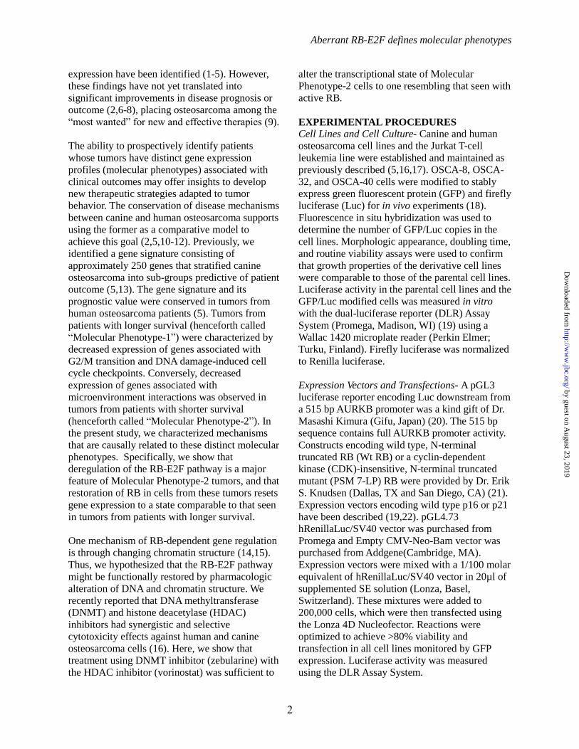
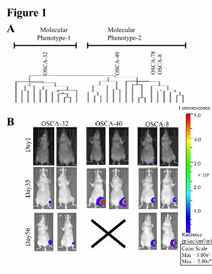
predictive of tumor behavior and outcome (Fig.
1A) (5). To identify potential candidates, we used
the Upstream Regulator Analysis within the IPA
Suite. The direction of gene expression changes in
the Molecular Phenotype-2 samples (shorter
median survival times) was consistent with
inactive RB and p53 tumor suppressor genes
(Table 1, activation Z-scores of -3.801 and -3.791,
respectively). Other significant, predicted altered
regulators included E2F transcription factors and
chromatin remodelers: E2F-1, E2F-2, E2F-3,
E2F4, E2F6, SMARCB1, KDM5B, and HDAC1.
E2F4 was the most significantly altered
transcriptional regulator of the gene signature
(Table 1). However, since E2F4 up-regulates some
of these genes and down-regulates others, a
direction of activity (Z-score) could not be
by guest on August 23, 2019
http://ww
w.jbc.org/
Dow
nloaded from

Aberrant RB-E2F defines molecular phenotypes
5
determined.
An important role for E2F-regulation of the target
genes in the gene signature became more evident
when we searched for conserved DNA response
elements in 5’ upstream promoter sequences. More
than 70% of the 5’ upstream promoter sequences
of the genes comprising the prognostic signature
contained the E2F consensus binding motif
sequence, CCAGGCTGG (data not shown). The
CCAGGCTGG sequence was present in 106 of
143 promoters of genes in the G2/M cluster (E
value 8.1E-105) and in 80 of 108 promoters of the
genes associated with microenvironment
interactions (E value 5.0E-32) (5). Importantly, the
9 base-pair dyad sequence also is one of the most
common sequence motifs in promoters of E2F4
target genes (28).
Orthotopic xenografts using cell lines derived
from Molecular Phenotype-1 and Molecular
Phenotype-2 osteosarcomas recapitulate their
clinical behavior- To test the tumorigenic potential
and outcome of cells from tumors from each
molecular phenotype and to establish suitable cell
lines for downstream functional studies, we
evaluated tumor growth in orthotopic xenografts.
The pattern of outcomes and tumor behavior was
maintained in vivo: the OSCA-32 cell line, which
was derived from a Molecular Phenotype-1 tumor
progressed more slowly and generated less local
bone destruction than the OSCA-40 and OSCA-8
cell lines, which were derived from dogs with
Molecular Phenotype-2 (shorter median survival
times) tumors (Fig 1B.). Microscopic findings for
these tumors were consistent with clinical
outcome. The OSCA-32 tumor cells showed
relatively well differentiated tumor cells laying
down osteoid seams in an orderly fashion (Fig.
2A, C); in contrast, OSCA-40 had highly
anaplastic cells embedded in a poorly organized
osteoid matrix, and extensive areas of necrosis
(Fig. 2B, D)
Characterization of representative cell lines of
each osteosarcoma phenotype- The results from
the orthotopic xenografts supported the use of
these cell lines to elucidate pathogenetic
mechanisms responsible for the biological
behavior of the two molecular phenotypes of
osteosarcoma. Deregulation of E2F transcriptional
activity could result from direct or indirect
mechanisms upon loss-of or reduced RB function.
This loss-of or reduced RB function, in turn, might
be due to mutations that decrease or eliminate RB-
1 expression or that render the RB protein
inactive; however, RB is a component of a
complex pathway and its activity can be
influenced by several regulatory factors (29). The
steady state levels of RB protein were
reproducibly lower in cell lines derived from
Molecular Phenotype-2 tumors (OSCA-40,
OSCA-78, OSCA-8) as compared to those seen in
the cell line derived from a Molecular Phenotype-
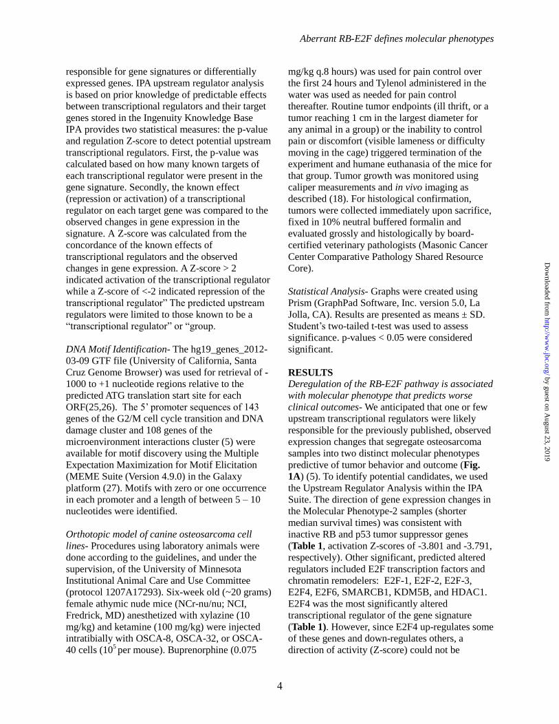
1 tumor (OSCA-32) (Fig. 3A).
To test the hypothesis that functional deregulation
of RB-E2F transcriptional regulation was causally
related to the osteosarcoma molecular phenotypes,
we measured the effect of RB protein abundance
on expression levels of Aurora Kinase B
(AURKB), a target gene that was among those
most highly expressed in Molecular Phenotype-2
samples (5). As shown in Fig. 3B, differential
expression of AURKB between the Molecular
Phenotypes 1 and 2 was maintained; Molecular
Phenotype-1 cells had lower expression levels of
AURKB while Molecular Phenotype-2 cells had
higher expression levels, providing the rationale to
next assess the activity of a AURKB-Luc reporter
construct (20). The AURKB 515 base pair
minimal promoter alone showed basal activity in
all four osteosarcoma cell lines that was consistent
with observed endogenous AURKB transcript
abundance (Fig. 3C). Reporter activity was not
further enhanced when we used an AURKB
promoter that included 1000 base pairs upstream
from the transcriptional start site (20), indicating
that the full complement of activity for this
promoter in osteosarcoma cells was contained
within the minimal promoter sequence.
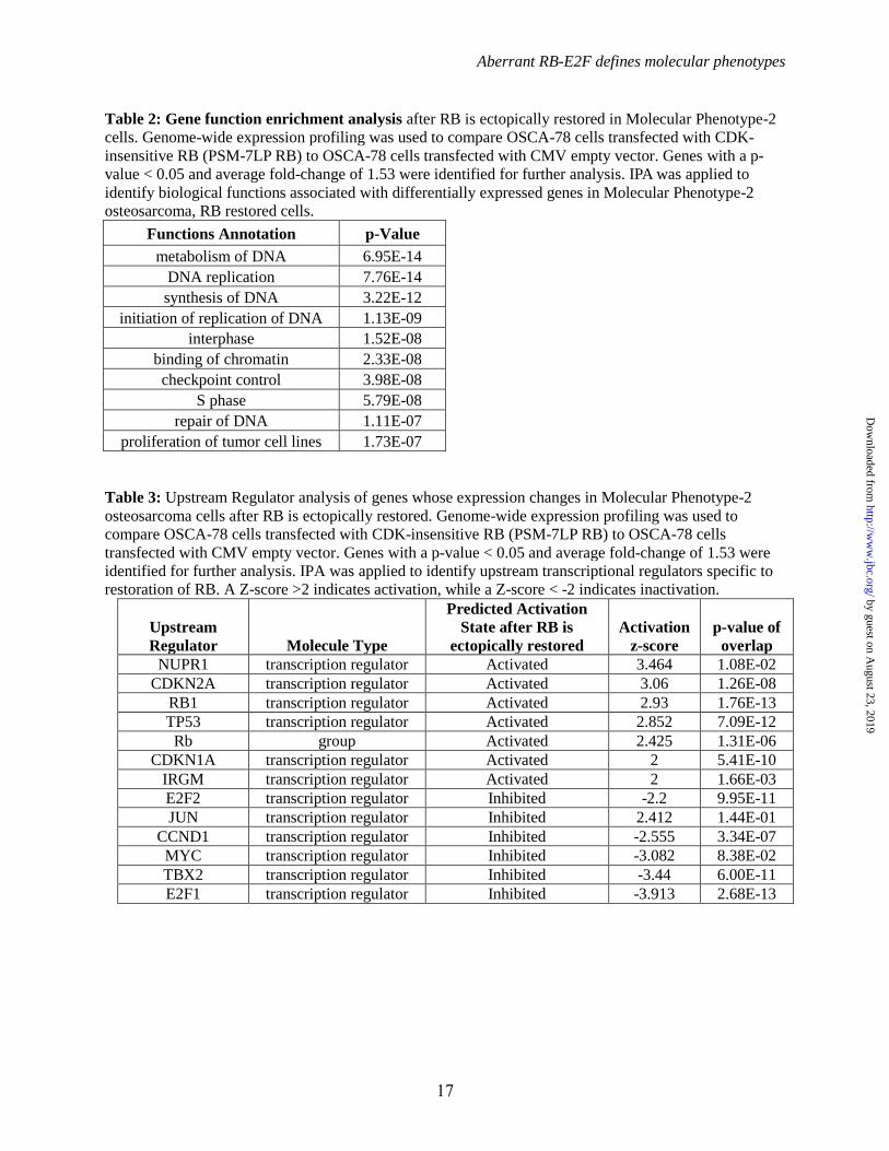
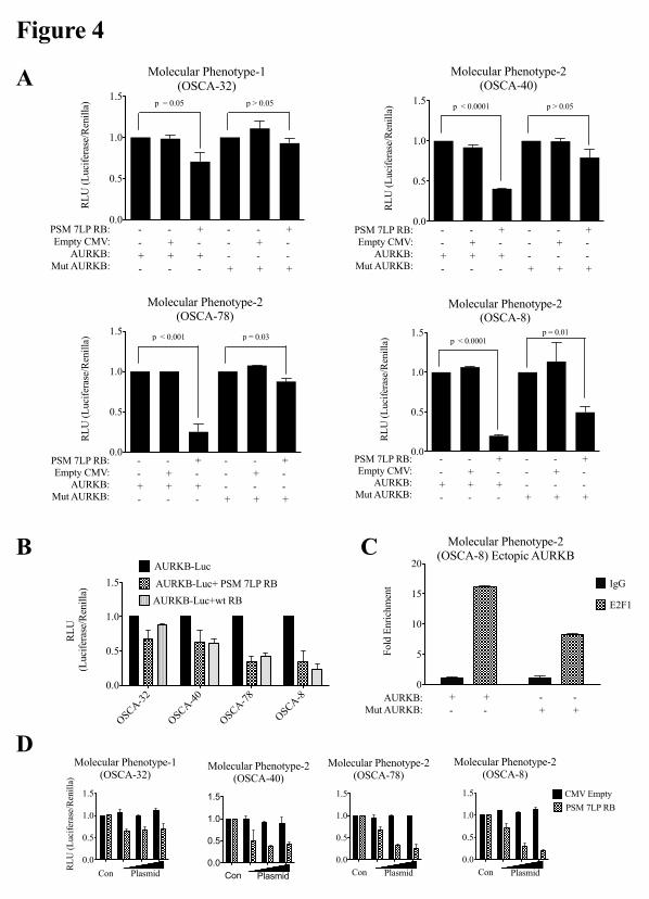
Ectopic RB partially rescues the effects of RB-E2F
deregulation in vitro- The minimal AURKB
promoter reporter does not contain the
CCAGGCTGG sequence or the canonical E2F
binding DNA motif (TTTCCCGC). However, it
contains the cell cycle dependent element (CDE;
GGGCGG) that is responsive to E2F-mediated
transcriptional activation (20,30). In order to
evaluate the role of RB-E2F1 we used both the
by guest on August 23, 2019
http://ww
w.jbc.org/
Dow
nloaded from
Aberrant RB-E2F defines molecular phenotypes
6
AURKB-Luc reporter and a CDE-mutant
AURKB-Luc reporter.
Canine osteosarcoma cell lines were co-
transfected with AURKB reporter and an N-
terminally truncated, RB pocket protein domain
(Wt RB), or the same construct containing seven
mutations that render the protein insensitive to
CDK inactivation (PSM-7LP RB). Introduction of
a CMV empty vector control (21) did not alter the
activity of the AURKB reporter, whereas ectopic
expression of RB inhibited AURKB promoter
activity in the four cell lines (Fig. 4A-B).
However, only the CDK-insensitive RB construct
decreased the activity of the AURKB reporter in
OSCA-32 cells, and the effect was modest when
compared to that observed in the OSCA-40,
OSCA-78, and OSCA-8 cell lines, where both RB
plasmids showed approximately equal repression
(Fig. 4B).
Repression by ectopic PSM-7LP RB was
significantly attenuated in each of the four cell
lines when we used a CDE-mutant AURKB
promoter, although partial repression was still
observed in the Molecular Phenotype-2 cell lines
(Fig. 4C). ChIP analysis confirmed that
endogenous canine E2F1 binds to the CDE in the
AURKB reporter, and that E2F1 had lower affinity
for the mutant CDE site (Fig. 4C).
The effect of ectopic PSM-7LP RB to suppress the
AURKB reporter also was rapidly saturable in
OSCA-32 cells, consistent with the presence of
active endogenous RB (Fig. 4D). In contrast,
ectopic PSM-7LP RB showed dose-dependent
suppression in OSCA-40, OSCA-78, and OSCA-8
cells (Fig. 4D), as would be predicted by absence
of endogenous functional RB.
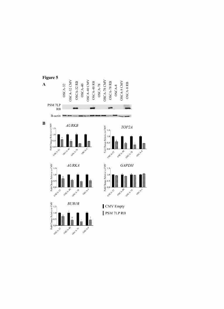
Ectopic RB displaces E2F1 from endogenous
promoters - Given the repressive effect of ectopic
PSM-7LP RB on the ectopic AURKB vector in
cells representing the two molecular phenotypes of
osteosarcoma, we investigated whether these
effects were relevant and reproducible in the
endogenous context. Ectopic PSM-7LP RB protein
was detectable in all of the cell lines (Fig. 5A),
and the presence of ectopic RB consistently
reduced transcript abundance of AURKB and three
other genes (AURKA, BUB1B, and TOP2A) that
were part of the signature that identified
osteosarcoma molecular phenotypes (5) GAPDH
transcript abundance was not affected by the
presence of ectopic PSM-7LP RB (Fig. 5B).
To further validate the importance of the E2F-
dependent interactions in the study context, we
next confirmed that E2F1 was bound to the
endogenous TTTCCAGCCAAT motif in the
canine AURKB promoter, and that its presence on
the promoter contributed to transcription of
AURKB. ChIP assays (Fig. 6A) showed greater
enrichment for E2F1 bound to endogenous
AURKB promoter in OSCA-8 cells (Molecular
Phenotype-2) than in OSCA-32 cells (Molecular
Phenotype-1). Furthermore, E2F1 binding to the
promoter in both molecular phenotypes was
reduced in the presence of ectopic PSM-7LP RB.
Quantitative assessment of AURKB transcript
abundance by qRT-PCR was consistent with what
was observed in our ChIP data, showing that
ectopic PSM-7LP RB caused a greater magnitude
of reduction of AURKB transcript in OSCA-8 cells
versus OSCA-32 cells (data not shown). We
observed a similar effect upon analysis of E2F1
binding to the CGGCGCTAAA motif of the
endogenous CHEK1 promoter (Fig. 6B),
illustrating that ectopic PSM-7LP RB was
repressing the binding of E2F1 protein to genes
associated with G2/M progression and the DNA
damage checkpoint.
Importantly, we did not see any difference in E2F1
protein abundance in the cells transfected with
either the ectopic PSM-7LP RB or CMV plasmids
suggesting that ectopic RB was not simply
reducing E2F1 protein levels. PSM-7LP RB also
was not found in complexes bound to the E2F1-
response elements in the endogenous AURKB
promoter as determined by ChIP in OSCA-8 cells
using an antibody that specifically recognized the
ectopic human RB protein (data not shown). We
similarly did not find evidence of complex
formation between ectopic PSM-7LP RB and
endogenous E2F1 in co-immunoprecipitation
assays; yet, we also did not see a quantitative
difference in the amount of immunoprecipitated
E2F1 protein in cells from either molecular
phenotype transfected with PSM-7LP RB or with
the CMV empty vector, suggesting that ectopic
PSM-7LP RB did not compete with or sterically
by guest on August 23, 2019
http://ww
w.jbc.org/
Dow
nloaded from
Aberrant RB-E2F defines molecular phenotypes
7
hinder binding of the anti-E2F1 antibody (data not
shown). Thus, we favor the interpretation that
displacement of E2F1 from the AURKB promoter
was an indirect effect of RB.
Ectopic RB alters the transcriptional landscape of
Molecular Phenotype-2 osteosarcoma cells
towards one resembling Molecular Phenotype-1-
The selective effects of RB on Molecular
Phenotype-2 osteosarcoma cells led us to
hypothesize that restoration of RB in these cells
would shift the genome-wide transcriptional state
in these cells to one resembling that of Molecular
Phenotype-1 cells. Genome-wide expression
profiling highlighted 78 genes that were
differentially expressed in OSCA-78 cells
transfected with PSM-7LP RB versus the CMV
control: these genes were significantly associated
with functions of DNA replication, metabolism of
DNA, and binding of chromatin (Table 2). The
most significant canonical pathway identified by
IPA was cell cycle control (data not shown).
Importantly, active RB, and inactive E2F1 and
E2F1 were among the most significant predicted
transcriptional regulators associated with the
differential expression of these 78 genes (Table 3).
In addition, IPA yielded a number of predicted
inactivated oncogenes, which have been shown to
be aberrantly expressed in osteosarcoma, including
MYC (31), as well as transcriptional regulators
associated with DNA damage repair and the
mitotic checkpoint, including TBX2 (32) (Table
3). Intriguingly, the gene expression profiles
resulting from restored RB activity in these cells
reflected an apparent recovery of activity for the
TP53 tumor suppressor gene (Table 3).
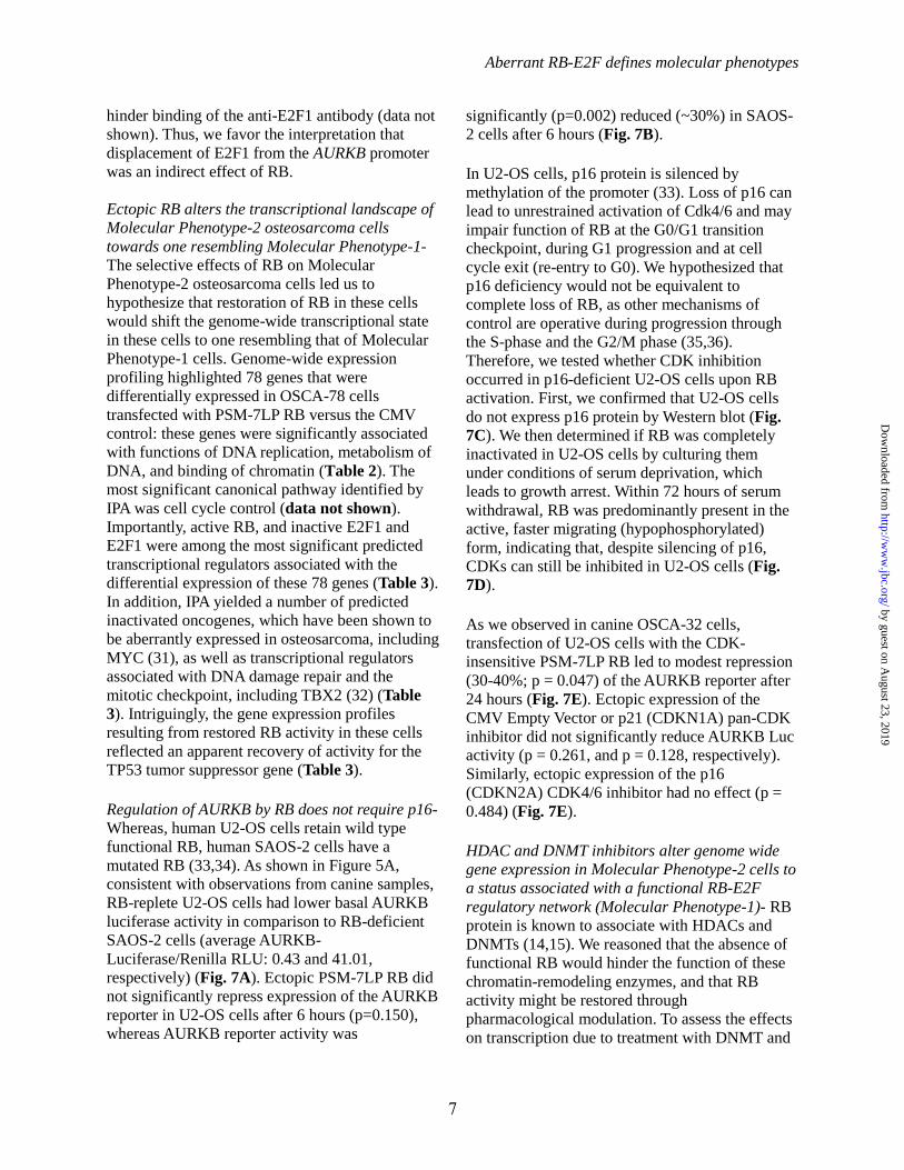
Regulation of AURKB by RB does not require p16-
Whereas, human U2-OS cells retain wild type
functional RB, human SAOS-2 cells have a
mutated RB (33,34). As shown in Figure 5A,
consistent with observations from canine samples,
RB-replete U2-OS cells had lower basal AURKB
luciferase activity in comparison to RB-deficient
SAOS-2 cells (average AURKB-
Luciferase/Renilla RLU: 0.43 and 41.01,
respectively) (Fig. 7A). Ectopic PSM-7LP RB did
not significantly repress expression of the AURKB
reporter in U2-OS cells after 6 hours (p=0.150),
whereas AURKB reporter activity was
significantly (p=0.002) reduced (~30%) in SAOS-
2 cells after 6 hours (Fig. 7B).
In U2-OS cells, p16 protein is silenced by
methylation of the promoter (33). Loss of p16 can
lead to unrestrained activation of Cdk4/6 and may
impair function of RB at the G0/G1 transition
checkpoint, during G1 progression and at cell
cycle exit (re-entry to G0). We hypothesized that
p16 deficiency would not be equivalent to
complete loss of RB, as other mechanisms of
control are operative during progression through
the S-phase and the G2/M phase (35,36).
Therefore, we tested whether CDK inhibition
occurred in p16-deficient U2-OS cells upon RB
activation. First, we confirmed that U2-OS cells
do not express p16 protein by Western blot (Fig.
7C). We then determined if RB was completely
inactivated in U2-OS cells by culturing them
under conditions of serum deprivation, which
leads to growth arrest. Within 72 hours of serum
withdrawal, RB was predominantly present in the
active, faster migrating (hypophosphorylated)
form, indicating that, despite silencing of p16,
CDKs can still be inhibited in U2-OS cells (Fig.
7D).
As we observed in canine OSCA-32 cells,
transfection of U2-OS cells with the CDK-
insensitive PSM-7LP RB led to modest repression
(30-40%; p = 0.047) of the AURKB reporter after
24 hours (Fig. 7E). Ectopic expression of the
CMV Empty Vector or p21 (CDKN1A) pan-CDK
inhibitor did not significantly reduce AURKB Luc
activity (p = 0.261, and p = 0.128, respectively).
Similarly, ectopic expression of the p16
(CDKN2A) CDK4/6 inhibitor had no effect (p =
0.484) (Fig. 7E).
HDAC and DNMT inhibitors alter genome wide
gene expression in Molecular Phenotype-2 cells to
a status associated with a functional RB-E2F
regulatory network (Molecular Phenotype-1)- RB
protein is known to associate with HDACs and
DNMTs (14,15). We reasoned that the absence of
functional RB would hinder the function of these
chromatin-remodeling enzymes, and that RB
activity might be restored through
pharmacological modulation. To assess the effects
on transcription due to treatment with DNMT and
by guest on August 23, 2019
http://ww
w.jbc.org/
Dow
nloaded from

Aberrant RB-E2F defines molecular phenotypes
8
HDAC inhibitors, we did genome-wide expression
profiling of Molecular Phenotype-2 cells treated
with Zeb and SAHA. Figs. 8A and 8B show that
in Molecular Phenotype-2 cells (OSCA-78) cells
the transcriptional state of the defining prognostic
signature shifted to a state that resembled
Molecular Phenotype-1 (OSCA-32) after treatment
with Zeb and SAHA (5). Next, we examined if the
observed effect was associated with displacement
of E2F1 from relevant promoters. ChIP showed
that the amount of E2F1 bound to the endogenous
AURKB (Fig. 8C) promoter was decreased by
approximately 90% in Molecular Phenotype-1
cells, and 95% in Molecular Phenotype-2 cells,
after treatment with these drugs. Similarly, the
amount of E2F1 bound to the endogenous CHEK1
promoter decreased by approximately 80% in both
Molecular Phenotype-1 and Molecular Phenotype-
2 cells after treatment (Fig. 8C).
In addition, we identified 1047 statistically
significant differentially expressed genes (p< 0.05
and average fold-change of 2.0) in untreated and
treated OSCA-78 cells. These genes clustered into
two groups (data not shown). The first group of
genes, which was significantly associated with
functions of cell cycle progression and
proliferation, was down regulated in treated cells.
The second group, which was significantly
associated with functions of cellular organization,
maintenance, and cell-cell interactions, was up
regulated in treated cells. As a single group, these
genes were significantly associated with functions
related to cell cycle, proliferation, and cancer
functions (data not shown).
The IPA transcription factor module was used to
predict upstream regulators of the 1047 genes that
were differentially expressed between cells treated
with DNMT and HDAC inhibitors and those that
were not. RB was a predicted upstream
transcriptional regulator (Table 4) and was
predicted as being active in treated cells. The
predicted activity of other upstream transcriptional
regulators for these 1047 genes paralleled that
observed in Molecular Phenotype-1 cells (Table 1)
as well as that seen upon ectopic reintroduction of
RB into Molecular Phenotype-2 cells (Table 2).
SMARCB1 was predicted as being activated in
treated cells while E2F1, MYC, and FOXM1 were
predicted as being inactivated (Table 4).
DISCUSSION
Here we provide insight into mechanisms that
account for a prognostic gene signature that
reduces the heterogeneity associated with
osteosarcoma. As shown in our model Fig. 9, the
signature allowed us to group the disease into two
subgroups (Molecular Phenotype-1 and Molecular
Phenotype-2) that differ in their biological
behavior; i.e., time to progression and clinical
outcomes (5).
We observed that Molecular Phenotype-1 and
Molecular Phenotype-2 derived xenograft tumors
recapitulated the gross and histologic features of
spontaneous canine osteosarcoma. More
importantly, we show that Molecular Phenotype-2
xenograft tumors appeared to be phenotypically
more aggressive than Molecular Phenotype-1,
exhibiting more rapid growth at the primary tumor
site and a greater propensity for pulmonary
metastasis.
We determined that critical transcriptional
regulators of this evolutionary conserved signature
responsible for these two osteosarcoma
phenotypes are in the RB-E2F regulatory pathway.
In Molecular Phenotype-2 osteosarcoma (which
represents tumors from patients with worse
prognosis), the RB-E2F pathway is dysfunctional.
As a consequence, E2F-regulated genes including
those involved in G2/M transition and DNA
damage-induced cell cycle checkpoints are up
regulated and microenvironment-interacting genes
are down regulated.
Our results show that Molecular Phenotype-2 cells
are more responsive to RB restoration than
Molecular Phenotype-1 cells. The observation that
ectopic expression of constitutively active RB
resulted in transcriptional repression of E2F
targets and other genes associated with cell cycle
control and DNA replication is consistent with
previously defined mechanisms (15). However,
assessments of individual components of the RB
pathway have not always correlated with event
free or overall survival (37,38). Loss of
heterozygosity of RB has been proposed as an
indicator of poor prognosis in human
osteosarcoma patients (39), but our work is the
first to establish a direct link between a conserved,
by guest on August 23, 2019
http://ww
w.jbc.org/
Dow
nloaded from

Aberrant RB-E2F defines molecular phenotypes
9
prognostic genome-wide gene expression
signature and deregulation of the RB-E2F pathway
in osteosarcoma (5). More specifically, current
models of the RB-E2F pathway do not
consistently account for expression levels of genes
during the G2/M phase of cell cycle (40,41); yet,
many of the genes that are overexpressed in
Molecular Phenotype-2 osteosarcoma, including
AURKB, are associated with the G2/M transition.
Recent data suggest that, unlike canonical E2F
response elements that operate primarily in G1/S,
the binding of E2F to the CDE is stronger in G2/M
(30).
One interesting finding from our study was that
restoration of RB compensated for other common
genetic alterations (e.g., TP53 and MYC)
associated with osteosarcoma. The finding
suggests that RB loss or deregulation can modify
tumor behavior and disease progression
downstream of oncogenes that are often altered in
osteosarcoma. While some of these effects could
be due to direct modulation of E2F activity, it also
is likely that indirect changes in the epigenetic
landscape that are established by functional RB
contribute to these effects. The interpretation that
functional RB utilizes mechanisms that are
independent of E2F binding is consistent with the
observation that mutating the CDE sequence did
not completely abrogate reduced AURKB activity
upon re-introduction of a functional RB gene to
Molecular Phenotype-2 cells. Additional support
for our interpretation was the observation that
mutating the CDE sequence also did not
completely abolish E2F1 binding, and that we
did not see evidence of RB binding to the E2F-
responsive elements in the AURKB promoter.
An important finding from our study was that
treatment with DNMT and HDAC inhibitors was
sufficient to supplant RB-E2F pathway function in
Molecular Phenotype-2 cells. Treatment with these
inhibitors altered genome wide gene expression in
Molecular Phenotype-2 cells to a status associated
with a functional RB-E2F regulatory network (as
seen in Molecular Phenotype-1). Moreover, we
show that treatment with DNMT and HDAC
inhibitors achieved comparable transcriptional
changes as genetic restoration of RB, albeit
through regulation of somewhat different gene
sets. Still, these gene sets were concentrated in or
near control nodes for overlapping biochemical
pathways, which is not entirely unexpected, given
the expectation that chromatin-remodeling
enzymes would have a broader effect to modulate
gene expression.
The observation that p16-deficient U2-OS cells
maintained at least a partially functional RB,
possibly through compensation of other CDK
inhibitors like p21, indicates that by itself, the
status of p16 cannot explain the RB-dependent
heterogeneity of osteosarcoma. Specifically, our
findings show that RB-E2F pathway are still able
to regulate genes associated with the G2/M
transition in U2-OS cells, probably through
inhibition of the S-phase and G2/M CDKs.
Nevertheless, CDKN2A deletion or silencing could
contribute to deregulation of the E2F pathway in
osteosarcoma (42). Not surprisingly, CDKN2A
was a predicted transcriptional regulator of the
prognostic gene signature. The CDKN2A locus
was recently linked to osteosarcoma risk in dogs,
and that the risk allele is “fixed” in certain breeds
like Rottweilers and Irish Wolfhounds (43). This
finding illuminates the need to investigate an
explanation for why canine osteosarcoma is so
often a highly aggressive disease (5).
It is widely accepted that RB inactivation is not
necessary for the development of osteosarcoma,
but rather accelerates its development and
progression (44,45). Here, we show that the
integrity of the RB-E2F pathway is
mechanistically associated with the biological
behavior of tumor cells derived from spontaneous
canine osteosarcoma in vitro and in vivo, and that
the RB-E2F molecular regulatory network extends
to human osteosarcoma cells. Specifically, our
data suggest that alternative treatment options that
create a state analogous to that seen with
functional RB could improve outcomes in
osteosarcoma patients, especially those with the
worst prognoses. Our data provide support for
further evaluation of the mechanistic role of RB-
E2F pathway in chromatin remodeling and the
contribution of the epigenetic landscape in
osteosarcoma pathogenesis.
by guest on August 23, 2019
http://ww
w.jbc.org/
Dow
nloaded from
Aberrant RB-E2F defines molecular phenotypes
10
RESEARCH SUPPORT
Grant support for the project was provided by Morris Animal Foundation (D13CA-032), the American
Cancer Society (RSG-13-381-01), the Karen Wyckoff Rein in Sarcoma Foundation (2011-1), the Zach
Sobiech Fund for Osteosarcoma Research of the Children’s Cancer Research Fund, and the Comparative
Medicine Signature Program of the College of Veterinary Medicine, University of Minnesota. The NIH
Comprehensive Cancer Center Support Grant to the Masonic Cancer Center (P30 CA077598) provided
support for bioinformatics, genomics, flow cytometry, bioimaging, cytogenetics, and comparative
pathology services. Jamie Van Etten was supported by grant T32 CA09138, Cancer Biology Training
Grant from the National Institutes of Health, and Jaime F. Modiano is supported by the Alvin and June
Perlman Chair in Animal Oncology. The authors also gratefully acknowledge support from donors to the
Animal Cancer Care and Research Program of the University of Minnesota that helped support of the
project.
AKNOWLEDGEMENTS
We thank the Minnesota Supercomputing Institute for computational resources, and especially Dr. Ying
Zhang, for providing support and advice. We thank Dr. Aric Frantz for generation of OSCA-40-G/L cells,
Dr. Ramesh Kovi for assistance with pathological analyses and Mitzi Lewellen for assistance with in vivo
experiments, and LeAnn Oseth and the MCC cytogenetics core for assistance with FISH. We also thank
Rachit Gupta and Frances Phan for technical assistance and Drs. Dai Ito, Ali Khammanivong, and Siu
Chiu Chan for technical advice. We also thank Drs. Eric Knudsen and Masashi Kimura for providing
constructs and Drs. David Largaespada, Logan Spector, Scott Dehm, Tim Hallstrom, and Richard Gorlick
for helpful discussions and for review of the manuscript.
CONFLICT OF INTEREST
None.
AUTHOR CONTRIBUTIONS
MCS generated data, led the analysis and interpretation of data, prepared manuscript figures, and wrote
the paper. ALS generated data, contributed to bioinformatics data analysis and interpretation, and
performed a critical analysis of the manuscript. HT conducted xenograft and co-immunoprecipitation
experiments, provided technical assistance, and contributed to the preparation of the xenograft figures and
text. JLVE and JV conducted qRT-PCR analysis and revised the manuscript. IC and MGO did
histopathology of xenograft tumors and prepared the histopathology figure and text. SS and JFM
conceived and designed the study. SS offered advice on interpretation of data and gave a critical analysis
of the manuscript during its preparation. JFM supervised laboratory experiments, and assisted in writing
the manuscript. All authors reviewed the results and approved the final version of the manuscript.
by guest on August 23, 2019
http://ww
w.jbc.org/
Dow
nloaded from

Aberrant RB-E2F defines molecular phenotypes
11
REFERENCES
1. Martin, J. W., Squire, J. A., and Zielenska, M. (2012) The genetics of osteosarcoma. Sarcoma
2012, 627254
2. Dobson, J. M. (2013) Breed-predispositions to cancer in pedigree dogs. ISRN veterinary science
2013, 941275
3. Tan, M. L., Choong, P. F., and Dass, C. R. (2009) Osteosarcoma: Conventional treatment vs.
gene therapy. Cancer biology & therapy 8, 106-117
4. Thomas, R., Wang, H. J., Tsai, P. C., Langford, C. F., Fosmire, S. P., Jubala, C. M., Getzy, D.
M., Cutter, G. R., Modiano, J. F., and Breen, M. (2009) Influence of genetic background on
tumor karyotypes: evidence for breed-associated cytogenetic aberrations in canine appendicular
osteosarcoma. Chromosome research : an international journal on the molecular,
supramolecular and evolutionary aspects of chromosome biology 17, 365-377
5. Scott, M. C., Sarver, A. L., Gavin, K. J., Thayanithy, V., Getzy, D. M., Newman, R. A., Cutter,
G. R., Lindblad-Toh, K., Kisseberth, W. C., Hunter, L. E., Subramanian, S., Breen, M., and
Modiano, J. F. (2011) Molecular subtypes of osteosarcoma identified by reducing tumor
heterogeneity through an interspecies comparative approach. Bone 49, 356-367
6. Gorlick, R., Janeway, K., Lessnick, S., Randall, R. L., Marina, N., and Committee, C. O. G. B. T.
(2013) Children's Oncology Group's 2013 blueprint for research: bone tumors. Pediatric blood &
cancer 60, 1009-1015
7. Janeway, K. A., Barkauskas, D. A., Krailo, M. D., Meyers, P. A., Schwartz, C. L., Ebb, D. H.,
Seibel, N. L., Grier, H. E., Gorlick, R., and Marina, N. (2012) Outcome for adolescent and young
adult patients with osteosarcoma: a report from the Children's Oncology Group. Cancer 118,
4597-4605
8. Rainusso, N., Wang, L. L., and Yustein, J. T. (2013) The adolescent and young adult with cancer:
state of the art -- bone tumors. Current oncology reports 15, 296-307
9. Janeway, K. A., and Maki, R. G. (2012) New strategies in sarcoma therapy: linking biology and
novel agents. Clinical cancer research : an official journal of the American Association for
Cancer Research 18, 5837-5844
10. Ranieri, G., Gadaleta, C. D., Patruno, R., Zizzo, N., Daidone, M. G., Hansson, M. G., Paradiso,
A., and Ribatti, D. (2013) A model of study for human cancer: Spontaneous occurring tumors in
dogs. Biological features and translation for new anticancer therapies. Critical reviews in
oncology/hematology 88, 187-197
11. Withrow, S. J., and Wilkins, R. M. (2010) Cross talk from pets to people: translational
osteosarcoma treatments. ILAR journal / National Research Council, Institute of Laboratory
Animal Resources 51, 208-213
12. Modiano, J. F., Breen, M., Lana, S. E., Ehrhart, E. J., Schaack, J., Duke, R. C., Cutter, G. C., and
Bellgrau, D. (2006) Naturally occurring translational models for development of cancer gene
therapy. Gene Ther Mol Bio 10, 31-40
13. Sarver, A. L., Thayanithy, V., Scott, M. C., Cleton-Jansen, A. M., Hogendoorn, P. C., Modiano,
J. F., and Subramanian, S. (2013) MicroRNAs at the human 14q32 locus have prognostic
significance in osteosarcoma. Orphanet journal of rare diseases 8, 7
14. Fiorentino, F. P., Marchesi, I., and Giordano, A. (2013) On the role of retinoblastoma family
proteins in the establishment and maintenance of the epigenetic landscape. Journal of cellular
physiology 228, 276-284
15. Harbour, J. W., and Dean, D. C. (2000) Chromatin remodeling and Rb activity. Current opinion
in cell biology 12, 685-689
16. Thayanithy, V., Park, C., Sarver, A. L., Kartha, R. V., Korpela, D. M., Graef, A. J., Steer, C. J.,
Modiano, J. F., and Subramanian, S. (2012) Combinatorial treatment of DNA and chromatin-
by guest on August 23, 2019
http://ww
w.jbc.org/
Dow
nloaded from

Aberrant RB-E2F defines molecular phenotypes
12
modifying drugs cause cell death in human and canine osteosarcoma cell lines. PloS one 7,
e43720
17. Jubala, C. M., Wojcieszyn, J. W., Valli, V. E., Getzy, D. M., Fosmire, S. P., Coffey, D., Bellgrau,
D., and Modiano, J. F. (2005) CD20 expression in normal canine B cells and in canine non-
Hodgkin lymphoma. Veterinary pathology 42, 468-476
18. Kim, J. H., Frantz, A. M., Anderson, K. L., Graef, A. J., Scott, M. C., Robinson, S., Sharkey, L.
C., Obrien, T. D., Dickerson, E. B., and Modiano, J. F. (2014) Interleukin-8 promotes canine
hemangiosarcoma growth by regulating the tumor microenvironment. Exp Cell Res 232, 155-164
19. Ritt, M. G., Mayor, J., Wojcieszyn, J., Smith, R., Barton, C. L., and Modiano, J. F. (2000)
Sustained nuclear localization of p21/WAF-1 upon growth arrest induced by contact inhibition.
Cancer letters 158, 73-84
20. Kimura, M., Uchida, C., Takano, Y., Kitagawa, M., and Okano, Y. (2004) Cell cycle-dependent
regulation of the human aurora B promoter. Biochemical and biophysical research
communications 316, 930-936
21. Knudsen, E. S., and Wang, J. Y. (1997) Dual mechanisms for the inhibition of E2F binding to RB
by cyclin-dependent kinase-mediated RB phosphorylation. Molecular and cellular biology 17,
5771-5783
22. Modiano, J. F., Mayor, J., Ball, C., Fuentes, M. K., and Linthicum, D. S. (2000) CDK4
expression and activity are required for cytokine responsiveness in T cells. Journal of
immunology (Baltimore, Md. : 1950) 165, 6693-6702
23. Ritt, M. G., Wojcieszyn, J., and Modiano, J. F. (1998) Functional loss of p21/Waf-1 in a case of
benign canine multicentric melanoma. Veterinary pathology 35, 94-101
24. Modiano, J. F., Bellgrau, D., Cutter, G. R., Lana, S. E., Ehrhart, N. P., Ehrhart, E., Wilke, V. L.,
Charles, J. B., Munson, S., Scott, M. C., Pozniak, J., Carlson, C. S., Schaack, J., and Duke, R. C.
(2012) Inflammation, apoptosis, and necrosis induced by neoadjuvant fas ligand gene therapy
improves survival of dogs with spontaneous bone cancer. Molecular therapy : the journal of the
American Society of Gene Therapy 20, 2234-2243
25. Kent, W. J., Sugnet, C. W., Furey, T. S., Roskin, K. M., Pringle, T. H., Zahler, A. M., and
Haussler, D. (2002) The human genome browser at UCSC. Genome research 12, 996-1006
26. Group, G. B. (2002) UCSC Genome Bioinformatics.
27. Goecks, J., Nekrutenko, A., Taylor, J., and Galaxy, T. (2010) Galaxy: a comprehensive approach
for supporting accessible, reproducible, and transparent computational research in the life
sciences. Genome biology 11, R86
28. Weinmann, A. S., Yan, P. S., Oberley, M. J., Huang, T. H., and Farnham, P. J. (2002) Isolating
human transcription factor targets by coupling chromatin immunoprecipitation and CpG island
microarray analysis. Genes & development 16, 235-244
29. Chitko-McKown, C. G., and Modiano, J. F. (1997) Clues to immune function and oncogenesis
provided by events that activate the cell cycle machinery in normal human T cells. Journal of
leukocyte biology 62, 430-437
30. Muller, G. A., and Engeland, K. (2010) The central role of CDE/CHR promoter elements in the
regulation of cell cycle-dependent gene transcription. The FEBS journal 277, 877-893
31. Thayanithy, V., Sarver, A. L., Kartha, R. V., Li, L., Angstadt, A. Y., Breen, M., Steer, C. J.,
Modiano, J. F., and Subramanian, S. (2012) Perturbation of 14q32 miRNAs-cMYC gene network
in osteosarcoma. Bone 50, 171-181
32. Wansleben, S., Davis, E., Peres, J., and Prince, S. (2013) A novel role for the anti-senescence
factor TBX2 in DNA repair and cisplatin resistance. Cell death & disease 4, e846
33. Park, Y. B., Park, M. J., Kimura, K., Shimizu, K., Lee, S. H., and Yokota, J. (2002) Alterations in
the INK4a/ARF locus and their effects on the growth of human osteosarcoma cell lines. Cancer
genetics and cytogenetics 133, 105-111
by guest on August 23, 2019
http://ww
w.jbc.org/
Dow
nloaded from

Aberrant RB-E2F defines molecular phenotypes
13
34. Rogatsky, I., Trowbridge, J. M., and Garabedian, M. J. (1997) Glucocorticoid receptor-mediated
cell cycle arrest is achieved through distinct cell-specific transcriptional regulatory mechanisms.
Molecular and cellular biology 17, 3181-3193
35. Lukas, J., Herzinger, T., Hansen, K., Moroni, M. C., Resnitzky, D., Helin, K., Reed, S. I., and
Bartek, J. (1997) Cyclin E-induced S phase without activation of the pRb/E2F pathway. Genes &
development 11, 1479-1492
36. Mann, D. J., and Jones, N. C. (1996) E2F-1 but not E2F-4 can overcome p16-induced G1 cell-
cycle arrest. Current biology : CB 6, 474-483
37. Heinsohn, S., Evermann, U., Zur Stadt, U., Bielack, S., and Kabisch, H. (2007) Determination of
the prognostic value of loss of heterozygosity at the retinoblastoma gene in osteosarcoma.
International journal of oncology 30, 1205-1214
38. Maitra, A., Roberts, H., Weinberg, A. G., and Geradts, J. (2001) Loss of p16INK4a expression
correlates with decreased survival in pediatric osteosarcomas. Inter J of Cancer 95, 34-38
39. Feugeas, O., Guriec, N., Babin-Boilletot, A., Marcellin, L., Simon, P., Babin, S., Thyss, A.,
Hofman, P., Terrier, P., Kalifa, C., Brunat-Mentigny, M., Patricot, L. M., and Oberling, F. (1996)
Loss of heterozygosity of the RB gene is a poor prognostic factor in patients with osteosarcoma.
Journal of clinical oncology : official journal of the American Society of Clinical Oncology 14,
467-472
40. Dyson, N. (1998) The regulation of E2F by pRB-family proteins. Genes & development 12, 2245-
2262
41. Sadasivam, S., and DeCaprio, J. A. (2013) The DREAM complex: master coordinator of cell
cycle-dependent gene expression. Nature reviews. Cancer 13, 585-595
42. Ashizawa, S., Nishizawa, H., Yamada, M., Higashi, H., Kondo, T., Ozawa, H., Kakita, A., and
Hatakeyama, M. (2001) Collective inhibition of pRB family proteins by phosphorylation in cells
with p16INK4a loss or cyclin E overexpression. The Journal of biological chemistry 276, 11362-
11370
43. Karlsson, E. K., Sigurdsson, S., Ivansson, E., Thomas, R., Elvers, I., Wright, J., Howald, C.,
Tonomura, N., Perloski, M., Swofford, R., Biagi, T., Fryc, S., Anderson, N., Courtay-Cahen, C.,
Youell, L., Ricketts, S. L., Mandlebaum, S., Rivera, P., von Euler, H., Kisseberth, W. C., London,
C. A., Lander, E. S., Couto, G., Comstock, K., Starkey, M. P., Modiano, J. F., Breen, M., and
Lindblad-Toh, K. (2013) Genome-wide analyses implicate 33 loci in heritable dog osteosarcoma,
including regulatory variants near CDKN2A/B. Genome biology 14, R132
44. Berman, S. D., Calo, E., Landman, A. S., Danielian, P. S., Miller, E. S., West, J. C., Fonhoue, B.
D., Caron, A., Bronson, R., Bouxsein, M. L., Mukherjee, S., and Lees, J. A. (2008) Metastatic
osteosarcoma induced by inactivation of Rb and p53 in the osteoblast lineage. Proceedings of the
National Academy of Sciences of the United States of America 105, 11851-11856
45. Calo, E., Quintero-Estades, J. A., Danielian, P. S., Nedelcu, S., Berman, S. D., and Lees, J. A.
(2010) Rb regulates fate choice and lineage commitment in vivo. Nature 466, 1110-1114
by guest on August 23, 2019
http://ww
w.jbc.org/
Dow
nloaded from

Aberrant RB-E2F defines molecular phenotypes
14
FIGURE LEGENDS
Fig. 1. Osteosarcoma in vivo models recapitulates the biological behavior of two distinct molecular
phenotypes. A, Sample dendrogram from previously published gene expression profiling of canine
osteosarcoma showing two molecularly distinct sample clusters, denoted as Molecular Phenotype-1 and
Molecular Phenotype-2, which have significantly different survival times 14 month and 2.83 months
respectively (5). Representative cell lines from each phenotype (OSCA-32, OSCA-40, OSCA-78, OSCA-
8) were used to identify mechanisms driving sample stratification. B, Serial in vivo imaging of athymic
nude mice harboring intratibial xenografts of OSCA-32 cells (Molecular Phenotype-1) or OSCA-40 or
OSCA-8 cells (Molecular Phenotype-2).
Fig. 2. Histopathology findings. Panels A and B show low magnification photomicrographs of OSCA-32
and OS-40 tumors, respectively. Note orderly deposition of osteoid seams (indicated by asterisk) in an
OSCA-32 tumor (Panel A) in contrast to the poorly organized osteoid matrix in an OSCA-40 tumor
(Panel B); also note area of necrosis (indicated by asterisk). Bars = 200 µm.
Panels C and D show high magnification photomicrographs of OSCA-32 and OS-40 tumors, respectively.
Relatively well-differentiated osteoblastic cells lay down osteoid (indicated by asterisk) in an OSCA-32
tumor (Panel C). In contrast, note proliferating spindle cells with numerous mitotic figures (arrowheads)
within poorly organized matrix (osteoid) of an OSCA-40 tumor (Panel D). Bars = 50 µm.
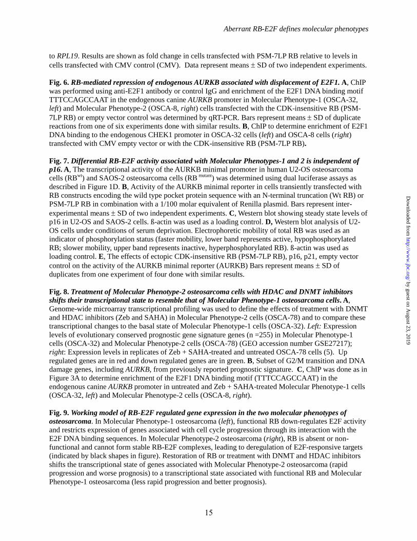
Fig. 3. Expression of AURKB in osteosarcoma phenotypes is inversely related to endogenous RB
protein and cells from tumors derived from dogs with shorter median survival times (Molecular
Phenotype-2) behave aggressively in vivo. A, Steady state levels of RB protein as determined by Western
blotting (ß-actin, loading control). B, Average value (SD) for AURKB expression from two Affymetrix
probes (GEO accession number GSE27217) (5). C, AURKB minimal promoter as determined by dual
luciferase assays in the four cell lines (means SD of duplicate experiments).
Fig. 4. Ectopic expression of RB represses activity of the AURKB minimal reporter through binding of
E2F1 to the cell cycle–dependent element (CDE). A, OSCA-32 (Molecular Phenotype-1), and OSCA-
40, OSCA-78, and OSCA-8 (Molecular Phenotype-2) cell lines were transiently transfected with CDK-
insensitive RB (PSM-7LP RB), or with CMV empty vector (Empty CMV) in combination with the
AURKB minimal reporter (AURKB) or the CDE site mutant AURKB reporter (Mut AURKB). All
reactions were done with a 1/100 molar equivalent of Renilla luciferase plasmid. Data show normalized
luciferase activity (Firefly/Renilla). Bars, represent inter-experimental means SD of two or more
independent experiments for each cell line. B, Cells were transiently transfected with RB constructs
encoding the wild type pocket protein sequence with an N-terminal truncation (Wt RB) or a CDK-
insensitive RB (PSM-7LP RB), in combination with the AURKB minimal reporter (AURKB) and a 1/100
molar equivalent of Renilla-Luc plasmid. Bars represent means SD of five independent experiments. C,
ChIP was performed using anti-E2F1 antibody or control IgG in cells transfected with the AURKB
reporter or the Mut AURKB reporter followed by PCR amplification of the CDE site of these reporters.
Bars show the mean fold enrichment SD of endogenous canine E2F1 normalized to IgG control in two
independent experiments. D, The experimental set-up described in (A) was used to determine the dose
response relationships of CDK-insensitive RB (PSM-7LP RB), or empty vector control (0- 0.25 µg per
2E5 cells) on the AURKB minimal reporter (AURKB-Luc).
Fig. 5. Ectopic RB decreases the abundance of prototypical E2F targets. A, Western blot (ß-actin,
loading control) showing steady state protein levels of ectopic PSM-7LP RB in cells transfected with
CDK-insensitive RB (PSM-7LP RB), or CMV empty vector. B, Transcript abundance of AURKB,
AURKA, BUB1B, TOP2A, and GAPDH in transfected cells was determined by qRT-PCR and normalized
by guest on August 23, 2019
http://ww
w.jbc.org/
Dow
nloaded from
Aberrant RB-E2F defines molecular phenotypes
15
to RPL19. Results are shown as fold change in cells transfected with PSM-7LP RB relative to levels in
cells transfected with CMV control (CMV). Data represent means SD of two independent experiments.
Fig. 6. RB-mediated repression of endogenous AURKB associated with displacement of E2F1. A, ChIP
was performed using anti-E2F1 antibody or control IgG and enrichment of the E2F1 DNA binding motif
TTTCCAGCCAAT in the endogenous canine AURKB promoter in Molecular Phenotype-1 (OSCA-32,
left) and Molecular Phenotype-2 (OSCA-8, right) cells transfected with the CDK-insensitive RB (PSM-
7LP RB) or empty vector control was determined by qRT-PCR. Bars represent means SD of duplicate
reactions from one of six experiments done with similar results. B, ChIP to determine enrichment of E2F1
DNA binding to the endogenous CHEK1 promoter in OSCA-32 cells (left) and OSCA-8 cells (right)
transfected with CMV empty vector or with the CDK-insensitive RB (PSM-7LP RB).
Fig. 7. Differential RB-E2F activity associated with Molecular Phenotypes-1 and 2 is independent of
p16. A, The transcriptional activity of the AURKB minimal promoter in human U2-OS osteosarcoma
cells (RBwt
) and SAOS-2 osteosarcoma cells (RB mutant
) was determined using dual luciferase assays as
described in Figure 1D. B, Activity of the AURKB minimal reporter in cells transiently transfected with
RB constructs encoding the wild type pocket protein sequence with an N-terminal truncation (Wt RB) or
PSM-7LP RB in combination with a 1/100 molar equivalent of Renilla plasmid. Bars represent inter-
experimental means SD of two independent experiments. C, Western blot showing steady state levels of
p16 in U2-OS and SAOS-2 cells. ß-actin was used as a loading control. D, Western blot analysis of U2-
OS cells under conditions of serum deprivation. Electrophoretic mobility of total RB was used as an
indicator of phosphorylation status (faster mobility, lower band represents active, hypophosphorylated
RB; slower mobility, upper band represents inactive, hyperphosphorylated RB). ß-actin was used as
loading control. E, The effects of ectopic CDK-insensitive RB (PSM-7LP RB), p16, p21, empty vector
control on the activity of the AURKB minimal reporter (AURKB) Bars represent means SD of
duplicates from one experiment of four done with similar results.
Fig. 8. Treatment of Molecular Phenotype-2 osteosarcoma cells with HDAC and DNMT inhibitors
shifts their transcriptional state to resemble that of Molecular Phenotype-1 osteosarcoma cells. A,
Genome-wide microarray transcriptional profiling was used to define the effects of treatment with DNMT
and HDAC inhibitors (Zeb and SAHA) in Molecular Phenotype-2 cells (OSCA-78) and to compare these
transcriptional changes to the basal state of Molecular Phenotype-1 cells (OSCA-32). Left: Expression
levels of evolutionary conserved prognostic gene signature genes (n =255) in Molecular Phenotype-1
cells (OSCA-32) and Molecular Phenotype-2 cells (OSCA-78) (GEO accession number GSE27217);
right: Expression levels in replicates of Zeb + SAHA-treated and untreated OSCA-78 cells (5). Up
regulated genes are in red and down regulated genes are in green. B, Subset of G2/M transition and DNA
damage genes, including AURKB, from previously reported prognostic signature. C, ChIP was done as in
Figure 3A to determine enrichment of the E2F1 DNA binding motif (TTTCCAGCCAAT) in the
endogenous canine AURKB promoter in untreated and Zeb + SAHA-treated Molecular Phenotype-1 cells
(OSCA-32, left) and Molecular Phenotype-2 cells (OSCA-8, right).
Fig. 9. Working model of RB-E2F regulated gene expression in the two molecular phenotypes of
osteosarcoma. In Molecular Phenotype-1 osteosarcoma (left), functional RB down-regulates E2F activity
and restricts expression of genes associated with cell cycle progression through its interaction with the
E2F DNA binding sequences. In Molecular Phenotype-2 osteosarcoma (right), RB is absent or non-
functional and cannot form stable RB-E2F complexes, leading to deregulation of E2F-responsive targets
(indicated by black shapes in figure). Restoration of RB or treatment with DNMT and HDAC inhibitors
shifts the transcriptional state of genes associated with Molecular Phenotype-2 osteosarcoma (rapid
progression and worse prognosis) to a transcriptional state associated with functional RB and Molecular
Phenotype-1 osteosarcoma (less rapid progression and better prognosis).
by guest on August 23, 2019
http://ww
w.jbc.org/
Dow
nloaded from

Aberrant RB-E2F defines molecular phenotypes
16
Table 1: Regulators of the gene signature that identifies two osteosarcoma phenotypes. IPA was
applied to identify upstream transcriptional regulators of the gene signature consisting of approximately
250 genes (5). A Z-score >2 indicates activation, while a Z-score < -2 indicates inactivation.
Upstream
Regulator Molecule Type
Predicted Activation State
in samples with worse
patient outcomes
Activation
z-score
p-value of
overlap
E2F4 transcription regulator NA NA 7.86E-39
Vegf group Activated 5.751 2.79E-30
E2F1 transcription regulator Activated 5.032 1.18E-31
TBX2 transcription regulator Activated 4.583 9.26E-27
FOXM1 transcription regulator Activated 4.127 3.70E-20
CCND1 transcription regulator Activated 3.755 6.74E-28
MED1 transcription regulator Activated 3.550 2.88E-09
FOXO1 transcription regulator Activated 3.503 4.12E-09
E2f group Activated 3.382 3.61E-19
MYC transcription regulator Activated 3.309 3.18E-10
STAT3 transcription regulator Activated 3.239 2.24E-04
E2F3 transcription regulator Activated 3.124 1.28E-23
NFKBIA transcription regulator Activated 3.011 2.16E-09
E2F2 transcription regulator Activated 3.000 1.20E-18
NRIP1 transcription regulator Inhibited -2.219 2.32E-06
SMARCB1 transcription regulator Inhibited -2.393 2.73E-16
TOB1 transcription regulator Inhibited -2.449 5.86E-06
ATF3 transcription regulator Inhibited -2.577 3.17E-07
HOXA10 transcription regulator Inhibited -2.588 2.20E-03
HDAC1 transcription regulator Inhibited -2.611 2.24E-11
E2F6 transcription regulator Inhibited -2.828 1.27E-11
TCF3 transcription regulator Inhibited -3.317 3.95E-08
Rb group Inhibited -3.348 2.94E-19
RBL1 transcription regulator Inhibited -3.379 8.36E-15
KDM5B transcription regulator Inhibited -3.734 9.06E-17
TP53 transcription regulator Inhibited -3.791 2.03E-38
RB1 transcription regulator Inhibited -3.801 2.51E-23
CDKN2A transcription regulator Inhibited -4.358 2.27E-16
NUPR1 transcription regulator Inhibited -4.849 6.56E-17
by guest on August 23, 2019
http://ww
w.jbc.org/
Dow
nloaded from
Aberrant RB-E2F defines molecular phenotypes
17
Table 2: Gene function enrichment analysis after RB is ectopically restored in Molecular Phenotype-2
cells. Genome-wide expression profiling was used to compare OSCA-78 cells transfected with CDK-
insensitive RB (PSM-7LP RB) to OSCA-78 cells transfected with CMV empty vector. Genes with a p-
value < 0.05 and average fold-change of 1.53 were identified for further analysis. IPA was applied to
identify biological functions associated with differentially expressed genes in Molecular Phenotype-2
osteosarcoma, RB restored cells.
Functions Annotation p-Value
metabolism of DNA 6.95E-14
DNA replication 7.76E-14
synthesis of DNA 3.22E-12
initiation of replication of DNA 1.13E-09
interphase 1.52E-08
binding of chromatin 2.33E-08
checkpoint control 3.98E-08
S phase 5.79E-08
repair of DNA 1.11E-07
proliferation of tumor cell lines 1.73E-07
Table 3: Upstream Regulator analysis of genes whose expression changes in Molecular Phenotype-2
osteosarcoma cells after RB is ectopically restored. Genome-wide expression profiling was used to
compare OSCA-78 cells transfected with CDK-insensitive RB (PSM-7LP RB) to OSCA-78 cells
transfected with CMV empty vector. Genes with a p-value < 0.05 and average fold-change of 1.53 were
identified for further analysis. IPA was applied to identify upstream transcriptional regulators specific to
restoration of RB. A Z-score >2 indicates activation, while a Z-score < -2 indicates inactivation.
Upstream
Regulator Molecule Type
Predicted Activation
State after RB is
ectopically restored
Activation
z-score
p-value of
overlap
NUPR1 transcription regulator Activated 3.464 1.08E-02
CDKN2A transcription regulator Activated 3.06 1.26E-08
RB1 transcription regulator Activated 2.93 1.76E-13
TP53 transcription regulator Activated 2.852 7.09E-12
Rb group Activated 2.425 1.31E-06
CDKN1A transcription regulator Activated 2 5.41E-10
IRGM transcription regulator Activated 2 1.66E-03
E2F2 transcription regulator Inhibited -2.2 9.95E-11
JUN transcription regulator Inhibited 2.412 1.44E-01
CCND1 transcription regulator Inhibited -2.555 3.34E-07
MYC transcription regulator Inhibited -3.082 8.38E-02
TBX2 transcription regulator Inhibited -3.44 6.00E-11
E2F1 transcription regulator Inhibited -3.913 2.68E-13
by guest on August 23, 2019
http://ww
w.jbc.org/
Dow
nloaded from

Aberrant RB-E2F defines molecular phenotypes
18
Table 4: Regulators of genes whose expression significantly changes in Molecular Phenotype-2
osteosarcoma cells after treatment with DNMT and HDAC inhibitors. Genome-wide expression
profiling was used to compare OSCA-78 cells treated with DNMT and HDAC inhibitors to untreated
OSCA-78 cells. Genes with a p-value < 0.05 and average fold-change of 1.53 were identified for further
analysis. IPA was applied to identify upstream transcriptional regulators specific to treatment with DNMT
and HDAC inhibitors. A Z-score >2 indicates activation, while a Z-score < -2 indicates inactivation.
Upstream
Regulator Molecule Type
Predicted Activation
Status after treatment
with DNMT and HDAC
inhibitors
Activation
z-score
p-value of
overlap
FOXO3 transcription regulator Activated 2.224 6.58E-06
RB1 transcription regulator Activated 2.200 1.19E-05
HNF4A transcription regulator Activated 2.114 1.17E-04
EPAS1 transcription regulator Activated 2.362 6.75E-04
Rb group Activated 2.528 4.46E-03
SMARCB1 transcription regulator Activated 2.877 1.89E-02
HIF1A transcription regulator Activated 2.063 2.19E-02
TBX2 transcription regulator Inhibited -3.053 4.34E-06
Vegf group Inhibited -2.882 1.23E-05
MYC transcription regulator Inhibited -3.386 3.16E-04
FOXM1 transcription regulator Inhibited -3.082 4.13E-04
E2F1 transcription regulator Inhibited -2.417 3.52E-03
EIF4E translation regulator Inhibited -2.517 5.61E-03
ZNF217 transcription regulator Inhibited -2.000 5.78E-03
Ras group Inhibited -2.150 1.59E-02
KLF5 transcription regulator Inhibited -2.197 3.19E-02
JUN transcription regulator Inhibited -2.013 5.03E-02
Hdac group Inhibited -2.345 7.14E-02
ETS1 transcription regulator Inhibited -2.076 8.35E-02
by guest on August 23, 2019
http://ww
w.jbc.org/
Dow
nloaded from
OSC
A-3
2
OSC
A-7
8 O
SCA
-8
OSC
A-4
0
Molecular Phenotype-1
Molecular Phenotype-2
OSCA-32
Day1
Day35
OSCA-40 OSCA-8
Day56
Luminescence 5.0 4.0 3.0 2.0 1.0 × 10⁹ Radiance (p/sec/cm2/sr) Color Scale Min = 5.00e7 Max = 5.00e9
B
Figure 1A
by guest on August 23, 2019
http://ww
w.jbc.org/
Dow
nloaded from

RB
B-actin
OSCA-32 OSCA-40 OSCA-78 OSCA-8
100200300
1000
1500
2000
AU
RK
B e
xpre
ssio
n (A
ffym
etrix
_Can
ine
2.0)
OSCA-32 OSCA-40 OSCA-78 OSCA-80
2
4
6
AU
RK
B F
irefly
/Ren
illa
RLU
OSCA-32 OSCA-40 OSCA-78 OSCA-8
B
C
A
Molecular Phenotype-1
Molecular Phenotype-2
Figure 3
by guest on August 23, 2019
http://ww
w.jbc.org/
Dow
nloaded from
Molecular Phenotype-1 (OSCA-32)
0.0
0.5
1.0
1.5R
LU (L
ucife
rase
/Ren
illa)
PSM 7LP RB:Empty CMV:
AURKB:Mut AURKB:
- - + - - +
+ + + - - -- - - + + +
- + - - + -
p > 0.05p = 0.05
Molecular Phenotype-2 (OSCA-78)
RLU
(Luc
ifera
se/R
enill
a)
0.0
0.5
1.0
1.5 p = 0.03p < 0.001
PSM 7LP RB:Empty CMV:
AURKB:Mut AURKB:
- - + - - +
+ + + - - -- - - + + +
- + - - + -
Molecular Phenotype-1(OSCA-32)
0.0
0.5
1.0
1.5
RLU
(Luc
ifera
se/R
enill
a)
Con Plasmid
Molecular Phenotype-2(OSCA-78)
0.0
0.5
1.0
1.5
Con Plasmid
Molecular Phenotype-2(OSCA-8) Ectopic AURKB
0
5
10
15
20
Fold
Enr
ichm
ent IgG
E2F1
AURKB:Mut AURKB:
+ + - -- - + +
Molecular Phenotype-2 (OSCA-40)
0.0
0.5
1.0
1.5
RLU
(Luc
ifera
se/R
enill
a) p > 0.05p < 0.0001
PSM 7LP RB:Empty CMV:
AURKB:Mut AURKB:
- - + - - +
+ + + - - -- - - + + +
- + - - + -
Molecular Phenotype-2 (OSCA-8)
RLU
(Luc
ifera
se/R
enill
a)
0.0
0.5
1.0
1.5 p = 0.01p < 0.0001
PSM 7LP RB:Empty CMV:
AURKB:Mut AURKB:
- - + - - +
+ + + - - -- - - + + +
- + - - + -
Molecular Phenotype-2(OSCA-40)
0.0
0.5
1.0
1.5
Con Plasmid
Molecular Phenotype-2(OSCA-8)
0.0
0.5
1.0
1.5 CMV EmptyPSM 7LP RB
Con Plasmid
A
D
B
Figure 4R
LU (L
ucife
rase
/Ren
illa)
OSCA-32
OSCA-40
OSCA-78
OSCA-80.0
0.5
1.0
1.5AURKB-Luc AURKB-Luc+ PSM 7LP RB
AURKB-Luc+wt RB
C
by guest on August 23, 2019
http://ww
w.jbc.org/
Dow
nloaded from

Molecular Phenotype-1Endogenous AURKB
012345
Fold
Enr
ichm
ent
IgGE2F1
Empty CVM: PSM 7LP RB:
+ + - -- - + +
Molecular Phenotype-1Endogenous CHEK1
012345
Fold
Enr
ichm
ent
IgGE2F1
Empty CVM: PSM 7LP RB:
+ + - -- - + +
Molecular Phenotype-2Endogenous AURKB
012345
Fold
Enr
ichm
ent
IgGE2F1
Empty CVM: PSM 7LP RB:
+ + - -- - + +
Molecular Phenotype-2Endogenous CHEK1
012345
Fold
Enr
ichm
ent
IgGE2F1
Empty CVM: PSM 7LP RB:
+ + - -- - + +
A
B
Figure 6
by guest on August 23, 2019
http://ww
w.jbc.org/
Dow
nloaded from
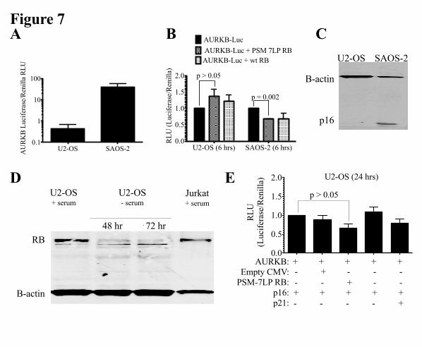
0.0
0.5
1.0
1.5
RLU
(Luc
ifera
se/R
enill
a) U2-OS
U2-OS - serum
48 hr 72 hr
Jurkat + serum
U2-OS + serum
RB
B-actin
U2-OS SAOS-20.1
1
10
100
AU
RK
B L
ucife
rase
/Ren
illa
RLU
U2-OS (6 hrs) SAOS-2 (6 hrs)0.0
0.5
1.0
1.5
2.0
RLU
(Luc
ifera
se/R
enill
a)
AURKB-Luc AURKB-Luc + PSM 7LP RB
AURKB-Luc + wt RB
p > 0.05
p = 0.002
0.0
0.5
1.0
1.5
2.0
RLU
(Luc
ifera
se/R
enill
a) U2-OS (24 hrs)
p > 0.05
AURKB:Empty CMV:
PSM-7LP RB:p16:p21:
+ + + + +- + - - -- - + - -+ + + + +- - - - +
A C
D E
Figure 7
B-actin
p16
U2-OS SAOS-2
B-actin
p16
U2-OS SAOS-2
B
by guest on August 23, 2019
http://ww
w.jbc.org/
Dow
nloaded from

Mol
ecul
ar P
heno
type
-1
Mol
ecul
ar P
heno
type
-2
Bla
nk
SAH
A+Z
eb
SAH
A+Z
eb
Unt
reat
ed
Unt
reat
ed
G2/
M tr
ansi
tion
an
d D
NA
dam
age
gene
s M
icro
envi
ronm
ent
gene
s
Molecular Phenotype-2
Subs
et o
f G2/
M tr
ansi
tion
and
DN
A d
amag
e ge
nes
Mol
ecul
ar P
heno
type
-1
Mol
ecul
ar P
heno
type
-2
Bla
nk
SAH
A+Z
eb
SAH
A+Z
eb
Unt
reat
ed
Unt
reat
ed
A B
C
Figure 8
Molecular Phenotype-1Endogenous AURKB
01234
Fold
Enr
ichm
ent
SAHA + Zeb
- - + +
Molecular Phenotype-2Endogenous AURKB
01234
Fold
Enr
ichm
ent
IgGE2F1
- - + +SAHA + Zeb
by guest on August 23, 2019
http://ww
w.jbc.org/
Dow
nloaded from
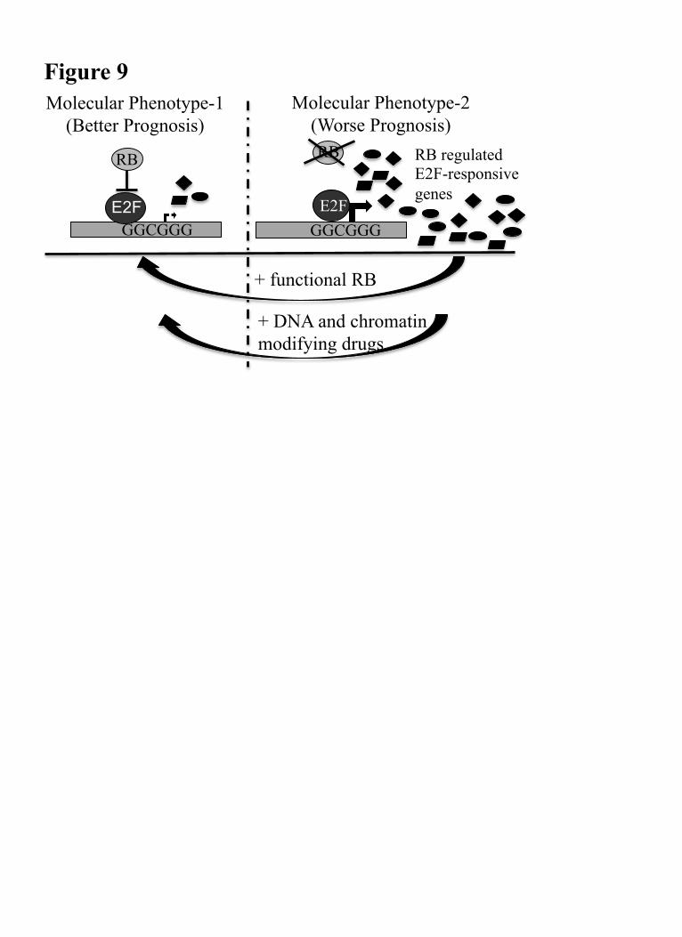
Figure 9
RB regulated E2F-responsive genes
Molecular Phenotype-1 (Better Prognosis)
Molecular Phenotype-2 (Worse Prognosis)
GGCGGG
RB RB
GGCGGG E2F
+ functional RB
+ DNA and chromatin modifying drugs
E2F
Jyotika Varshney, M. Gerard O'Sullivan, Subbaya Subramanian and Jaime F. ModianoMilcah Carol Scott, Aaron L. Sarver, Hirotaka Tomiyasu, Ingrid Cornax, Jamie Van Etten,
OsteosarcomaAberrant RB-E2F Transcriptional Regulation Defines Molecular Phenotypes of
published online September 16, 2015J. Biol. Chem.
10.1074/jbc.M115.679696Access the most updated version of this article at doi:
Alerts:
When a correction for this article is posted•
When this article is cited•
to choose from all of JBC's e-mail alertsClick here
by guest on August 23, 2019
http://ww
w.jbc.org/
Dow
nloaded from


