A new species of Leucopaxillus BOURS. (Agaricales) fromWestern Australia
N. L. BOUGHER
Botany Department, University of Western Australia,Nedlands, W. A. 6009.
Abs t rac t . - Leucopaxillus lilacinus is described as a distinctive new species ofthe indigenous eucalyptus (Jarrah) forest in southwestern Australia. Comparison ismade with the only other known representative of Leucopaxillus in Australia -Clitocybe eucalyptorura.
DescriptionLeucopaxillus lilacinus BOUGHER sp. nov. - Fig. 1, a-g; PL 1 a.Pileus 80-150 mm, convexus demum planus, margo sulcatus revolutus demum
planus, lilacinus demum atroviolaceus. Lamellae ad 5 mm latae, adnatae brevidentes,albae demum cremeae, margincs lucentes sub lens. Stipes 50-100 x 20-30 mm,cylindricus vel clavatus, pileus concolor, laevis vel tomentosus, siccus. Odor leniterfragrans. Sapor nullus. Sporae congregatae cremeae. Sporae 5(5.5)-6.5(7) x4-5(5.5) |im, ellipsoideae vel ovatae, verrucosae, ornamento valde amyloideo.Cheilocystidia 22-60 x 2-6 (im, filiformia vel cylindracea, tenuitunicata, hyalina,abundantes. Hyphae pileipellis densiter intricatae, cylindraceae, tenuitunicatae, hy-alinae, 2-6 [im latae. Fibulae frequentes. In foliis dejectiis in silvis Eucalypti mar-ginatae necnon Eucalypti calophyllae. Australia. BOUGHER (UWA 3014, Holotypus).
Pileus 80-150 mm; convex with inrolled margin expanding toplane with incurved margin, margin consistently grooved; uniformlylilac (16-B/3 or 16-B/4)*) soon dark violet (18-F/4 or 18-F/5) dar-kest at center; surface slightly velutinous, moist when wet thendrying with consistency of soft leather; pigmented cuticle layer verythin, easily removed, with whitish and rust patches (insect damage)and whitish cracks especially near margin at maturity, veil rem-nants absent. - Context white. - Lamellae up to 5 mm broad;adnate with short decurrent tooth (up to 1 mm), crowded; whitethen cream; edges concolorous, glistening under lens, entire becom-ing wavy to eroded with age; lamellules present between all lamel-lae, L : 30-50, 1 : —15 between. - Stipe 50- 100 x 20-30 mm;cylindrical to clavate; concolorous with the pileus except white atapex (top 3 mm) and white with irregular rust markings at base;surface smooth to tomentose, dry. Context white, solid. - Mac-
*) Colours quoted in the taxonomic description are those of Kornerup andWanscher (1978).
, Vol. XXXIX, 1986 17
©Verlag Ferdinand Berger & Söhne Ges.m.b.H., Horn, Austria, download unter www.biologiezentrum.at
rochemical tests: Melzer's reagent, 15% KOH, 10% FeSO4, 30%NH4OH all negative on stipe and pileus surface and context. -Odour weakly fragrant. - Taste none.
Spore print cream. - Spores in Melzer's reagent (5) 5.5-6.5(7)X 4—5(5.5) |xm, (mean of 30 spores 5.9 x 4.5 p i ) ; ellipsoid to ovoidwith a suprahilar applanation; verrucose with unevenly spaced andirregularly sized warts, ornamentation strongly amyloid and re-moved in 3% or 10% KOH (not removed in ammonia), suprahilarplage inconsistently present. - B a s i d i a 30-40 x 4-8 |o,m; clavate; 4-spored. - Subyhmenium narrow, ramose. - Pleurocyst idianone. - Cheilocystidia 22-60 x 2-6 \xm; filiform to cylindricalwith obtuse to pointed apices, thin-walled; hyaline; abundant, con-spicously protruding. - Lamellar t rama composed of undulating,subparallel, narrow, thin-walled hyphae, 2-5 fxm diameter. -Pileipell is of densely entangled, cylindrical, thin-walled, hyaline,clamped hyphae, 2-6 \im diameter with some elements perpendicu-lar to the surface, not gelatinised, pigment plasmatic and intercellu-lar dissolves in 3% KOH becomes reddish brown. - Pileus t ramaof similar but broader hyphae up to 10 \xm diameter. - S t i p i t i p e l -lis of narrow, entangled, thin-walled hyphae up to 4 [im diameter. -Clamp connections frequent, on all septa.
Habi ta t . - Among litter in mixed Myrtaceae-Jarrah (Eucalyp-tus marginata DoNNex SM.) and Marri (Eucalyptus calophylla R. Bn.ex LINDL.) forests, southwestern Western Australia.
Mater ial examined. - Southwestern Western AUSTRALIA:Dwellingup, Amphion Block, 29 May 1985, BOUGHER, UWA 3014(holotype); ZT 2663 (isotype). - Dwellingup, Amphion Block, 18 May1983, BOUGHER & DARLING, UWA 2898, topotype. - Donnybrook,Newlands, 24 June 1985, BOUGHER, UWA 3018.
DiscussionL. lilacinus is most readily recognised in the field by its large
size, lilac pileus and stipe darkening to violet with age, and crowdedwhite or cream lamellae.
The possession of spores with strongly amyloid ornamentationclearly places the species within either the genus LeucopaxillusBOURS. or Melanoleuca PAT. The presence of clamp connectionsexcludes it from the latter. Clamp connections were found to beabundant in basidiomes of L. lilacinus and also in mycelium encom-passed in the clumps at the base of their stipes.
Spores of L. lilacinus are no longer amyloid following treatmentwith KOH, or concentrated acid (HC1 or H2SO4). However this effectis not seen with ammonia in which the ornamentation and henceamyloidy are retained.
18
©Verlag Ferdinand Berger & Söhne Ges.m.b.H., Horn, Austria, download unter www.biologiezentrum.at
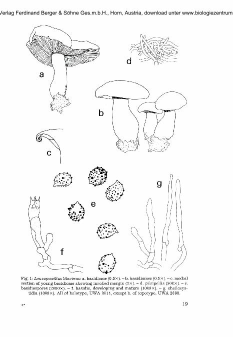
Fig. 1: Leucopaxillus Ulacinus: a. basidiome (0.5x). - b . basidiomes (0.5x). - c. medialsection of young basidiome showing inrolled margin (2x). - d. pileipellis (500x). - e.basidiospores (2000X). — f. basidia, developing and mature (lOOOx). - g. cheilocys-
tidia (lOOOx). All of holotype, UWA 3014, except b. of topotype, UWA 2898.
2- 19
©Verlag Ferdinand Berger & Söhne Ges.m.b.H., Horn, Austria, download unter www.biologiezentrum.at
The only other recorded species of Leucopaxillus in Australia iscurrently placed in Clitocybe**). This species - Clitocybe eucalyp-torum CLEL., is found in southeastern Australia. It differs from L.lilacinus in the absence of lilac or violet pigmentation and in beingdevoid of conspicuous cheilocystidia. The pileus is described byCLELAND (1934) as "drab when young to browner than Tawny Olive".Air-dried specimens of C. eucalyptorum (holotype, AD 3522) areindeed drab on the pileus, with brownish gills. In contrast, thepigmentation typical of fresh basidiomes of L. lilacinus persists asintense shades of violet on both freeze-dried and air-dried speci-mens. Another difference between the two species is the absence ofconspicuous cheilocystidia in C. eucalyptorum. In L. lilacinuscheilocystidia are very abundant, and conspicuous to the eye as wellas under the microscope. There is no mention of a glistening gill edgefor C. eucalyptorum by CLELAND (1934), and a microscopic examina-tion of the holotype has failed to reveal cheilocystidia. C. eucalyp-torum shares with L. lilacinus very similar ornamented basidio-spores (PL 1, b). The ornaments behave identically, being removed inKOH and acids but not in ammonia. Under the scanning electronmicroscope the similarity between the spores of the two species isemphasised (Plate 1). The spores have a surface of variously sizedand spaced dome-shaped verrucae, and a suprahilar plage whichcan be smooth or with one or more low verrucae. The plage of thesespecies is comparable to that observed by BIGELOW & ROWLEY (1968)for L. lentus (POST) SING.
The author gratefully acknowledges Dr. E. HOEAK and R. N. HILTON for theiradvice, and C. GRGURINOVIC for loan of type material of C. eucalyptorum. Work wascarried out during the tenure of a Commonwealth Postgraduate Award.
ReferencesBIGELOW, H. E. & ROWLEY, J. R. (1968). Surface replicas of the spores of fleshy fungi. -
Mycologia 60: 869-887.CLELAND, J. B. (1934). Toadstools and Mushrooms and other Larger Fungi of South
Australia. - Government Printer, Adelaide, South Australia.KORNERUP, A. & WANSCHER, J. H. (1978). Methuen handbook of colour. - (Third
edition). - Methuen and Co Ltd, London. 243 pp.
**) To be transferred in a forthcoming revision of fungi collected by J. B.CLELAND.
20
©Verlag Ferdinand Berger & Söhne Ges.m.b.H., Horn, Austria, download unter www.biologiezentrum.at
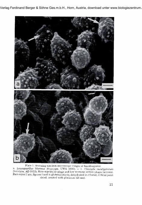
Plate 1: Scanning electron microscope images of basidiospores:a. Leucopaxillus lilacinus (topotype, UWA 2898). - b. Clitocybe eucalyptorum(holotype, AD 3522). Note suprahilar plage and low verrucae within plages (arrows).Bars equal 2 urn. Spores fixed in glutaraldehyde, dehydrated in ethanol, critical point
dried, croated with platinum (20 nm).
21
©Verlag Ferdinand Berger & Söhne Ges.m.b.H., Horn, Austria, download unter www.biologiezentrum.at


