
56 yo F with RUQ pain and jaundice


US – presumed panc head mass, dilated thickwalled CBD terminates in
“mass”

CT – bulbous panc head – no mass

CT – pancreas diffusely thickened with decreased normal lobulations.
Hypodense capsule
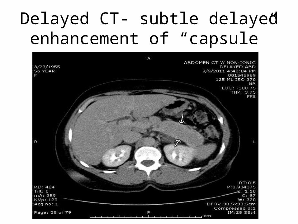
Delayed CT- subtle delayed enhancement of “capsule”

Dual-Phase CT of Autoimmune Pancreatitis: A Multireader Study
• The characteristic CT appearance of autoimmune pancreatitis has been described as diffuse enlargement of the pancreas with a capsule-like rim [5–7].
• AJR February 2008 vol. 190 no. 2 280-286
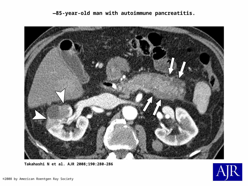
—85-year-old man with autoimmune pancreatitis.
Takahashi N et al. AJR 2008;190:280-286
©2008 by American Roentgen Ray Society
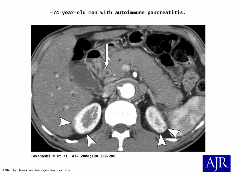
—74-year-old man with autoimmune pancreatitis.
Takahashi N et al. AJR 2008;190:280-286
©2008 by American Roentgen Ray Society


![[Cartilha] Cartilha PANC Viveiros Comunitários](https://cdn.vdocuments.mx/doc/165x107/5695d0921a28ab9b02930095/cartilha-cartilha-panc-viveiros-comunitarios.jpg)
