4/26/2016
1
Non-IPF Interstitial Lung Disease
Tom Schaumberg, M.D.Pulmonary & Critical Care MedicineThe Oregon Clinic
Non-IPF Interstitial Lung Disease • Disclosures
– PI for Intermune trials for Perfenidone– PI for Boeringengehim trial of Nintedanib– PI for Gilliad trial for Sustimamab– Investigator for Genentech for Lebrikizumab– Investigator for Bristol-Meyers Squibb for Lysoposhatidic Acid
Receptor Antagonist
– Contractual agreements for recruiting, screening and conducting clinical trials with patients who have IPF
– I do not receive any money for services that are not directly related to patient care.
4/26/2016
2
Non-IPF Interstitial Lung Disease

4/26/2016
3
IPF: 3-5 year 50% mortality from the time of diagnosis 6.7 years from onset of symptoms
IPF vs Non-IPF ILD
• IPF is fatal with a 50% mortality in 3-5 years
• IPF is fibro-proliferative lung disease
• IPF is limited to the lungs
• The prognosis is good– Dependent on the
specific diagnosis
• Inflammatory conditions
• Often associated with systemic conditions
4/26/2016
4
IPF vs Non-IPF ILD
• Steroids and immunosuppressive medications cause harm
• Pirfenidone and Nintedanib slow the progression of IPF– Small treatment effect– The results are not
predictable– Cost $90,000-95,000/
year
• Steroids and immunosuppressive can have dramatic benefit in some forms of ILD– The type and duration of
immunosuppression depends on the diagnosis
• Pirfenidone and Nintedanib are of no benefit in Non-IPF diseases
Clinical Presentation of Interstitial Lung Disease
• Prevalence of all ILD 74/100,000– 3,000 Oregonians
– 42/100,000 new colorectal cancers per year
• 35% of all ILDs are secondary to IPF
AJRCCM 1994;150:967-972, http://seer.cancer.gov/statfacts/html/colorect.html, Chest 2010; 137(1): 129-137
4/26/2016
5
Clinical Presentation of Interstitial Lung Disease
• Chronic unexplained cough
• Unexplained dyspnea
• “Velcro” rales on exam
• Abnormal CXR or CT scan– Beware of the term “Stable fibrosis”
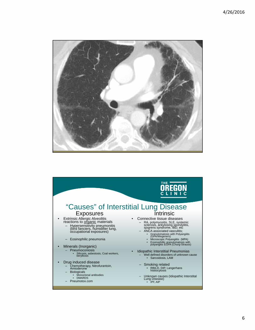
68 y/o man referred for second opinion regarding IPF
• Sub-acute DOE now at < 50 yards• NP Cough• Hx GERD, OSA, TIA with + anticardiolipin antibody• 22 p/y smoking D/C 1985• Retired general contractor
– Incidental asbestos exposure • Rales at bases• Nl CBC, BMP, EKG, Echo• FEV1/FVC .71, FVC 83%, TLC 79%, DLCO 38%
4/26/2016
6
High Resolution CT Scan
PatternsGround glass opacities
Reticular “fibrotic” changes
Honey combing
Cysts
Nodules/Micro-nodules
Mosaic attenuation
LocationDiffuse
Focal
Patchy
Upper lobe predominant
Lower lobe dominant
Peripheral
Peribronchovascular
Bronchiectasis
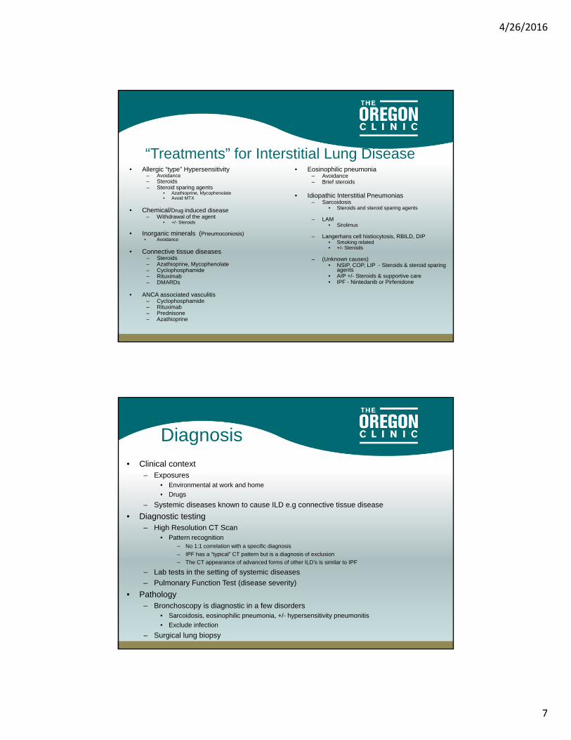
“Causes” of Interstitial Lung DiseaseExposures
• Extrinsic Allergic Alveolitisreactions to organic materials – Hypersensitivity pneumonitis
(bird fanciers, humidifier lung, occupational exposures)
– Eosinophilic pneumonia
• Minerals (Inorganic)– Pneumoconiosis
• Silicosis, asbestosis, Coal workers, berylliosis
• Drug induced disease– Chemotherapy, Nitrofurantoin,
Amioderone– Biologicals
• Monoclonal antibodies• DMARDS
– Pneumotox.com
Intrinsic • Connective tissue diseases
– RA, polymyositis, SLE, systemic sclerosis, ankylosing spondylitis, sjogrens syndrome, IBD, etc
– ANCA associated vasculitis; • Granulomatosis with Polyangiitis
(GPA/Wegeners)• Microscopic Polyangiitis (MPA)• Eosinophillic granulomatosis with
polyangitis EGPA (Churg-Strauss)
• Idiopathic Interstitial Pneumonias – Well defined disorders of unknown cause
• Sarcoidosis, LAM
– Smoking related• RBILD, DIP, Langerhans
histiocytosis
– Unknown causes (Idiopathic InterstitalLung Disease)
• IPF, AIP
4/26/2016
7
“Treatments” for Interstitial Lung Disease• Allergic “type” Hypersensitivity
– Avoidance – Steroids– Steroid sparing agents
• Azathioprine, Mycophenolate• Avoid MTX
• Chemical/Drug induced disease– Withdrawal of the agent
• +/- Steroids
• Inorganic minerals (Pneumoconiosis)• Avoidance
• Connective tissue diseases– Steroids– Azathioprine, Mycophenolate– Cyclophosphamide– Rituximab– DMARDs
• ANCA associated vasculitis– Cyclophosphamide– Rituximab– Prednisone– Azathioprine
• Eosinophilic pneumonia– Avoidance– Brief steroids
• Idiopathic Interstitial Pneumonias – Sarcoidosis
• Steroids and steroid sparing agents
– LAM• Sirolimus
– Langerhans cell histiocytosis, RBILD, DIP • Smoking related• +/- Steroids
– (Unknown causes)• NSIP, COP, LIP - Steroids & steroid sparing
agents• AIP +/- Steroids & supportive care• IPF - Nintedanib or Pirfenidone
• Clinical context– Exposures
• Environmental at work and home
• Drugs
– Systemic diseases known to cause ILD e.g connective tissue disease
• Diagnostic testing – High Resolution CT Scan
• Pattern recognition– No 1:1 correlation with a specific diagnosis
– IPF has a “typical” CT pattern but is a diagnosis of exclusion
– The CT appearance of advanced forms of other ILD’s is similar to IPF
– Lab tests in the setting of systemic diseases
– Pulmonary Function Test (disease severity)
• Pathology– Bronchoscopy is diagnostic in a few disorders
• Sarcoidosis, eosinophilic pneumonia, +/- hypersensitivity pneumonitis
• Exclude infection
– Surgical lung biopsy
Diagnosis
4/26/2016
8
• HRCT alone cannot provide a diagnosis – Pattern
• Ground Glass, linear reticulation, micro-nodular, “tree-in-bud”, consolidation, cysts
– Location• Peripheral, peri-bronchial, basilar, sub-pleural, diffuse
– Airway involvement• Traction bronchiectasis, peri-bronchial
– Lack of effusions• Except LAM
– Lack of enlarged lymph nodes• Except Sarcoidosis and Lyphangitic spread of cancer
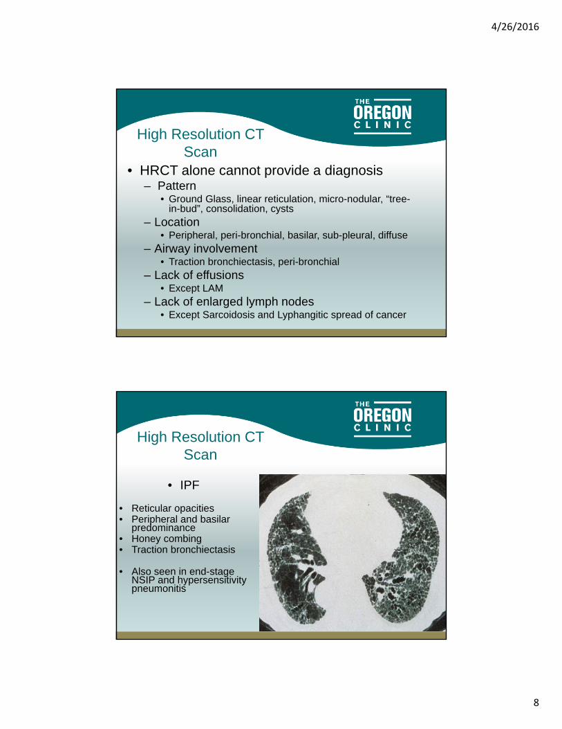
High Resolution CT Scan
• IPF
• Reticular opacities• Peripheral and basilar
predominance• Honey combing• Traction bronchiectasis
• Also seen in end-stage NSIP and hypersensitivity pneumonitis
High Resolution CT Scan

4/26/2016
9
• IPF
• Reticular opacities• Peripheral and basilar
predominance• Honey combing• Traction bronchiectasis
• Also seen in end-stage NSIP and hypersensitivity pneumonitis
High Resolution CT Scan
• Characteristic not found in IPF– Ground glass opacities > reticular “fibrotic” changes– Preibronchovascular– Upper lobe predominance– Focal consolidation– Lymphadenopathy– Pleural plaques– Cysts– Pleural effusions– Nodules/Micronodules– Mosaic attenuation
HRCT ScanIPF vs Non-IPF

4/26/2016
10
• Hypersensitivity pneumonitis
• Desquamative interstitial pneumonitis (DIP)
• Respiratory bronchiolitisassociated ILD (RB-ILD)
• Drug toxicity• Pulmonary hemorrhage
HRCT “Ground Glass”
Nodules
• Sarcoidosis• Hypersensitivity
pneumonitis• Respiratory bronchiolitis
associated ILD (RB-ILD)• Pulmonary histiocytosis X• Silicosis• Pneumoconiosis• Metastatic cancer
4/26/2016
11
Focal Consolidation
• Cryptogenic Organizing Pneumonia (COP, formally known as BOOP
• Infection
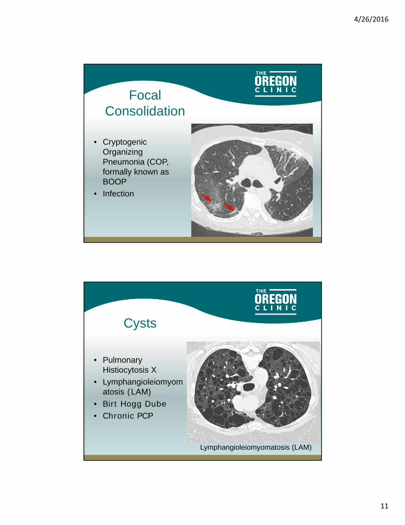
Lymphangioleiomyomatosis (LAM)
Cysts
• Pulmonary Histiocytosis X
• Lymphangioleiomyomatosis (LAM)
• Birt Hogg Dube• Chronic PCP
Lymphangioleiomyomatosis (LAM)
4/26/2016
12
Serologic Testing
• The ATS recommends screening all patients with ILDANA RF, Anti-CCP
• Additional testing based on Systemic symptomsAldolaseCPK Anti-synthitase antibodies (e.g. Jo-1)Sjogren’s Antibodies SS-A, SS-BScleroderma Antibodies (scl-70, PM-1)
American Journal of Respiratory and Critical Care Medicine, Vol. 183, No. 6 (2011), pp. 788-824.
PFTs• Disease severity• Restrictive physiology
– Spirometry• Nl or increased FEV1/FVC ratio• Decreased FVC• FEV1 decreased but in proportion to FVC
– Lung Volumes• Decreased TLC• Decreased RV
– DLCO• Decreased

4/26/2016
13
• Pathology alone does not provide a specific diagnosis/etiology
Pathology
• Pathology alone does not provide a specific diagnosis
- Interstitial Pneumonias• Usual interstitial pneumonia (UIP) = IPF ( Occasionally with hypersensitivity pneumonitis, rheumatoid arthritis,
scleroderma, and asbestosis)• Nonspecific interstitial pneumonia (NSIP) often associated with connective tissue diseases, occasionally with viral
pneumonias, hypersensitivity pneumonitis, and drug-induced toxicity• Respiratory bronchiolitis (RB) Cigarette smoke, inhaled minerals, viral infections ,connective tissue disease• Desquamative interstitial pneumonia (DIP) Associated with smoking• Organizing pneumonia Cryptogenic organizing pneumonia (COP)/(BOOP) Connective tissue disease, infections,
drug reactions • Lymphoid interstitial pneumonia (LIP)• Diffuse alveolar damage (DAD) Any form of acute lung injury e.g. ARDS
– Granulomatous lung diseases• Sarcoidosis• Vasculitis GPA (Wegener's), MPA , EGPA (Churg-Strauss)• Hypersensitivity pneumonitis - environmental exposures and Drugs • Langerhans cell granulomatosis - associated with smoking • Foreign body granulomatosis• Chronic beryllium disease• Bronchocentric granulomatosis
– Alveolar filling parenchymal lung diseases• Eosinophilic pneumonia• Pulmonary hemorrhage syndromes• Pulmonary alveolar proteinosis
– Proliferative lung diseases • Pulmonary amyloidosis• Smooth muscle proliferation (LAM)
Pathology
4/26/2016
14
• Clinical context– Exposures
• Environmental at work and home
• Drugs
– Systemic diseases known to cause ILD e.g connective tissue disease
• Diagnostic testing – Lab tests in the setting of systemic diseases
– High Resolution CT Scan• Pattern recognition
• IPF is a diagnosis of exclusion and a “typical” CT pattern
• The CT appearance of advanced forms of other ILD’s is similar to IPF
– Pulmonary Function Test (disease severity)
• Pathology– Bronchoscopy is diagnostic in a few disorders
• Sarcoidosis, eosinophilic pneumonia, +/- hypersensitivity pneumonitis
• Exclude infection
– Surgical lung biopsy
Diagnosis
Connective Tissue Disease Related ILD
• General Principles– ILD rarely presents prior to systemic symptoms– No CT pattern or pathologic finding is specific to ILD related to
connective tissue diseases– Serositis > Interstitial Lung Disease – Infections are common with immunosuppressive therapy – Methotrexate is associated with the development of ILD
independent of underlying Dx– Hydroxychloroquine, sulfasalazine are not effective in treating
ILD– Aspiration is common with scleroderma/Systemic sclerosis
1. Am J Respir Crit Care Med. 2011;183(3):372; 2. Am J Respir Crit Care Med. 1997;156(2 Pt 1):528; 3. Thorax. 2001;56(8):622.
4/26/2016
15
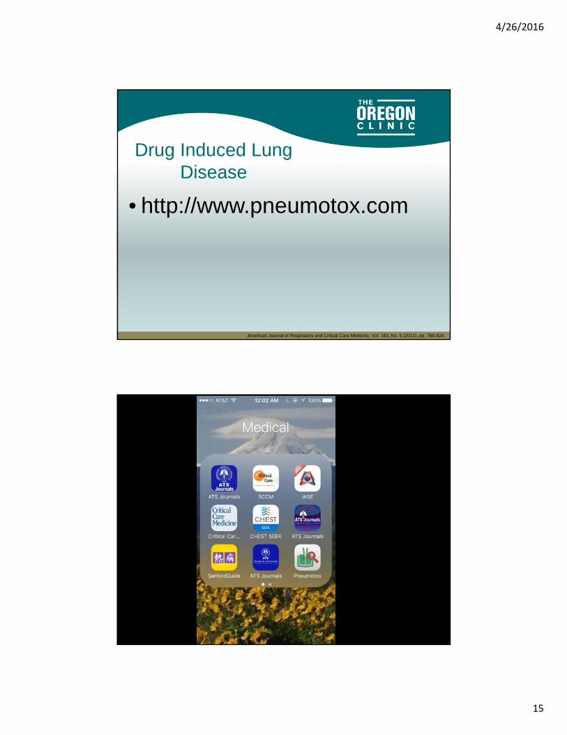
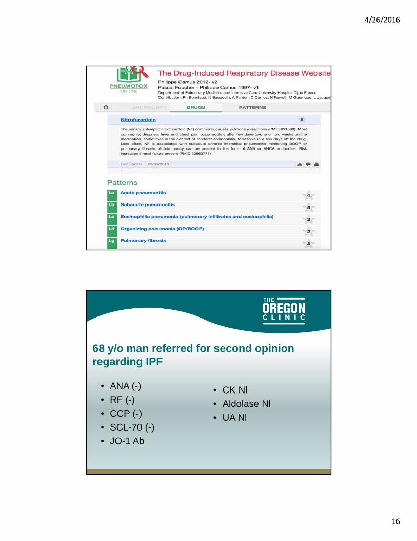
Drug Induced Lung Disease
• http://www.pneumotox.com
American Journal of Respiratory and Critical Care Medicine, Vol. 183, No. 6 (2011), pp. 788-824.
4/26/2016
16
69 y/o woman with referred by her rheumatologist for
• Daily nitrofurantoin 10/15-3/2016
68 y/o man referred for second opinion regarding IPF
• ANA (-)
• RF (-)
• CCP (-)
• SCL-70 (-)
• JO-1 Ab
• CK Nl
• Aldolase Nl
• UA Nl
4/26/2016
17
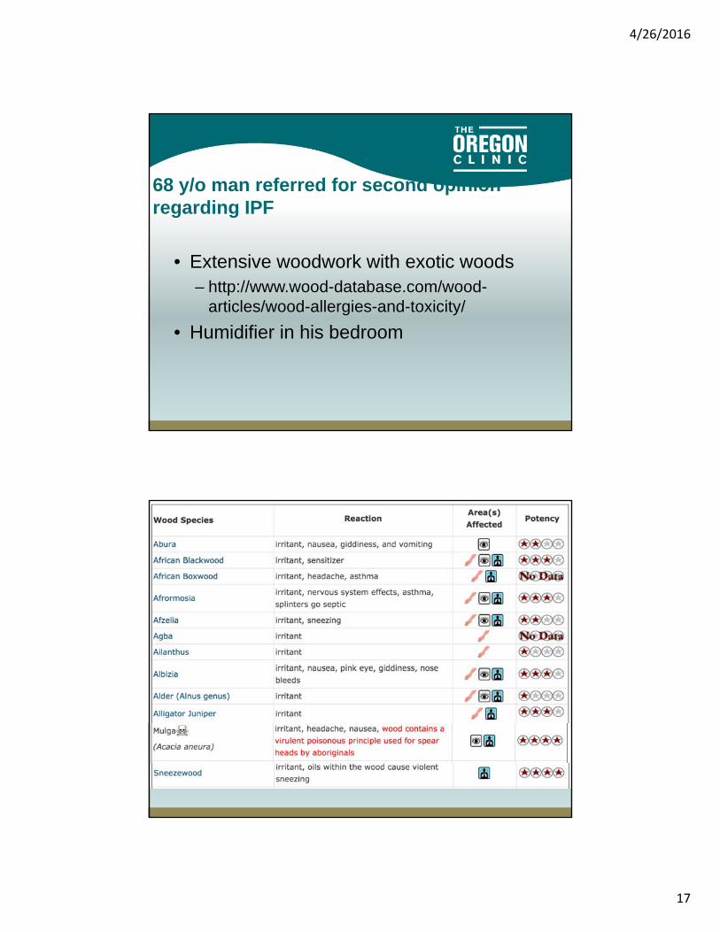
68 y/o man referred for second opinion regarding IPF
• Extensive woodwork with exotic woods– http://www.wood-database.com/wood-
articles/wood-allergies-and-toxicity/
• Humidifier in his bedroom

4/26/2016
18
68 y/o man referred for second opinion regarding IPF
• (+) Hypersensitivity pneumonitis panel– Aureobasdium Pullulan
• VATS Biopsy– UIP and Hypersensitivity pneumonitis
68 y/o man referred for second opinion regarding IPF
• Avoidance
• Prednisone and Mycophenolate
• FVC 79% => 100%
• DLCO 38% => 53%

4/26/2016
19
Non-IPF Interstitial Lung DiseaseConclusions
• You will see patients with ILD– Cough, SOB, Abnl CT
• IPF is only one type of ILD– Progressive, fatal, treated with Pirfenidone or
Nintedanib
• The prognosis and treatment of other types of ILD depends on the specific diagnosis
• Consider medications– Pneumotox.com– “Biologic Agents”
• Immunosuppression• Biologic effect• Drug Rxn
• Environmental exposures work and home– Intense or highly repetitive exposures; birds, decaying organic materials
• Underling diseases and systemic symptoms– Connective tissue disease – Cancer and it’s treatments
• Consider non-bacterial infections– Viruses, PJP– Molds rarely cause infections in Nl hosts, may cause hypersensitivity reactions
Non-IPF Interstitial Lung DiseaseConclusions

4/26/2016
20
• Avoid “Theraputic/Diagnostic trials” of steroids– Dysphoria– Delay in diagnosis– Duration?– Dose? – Which steroid sparing agent is most
appropriate?
Non-IPF Interstitial Lung DiseaseConclusions


