
Intracranial Hypertension
from Traumatic Brain Injury
Axel J. Rosengart, MD PhD
Director
Neurosciences Critical Care and
Emergency Neurology and Neurosurgery
Departments of Neurology, Neurosciences and
Neurosurgery
New York Presbyterian Hospital –
Weill Cornell Medical College
New York, NY, USA

Outline
ICP: Vital Sign in Brain Injury
Brain Trauma and Herniation
Syndromes
=> Central Herniation
=> Lateral Brain Herniation
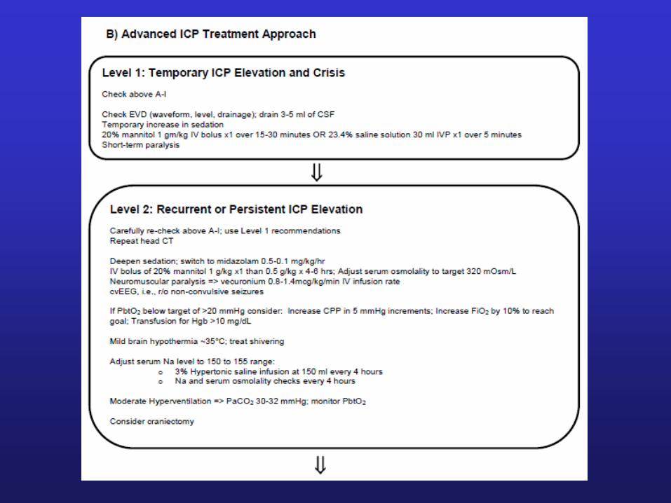
Treatment Options
Intracranial Pressure
• Normal ICP: 5-10 mm Hg
• Intracranial Hypertension:
Sustained > 15 mm Hg
• Basic Concepts….

Intracranial Contents
Content
• Brain Parenchyma
• Cerebrospinal Fluid
& Interstitial Fluid
• Blood: Arterial,
Capillary, Venous
Abnormality
=> Tumor, ICH
=> Hydrocephalus & Cerebral Edema
=> Venous Sinus Thrombosis

Pressure-Volume Curve
Compliance: in volume/pressure
Increasing intracranial volumes
A0: Normal ICP
A1: IPC high normal
B: ICP rapidly increases
First CSF, second CBV translocation
Raised ICP
CBF and ischemia
Pressure gradients: herniation
(compartmental alterations)
CSF Displacement
Blood Displacement
1
2
3
1: Optimal – 2: Spatial compensation – 3: Spatial decompensation
Herniation

ICP Waveform
P1: Percussion wave => choroid pulsations
P2: Dicrotic wave => pulsations of major arteries
Tidal wave => blood perfusion ‘wave’
Increased ICP
P2 > P1
P1 and tidal wave
(waveform ‘rounded’)

Measuring ICP


‘Bedside’ Camino

Bedside Monitoring

Camino on CT

Ventricular Catheters
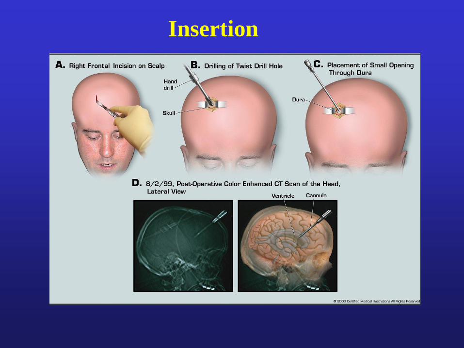
Insertion

Ventricular
Drainage
Systems
Leveling to
EAC

Cerebral Perfusion Pressure
• CPP = Cerebral Perfusion Pressure
• CPP = MAP – ICP
• Goal CPP = 70 – 100 mm Hg
Depressed Consciousness and Raised ICP
• ICP => CPP => Cerebral Ischemia
• Decreased LOC from ICP =>
ischemic encephalopathy

CSF Displacement
First
CSF displacement compensates for ICP
Dynamic equilibrium: absorption = production
CSF total 90 to 150 ml; 20ml/h or 500ml/d
Production: steady despite ICP!
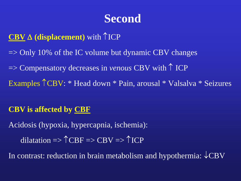
Second
CBV (displacement) with ICP
=> Only 10% of the IC volume but dynamic CBV changes
=> Compensatory decreases in venous CBV with ICP
Examples CBV: * Head down * Pain, arousal * Valsalva * Seizures
CBV is affected by CBF
Acidosis (hypoxia, hypercapnia, ischemia):
dilatation => CBF => CBV => ICP
In contrast: reduction in brain metabolism and hypothermia: CBV

Auto(dys)regulation
Decrease in MAP
Cerebral Vasodilatation (intact autoregulation)
Vasoparalysis (dysregulation)
Increase Decrease
in cerebral blood
volume flow
Further increase in ICP
Compromise of CPP => Ischemia

Herniation Syndromes
Differentiate two commonly observed herniation
syndromes:
Central herniation
Lateral displacement/herniation
Initial therapy similar: “stabilize the ICP”
Subsequent therapy: different!
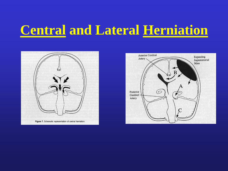
Central and Lateral Herniation
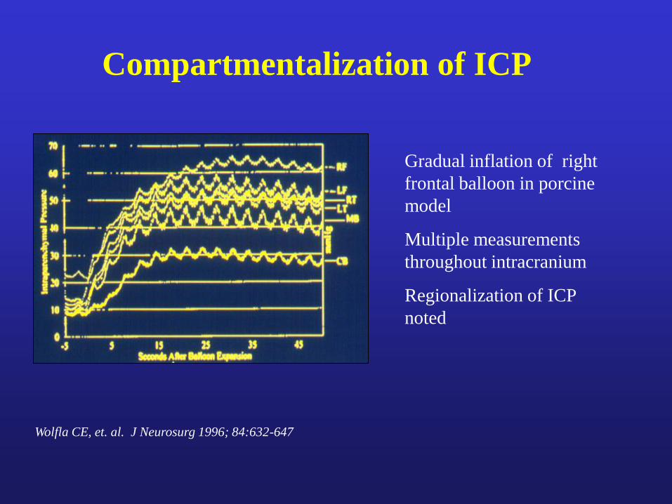
Compartmentalization of ICP
Wolfla CE, et. al. J Neurosurg 1996; 84:632-647
Gradual inflation of right
frontal balloon in porcine
model
Multiple measurements
throughout intracranium
Regionalization of ICP
noted
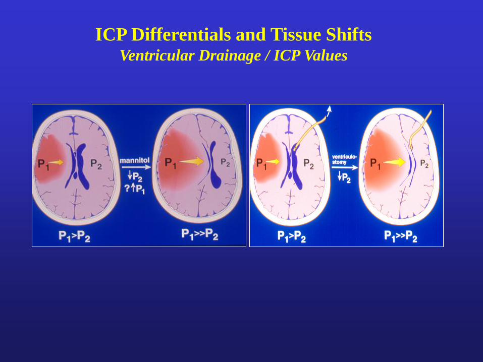
ICP Differentials and Tissue Shifts Ventricular Drainage / ICP Values


CT 20 hrs
post IA t-PA
Rapid LOC
AS Shift: 12mm
P Shift: 6mm Protrusion of uncus
Open ambien
cisterns
Distorted midbrain
Aqueduct open
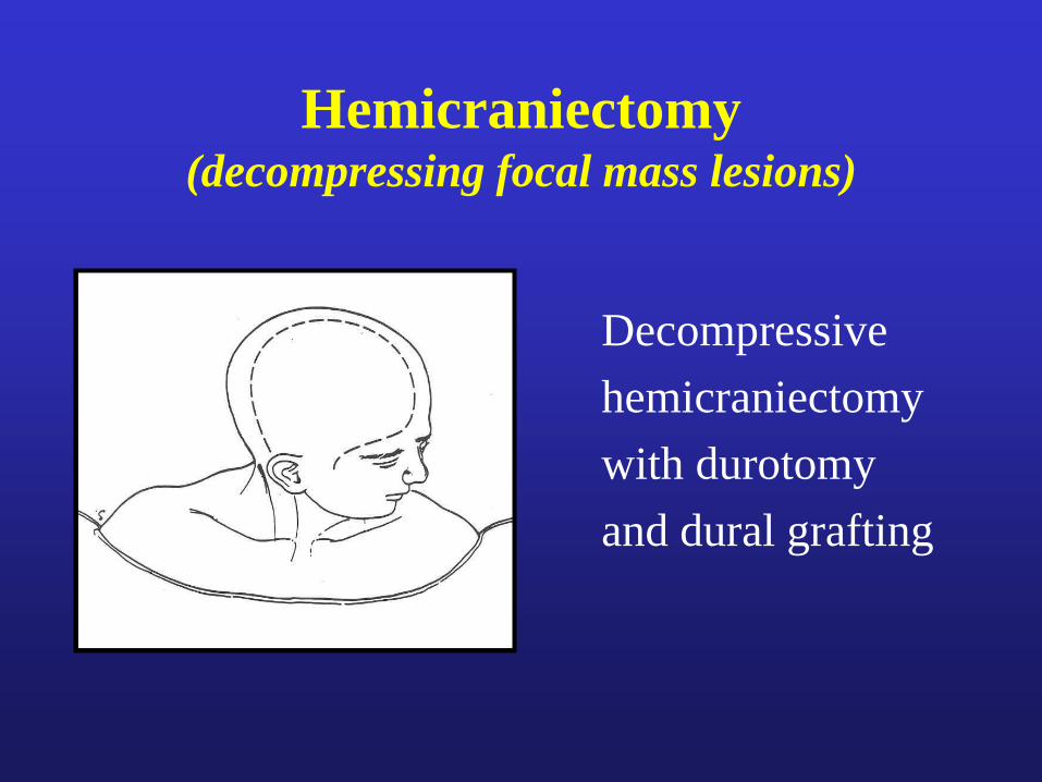
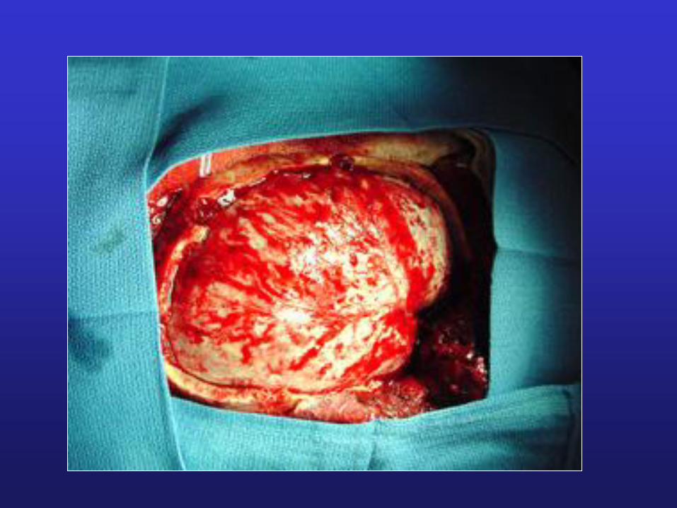
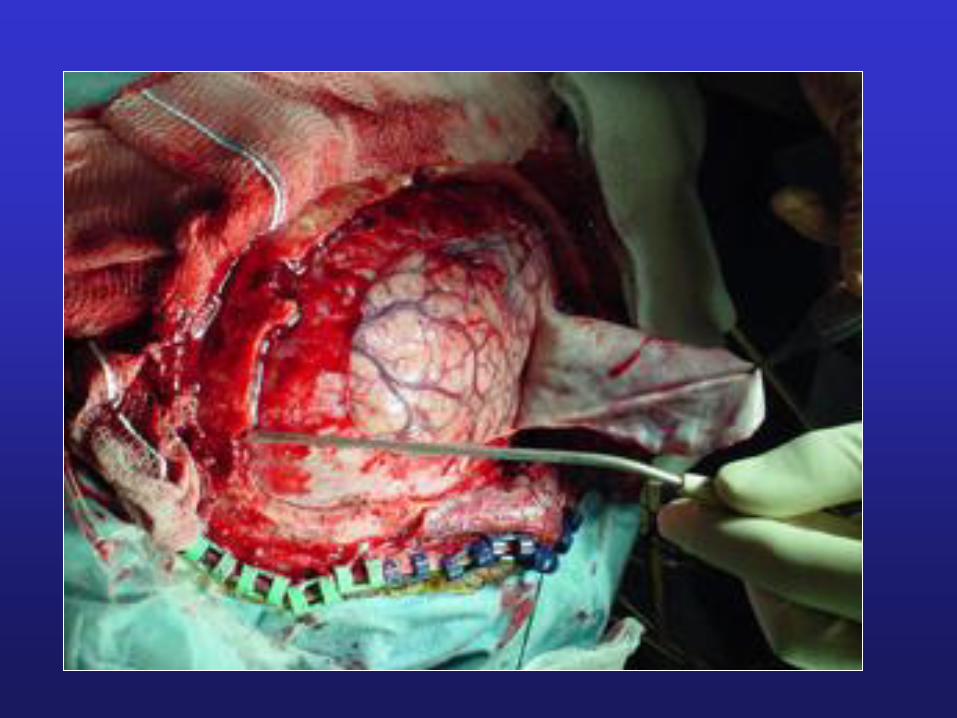
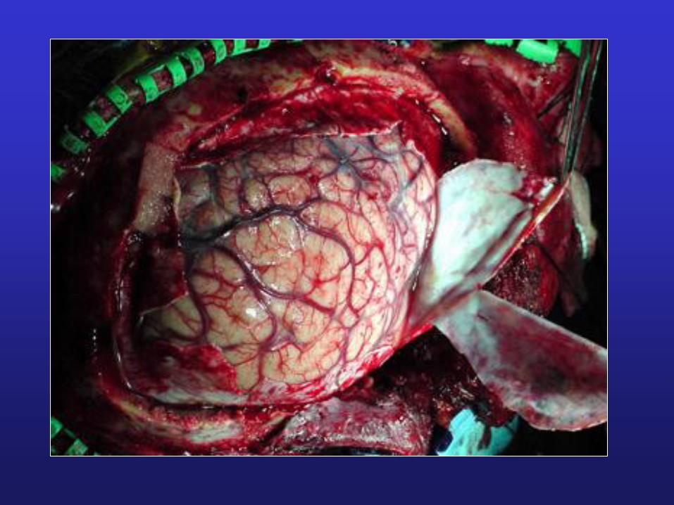
Hemicraniectomy (decompressing focal mass lesions)
Decompressive
hemicraniectomy
with durotomy
and dural grafting

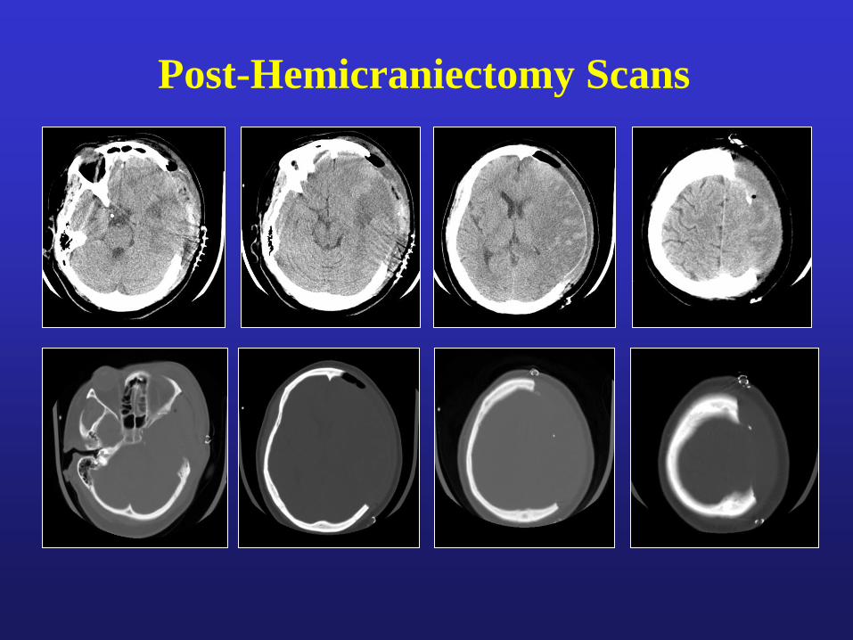
Post-Hemicraniectomy Scans

S/P Bone Flap Replacement
(About 2 ms later)


Summary
TRAUMATIC BRAIN INJURY ALGORITHM INCLUDES
• Acute recognition and empiric Tx of increased ICP
• Neurosurgical removal of acute mass lesions
• Measuring ICP and monitoring CPP
• Stabilizing ICP crisis
• Repeated CT scanning if needed

Thank you
for your attention!


