
Challenging the prescribed enrofloxacin treatment regimen of bacterial respiratory
disease caused by Ornithobacterium rhinotracheale and Escherichia coli in
turkeys
An Garmyn
Thesis submitted in fulfillment of the requirements for the degree of Doctor in Veterinary Sciences (PhD), Faculty of Veterinary Medicine, Ghent University, December 2009
Promoters: Prof. Dr. A. Martel
Prof. Dr. F. Pasmans Prof. Dr. F. Haesebrouck
Faculty of Veterinary Medicine Department of Pathology, Bacteriology and Poultry Diseases
FACULTEIT DIERGENEESKUNDE

This work was printed by DCL Print & Sign www.dclsigns.be
ISBN: 9789058642042


Table of contents ___________________________________________________________________________
1
TABLE OF CONTENTS
___________________________________________________________________________
Table of contents ___________________________________________________________________________
2
TABLE OF CONTENTS
LIST OF ABBREVIATIONS 5
CHAPTER 1. GENERAL INTRODUCTION 9
1.1. Enrofloxacin, a fluoroquinolone compound 10
1.2. Multicausal respiratory disease in turkeys 23
CHAPTER 2. AIMS 51
CHAPTER 3. EXPERIMENTAL STUDIES 55
3.1. Efficacy of four enrofloxacin treatment regimens against experimental infection in
turkey poults with avian metapneumovirus and Ornithobacterium rhinotracheale 57
3.2. Effect of multiple and single day enrofloxacin medications against dual
experimental infection with avian metapneumovirus and Escherichia coli in turkeys
77
3.3. Effect of reduced treatment time and dosage of enrofloxacin on the course of
respiratory disease caused by avian metapneumovirus and Ornithobacterium
rhinotracheale 97
CHAPTER 4. GENERAL DISCUSSION 123
SUMMARY 145
SAMENVATTING 149
CURRICULUM VITAE 153

Table of contents ___________________________________________________________________________
3
PUBLICATIONS 155
DANKWOORD 157

1

List of abbreviations ___________________________________________________________________________
5
LIST OF ABBREVIATIONS
________________________________________________________________

List of abbreviations ___________________________________________________________________________
6
LIST OF ABBREVIATIONS
APEC avian pathogenic Escherichia coli
APV avian metapneumovirus
AUC area under the curve
BHI brain heart infusion
BW body weight
Clb body clearance
Cmax maximum serum concentration
CD50 ciliostatic dose
cfu colony forming units
CO2 carbon dioxide
DNA desoxyribonucleic acid
E5-5D treatment group receiving 5 mg enrofloxacin/kg bodyweight for 5 days
E10-5D treatment group receiving 10 mg enrofloxacin/kg bodyweight for 5 days
E15-3D treatment group receiving 15 mg enrofloxacin/kg bodyweight for 3 days
alternated each time by a non treating day
E20-5D treatment group receiving 20 mg enrofloxacin/kg bodyweight for 5 days
E25-2D treatment group receiving 25 mg enrofloxacin/kg bodyweight for 2 days
alternated by a non treating day
E50-5H treatment group receiving 50 mg enrofloxacin/kg bodyweight for 5 hours
E50-10H treatment group receiving 50 mg enrofloxacin/kg bodyweight for 10 hours
E50-20H treatment group receiving 50 mg enrofloxacin/kg bodyweight for 20 hours
ELISA enzyme-linked immunosorbent assay
h hour
HEPA high efficiency particulate air
kg kilogram
l liter
LPS lipopoly-saccharides
M molar
MBC minimal bactericidal concentration
mg milligram

List of abbreviations ___________________________________________________________________________
7
MIC50 minimum inhibitory concentration required to inhibit the growth of 50% of the
organisms
MIC90 minimum inhibitory concentration required to inhibit the growth of 90% of the
organisms
ml milliliter
MPC mutant prevention concentration
MRT mean residence time
NC negative control group
Omp outer membrane protein
P P-value
PAE post-antibiotic effect
PALE post-antibiotic leukocyte enhancement effect
pbi post bacterial infection
PBS phosphated buffered saline
Pc partition coefficient
PC positive control group
PCR polymerase chain reaction
pKa acid dissociation constant
ppm parts per million
pvi post viral infection
Qnr gene quinolone resistance gene
QRDR quinolone resistance-determining region
RNA ribonucleic acid
rpm rotates per minute
SM-PAE subMIC- post-antibiotic effect
SPF specified pathogen free
t1/2 elimination half life
TNFα tumor necrosis factor α
TOC tracheal organ culture
µg microgram
µl microliter

8
General introduction ___________________________________________________________________________
9
CHAPTER 1: GENERAL INTRODUCTION
1.1. ENROFLOXACIN, A FLUOROQUINOLONE COMPOUND
1.2. MULTICAUSAL RESPIRATORY DISEASE IN TURKEYS
________________________________________________________________

General introduction ___________________________________________________________________________
10
GENERAL INTRODUCTION
1.1. ENROFLOXACIN, A FLUOROQUINOLONE ANTIMICROBIAL COMPOUND
1.1.1. General considerations of enrofloxacin use in turkeys
In modern poultry industry, turkeys have been selected for growth rate and feed
efficiency rather than for robustness and resistance to disease. Disease outbreaks often lead to
extensive carcass condemnations at processing, resulting in significant economic losses. To
minimize these economic losses due to microbial disease, therapeutic programs are needed
using agents that have the ability to rapidly kill any bacterial pathogen that invades the poultry
flocks (Tanner, 2000). Other important matters to consider are the cost-to-benefit ratio of the
treatment, the available administration route, the drug withdrawal time, and the antimicrobial
susceptibility of the target pathogen (Tanner, 2000).
After flumequine, enrofloxacin was the second antimicrobial fluoroquinolone compound used
in veterinary medicine. The drug is active against pathogenic bacteria that play a significant
role in turkey respiratory disease or septicaemia: Mycoplasma sp., Avibacterium
paragallinarum, Pasteurella sp., Escherichia coli, Staphylococcus sp. and Erysipelotrix
rhusiopathiae. The current treatment schedule advises administration of enrofloxacin at a
dose of 10 mg/kg body weight for 3 to 5 continuous days. When mixed bacterial infections
are involved treatment should last for 5 continuous days. After such treatment several clinical
trials have observed a rapid decrease in mortality, clinical symptoms and respiratory tract
lesions. In most cases, a complete eradication of the causative bacterial organism was reported
(Braunius, 1987; Behr et al., 1988; Hafez et al., 1992; Cargill, 1995; Gautrais and Copeland,
1997; Marien et al., 2007). When involved in the infection, complete eradication of
Mycoplasma sp. was not accomplished, but enrofloxacin treatment reduced the mycoplasma
excretion and mortality rates (Hinz and Rottmann, 1990; Rainhardt et al., 2005).
Enrofloxacin can be administered through drinking water. This route of administration proves
most effective in clinical outbreaks of microbial disease (Tanner, 2000). Advantages of water
medication are the rapid response, medication reaching the birds within short time and
convenience of administration. Possible disadvantages are that impurities in the water (bi- or
trivalent cations) may reduce absorption. The acrid taste of enrofloxacin which may
contribute to variations in water consumption in pigs, does not affect the water palatability in
poultry. Parenteral injection delivers a correct dose but is labour-intensive and results in

General introduction ___________________________________________________________________________
11
considerable stress in the birds. Feed incorporation of antimicrobials for therapeutic purpose
is less effective because of the time to manufacture, the inappetence and the inability to
compete for feed in sick animals. Antimicrobial sensitivity testing should be conducted to
support the antimicrobial treatment and should be repeated if no clinical improvement is
noticed after two or three days of treatment.
1.1.2. Structure/activity relationships and physicochemical properties of enrofloxacin
Enrofloxacin is a fluoroquinolone antimicrobial drug derived from nalidixic acid. Like
all quinolones, it is manufactured synthetically. Nalidixic acid, discovered in 1962, was the
first clinically approved 4-quinolone-type compound, but had several limitations. The drug
showed only modest activity against Gram-negative bacteria, restricted to the
Enterobacteriaceae, and possessed poor pharmacokinetic properties regarding absorption and
distribution. Therefore, nalidixic acid proved only effective against urinary tract infections. In
addition, the molecule had a tendency to select for resistant organisms and induced several
toxic effects (Appelbaum and Hunter, 2000; Walker, 2000b; Martinez et al., 2006). Hence,
several structural modifications have been implemented to the “original” molecule of
nalidixic acid, to improve its antibacterial activity and its pharmacokinetic features (Chu and
Fernandes, 1989; Sarkozy, 2001).
Figure 1. Structural formulas of nalidixic acid and enrofloxacin (adapted from Sarkozy, 2001).
First, the naphthyridone nucleus (N at position 1 and 8) was transformed into a quinolone
nucleus, a ring structure containing only one nitrogen in position 1. Halogenating position 6
of the quinolone nucleus with a fluorine atom substantially broadened the antibacterial
activity spectrum by improving the clinical activity against Gram-positive bacteria and
enhanced oral bioavailibility and tissue penetration (Chu and Fernandes; 1989; Sarkozy,
Nalidixic acid
Enrofloxacin

General introduction ___________________________________________________________________________
12
2001). The introduction of the piperazinyl (a heterocyclic nitrogen-containing ring) side chain
at position 7 of the quinolone nucleus enhanced activity by increasing the ability of the drug
to penetrate the bacterial cell wall improving once more activity against Gram-negatives,
including Pseudomonas sp. (Chu and Fernandes; 1989; Sarkozy, 2001). Adding a
cyclopropyl-group at position 1 of the nitrogen ring and an etylgroup to the piperazinyl ring
increased the lipid solubility and the volume of distribution of the compounds (Sarkozy,
2001) again enhancing potency against members of the family Enterobacteriaceae and
Pseudomonas aeruginosa (Chu and Fernandes, 1989).
All these transformations lead to the construction of 1-cyclopropyl-6-fluoro-1,4-dihydro-4-
oxo-7-(4-ethyl-1-piperazinyl)-3-quinolone-carboxilic acid, or enrofloxacin, with the following
physicochemical properties. The dissociation constants of enrofloxacin are pKa1=5.94 and
pKa2=8.70 corresponding to the carboxylic acid group on the 3- position and to the basic
piperazynil group in the 7-position, respectively (Lizondo et al., 1997). These constants help
define the state of ionization of enrofloxacin since the molecule can exist in four possible
analogues depending on the environmental pH: an acidic cation, a neutral un-ionized species,
an intermediate zwitterion and a basic ion (Lizondo et al., 1997). Maximal transfer of
enrofloxacin from the aqueous to the lipid phase occurs at pH 7.0. Below and above this pH,
partition coefficients (Pc) of the drug decrease (i.e. Pc<3.48) indicating higher polarity and,
therefore, minor transfer (Lizondo et al., 1997; Martinez et al., 2006). This lipophility of
enrofloxacin facilitates its diffusion into biological tissues, including bacterial cells. The
aqueous solubility of enrofloxacin is low between pH 6.0-8.0, which is near its isoelectric
point. Maximal aqueous solubility (i.e. 10.42 mg/ml) is achieved at pH 5.02. When a
concentrated acetate buffer is used, the amount of enrofloxacin solubilized can be increased
(> 100 mg enrofloxacin/ml using a 1.178 M acetate buffer) (Lizondo et al., 1997). According
to these physicochemical properties with moieties of the molecule being lipophilic and
lipophobic as well as having different areas of positive and negative charge (zwitterion
properties) enrofloxacin has the capacity to cross biological membranes easily and achieve
high concentrations in body tissues.
1.1.3. Bacterial topoisomerases and the mechanisms of enrofloxacin action
The bacterial chromosome is a continuous, circular, double-stranded DNA molecule
approximately 1.000 times longer than the bacteria in which it is contained. To fit into the
cell, the DNA ring must be condensed into a negative supercoil through repeated twisting
(Walker, 2000b; Hawkey, 2003). Each one of such twists results in one superhelical turn,
General introduction ___________________________________________________________________________
13
which can only be released by breaking the covalent bonds of the DNA backbone. Enzymes
that catalyze such conversions are called topoisomerases (Messer, 1999; Walker 2000b;
Hawkey, 2003). Enrofloxacin inhibits the activity of two of those enzymes: DNA gyrase and
topoisomerase IV (Hawkey, 2003).
DNA gyrase (or topoisomerase II) is a tetramer of two A and B subunits A2B2, encoded by
the gyrA and gyrB genes, respectively (Hawkey, 2003; Martinez et al., 2006). The enzyme
introduces negative supercoils into DNA, which is important for initiation of DNA
replication, and relieves torsional stress expected to accumulate ahead of transcription and
replication complexes (Drlica, 1999; Hooper, 2000; Hawkey, 2003). This is obtained by
wrapping DNA into a positive supercoil and then passing one region of duplex DNA through
another via DNA breakage and rejoining (Hawkey, 2003). In addition, the enzyme removes
knots from the DNA and helps bending and folding it (Drlica and Zhao, 1997). DNA gyrase is
the primary target of enrofloxacin for Gram-negative bacteria (Hawkey, 2003).
Composed of two C and E subunits and encoded by the parC and parE genes respectively,
topoisomerase IV is similarly structured as DNA gyrase. Also the reaction mechanism is
similar to that of DNA gyrase, although topoisomerase IV binds to DNA crossovers rather
than wrapping DNA. The enzyme is primarily involved in decatenation, the unlinking of
daughter chromosomes following replication so that segregation into the daughter cells can
occur (Hawky, 2003). Topoisomerase IV is the primary target of enrofloxacin in Gram-
positive bacteria (Hawkey, 2003).
Enrofloxacin induces a conformational change in these two topoisomerases which traps them
on the bacterial DNA as drug/enzyme/DNA-complexes. This prevents religation of the broken
DNA strands and thus DNA synthesis and cell growth (Drlica, 1999).The bacteriostatic action
of enrofloxacin is related to this complex formation. The bactericidal action of enrofloxacin,
however, is thought to be a separate event and three modes of cell killing are suggested
(Brown and Reeves, 1997; Martinez et al., 2006).
The first mode involves removal of gyrase-drug complexes from DNA and liberation of lethal
double strand DNA breaks. This mechanism requires RNA and protein synthesis and is only
effective against dividing bacteria. The second mode of action postulates that DNA ends are
released, albeit with the gyrase subunits attached. This mechanism does not require RNA and
protein synthesis (since it is chloramphenicol insensitive), can act on bacteria that are unable
to multiply and becomes more prominent as drug concentrations increase. The third mode
requires RNA and protein synthesis but does not require cell division. This mechanism may
correlate with trapping of topoisomerase IV complexes on DNA.

General introduction ___________________________________________________________________________
14
Some aspects of bactericidal action fail to fit into these schemes. High concentrations of the
drug (quinolones), for example, are not as effective in killing bacteria as moderate
concentrations (Drlica and Zhao, 1997). It has been suggested that inhibition of RNA
synthesis, which occurs at high concentrations might interfere with lethal removal of
complexes. However, at high concentrations, ciprofloxacin, a metabolite of enrofloxacin,
predominantly kills by a mode that is not blocked by protein synthesis (Drlica and Zhao,
1997). A satisfactory explanation for this behaviour has not yet been found and therefore the
precise mode of action of the drug is not yet completely understood.
Additionally, enrofloxacin exhibits immunomodulatory effects. In vitro, it stimulates the
oxidative capacity of phagocytic neutrophils (Hoeben et al., 1997a; 1997b) enhancing their
phagocytic killing and therefore strengthening the innate host defense. Ciprofloxacin, a
metabolite of enrofloxacin, downregulates the release of cytokines such as TNFα (Khan et al.,
2000; Purswani et al., 2002) reducing the acute phase response which is a cascade of harmful
inflammatory reactions caused by exposure to bacterial endotoxin (also known as LPS
compound of the Gram-negative cell wall, released in the blood stream during bacteraemia or
in inflammatory tissue).
1.1.4. Acquired resistance to enrofloxacin
Acquired resistance to fluoroquinolone antimicrobials, including enrofloxacin, is most
often chromosomally mediated. The major mechanisms are mutations in the drug targets,
over-expression of efflux pumps and loss of porins (Brown and Reeves, 1997; Hooper, 2001a,
2001b; Chen and Lo, 2003). After acquirement the organism passes the resistance mechanism
vertically to surviving progeny (Robicsek et al., 2006a).
Mutations in the drug targets are the primary mechanism for enrofloxacin resistance which
lead to inefficient binding of enrofloxacin to the target enzymes. In Gram-negative bacteria
(e.g. E. coli) the primary target (for the first step mutations) is the gyrA gene of the DNA
gyrase (Hooper, 2001a). Mutations in the gyrA gene are located in a small region called the
quinolone resistance-determining region (QRDR). Two amino acids most commonly
substituted are Ser83 and Asp87 (Hooper, 2001a). Recently, substitution of the same amino
acids after mutation in the gyr A gene have been reported in O. rhinotracheale, resulting in
isolates with reduced susceptibility against enrofloxacin (Marien et al., 2006a). In some
Gram-positive bacteria (e.g. S. aureus) the primary target is the parC gene of topoisomerase
IV (Ferrero et al., 1994; Hooper, 2001a). For this parC gene a similar QRDR has been

General introduction ___________________________________________________________________________
15
reported. Mutations in the gyrB and parE genes are less common (Chen and Lo, 2003;
Hooper, 2001a).
In most bacteria, first step mutations lead to only small (2 to 8) increases in minimal
inhibitory concentrations (MICs). The organism becomes less susceptible to enrofloxacin, a
condition that is not clinically detectable. Only after accumulation of additional point
mutations in different segments of the bacterial genome encoding for the topoisomerases
resistance is conferred. Single-step mutation frequencies may vary among bacterial species. In
E. coli the single step mutation frequency is <10-7 (Lindgren et al., 2005), in Mycobacterium
sp. 10-7 to 10-8 (Sindelar, 2000) and in Chlamydia sp. 10-8 (Rupp et al., 2008). Consequently,
frequencies at which bacteria develop resistance to a fluoroquinolone compound (i.e. after
two concurrent, independent target mutations) are low and rated at 10-14 to 10-16 (Drlica, 2003;
Strahilevitz and Hooper, 2005). Resistance after single step mutations is only seen in
Campylobacter sp. (Payot et al., 2002) and in species with MICs already high like
Pseudomonas sp. and Staphylococcus sp. (Brown and Reeves, 1997). However, when
resistance to one quinolone is acquired, at least some degree of resistance to all the other
drugs in this group has been observed (Brown and Reeves, 1997).
Increasing the activity of efflux pumps does not confer but may contribute to resistance to
fluoroquinolones (Brown and Reeves, 1997; Hooper, 2001a; Chen and Lo, 2003). These
efflux systems are typically capable of causing resistance to non-structure-related
antimicrobials and are referred to as multi drug resistant pumps (Hooper, 2001a). Efflux pump
systems leading to fluoroquinolone resistance identified in Gram-negative bacteria are MexA-
MexB-OprM, MexE-MexF-OprN and MexC-MexD-OprJ in Pasteurella aeruginosa, and
AcrA-AcrB-TolC and marR–efflux system in E. coli (Hooper, 2001a; Chen and Lo, 2003).
Efflux pumps in Gram-positive bacteria contributing to fluoroquinolone resistance are NorA
pumps (Staphylococcus aureus), LfrA efflux pumps (Mycobacterium sp.) and an unidentified
energy-dependent efflux pump (Enterococcus faecalis and E. faecium) (Hooper, 2001a; Chen
and Lo, 2003).
Another way to resistance is achieved by loss of porins, preventing enrofloxacin from
entering the bacterial cell and reducing intracellular drug accumulation (Chen and Lo, 2003).
This is achieved by decreased production of outer membrane proteins (Omps), alterations to
the structure or composition of the Omps or the synthesis of novel Omps. Particularly OmpF
and OmpC are involved (Chen and Lo, 2003). This mechanism also leads to decreased
permeability to unrelated antibiotics (i.e. β-lactams, aminoglycosides, tetracyclines and
chloramphenicol) (Brown and Reeves, 1997). However, this mechanism is not universal on

General introduction ___________________________________________________________________________
16
fluoroquinolone resistance as Salmonella sp. lacking OmpF porins did not show a decrease in
fluoroquinolone accumulation (Chen and Lo, 2003).
Recently, plasmid-mediated horizontally transferable genes encoding quinolone resistance
(Qnr genes) have been discovered (Robicsek et al., 2006a). QnrA has been shown to bind
DNA gyrase directly as well as topoisomerase IV. Hereby, QnrA minimizes opportunities for
the quinolones to stabilise the lethal gyrase-DNA-quinolone cleavage complex (Tran et al.,
2005). According to several epidemiological surveys, QnrA was found in all populated
continents (except South America) and in most clinically common Enterobacteriaceae
(Robicsek et al., 2006a).
Although these mechanisms do not allow a population to survive in the presence of a
quinolone, as they only confer low-level resistance, they substantially enhance the number of
resistant mutants that can be selected from the population. To define the proportion of clinical
Enterobacteriaceae harbouring such low-level resistance and the effect of these genes on
clinical outcome, further investigations are to be carried out (Robicsek et al., 2006a).
1.1.5. Drug interactions and toxic effects of enrofloxacin in poultry and turkeys
To prevent a reduction in the antibacterial activity expected from enrofloxacin, and to
optimize its clinical efficacy, some precautions must be observed when administering the drug
to turkeys. Despite of analogue formulations, some enrofloxacin products lack bioequivalence
which may result in lower maximum serum concentrations (Sumano et al., 2001, 2006).
Enrofloxacin is a photosensitive molecule. Exposure of medicated water to direct light must
be prevented (Sumano et al., 2004). Compounds containing divalent or trivalent cations, such
as aluminium, calcium, iron, magnesium or zinc, administered concurrently with
enrofloxacin, may reduce absorption of the fluoroquinolone (Walker, 2000b). Therefore, the
use of galvanized water tanks, hard water as a vehicle, or food rich in magnesium or calcium
should be avoided (Tanner, 2000; Sumano et al., 2004).
Only few drug interactions with enrofloxacin are reported and most are of no veterinary
significance. In chickens, enrofloxacin has been shown to inhibit liver microsomal mixed-
function oxidases in broiler chickens, including aniline hydroxylase and aminopyrine N-
demethylase (Shlosberg et al., 1995) resulting in reduced elimination of drugs that depend on
liver metabolism for excretion. Adverse interactions with ionophore antibiotics may therefore
be anticipated. Cytochrome P450 enzyme activity was not significantly affected in chickens
(Shlosberg et al., 1995). In turkeys, reports concerning enrofloxacin depressing hepatic
enzyme activity can not be found, but since the pharmacokinetics of enrofloxacin in turkeys

General introduction ___________________________________________________________________________
17
can be characterized as similar to those in chickens (Dimitrova et al., 2006), the same
measures should be taken.
Fluoroquinolones are relatively safe antimicrobials (Anonymous, 2003). Administered at
therapeutic doses, toxic effects are mild and generally limited to gastrointestinal disturbances
(Walker, 2000b). However, increased uptake of water medicated with enrofloxacin (e.g.
because the temperatures in the stable are fluctuating) can cause cartilage lesions leading to
arthropathies (Enrofloxacin package insert, Baytril, Bayer-US, Rev 11/00. Rec 6/10/02).
Overdoses of enrofloxacin have been reported after administration of 626 ppm enrofloxacin in
the drinking water to 1–day old turkey poults for 21 days. In the first 10 days, 11 out of 40
turkeys died. Surviving birds showed signs of listlessness and decreased body weight gain
(Veterinary Healthcare Communications, 2001). For enrofloxacin no evidence of
carcinogenicity was found in any study of laboratory animal models (Veterinary health care
communications, 2001). The effect on reproduction in turkeys has not yet been established. In
chickens no effects were noted measuring the reproductive parameters (egg production, egg
weight, hatchability, chick viability and reproductive histology of treated birds and their
progeny). In this study, male and female chickens were given enrofloxacin at a dose of 150
ppm in drinking water for 7 days, at different ages between 1-206 days (Veterinary Healthcare
Communications, 2001). Because enrofloxacin is irritating to eyes and skin, safety
precautions should be taken when administering the drug.
1.1.6. Pharmacokinetic parameters of enrofloxacin
The pharmacokinetic parameters of a drug are largely governed by its chemical nature
and related physicochemical properties (Baggot, 2000). They include the route of
administration, the rate of absorption, the rate of distribution, the volume of distribution, the
protein binding capacity of the drug and the route and rate of elimination (Walker, 2000b).
Many reports can be found dealing with the pharmacokinetic data of enrofloxacin and
species-specific differences have been observed (Haritova et al., 2009). Because the
experiments in this dissertation are performed in turkeys, mainly the pharmacokinetic
properties of enrofloxacin in this bird will be discussed.
The pharmacokinetic values of enrofloxacin reported below were obtained after single
(oral/intravenous) pulse administration (Haritova et al., 2004; Dimitrova et al., 2007) or after
continuous oral dosing (Fraatz et al., 2006) of turkeys at a dose of 10 mg/kg body weight/day.
No significant differences were noticed between sexes (Dimitrova et al., 2006).

General introduction ___________________________________________________________________________
18
After oral administration, enrofloxacin is well absorbed. The oral bioavailability of the drug
proved to be 69.20% (Dimitrova et al., 2007). The mean absorption time was 2 hours and 45
minutes and maximum serum concentrations (i.e. 1.23 µg/ml) were reached approximately 6
hours after treatment (Dimitrova et al., 2007). From 10 minutes until the 24th hour inclusive,
serum levels remained above 0.170 µg/ml. After intravenous injection serum levels were
constantly higher than 0.20 µg/ml (Dimitrova et al., 2007). After continuous drinking water
medication (10 mg/kg for 3 continuous days), mean serum concentrations at day three of
treatment ranged between 0.32-0.45 µg/ml (Fraatz et al., 2006). Following absorption,
enrofloxacin is well distributed in different tissues. Volume distributions of approximately 4
l/kg were observed (Haritova et al., 2004; Dimitrova et al., 2007). At the third day of
continuous enrofloxacin administration, mean concentrations in liver and lung ranged
between 3.49-3.58 and 0.95-1.09 µg/ml, two to three times higher than the mean
concentrations found in serum (Fraatz et al., 2006), indicating very good drug penetration into
the deep respiratory tract. Enrofloxacin concentrations are higher in infected tissues compared
to healthy tissues, probably because fluoroquinolones rapidly accumulate in leukocytes. In
addition, because they are distributed into the cytosol, intracellular pathogens can be reached
(Papich and Riviere, 2001). The elimination half-life (t1/2) of enrofloxacin is long, making the
drug ideal for q12-q24-hour dosing regimens. After single oral administration at 10 mg/kg
body weight t1/2 was 6.92 +/-0.97 h. The mean residence time (MRT) was 11.91 +/- 1.87 h.
After intravenous injection t1/2 was 6.64 h (+/- 0.90 h) and MRT 8.96 h (+/- 1.18 h)
(Dimitrova et al., 2007). Enrofloxacin is partially metabolized in the liver, yielding the
metabolites ciprofloxacin, dioxociprofloxacin, oxociprofloxacin, N-formyl ciprofloxacin,
enrofloxacin amide, desethylene enrofloxacin, oxoenrofloxacin and hydroxyl oxoenrofloxacin
(Dimitrova et al., 2007). The major metabolite is ciprofloxacin, the only metabolite with
antimicrobial activity (Anonymous, 2003). In turkeys, a fast but low-level biotransformation
of ciprofloxacin can be observed with serum concentrations being < 7.68% of the parent
substance (Fraatz et al., 2006; Dimitrova et al., 2007). Also in body tissues, ciprofloxacin
levels were low (< 5% of the total antimicrobial concentration) except in the liver where
levels exceeded 40% (Fraatz et al., 2006). However, because of the usually lower MIC of
ciprofloxacin for pathogens, Dimitrova et al. (2007) evaluate the combined action of
enrofloxacin and ciprofloxacin as important. Besides the bile, enrofloxacin is mainly excreted
in the urine (Dimitrova et al., 2007). Reported body clearance (Clb) values are 0.47 l/kg/h
(Haritova et al., 2004) and 0.41 l/kg/h (Dimitrova et al., 2007).

General introduction ___________________________________________________________________________
19
The pharmacokinetics of enrofloxacin can be characterized as similar to those in chickens
(Dimitrova, 2007). Half-life and mean maximum serum concentrations are almost the same
as in chickens (Bugyei et al., 1999; Garcia Ovando et al., 1999; Sumano et al., 2001). In
contrast with chickens, the process of absorption after oral administration seems delayed and
also the biological half-life and mean residence time are longer (Anadon et al., 1995; Abd- El-
Aziz et al., 1997; Bugyei et al., 1999; Knoll et al., 1999; Sumano et al., 2001). A hypothetical
cause for these differences could be the experimental conditions, the species and breed
peculiarities, and the difference in the body weight of the treated birds (Dimitrova et al.,
2007).
1.1.7. Pharmacodynamic parameters of enrofloxacin
Antimicrobial pharmacodynamics describe the impact of an antimicrobial agent on a
target pathogen and are based on the drug’s pharmacokinetics and microbiological activity
toward that pathogen, together with the pathogen’s susceptibility (Rybak, 2006).
Pharmacodynamic properties include drug concentration over time in the tissue and other
body fluids, drug concentration over time at the site of infection, and antimicrobial effect at
the site of infection. Pharmacodynamic antimicrobial measures and effects at the site of
infection include minimal inhibitory concentration (MIC), minimal bactericidal concentration
(MBC), concentration-dependent killing effect, post-antibiotic effect (PAE), sub-MIC post-
antibiotic effect (SM-PAE), and post-antibiotic leukocyte enhancement effect (PALE)
(Walker, 2000b).
The minimal inhibitory concentration or MIC of an antimicrobial is defined as the lowest
concentration of an antimicrobial that will inhibit the visible growth of a standardised
inoculum of approximately 105 cfu/ml micro-organisms after overnight incubation (Andrews,
2001). The MICs that inhibit 50% and 90% of a collection of field isolates are defined as
MIC50 and MIC90, respectively. MICs have a pivotal role as prime pharmacodynamic
predictor factor in designing dosing regimens (Frimodt-Moller, 2002). Although low MICs
generally indicate greater in vitro potency, these values must be interpreted in relation to
achievable serum and tissue concentrations of the drug (Walker, 2000a). Bacteria with MICs
< 0.25 µg/ml are regarded susceptible to enrofloxacin, bacteria with MICs between 0.5-1
µg/ml intermediate susceptible and bacteria with MICs > 2 µg/ml considered resistant to
enrofloxacin (CSLI guidelines, M31-A3, 2008).
Enrofloxacin exhibits bactericidal action. The minimal bactericidal concentration (MBC) is
the lowest concentration of an antimicrobial drug that causes at least a 3 log10 reduction in the

General introduction ___________________________________________________________________________
20
number of surviving bacterial cells compared to the initial pre-incubation inoculum after 18-
24 hours of incubation (Prescott and Walker, 2000). For enrofloxacin, the bactericidal
concentrations are equal to or barely twice as high as the MIC (Pirro et al., 1997).
Enrofloxacin exerts a concentration-dependent killing. Whenever the concentration of the
fluoroquinolone increases above the MIC of the pathogen, the number of viable organisms
decreases dramatically, because of increased killing (Prescott and Walker, 2000). As the ratio
of enrofloxacin concentration to MIC increases from 1:1 to the bactericidal concentration, a
bacterial reduction of more than 3 log10 cfu/ml (i.e. 99.9% of the bacteria) is achieved within
6 hours of exposure (Maxwell and Critchlow, 1998). Unfortunately, fluoroquinolones also
exhibit a biphasic dose response curve. They are less active at concentrations below or much
higher than the MIC (Brown, 1996). As mentioned before, the decrease in antimicrobial
activity at high drug concentrations is thought to be caused by inhibition of protein or RNA
synthesis, resulting in bacteriostasis (Smith and Zeiler, 1998).
The post-antibiotic effect (PAE) of enrofloxacin refers to the persistent suppression of
bacterial growth following initial exposure and subsequent removal of the drug. The length of
the PAE depends on the microorganism, the duration of exposure and the concentration of the
drug to which the bacteria are exposed (Prescott and Walker, 2000). At concentrations of 10
times the MIC prior to drug withdrawal, the PAE of enrofloxacin for Pasteurella multocida
was 3.2 h (Fera et al., 2002). At concentrations of 8 times the MIC prior to drug withdrawal,
the PAE of enrofloxacin for E. coli was 1.9 h (Wang et al., 2003).
When there is a slow removal of the drug from the site of infection, a sub-inhibitory drug
concentration may continue to inhibit DNA and protein synthesis (Prescott and Walker,
2000). This is called the sub-MIC post antibiotic effect and can be measured by exposing the
bacterium to a concentration corresponding to half the MIC, following primary drug exposure
and drug removal (Prescott and Walker, 2000). This effect is important for pulsed-dose
medication, as enrofloxacin concentrations decline for some time below level of bacteriostatic
or bactericidal activity between successive medication days (Baggot, 2000). Because of its
variable duration, generally 1-6 hours, PAE is not taken into account when calculating dosage
regimens (Baggot, 2000).
Finally, for enrofloxacin a post-antibiotic leukocyte enhancement effect (PALE) is reported.
PALE describes a bacterium’s increased susceptibility to phagocytosis and intracellular
killing following drug exposure (Prescott and Walker, 2000) and can be ascribed to the
immunomodulatory effects of enrofloxacin.

General introduction ___________________________________________________________________________
21
1.1.8. PK/PD parameters defining dosing strategies
To predict the efficacy of an antimicrobial compound, specific PK/PD parameters are
commonly used. For fluoroquinolones, which are concentration-dependent, these parameters
are the ratio of maximum serum concentration to the minimum inhibitory concentration
Cmax/MIC and the ratio of the area under the curve of the plasma concentration over time
(AUC) to the MIC (AUC/MIC) (Frimodt-Moller, 2002; Ryback, 2006). The higher these
ratios are, the better the cure rate (reduction of clinical symptoms, clearance of the pathogen
from the site of infection) and the lower the chance that less drug-sensitive or drug-resistant
microorganisms will emerge (Frimodt-Moller, 2002). Athough a strong correlation exists
between both ratios (correlation coefficient of 0.92) (Forrest et al., 1993), the AUC/MIC ratio
correlates best with clinical outcome and the Cmax/MIC ratio is the more appropriate measure
for assessing the potential for emergence of less susceptible or resistant microbial
subpopulations (Ambrose, 2003).
Many studies, primarily performed in Gram-negative bacteria, have concluded that for clinical
and microbiological success and to limit the development of bacterial resistance AUC/MIC
ratios of >100-125 or Cmax/MIC ratios of >10 are required (Blaser et al., 1987; Madaras-Kelly
et al., 1996, Frimodt-Moller, 2002). In turkeys, the pharmacodynamic predictors of
enrofloxacin, orally given at 10 mg/kg body weight, for clinically significant microorganisms
whose MIC varied from 0.008 µg/ml to 0.125 µg/ml were determined by Dimitrova et al.
(2007). Cmax/MIC ranged from 161.23 +/- 5.9 h to 12.90 +/-0.5 h and AUC0→24/MIC rated
from 2153.44 +/- 66.6 h to 137.82 +/- 4.27 h. Similar results were found by Fraatz et al.
(2006) whereas mean enrofloxacin concentrations in the lung after continuous drinking
medication at 10 mg/kg body weight exceeded MIC90 values for E. coli (0.06 µg/ml) by
approx four log2, for P. multocida (0.16 µg/ml) by six steps, for O. rhinotracheale (0.25
µg/ml) by two steps, for M. gallisepticum (0.1 µg/ml) by three steps and M. synoviae (0.5
µg/ml) by one step. These high values should provide a maximal clinical and microbiological
effect, and a good condition to minimize the risk of appearance of antimicrobial resistance in
this bird species.
Certain limitations are involved with the application of the pharmacodynamic predictors in
designing dosing strategies. First, there is a considerable variation in pharmacodynamic
measures and outcome parameters appear to be quinolone and pathogen specific. Secondly,
unsolved questions regarding optimal pharmacodynamic outcome predictors for Gram-
positive bacteria, anaerobes and atypical respiratory pathogens remain (Wright et al., 2000).

General introduction ___________________________________________________________________________
22
In addition, Zhao and Drlica (2008) argue that dosing strategies based on the previous
predictors fail to consider resistant mutant subpopulations as only bulk population
susceptibility, measured by MIC, is being addressed. Therefore, another strategic approach is
to use the mutant selection window hypothesis (Drlica, 2003). The mutant selection window
is an antimicrobial concentration range extending from the minimal concentration required to
block the growth of wild-type bacteria (MIC99) up to that required to inhibit the growth of the
least susceptible, single-step mutant. The upper boundary is called the mutant prevention
concentration (MPC). Consideration of the mutant selection window leads to the suggestion
that antimicrobial concentrations between MIC and MPC enrich mutant subpopulations
selectively. If antimicrobial concentrations are kept above mutant prevention concentration
throughout therapy, mutant subpopulation (single-step mutants) amplification will be
inhibited (Zhao and Drlica, 2008). For fluoroquinolones the MPC corresponds to AUC24/MIC
> 200 (Drlica, 2003). However, when an antimicrobial agent like enrofloxacin for example
has two intracellular targets (DNA gyrase and topoisomerase IV), a bacterial cell would have
to acquire concurrent mutations for growth. As mutations occur at a rate of 10-14 and bacterial
populations in infected individuals only reach 1010 cells, resistant mutants would be rarely
recovered as the mutant prevention window would be closed (MIC = MPC) (Ng et al.,1996,
Pan et al., 1996). Whether exceeding the MPC, which is an in vitro concept, is sufficient to
restrict the development of resistance in vivo requires clinical testing.
Another dosing strategy aiming at slowing down the emergence of mutational resistance is
dose escalation modelling. It involves mathematical modelling of data obtained with
escalating doses to identify the susceptible population-based drug exposure (AUC24/MIC) that
blocks amplification of resistant mutant subpopulations and kills most susceptible cells. As in
the first strategy, problems arise from using bulk population susceptibility rather than mutant
subpopulations susceptibility (Jumbe et al., 2003). Instead, using the MPC-based threshold
AUC24/MPC as a target parameter for blocking resistant subpopulation proliferation would
improve the model (Zhao and Drlica, 2008). This index for susceptible cells should apply to
resistant subpopulations if MPC is substituted for MIC, as MPC correlates with the MIC of
the least-susceptible single (next)-step mutant. However, more in vivo studies must be carried
out to confirm these expectations.

General introduction ___________________________________________________________________________
23
1.2. MULTICAUSAL RESPIRATORY DISEASE IN TURKEYS
Respiratory disease in turkeys is complex and involves multiple etiologies. Unfavourable
environmental and husbandry factors such as high atmospheric ammonia levels, high dust
levels and changes in temperature predispose for infectious respiratory agents (Kleven, 2008).
In addition, immunosuppressive agents like hemorrhagic enteritis virus or digestive disorders
may weaken the immune system rendering the turkeys more susceptible to respiratory
infections (Kleven, 2008). These respiratory infections can be implicated by several
pathogenic agents (Kleven, 2008). Avian metapneumovirus, Newcastle disease,
Paramyxovirus 1, 2 and 3 and avian influenza are relevant associated viral infections. Also
severe or prolonged vaccination reactions following the use of live ND or infectious
bronchitis vaccines may result in the development of respiratory disease. Bacterial species
commonly involved are Mycoplasma sp., E. coli, O. rhinotracheale, P. multocida, Riemerella
anatipestifer, Bordetella avium and Chlamydophila psittaci. Finally moulds like Aspergillus
fumigatus can also be implicated in turkey respiratory disease. However, certain infections are
more encountered in the commercial turkey production than others. The viral agent currently
most relevant for commercial turkey production and acting as a primarily triggering agent is
avian metapneumovirus. E. coli and O. rhinotracheale are the two most frequently
encountered micro-organisms causing secondary bacterial infections and severely aggravate
the turkey respiratory disease and worsen its economic impact. In the next paragraphs these
two important bacterial species are more thoroughly reviewed.
1.2.1. Ornithobacterium rhinotracheale infections in turkeys
1.2.1.1. A short history and general characteristics of O. rhinotracheale infections
In 1991 a new respiratory disease characterized by a foamy white, yoghurt-like
exudate in the air sacs in South African broiler chickens was reported (van Beek et al., 1994).
Bacterial examination revealed a “Pasteurella-like” bacterium, that could not be classified as
any of the known bacterial species. Thereupon, organisms with the same feature were isolated
out of poultry, including turkeys, in the Netherlands and Germany (Hafez et al., 1993; Hinz et
al., 1994; Van Beek et al., 1994). The new organism was designated several names. It was
referred to as Pasteurella-like bacterium (Hafez et al., 1993) and Kingella-like bacterium
(Van Beek et al., 1994). Charlton et al. (1993), the first ones to characterize the bacterium,
called it a pleomorphic Gram-negative rod and Bisgaard (van Beek et al., 1994) stated that it

General introduction ___________________________________________________________________________
24
could be classified within a group of bacteria designated TAXON 28. In 1994 Vandamme et
al. (1994) proposed its custom name -Ornithobacterium rhinotracheale- after identifying its
phylogenetic position and various genotypic, chemotaxonomic, and classical phenotypic
characteristics. O. rhinotracheale is a member of the rRNA superfamily V within the
Cythophaga-Flavobacterium-Bacteroides phylum and closely related to Riemerella
anatipestifer and Coenonia anatine. Afterwards, investigations of culture collections (Hinz
and Hafez, 1997) revealed that O. rhinotracheale was already isolated from turkeys and rooks
suffering from respiratory disease in Germany, England and California in the early eighties,
years before the first observation in South Africa.
Since its recognition in 1994, numerous isolations of O. rhinotracheale have been reported
from many countries worldwide and out of a whole range of avian species: partridge,
pheasant, pigeon, rook, quail, duck, chuckar partridge, ostrich, goose, guinea fowl, chicken
and turkey (Charlton et al., 1993; Vandamme et al., 1994; Devriese et al., 1995; Buys, 1996;
Abdul-Aziz et al., 1999; Joubert et al., 1999; Sakai et al., 2000). Thus, it can be assumed that
the organism is spread all over the world.
After incubation, oxidase-positive, catalase-negative, nonhemolytic, circular, grey to grey-
white, 1-2 mm convex colonies with an entire edge pin point and a butyric acid odor will
appear (Chin et al., 2008). After Gram staining, a characteristic pleomorphic, rod-shaped,
nonsporulating, Gram-negative bacterium measuring 0.2-0.9 µm in width and 0.6-5 µm in
length is noticed (Chin et al., 2008). The most characteristic biochemical properties of O.
rhinotracheale are: positive for oxidase, urease, β-galactosidase, arginine dehydrolase,
alkaline phosphatase, esterase lipase, leucine arylamidase, cystine arylamidase, acid
phosphatase, phosphohydrolase, α-glucosidase, N-acetyl-β-glucosaminidase, and acid
production from glucose, fructose, lactose and galactose. The bacterium is negative for nitrate
reduction, catalase, growth on McConkey agar, motility, lysine decarboxylase, indole
production, gelatinase, esterase, lipase, chymotrypsin, β-glucuronidase, β-glucosidase, α-
mannosidase, α-fucosidase, and acid production from maltose, sucrose, fructose and ribose
(van Empel and Hafez, 1999).
Currently, 18 serotypes (A through R) of O. rhinotracheale have been determined. In turkeys,
61% of the isolates belong to serotype A. Since serotype C could only be isolated from
turkeys in South Africa and the United States, there appears to be a correlation between the
geographic origin of the isolates and their serotype. There is no indication of host specificity
of the serotypes (Chin et al., 2008).

General introduction ___________________________________________________________________________
25
1.2.1.2. Pathogenesis and epidemiology of O. rhinotracheale infections
Respiratory disease has been and continues to be a significant problem for the turkey
industry. Like in all poultry species, infections of the respiratory tract in turkeys can rarely be
attributed to a single agent. Under commercial conditions complicated infections involving
multiple etiologies with viruses, mycoplasmas, other bacteria, immunosuppressive agents and
unfavourable environmental conditions are the rule rather than the exception. Besides, the use
of live viral vaccines against Newcastle Disease or Infectious Bronchitis play a major role in
the development of respiratory disease (Kleven, 2008).
Taking in consideration the many reports of field cases of O. rhinotracheale infections, it is
clear that the organism, twenty years after its discovery as the cause of a new respiratory
disease in poultry (van Beek et al., 1994), plays a significant role in the respiratory disease
complex. As mentioned above case reports of uncomplicated O. rhinotracheale infections in
turkeys are scarce and usually concomitant infections with other respiratory pathogens such as
Newcastle disease virus, avian metapneumovirus, Mycoplasma synoviae, Mycoplasma
gallisepticum, Escherichia coli, Bordetella avium and Chlamydophila psittaci are seen in the
field. Several researchers (DeRosa et al., 1997; Chin and Droual, 1997; van Empel and Hafez,
1999, Zorman-Rojs, 2000, Van Loock et al., 2005; Marien, 2007) have succeeded in
demonstrating the synergistic effect between O. rhinotracheale and these other respiratory
pathogens frequently encountered in the field showing aggravating clinical symptoms when
turkeys inoculated with O. rhinotracheale were challenged with concomitant infections.
The precise role of O. rhinotracheale as either a primary or secondary pathogen is not clear
yet. Most experimental studies have concluded that, when O. rhinotracheale is experimentally
inoculated as single agent, the organism causes minimal pathological lesions (Back et al.,
1997; De Rosa et al., 1997; Droual and Chin, 1997; Jirjiss et al., 2004; Marien et al., 2005).
Upon aerosol, oculonasal or intra-tracheal inoculation with O. rhinotracheale of specified
pathogen free (SPF) birds, pneumonia or airsacculitis were never induced with O.
rhinotracheale alone. Only after intravenous challenge meningitis, osteitis and a mortality rate
of 20% could be induced in SPF chickens (Goovaerts et al., 1999). Airsacculitis, however, a
lesion often observed in the field, was not reported in the experiment. In contrast with the
situation in SPF birds, some research groups did observe pathological lesions similar to those
seen in field cases after experimental infection of commercial birds with O. rhinotracheale.
Ryll et al. (1996) and Sprenger et al. (1998), for example, observed airsacculitis, pneumonia
and increased mortality after aerosol, intra-tracheal, intravenous and/or intra-thoracic O.
rhinotracheale inoculation. Also Van Empel et al. (1996) could evoke airsacculitis and

General introduction ___________________________________________________________________________
26
growth retardation after aerosol and intra-air sacs challenge. However, using commercial
birds in experimental studies implies that their microbiological and immunological status are
uncertain, which might have contributed to the development of lesions. Also the route of
infection used such as intrasaccular inoculation may bypass the bird’s innate defence
mechanisms situated at the upper respiratory tract.
Although the question whether O. rhinotracheale is or is not a primary respiratory pathogen is
still under debate, several studies (mostly conducted in chickens) have reported differences in
pathogenicity between isolates of O. rhinotracheale. After inoculation into the air sacs of
broilers, three South African isolates showed significant differences in the production of
airsacculitis and arthritis (Travers et al., 1996). In another study in chickens, an American O.
rhinotracheale isolate proved to be less pathogenic compared to Dutch and South African
isolates (Van Veen et al., 2000). In addition, Hafez and Popp (2003) classified several O.
rhinotracheale isolates as pathogenic, moderately pathogenic or highly pathogenic on the
basis of embryo lethality tests in chickens and turkeys. The existence of pathogenicity
difference between O. rhinotracheale isolates could be a plausible explanation why some O.
rhinotracheale strains appear to produce respiratory disease on their own and other isolates do
not.
Knowledge of the mechanisms of virulence of O. rhinotracheale is still very limited.
Currently no special structures or properties such as pili, fimbriae or specific toxic activities
have been reported (Chin et al., 2008). A study investigating the role of outer membrane
proteins (Omp) in the interaction of O. rhinotracheale with tracheal epithelium showed that
antibodies against a 53 kDa Omp resulted in a significant inhibition of bacterial adherence to
tracheal epithelium of up to 78% (Nofouzi et al., 2007). Another study, investigating the iron
acquisition mechanism of O. rhinotracheale, observed that several field strains of O.
rhinotracheale were resistant to the iron chelator 2,2’-dipyridyl, suggesting that this attribute
may be related to disease-producing potential of these strains (Tabatabai et al., 2008). It has
been reported that one strain of O. rhinotracheale, belonging to the rare serotype K, carries a
cryptic plasmid pOR1 (Back et al., 1997; Jansen et al., 2004). The plasmid was probably
introduced on a single recent occasion as it has not spread in the population. However, based
on this cryptic plasmid pOR1, a construct plasmid shuttle vector pOREC1 has recently been
constructed (Jansen et al., 2004). This transformation of O. rhinotracheale could pave the
way to genetic manipulation of this bacterium and could aid in a better understanding of the
virulence producing potential of O. rhinotracheale strains.

General introduction ___________________________________________________________________________
27
Transmission of O. rhinotracheale occurs horizontally by direct contact or indirectly through
aerosols or drinking water. Since the bacterium has been isolated from the sexual apparatus
(ovaries and oviduct), hatching eggs, infertile eggs, dead embryos and dead-in-shell turkeys,
O. rhinotracheale also seems to spread vertically (Chin et al., 2008), which was confirmed by
experimental studies performed by van Veen et al. (2004). Outbreaks of O. rhinotracheale
infections occur more often during the winter months (Chin et al., 2008). This is explained by
the longer survival period of the organism at lower temperatures (6 days at 22°C, 40 days at
4°C) (Lopes et al., 2002c).
1.2.1.3. Clinical symptoms observed during O. rhinotracheale outbreaks in turkeys
O. rhinotracheale can cause an acute, highly contagious disease in turkeys. The
severity of clinical signs, the duration of the disease and the mortality rate of O.
rhinotracheale outbreaks are, however, extremely variable. They can be influenced by
concurrent diseases, secondary infections, and possible also due to a pathogenicity difference
between O. rhinotracheale strains. Viruses affecting the respiratory tract like avian
metapneumovirus, Newcastle disease virus or Infectious bronchitis virus are regarded as
important triggering agents. In addition, environmental factors such as poor management,
inadequate ventilation, high stocking density, poor litter conditions, poor hygiene, high
ammonia levels, can seriously aggravate clinical disease (Van Empel and Hafez, 1999; Chin
et al. 2008).
Generally, the clinical disease observed in turkeys is more severe than in broilers (Chin et al.,
2008). Initial symptoms are coughing, sneezing and nasal discharge, followed, in some cases,
by severe respiratory disease, prostration and sinusitis. These symptoms are accompanied
with a reduction of feed consumption and water intake (Chin et al, 2008). In turkey breeder
flocks, a decrease in egg production and an increase in the number of unsuitable hatching
eggs can be observed (De Rosa et al, 1996; Van Empel and Hafez, 1999). In addition, O.
rhinotracheale has been reported to cause nervous signs, paralysis (motor disturbances,
recumbency, abnormally carriage of the head), arthritis, meningitis, osteitis and osteomyelitis
(Van Empel and Hafez, 1999; Szalay et al., 2002). Turkeys are mostly affected between 2 and
8 weeks of age and also when older than 14 weeks (Chin et al., 2008). Mortality rates of up to
50% are reported, but normally mortality ranges between 1-15% (Chin et al., 2008). Clinical
disease is more severe in heavier birds. The higher mortality in toms than in hens has
therefore been associated with their higher weight (DeRosa et al., 1996).

General introduction ___________________________________________________________________________
28
1.2.1.4. Macroscopic and microscopic lesions associated with O. rhinotracheale infections
in turkeys
Gross lesions associated with O. rhinotracheale infections in turkeys are rhinitis,
edema and unilateral or bilateral consolidation of the lungs and fibrinous exudate in the air
sacs and on the pleura. In addition, mild tracheitis, peritonitis and pericarditis occasionally
occur (van Empel and Hafez, 1999; Chin et al. 2008). Besides, yoghurt-like exudate in the air
sacs, a feature usually seen in chickens, is sometimes observed (personal observation). In
some cases, swelling of the liver and spleen, as well as degeneration of heart muscles, have
been noticed (van Empel and Hafez, 1999). In older birds infection of the joints and vertebrae
can be seen (Chin et al., 2008) and Szalay et al. (2002) report fibrinopurulent inflammation of
the cranial bones and meningitis.
The following microscopic lesions have been associated with O. rhinotracheale infections in
turkeys in field cases (De Rosa et al., 1996; Chin et al., 2008): congested lungs, large
collections of fibrin admixed with macrophages and heterophils lying free within the lumen of
air capillaries and parabronchi through the parenchyma, pronounced and diffuse interstitial
infiltrates of macrophages with smaller numbers of heterophils, coalescing foci of necrosis
usually filled with dense aggregates of necrotic heterophilic infiltrate or exudate and often
centered within the lumen of parabronchi with extension of the necrosis into the adjacent
parenchyma. Within these necrotic foci, small clusters of bacteria can be scattered. Also
severely thickened and edematous pleura and air sacs with interstitial fibrin deposits, diffuse
heterophilic infiltrate, scattered small foci of necrotic heterophilic infiltrate, and fibrosis have
been seen (De Rosa et al., 1996; Chin et al., 2008). Experimental infections with O.
rhinotracheale in turkeys (Marien et al., 2005) also describe an inflammatory reaction in the
mucosa, loss of cilia and degeneration of mucous glands of the turbinates and trachea. These
experiments, however, showed that O. rhinotracheale alone was only able to cause minor
histological abnormalities. Viral triggering (avian metapneumovirus) resulted in histological
lesions more comparable to those described in the field cases of De Rosa et al. (1996).
1.2.1.5. Diagnosis of O. rhinotracheale infections in turkeys
Lesions associated with O. rhinotracheale infections in turkeys are similar to those
caused by numerous other bacteria affecting the respiratory tract, such as Escherichia coli,
Pasteurella multocida, Riemerella anatipestifer and Chlamydophila psittaci. Therefore,
clinical signs and necropsy findings are of little value for diagnosis (Van Empel and Hafez,
1999; Chin et al., 2008) and bacterial culture is best carried out.

General introduction ___________________________________________________________________________
29
Isolation of O. rhinotracheale occurs preferably from the trachea, tracheal swabs, lungs and
air sacs. Also the nasal cavity and the infraorbital sinuses can be used for isolation. However,
because of overgrowth of other bacteria, O. rhinotracheale can be easily masked (Van Empel
and Hafez, 1999; Chin et al., 2008). Under field conditions, O. rhinotracheale has also been
isolated out of the pericardium and peritoneum (De Rosa et al., 1996) but until now never
from the heart, spleen or liver (van Empel and Hafez, 1999; Chin et al., 2008). After
experimental O. rhinotracheale infection, however, positive cultures from these organs have
been obtained as well as from the joints, brains, ovary and oviduct and kidney (van Beek et
al., 1994; Back et al., 1998b; Sprenger et al., 1998; Marien et al., 2005).
The conditions for optimal growth are incubation on Columbia agar with 5% sheep blood
(sheep blood agar) for at least 48 hours in a CO2-enriched atmosphere (5-10% CO2). To
suppress overgrowth by fast-growing bacteria, gentamicin and polymyxin can be added to the
sheep blood agar, both at 5 µg/ml. However, sheep blood agar without these additives should
always be included, since only 90% of O. rhinotracheale strains are resistant to these
antimicrobials (van Empel and Hafez, 1999). O. rhinotracheale grows readily on tryptose soy
agar, on chocolate agar as well as in peptone water and pasteurella broth aerobically and
anaerobically (Hafez, 1998; Chin et al., 2008).
Based on its biochemical properties, identification of O. rhinotracheale can be done using the
API-20NE system (BioMèrieux, France) after which 99.5% of the O. rhinotracheale strains
result in a code 0-2-2-0-0-0-4 or 0-0-2-0-0-0-4 (Hafez, 1998). The API-ZYM systems
(BioMèrieux, France) result consistently in negative reactions for lipase, β-glucuronidase, β-
glucosidase, α-mannosidase, α-fucosidase and high identification scores (4-7-2-2-6-4, 4-7-6-
2-6-4, 6-7-6-2-6-4, 6-7-2-2-6-4) were also seen using the RapID NF Plus system (Innovative
Diagnostics, USA) (Post et al., 1999). In addition, identification of the fatty acid profile
(Charlton et al., 1993), a rapid slide agglutination test (Back et al., 1998) and an agar gel
precipitation (van Empel et al., 1997) can be used to identify O. rhinotracheale isolates.
Immunohistochemical staining, immunofluorescence staining and PCR assays are also useful
for identification purposes (van Empel and Hafez, 1999; Chin et al., 2008). 16sRNA gene
sequencing and also repetitive-PCR fingerprinting techniques using the primers ERIC
(enterobacterial repetitive intergenic consensus) 1R and M13 have been used as well (van
Empel and Hafez, 1999). Both ERIC 1R and M13 revealed different rep-PCR patterns, but the
M13 fingerprinting technique was found to be more discriminating in differentiating O.
rhinotracheale isolates to the serotype level (Amonsin et al., 1997; Thachil et al., 2007).

General introduction ___________________________________________________________________________
30
Finally, serology is efficient as an aid in the diagnosis of O. rhinotracheale and is also useful
for flock monitoring. Testing for antibodies has been described by serum plate agglutination
(Back et al., 1998a), agar gel precipitation, immunoperoxidase test (van Empel et al., 1997),
DOT-immunobinding assay (Erganis et al, 2002) and enzyme-linked immunosorbent assays
(also commercial ELISA kits are available (Biocheck, Gouda)) (Hafez, 2002).
1.2.1.6. Treatment of O. rhinotracheale infections in turkeys and preventive measures
Several antimicrobial sensitivity studies have demonstrated that acquired resistance
against antimicrobials commonly used in poultry such as amoxicillin, ampicillin, doxycycline,
enrofloxacin, flumequine, gentamicin, lincomycin, trimethoprim-sulfonamide, tetracycline
and tylosin is very high (Devriese et al., 1995; 2001; Dudouyt et al., 1995; van Veen et al.,
2001; Soriano et al., 2003). As the susceptibility of O. rhinotracheale to antimicrobials is
highly inconsistent and seems to depend on the geographical location of the strain (Chin et al.,
2008), antimicrobial treatment of O. rhinotracheale infections in turkeys and poultry should
always go with vigilant sensitivity testing.
Several studies on vaccination of turkeys against O. rhinotracheale infection have been
reported. Vaccinations based on inactivated whole-cell formulations (bacterins in oil
adjuvant) have shown to induce protective immunity against homologous O. rhinotracheale
infections in turkeys (van Empel and Hafez, 1999; Sprenger et al., 2000, Van Veen et al.,
2004). Vaccination of turkey breeders with an injectable bacterin reduced vertical
transmission but vaccination of the progeny is needed to resist challenge at 6 weeks of age
(Van Veen et al., 2004). Unfortunately, most of these bacterins in oil adjuvans do not induce
cross-protection against other serovars (van Empel and Hafez, 1999). However, because of
the high variety of serotypes in turkeys (only 61% of the O. rhinotracheale isolates in turkeys
belongs to serotype A compared to 97% in chickens), protection against multiple serotypes is
necessary. Shuijffel et al. (2005; 2006) succeeded in developing a four component subunit
vaccine, based on recombinant cross-protecting antigens, that protects against challenge with
a heterologous O. rhinotracheale serotype. A disadvantage of oil adjuvant vaccines is that
they can cause local reactions and sometimes have negative effects on the performance of the
birds (Chin et al., 2008). Also, the way of administering these inactivated vaccines
(subcutaneous injection) is costly and impractical in commercial flocks. Vaccination with
attenuated O. rhinotracheale strains could avoid these negative side effects. Sprenger et al.
(2000) vaccinated turkeys intranasally with a live O. rhinotracheale vaccine resulting in the
protection of turkeys against pathologic changes after an intratracheal challenge with a

General introduction ___________________________________________________________________________
31
virulent O. rhinotracheale. Lopes et al. (2002a, 2002b) developed a temperature sensitive
mutant of O. rhinotracheale and used it as a live vaccine administered in the drinking water to
turkeys minimizing pathological changes after O. rhinotracheale infection. Roepke (2001)
administered an autogenous live vaccine orally protecting turkeys against O. rhinotracheale
infection even when the turkeys were simultaneously vaccinated with a live paramyxovirus 1
vaccine. This finding is remarkable, since in practice all investigated O. rhinotracheale strains
have been pathogenic after viral priming (van Empel and Hafez, 1999) making practical use
so far impossible.
1.2.2. Avian pathogenic Escherichia coli infections in turkeys
E. coli is a Gram negative, non-sporulating, rod-shaped bacterium of the family
Enterobacteriaceae present in the normal microbiota of the avian intestinal tract. A certain
number of these strains, however, designated as avian pathogenic E. coli (APEC), possess
particular virulence attributes which enables them to cause disease responsible for severe
economic losses to all poultry producers worldwide (Dho-moulin and Fairbrother, 1999;
Vandekerchove, 2004; Barnes et al., 2008; Dziva and Stevens, 2008).
Colibacillosis refers to any localized or systemic infection caused primarily or secondarily by
APEC, and may include colisepticemia, air sac disease or chronic respiratory disease (CRD),
swollen-head syndrome, osteomyelitis/synovitis or turkey osteomyelitis complex,
coligranuloma aka Hjarre’s disease, venereal colibacillosis, coliform cellulitis, peritonitis,
salpingitis, orchitis, panophtalmitis, omphalitis/yolk sac infection and enteritis (Barnes et al.,
2008). Since already several thorough reviews on APEC and colibacillosis in poultry exist
(Barnes et al., 1994, 2008; Gross, 1994; Dho-Moulin and Fairbrother, 1999; Vandemaele et
al., 2002, Awers et al., 2003; La Ragione et al., 2004; Vandekerchove, 2004; Rodriguez-Siek
et al., 2005; Dziva and Stevens, 2008) the following paragraphs will focus on respiratory
colibacillosis in turkeys.
1.2.2.1. Respiratory colibacillosis
Respiratory colibacillosis is the most common feature of colibacillosis in chickens and
turkeys. Starting as a respiratory tract infection, it may evolve into bacteraemia and a
generalized infection which manifests as a polyserositis (Barnes et al., 2008; Dho-Moulin and
Fairbrother, 1999). When the respiratory mucosa is damaged, APEC strains easily succeed in
entering the blood stream (Barnes et al., 2008). Excessive ammonia levels from poor

General introduction ___________________________________________________________________________
32
ventilation or faecal degradation in wet litter and fluctuations between low and high
temperatures are non infectious agents that can primarily affect the respiratory tract. In
addition, infectious agents such as avian metapneumovirus and Mycoplasma sp., but also live
vaccine strains of Infectious bronchitis virus or Newcastle disease virus (Ficken et al., 1987;
Kleven et al., 2008) can typically predispose the respiratory tract in turkeys. An increased
susceptibility of turkeys to colisepticemia is also promoted by other bacterial respiratory
pathogens like O. rhinotracheale and Chlamydophila psittaci (Barnes et al. 2008; Kleven et
al., 2008; Marien, 2008).
Lesions are prominent in the trachea, lungs, air sacs. Infected airsacs are thickened and often
have caseous exudate in the respiratory surface. Common in turkeys are pneumonia and
pleuropneumonia. Lesions in the pericardial sac and peritoneal cavities are often observed in
the subacute polyserositis stage of colibacillosis (Barnes et al., 2008). Microscopically, first
changes consist of edema and heterophil infiltration. Macrophages are seen from 12 hours
after inoculation and become the predominant inflammation cells, with giant cells along
margins of necrotic areas. Lesions are further characterized by fibroplast proliferation and
accumulation of vast numbers of necrotic heterophils in caseous exudate, in which numerous
E. coli bacteria are present (Barnes et al., 2008).
O-serotyping is the most frequently used typing method for diagnostic purposes and therefore
often used for APEC description. E. coli serotypes mostly identified in disease outbreaks are
O1, O2 and O78 (Barnes et al., 2008; Dziva and Stevens, 2008). However, these serotypes
can also be recovered from the faeces of healthy birds (Rodriguez-Siek et al., 2005),
reinforcing the suggestion that the intestinal tract may be an important natural reservoir for
APEC and that predisposing factors may be required to produce disease.
1.2.2.2. Virulence associated factors in APEC
As mentioned before, the respiratory tract is a significant route of entry into the host,
whilst the intestinal tract has been reported to be a reservoir for both pathogenic and non-
pathogenic strains. APEC have shown to express several adhesins (fimbrial or non fimbrial)
that could enhance preferential colonization of different sites. Colonization of multiple organs
occurs in birds that survive septicaemia (Dziva and Stevens, 2008).
How APEC reaches the bloodstream is not yet clarified. Whether there is a direct invasion
after damage of the respiratory epithelia or APEC gain entry following uptake by
macrophages is unknown (Dziva and Stevens, 2008). In O2 strains, the adhesin ibeA seems to

General introduction ___________________________________________________________________________
33
play a role (Germon et al., 2005). However, ibeA is also detected in APEC O78 from healthy
birds (Rodriguez-Siek et al., 2005).
Although tending to be less toxic than mammalian pathogenic E. coli, APEC has been shown
to produce several toxins: a vacuolating autotransport toxin, enterohaemolysin, cytotoxic
necrotic factor 1, cytolethal distending toxin and cytotoxin designated VT2y (Barnes et al.,
2008; Dziva and Stevens, 2008). In addition, APEC posses genes that encode iron acquisition
mechanisms (such as aerobactin, yersiniabactin, sit, and iro systems) to sequester iron from
the body fluids. These genes are considered pivotal for virulence and are significantly less
common in commensal E. coli strains (Rodriguez-Siek et al., 2005). Also believed to play an
important role in APEC virulence are serum resistance mechanisms protecting against
complement-mediated lysis and opsonophagocytosis. Resistance is related to structural factors
such as a smooth LPS layer or a particular LPS type, certain outer membrane proteins and
specific capsule types (Barnes et al., 2008; Dziva and Stevens, 2008). Finally, other
mechanisms that have been reported are biofilm formation (Barnes et al., 2008) and virulence
gene regulators (Dziva and Barnes, 2008).
It is generally believed that respiratory colibacillosis is a secondary disease. However, having
acquired all these genes that encode virulence factors suggests that certain clones of APEC
may be especially adapted to a life as a pathogen (Rodriguez-Siek et al., 2008).
Vandekerchove et al. (2005) observed that certain virulence associated traits were
significantly more prevalent in APEC isolates from colibacillosis outbreaks with high
mortality. Nevertheless, a virulence factor existing in APEC but not detected in faecal E. coli
strains from clinically healthy poultry, has not yet been identified (Barnes et al., 2008).
1.2.2.3. Diagnosis, treatment and prevention of respiratory colibacillosis
The clinical signs and the presence of the typical macroscopic lesions are a first
indication for respiratory colibacillosis. Isolation of APEC from the affected organs on Mac
Conkey agar, eosin-methylene blue or drigalki agar confirms the diagnosis. Further
identification is based on biochemical tests. Slide agglutination tests can be performed to
verify the serotype of the strain. Recently, a multiplex PCR has been developed to detect a
specific constellation of 6 virulence genes (sitA, iroN, hlyF, iss, iutA, and etsA) that appear to
characterize virulent strains identified in >74% of APEC isolates, but occur infrequently in
commensal strains (Barnes et al., 2008).
Respiratory colibacillosis is mainly treated with antimicrobials, but APEC isolates are
frequently resistant to one or more drugs (Barnes et al., 2008). Avoiding the introduction of
General introduction ___________________________________________________________________________
34
pathogens that promote APEC infection in the turkeys, vaccinating the birds against
respiratory pathogens and maintaining optimal environmental conditions are highly important
in reducing the risk of respiratory colibacillosis. Measures that minimize dust and ammonia
accumulation such as proper ventilation and good litter quality are fundamental since
ammonia affects the clearance capacity of the respiratory tract and inhalation of dust
contaminated with APEC is the most important source for infecting turkeys (Dho-Moulin and
Fairbrother, 1999; Barnes et al., 2008).
Some vaccines against APEC have been developed in turkeys. Effective inactivated vaccines
have been produced (Arp, 1992; Trampel et al., 1997), but they do not provide significant
cross-protection against heterologous serogroups. Turkeys vaccinated with a carAB mutation
of a virulent O2 serotype were protected against collibacillosis in a wildtype challenge model
(Kwaga et al., 1994). In addition, antiserum prepared in rabbits against iron-regulated outer
membrane proteins of E. coli protected turkeys against mortality and reduced significantly
macroscopic lesions and bacterial multiplication in the respiratory tract (Bolin and Jensen,
1987) (Barnes et al., 2008). Although in Europe, a commercial vaccine is available for broiler
breeders containing F11 fimbrial antigen and flagellar antigen, currently, no procedure has
proved to be highly efficacious in commercial production and no product is used widely in the
industry at the present time (Barnes et al., 2008).

General introduction ___________________________________________________________________________
35
References
Abd el-Aziz, M.I., Aziz, M.A., Soliman, F.A., and Afify, N.A. (1997). Pharmacokinetic
evaluation of enrofloxacin in chickens. British Poultry Science, 38, 164-168.
Abdul-Aziz, T.A., and Weber, L.J. (1999). Ornithobacterium rhinotracheale infection in a
turkey flock in Ontario. The Canadian Veterinary Journal, 40, 349-350.
Ambrose, P.G., Bhavani, S.M.,and Owens, R.C. (2003). Clinical pharmacodynamics of
quinolones. Infectious Disease Clinics of North America, 17, 529-543.
Amonsin, A., Welleehan, J.F., Li, L.L., Vandamme, P., Lindeman, C., Edman, M., Robinson,
R.A., and Kapur, V. (1997). Molecular epidemiology of Ornithobacterium
rhinotracheale. Journal of Clinical Microbiology, 35, 2894-2898.
Anadon, A., Martinez-Larranaga, M.R., Diaz, M.J., Bringas, P., Martinez, M.A., Fernandez-
Cruz, M.L., Fernandez, M.C., and Fernandez, R. (1995). Pharmacokinetics and
residues of enrofloxacin in chickens. American Journal of Veterinary Research, 56,
501-506.
Andrews, J.A. (2001). Determination of minimum inhibitory concentrations. Journal of
Veterinary Chemotherapy, 48, (Suppl.1), 5-16.
Anonymous (2003). USP-veterinary pharmacoceutical monographs,
fluoroquinolones/veterinary-systemic. Journal of Veterinary Pharmacology and
Therapeutics, 26, (Suppl. 2), 87-108
Appelbaum, P.C., and Hunter, P.A. (2000). The fluoroquinolone antibacterials: past, present
and future perspectives. International Journal of Antimicrobial Agents, 16, 5-15.
Arp, L.H. (1982). Effect of passive immunization on phagocytosis of blood-borne Escherichia
coli in spleen and liver of turkeys. American Journal of Veterinary Research, 43,
1034-1040.
Awers, C., Janssen T., and Wieler L.H. (2003). Aviare pathogene Escherichia coli (APEC).
Berliner und Munchener Tierarztliche Wochenschrift, 116, 381-395.
Back, A., Halvorson, D., Rajashekara, G., and Nagaraja, K.V. (1998a). Development of a
serum plate agglutination test to detect antibodies to Ornithobacterium rhinotracheale.
Journal of Veterinary Diagnostic Investigation, 10, 84-86.
Back, A., Rajashekara, G., Jeremiah, R.B., Halvorson, D.A., and Nagaraja, K.V. (1998b).
Tissue distribution of Ornithobacterium rhinotracheale in experimentally infected
turkeys. The Veterinary Record, 143, 52-53.
Back, A., Sprenger, S., Rajashekara, G., Halvorson, D.A., and Nagaraja, K.V. (1997). Abstr.
48th North Central Avian Disease Conference, Des Moines, Iowa, p.15-18.

General introduction ___________________________________________________________________________
36
Baggot, J.D. (2000). Principles of antimicrobial drug bioavailability and disposition. In:
Prescott, J.F., Baggot, J. D., and Walker, R. D. (ed.). Antimicrobial Therapy in
Veterinary Medicine, 3rd edition. Ames, Iowa, pp. 50-87.
Barnes, H.J., and Lozano, F. (1994) Colibacillosis in poultry. Pfizer Veterinary Practicum,
Pfizer Animal Health, Lee’s Summit, MO, 45
Barnes, H.J., Nolan L.K., and Vaillancourt J.-P. (2008). Colibacillosis. In: Saif, Y.M, Fadly,
A.M., Glisson, J.R., Mcdougald, L.R., Nolan, L.K., and Swayne, D.E. (ed.). Diseases
of Poultry, 12th edition. Iowa State Press, Iowa, USA, pp.691-737.
Behr, K.P., Friedrichs, M., Hinz, K.-H., Lüders, H., Siegmann (1988). Klinische Erfahrungen
mit dem Chemotherapeutikum Enrofloxacin in Hühner- und Putenherden. Tierärztl
Umschau 43, 507-515.
Blaser, J., Stone, B.B., Groner, M.C., and Zinner, S.H. (1987). Comparative study with
enoxacin and netilmicin in a pharmacodynamic model to determine importance of
ratio of antibiotic peak concentration to MIC for bactericidal activity and emergence
of resistance. Antimicrobial Agents and Chemotherapy
Bolin, C.A., and Jensen, A.E. (1987). Passive immunization with antibodies against iron-
regulated outer membrane proteins protects turkeys from Escherichia coli septicemia.
Infection and Immunity, 55, 1239-1242.
, 31, 1054-1060.
Braunius, W.W. (1987). Effect van Baytrill (Bay Vp 2674) op jonge kalkoenen lijdende aan
luchtweginfecties. Tijdschrift Diergeneeskunde 112, 531-533.
Brown, S.A. (1996). Fluoroquinolones in animal health. Journal of Veterinary Pharmacology
and Therapies, 19, 1-14.
Brown, E.M., and Reeves, D.S. (1997). Quinolones. In: F. O’Grady, F., Lambert, H.P., Finch
R.G., and Greenwood, D. (ed.). Antibiotic and Chemotherapy, 7th edition. Churchill
Livingstone, New York, pp. 119-452.
Bugyei, K., Black, W.D., and McEwen, S. (1999). Pharmacokinetics of enrofloxacin given by
the oral, intravenous and intramuscular routes in broiler chickens. Canadian Journal
of Veterinary research, 63, 193-200
Buys, S. (1996). Ornithobacterium rhinotracheale an emerging disease in South Africa.
Aerosols, Newsletter of the World Veterinary Poultry Association, 1996, 8-10.
Cargill, P.W. (1995). Amoxicillin and enrofloxacin efficacy comparison in turkey poults. 1st
Bayer European Poultry Symposium, Leverkusen, Germany, 47-51.
General introduction ___________________________________________________________________________
37
Charlton, B.C., Channing-Santiago, S.E., Bickford, A.A., Cardonna, C.J., Chin, R.P., Cooper,
G.L., Droual, R., Jeffrey, J.S., Meteyer, C.U., Shivaprasad, H.L., and Walker, R.L.
(1993). Preliminary Characterization of a pleomorphic gram-negative rod associated
with avian respiratory disease. Journal of Veterinary Diagnostic Investigations, 5, 47-
51.
Chen, F.-J., and Lo, H.-J. (2003). Molecular mechanisms of fluoroquinolone resistance.
Journal of Microbiology, Immunology, and Infection, 36, 1-9.
Chin, R.P., and Droual, R. (1997). Ornithobacterium rhinotracheale infection. Emerging
Diseases, 37, 1012-1015.
Chin, R.P., van Empel, C.M., and Hafez, H.M. (2008). Ornithobacterium rhinotracheale
infection. In: Saif, Y.M, Fadly, A.M., Glisson, J.R., Mcdougald, L.R., Nolan, L.K.,
and Swayne, D.E. (ed.). Diseases of Poultry, 12th edition. Iowa State Press, Iowa,
USA, pp.765-774.
Chu, D.W., and Fernandes, P.B. (1989). Structure-activity relationships of the
fluoroquinolones. Antimicrobial Agents and Chemotherapy, 33, 131-135.
CSLI Guidelines. 2008. Performance Standards for Antimicrobial Disk and Dilution
Susceptibility Tests for Bacteria Isolated from Animals; Approved Standard, 3rd
edition, M31-A3. p.69.
De Rosa, M., Droual, R., Chin, R.P., Shivaprasad, H.L., and Walker, R.L. (1997).
Ornithobacterium rhinotracheale infection in turkey breeders. Avian Diseases, 40,
865-874.
Devriese, L., Hommez, J., Vandamme, P., Kersters, K., and Haesebrouck, F. (1995). In vitro
antibiotic sensitivity of Ornithobacterium rhinotracheale strains from poultry and wild
birds. The Veterinary Record, 137, 435-436.
Devriese, L.A., De Herdt, P., and Heasebrouck, F. (2001). Antibiotic sensitivity and
resistance in Ornithobacterium rhinotracheale strains from Belgian broiler chickens.
Avian Pathology, 30, 197-200.
Dho-Moulin, M., and Fairbrother, J.M. (1999). Avian Pathogenic Escherichia coli (APEC).
Veterinary Research, 30, 299-316.
Dimitrova, D.J., Lashev, L.D., Yanev, S.G., and Pandova, B. (2006). Pharmacokinetics of
enrofloxacin and its metabolite ciprofloxacin in male and female turkeys following
intravenous and oral administration. Veterinary Research Comminications, 30, 415-
422.
General introduction ___________________________________________________________________________
38
Dimitrova, D.J., Lashev, L.D., Yanev, S.G., and Pandova, B. (2007). Pharmacokinetics of
enrofloxacin in turkeys. Research in Veterinary Science, 82, 392-397.
Dudouyt, J., Léorat, J., van Empel, P., Gardin, Y., and Céline, D. (1995). Isolement d’un
nouvel pathogene chez la dinde: Ornithobacterium rhinotracheale; Conduite à tenir.
In: Proceedings of the Journées de la Recherche Avicole. Angers, France. pp. 240-243.
Drlica, K. (1999). Mechanisms of fluoroquinolone action. Current Opinion in Microbiology,
2, 504-508.
Drlica, K. (2003). The mutant selection window and antimicrobial resistance. Journal of
antimicrobial chemotherapy, 52, 11-17.
Drlica, K., and Malik, M. (2003). Fluoroquinolones: action and resistance. Current Topics in
Medicinal Chemistry, 3, 249-282.
Drlica, K., and Zhao, X. (1997). DNA gyrase, topoisomerase IV, and the 4-quinolones.
Microbiology and Molecular Biology Reviews, 61, 377-392.
Dziva, F., and Stevens M.P. (2008). Colibacillosis in poultry: unravelling the molecular basis
of virulence of avian pathogenic Escherichia coli in their natural hosts. Avian
Pathology, 37, 355-366.
Erganis, O., Hadimli, H.H., Kav, K., Corlu, M., and Öztürk, D. (2002). A comparative study
on detection of Ornithobacterium rhinotracheale antibodies in meat-type turkeys by
dot immunobinding assay, rapid agglutination test and serum agglutination test. Avian
Pathology, 31, 201-204.
Fera, M.T., Losi, E., Peninisi, M.G., Masucci, M., Giannone, M., Maugeri, T.L., ans Carbone,
M. (2002). Potency and postantibiotic effect of four fluoroquinolones against feline
Pasteurella multocida isolates. The Veterinary Record, 151, 180-181.
Ferrero, L., Cameron, B., and Crouze, J. (1995). Analysis of gyrA end grlA mutations in
stepwise-selected ciprofloxacin-resistant mutants of Staphylococcus aureus.
Antimicrobial Agents and Chemotherapy, 39, 1554-1558.
Ficken M.D., Edwards J.F., Lay J.C., and Tveter D.E. (1987). Tracheal mucus transport rate
and bacterial clearance in turkeys, exposed by aerosol to La Sota strain of Newcastle
disease virus. Avian Diseases, 31, 241-248.
Forrest A., Nix, D.E., Ballow, C.H., Goss, T.F., Birmingham, M.C., and Schentag, J.J. (1993).
Pharmacodynamics of intravenous ciprofloxacin in seriously ill patients.
Antimicrobial Agents and Chemotherapy, 37, 1073-1081.

General introduction ___________________________________________________________________________
39
Fraatz, K., Froyman, R., and Krebber, R. (2006). Pharmacokinetics of enrofloxacin and
florfenicol in turkeys during continuous drinking water medication. Journal of
Veterinary Pharmacology and Therapeutics, 29 (suppl. 1), 239-301.
Frimodt-Moller, N. (2002). How predictive is PK/PD for antibacterial agents? International
Journal of Antimicrobial Agents, 19, 333-339.
García Ovando, H., Gorla, N., Luders, C., Poloni, G., Errecalde, C., Prieto, G., and Puelles, I.
(1999). Comparative pharmacokinetics of enrofloxacin and ciprofloxacin in chickens.
Journal of Veterinary Pharmacology and Therapeutics, 2
Gautrais, B., and Copeland, D. (1996). Use of enrofloxacin in turkeys; a worldwide
experience. 46th Western Poultry Disease Conference, Sacramento, California, USA,
79.
2, 209-12.
Germon, P., Chen Y.H.? He, L., Blanco, J.E., Brée, A., Schouler, C., Huang, S.H. and
Moulin-Schouleur, M. (2005). ibeA, a virulence factor of avian pathogenic
Escherichia coli. Microbiology, 151, 1179-1186
Goovaerts, D., Vrijenhoek M., and van Empel, P. (1999). Immunohistochemical and
bacteriological investigation of the pathogenesis of Ornithobacterium rhinotracheale
infection in chickens with osteitis and encephalitis syndrome. Proceedings of the 48th
Western Poultry Disease Conference, Vancouver, Canada, p. 79.
Gross, W.B. (1994). Diseases due to Escherichia coli in poultry. In C.L. Gyles (ed.).
Escherichia coli in Domestic Animals and Humans. CAB Int’l, Wallingford, UK, 237-
260.
Hafez, H.M. (1998). Current status on the laboratory diagnosis of Ornithobacterium
rhinotracheale “ORT” in poultry. Berliner und Münchener Tierärztliche
Wochenschrift, 111, 143-145.
Hafez, H.M. (2002). Diagnosis of Ornithobacterium rhinotracheale. International Journal of
Poultry Science, 1, 114-118.
Hafez, H.M., Emele, J., and Kruse, W. (1992). Die Pasteurella multocida-Infeftion bei den
Puten: Literaturübersicht und praktische Erfahrungen in Putenbestanden. Archiv für
Geflügelkunde, 56, 45-52.
Hafez, H.M., Kruse, W., Emele, J., and Sting R. (1993). Eine Atemwegsinfektion bei
Mastputen durch Pasteurella-ähnliche Erreger: Klinik, Diagnostik und Therapie.
Proceedings of the International Conference of Poultry Diseases, Potsdam, Germany,
105-112.

General introduction ___________________________________________________________________________
40
Hafez, H.M., and Popp, C. (2003). Ornithobacterium rhinotracheale: Bestimmung der
Pathogenität an Hühnerembryonen. Proc. 64th Schrifthreihe der Deutsche
Veterinärmedizinische Gesellschaft, Fachgruppe “Geflügelkrankheiten” Deutsche
Veterinärmedizinische Gesellschaft, Giessen, Fachgespräch, Hannover, Germany, 79-
85.
Haritova, A., Djeneva, H., Lashev, L., Sotirova, P., Gyurov, B., Dyankov, V., and Stefanova,
P. (2004). Pharmacokinetics and PK/PD modelling of enrofloxacin in Meleagris
gallopavo and Gallus domesticus. Bulgarian Journal of Veterinary Medicine, 7, 139-
148.
Haritova, A.M., and Lashev, L.D. (2009). Comparison of the pharmacokinetics of seven
fluoroquinolones in mammalian and bird species using allometric analysis. Bulgarian
Journal of Veterinary Medicine, 12, 3-24.
Hawkey, P.M. (2003). Mechanisms of quinolone action and microbial response. Journal of
Antimicrobial Chemotherapy, 51, (Suppl. 1), 29-35.
Hinz, K.H., Blome, C., and Ryll, M. (1994). Acute exudative pneumonia and airsacculitis
associated with Ornithobacterium rhinotracheale in turkeys. The Veterinary Record,
135, 233-234.
Hinz, K.H., and Hafez H.M. (1997). The early history of Ornithobacterium rhinotracheale
(ORT). Archiv für Geflügelkunde, 61, 95-96.
Hinz, K.H., and Rottmann, S. (1990). Studies on the efficacy of enrofloxacin against
Mycoplasma gallisepticum. Avian Pathology, 19, 511-522.
Hoeben, D., Burvenich C., and Heyneman R. (1997a). Influence of antimicrobial agents on
bactericidal activity of bovine milk polymorphonuclear leucocytes. Veterinary
Immunology and Immunopathology, 56, 271-282.
Hoeben D., Dosogne H., Heyneman R., and Burvenich C. (1997b). Effect of antibiotics on the
phagocytic and respiratory burst activity of bovine granulocytes. European Journal of
Pharmacology, 332, 289-297.
Hooper, D.C. (2000). Mechanisms of action and resistance of older and newer
fluoroquinolones. Clinical Infection Diseases, 31, (Suppl. 2), 24-28.
Hooper, D.C. (2001a). Emerging mechanisms of fluoroquinolone resistance. Emerging
Infectious Diseases, 7, 337-341.
Hooper, D.C. (2001b). Mechanisms of action of antimicrobials: focus on fluoroquinolones.
Clinical Infectious Diseases, 32, (Suppl 1), 9-15.

General introduction ___________________________________________________________________________
41
Jansen, R., Chansiripornchai, N., Gaastra, W., and van Putten, J.P. (2004). Characterization of
plasmid pOR1 from Ornithobacterium rhinotracheale and construction of a shuttle
plasmid. Applied and environmental microbiology, 70, 5853-5858.
Jirjis, F.F., Noll, S.L., Halvorson, D.A., and Nagaraja, K.V., Martin, F., and Shaw, D.P.
(2004). Effects of bacterial coinfection on the pathogenesis of avian pneumovirus
infection in turkeys. Avian Diseases, 48, 34-49.
Johnson, T.K., Kariyawasam, S., Wannemuehler, Y., Mangiamele, P., Johnson, S.J., Doetkott,
C., Skyberg, J.A. Lynne, A.M., Johnson, J.R., and Nolan, L.K. (2007). The genome
sequence of avian pathogenic Escherichia coli strain O1:K1:H7 shares strong
similarity with human extraintestinal pathogenic E. coli genomes. Journal of
Bacteriology, 189, 3228-3236.
Joubert, P., Higgins, R., Laperle, A., Mikaelian, I., Venne, D., and Silim, A. (1999). Isolation
of Ornithobacterium rhinotracheale from turkeys in Quebeq, Canada. Avian Diseases,
43, 622-626.
Jumbe, N., Louie, A., and Leary, R. (2003). Application of a mathematical model to prevent
in vivo amplification of antibiotic-resistant bacterial populations during therapy.
Journal of clinical Investigation, 112, 275-285.
Khan, A.A., Slifer, T.R., Araujo, F.G., Suzuki Y., and Remington, J.S. (2000). Protection
against lipopolysaccharide induced death by fluoroquinolones. Antimicrobial Agents
and Chemotherapy, 44, 3169-3173.
Kleven, S.H. (2008). Multicausal Respiratory Diseases. In: Saif, Y.M, Fadly, A.M., Glisson,
J.R., Mcdougald, L.R., Nolan, L.K. and Swayne, D.E. (ed.). Diseases of Poultry, 12th
edition. Iowa State Press, Iowa, USA, pp.765-774.
Knoll, U., Glünder, G., and Kietzmann, M. (1999). Comparative study of plasma
pharmacokinetics and tissue concentrations of danofloxacin and enrofloxacin in broiler
chickens. Journal of veterinary Pharmacology and Therapies, 22, 239-246.
Kwaga, J.K., Allen B.J., van der Hurk, J.V., Seida H., and Potter A.A. (1994). A carAB
mutant of avian pathogenic Escherichia coli serogroup O2 is attenuated and effective
as a live oral vaccine against colibacillosis in turkeys. Infection and Immunity, 62,
3766-3772.
La Ragione, R.M., Cooley W.A., Parmar D.D., and Ainsworth, H.L. (2004). Virulence factors
of Escherichia coli serotypes associated with avian colisepticaemia. Research in
Veterinary Science, 73, 27-35.
General introduction ___________________________________________________________________________
42
Lindgren, P.K., Marcusson, L.L., Sandvag, D., Frimodt-Møller, N., and Hughes, D. (2005).
Biological cost of single and multiple norfloxacin resistance mutations in Escherichia
coli implicated in urinary tract infections. Antimicrobial Agents and Chemotherapy,
49, 2343-51.
Lizondo, M., Pons, M., Gallardo, M., and Estelrich, J. (1997). Physicochemical properties of
enrofloxacin. Journal of Pharmaceutical en Biomedical Analysis, 15, 1845-1849.
Lopes, V., Back, A., Halvorson, D.A., and Nagaraja, K.V. (2002a). Minimization of
pathological changes in Ornithobacterium rhinotracheale infection in turkeys by
temperature-sensitive mutant strain. Avian Diseases, 46, 177-185.
Lopes, V.C., Back, A., Shin, H.J., Halvorson, D.A., and Nagaraja, K.V. (2002b).
Development, characterization, and preliminary evaluation of a temperature-sensitive
mutant of Ornithobacterium rhinotracheale for potential use as a live vaccine in
turkeys. Avian Diseases, 46, 162-168.
Lopes, V., Velayudhan, B., Halvorson, D.A., and Nagaraja, K.V. (2002c). Survival of
Ornithobacterium rhinotracheale in sterilized poultry litter. Avian Diseases, 46, 1011-
1014.
Madaras-Kelly, K.J., Ostergaard, B.E., Hovde, L.B., and Rotschafer, J.C. (1996) Twenty-
four-hour area under the concentration-time curve/MIC ratio as a generic predictor of
fluoroquinolone antimicrobial effect by using three strains of Pseudomonas
aeruginosa and an in vitro pharmacodynamic model. Antimicrobial Agents and
Chemotherapy,
Marien, M. (2007). Mixed respiratory infections in turkeys, with emphasis on avian
metapneumovirus, Ornithobacterium rhinotracheale, Escherichia coli and
Mycoplasma gallisepticum. PhD thesis, University of Ghent, Belgium.
40, 627-632.
Marien, M., Decostere, A., Martel, A., Chiers, K., Froyman, R., and Nauwynck, H. (2005).
Synergy between avian pneumovirus and Ornithobacterium rhinotracheale in turkeys.
Avian Pathology, 34, 204-211.
Marien, M., Decostere, A., Nauwynck, H., Froyman, R., Devriese, L., and Haesebrouck, F.
(2006a). In vivo selection of reduced enrofloxacin susceptibility in Ornithobacterium
rhinotracheale and its resistance-related mutations in gyrA. Microbial Drug
Resistance, 12, 140-144.
Marien, M., Nauwynck, H., Duchateau, L., Martel, A., Chiers, K., Devriese, L., Froyman, R.,
and Decostere, A. (2006b). Comparison of the efficacy of four antimicrobial treatment

General introduction ___________________________________________________________________________
43
schemes against experimental Ornithobacterium rhinotracheale infection in turkey
poults pre-infected with avian pneumovirus. Avian Pathology, 35, 230-237.
Martinez, M., McDermott, P., and Walker, R. (2006). Pharmacology of the fluoroquinolones:
a perspective for the use in domestic animals. The Veterinary Journal, 172, 10-28.
Maxwell, A., and Critchlow, S.E. (1998). Mode of action. In: Kuhlman, J. (ed.). Quinolone
antibacterials. New York, Springer-Verlag. p 119.
Mehlhorn, A.J., and Brown, D.A. (2007). Safety concerns with fluoroquinolones. The Annals
of Pharmacotherapy, 41, 1859-1866.
Messer, W. (1999). DNA, chromosomes, and plamsids. In: Lengeler, J.W, Drews, G., and
Schlegel H.G. (ed.). Biology of the Prokaryotes. Blackwell Science, Oxford. pp. 343-
361.
Nagaraja, K.V., Emery D.A., Jordan, K.A., Sivanandan V., Newman, J.A., and Pomeroy, B.S.
(1984). Effect of ammonia on the quantitative clearance of Escherichia coli from
lungs, air sacs, and liver of turkeys aerosol vaccinated against Escherichia coli.
American Journal of Veterinary Research, 45, 392-395.
Ng, E.Y., Trucksis, M., and Hooper, D.C. (1996). Quinolone resistance mutations in
topoisomerase IV: relationship to the flqA locus and genetic evidence that
topoisomerase IV is the primary target and DNA gyrase is the secondary target of
fluoroquinolones in Staphylococcus aureus. Antimicrobial Agents and Chemotherapy,
40, 1881-1888.
Nofouzi, K., Seyfi Abad Shapouri, M.R., Jamshidian, M., Mayahi, M., Ghaforian, M. (2007).
The role of outer membrane proteins of Ornithobacterium rhinotracheale in
attachment to chicken tracheal epithelium. Pakistan Journal of Biological Science, 10,
470-473.
Pan, X.S., Ambler, J., Mehtar, S. (1996). Involvement of topoisomerase IV and DNA gyrase
as ciprofloxacin targets in Streptococcus pneumoniae. Antimicrobial Agents and
Chemotherapy, 40, 2321-2326.
Papich, M.G., and Riviere, J.E. (2001). Fluoroquinolone antimicrobial drugs. In: Adams H.R.
(ed.). Veterinary Pharmacology and Therapeutics, 8th edition. Iowa State University P
Press. pp. 898-912.
Payot, S., Cloeckaert, A., and Chaslus-Dancla, E. (2002). Selection and characterization of
fluoroquinolone-resistant mutants of Campylobacter jejuni using enrofloxacin.
Microbial Drug Resistance, 8, 335-343.

General introduction ___________________________________________________________________________
44
Pirro, F., Edingloh, M., and Schmeer, N. (1999). Bactericidal and inhibitory activity of
enrofloxacin and other fluoroquinolones in small animal pathogens. Supplementary
Compendium for Continuing Education for Practicing Veterinarians, 21 (12M), 19-
25.
Post, K.W., Murphy, S.C., Boyette, J.B., and Resseguie, P.M. (1999). Evaluation of a
commercial system for the identification of Ornithobacterium rhinotracheale. Journal
of Veterinary Diagnostic Investigations, 11, 97-99.
Prescott, J.F., and Walker, R.D. (2000). Principles of antimicrobial selection and use. In:
Prescott, J.F., Baggot, J.D., and Walker, R.D. (ed.) Antimicrobial Therapy in
Veterinary Medicine, 3rd edition. Ames, Iowa. pp. 88-104.
Purswani, M.U., Eckert, S.J., Arora, H.K, and Noel, G.J. (2002). Effect of ciprofloxacin on
lethal and sublethal challenge with endotoxin and on early cytokine responses in a
murine in vivo model. Journal of Antimicrobial Chemotherapy. 50, 51-58.
Reinhardt, A.K., Gautier-Bouchardon, A.V., Gicquel-Bruneau, M., Kobisch, M., and Kempf,
I. (2005). Persistence of Mycoplasma gallisepticum in chickens after treatment with
enrofloxacin without development of resistance. Veterinary Microbiology, 106, 129-
137.
Robicsek, A., Jacoby, G.A., and Hooper, D.C. (2006a). The worldwide emergence of
plasmid- mediated quinolone resistance. The Lancet Infectious Diseases, 6, 629-
640.
Rodriguez-Siek, K.E., Giddings C.W., Doetkott, C., Johnson T.J., and Nolan L.K. (2005).
Characterizing the APEC pathotype. Veterinary Research, 36, 241-256.
Roepke, D.C. (2001). Unpublished data.
Rupp, J., Solbach, W., and Gieffers, J. (2008). Variation in the mutation frequency
determining quinolone resistance in Chlamydia trachomatis serovars L2 and D.
Journal of Antimicrobial Chemotherapy, 61, 91-94.
Ryback, M.J. (2006). Pharmacodynamics: relation to antimicrobial resistance. The American
Journal of Medecine, 119, 37-S44.
Ryll, M., Hinz K.-H., Salisch H., and Kruse W. (1996). Pathogenicity of Ornithobacterium
rhinotracheale for turkey poults under experimental conditions. Veterinary Record,
139, 19.
Sakai, E., Tokuyama, Y., Nonaka, F., Ohishi, S., Ishikawa, Y., Tanaka, M., and Taneno, A.
(2000). Ornithobacterium rhinotracheale infection in Japan: preliminary
investigations. The Veterinary Record, 146, 502-503.

General introduction ___________________________________________________________________________
45
Sarkozy, G. (2001). Quinolones: a class of antimicrobial agents. Veterinarni Medicina Czech,
46, 257-274.
Schuijffel, D.F., van Empel, P.C., Pennings, A.M., Van Putten, J.P., and Nuijten, P.J. (2005).
Successful selection of cross-protective vaccine candidates for Ornithobacterium
rhinotracheale infection. Infection and immunity, 73, 6812-6821.
Schuijffel, D.F., van Empel, P.C., Segers, R.P., Van Putten, J.P., and Nuijten, P.J. (2006).
Vaccine potential of recombinant Ornithobacterium rhinotracheale antigens. Vaccine,
24, 1858-1867.
Shlosberg, A., Ershov, E., and Bellaiche, M. (1995). The effects of enrofloxacin on hepatic
microsomal mixed function oxidases in broiler chickens. Journal of Veterinary
Pharmacology and Therapies, 18, 311-313.
Sindelar, G., Zhao, X., Liew, A., Dong, Y., Lu, T., Zhou, J., Domagala, J., and Drlica, K.
(2000). Mutant prevention concentration as a measure of fluoroquinolone potency
against mycobacteria. Antimicrobial Agents and Chemotherapy, 44, 3337-3343.
Smith, J.T., and Zeiler, H.-J. (1998). History and introduction. In: Kuhlman J. (ed.).
Quinolone antibacterials. New York, Springer-Verlag, p.1.
Soriano, V.E., Vera, N.A., Salado, C.R., Fernandez, R.P., and Blackall, P.J. (2003). In vitro
susceptibility of Ornithobacterium rhinotracheale to several antimicrobial drugs.
Avian Diseases, 47, 476-480.
Sprenger, S.J., Back, A., Shaw, D.P., Kakambi, V., Nagaraja, V., Roepke, D.C., and
Halvorson, D.A. (1998). Ornithobacterium rhinotracheale infection in turkeys:
experimental reproduction of the disease. Avian Diseases, 42, 154-161.
Sprenger, S.J., Halvorson, D.A., Shaw, D.P., and Nagaraja, K.V. (2000). Ornithobacterium
rhinotracheale infection in turkeys: immunoprophylaxis studies. Avian Diseases, 44,
549-555.
Strahilevitz, J., and Hooper, D.C. (2005). Dual targeting of topoisomerase IV and gyrase to
reduce mutant selection: direct testing of the paradigm by using WCK-1734,
a new fluoroquinolone, and ciprofloxacin. Antimicrobial Agents and Chemotherapy,
49, 1949-1956.
Sumano, L.H., Gutierrez, O.L., Aguilera, R., Rosiles, M.R., Bernard, B.M.J., and Garcia, M.J.
(2004). Influence of hard water on the bioavailability of enrofloxacin in broilers.
Poultry Science, 83, 726-731.

General introduction ___________________________________________________________________________
46
Sumano, L.H., Gutierrez, O.L., and Ocampo, L. (2006). Bioequivalence comparison of
seventeen commercial oral enrofloxacins against the original pioneer preparation in
broilers. The Journal of Poultry Science, 43, 23-28.
Sumano, L.H., Gutierrez, O.L., and Zamora, M.A. (2001). Bioequivalence of four
preparations of enrofloxacin in poultry. Journal of veterinary Parmacology and
Therapeutics, 24, 309-313.
Szalay, D., Glavits, R., Nemes, C., Kosa, A., and Fodor, L. (2002). Clinical signs and
mortality caused by Ornithobacterium rhinotracheale in turkey flocks. Acta
Veterinaria Hungarica, 297-305.
Tabatabai, L.B., Zehr, E.S., Zimmerli, M.K., and Nagaraja, K.V. (2008). Iron acquisition by
Ornithobacterium rhinotracheale. Avian Diseases, 52, 419-425.
Tanner, A.C. (2000). Antimicrobial drug use in poultry. In: Prescott, J.F., Baggot J.D., and
Walker R.D. (ed.). Antimicrobial Therapy in Veterinary Medicine, 3rd edition. Ames,
Iowa. pp. 637-655.
Thachil, A.J., Velayudhan, B.T., Lopes-Berkas, V.C., Halvorson, D.A., and Nagaraja, K.V.
(2007). Application of polymerase chain reaction fingerprinting to differentiate
Ornithobacterium rhinotracheale isolates. Journal of Veterinary Diagnostic
Investigation, 19, 417-420.
Trampel, D.W., and Griffith R.W. (1997). Efficacy of aluminium hydroxide-adjuvanted
Escherichia coli bacterin in turkey poults. Avian Diseases, 41, 263-268.
Tran, J.H., Jacoby, G.A., and Hooper, D.C. (2005). Interaction of the plasmid-encoded
quinolone resistance protein QnrA with Escherichia coli topoisomerase IV.
Antimicrobial Agents and Chemotherapy, 49, 3050-3052.
Travers, A.F., Coetzee, L., and Gummow, B. (1996). Pathogenicity differences between South
African isolates of Ornithobacterium rhinotracheale. The Onderstepoort Journal of
Veterinary Research, 63, 197-207.
Van Beek, P.N., van Empel, P.C., van den Bosch, G., Storm, P.K., Bongers, J.H., and du
Preez, J.H. (1994). Ademhalingsproblemen, groeivertragingen en gewrichtsontsteking
bij kalkoenen en vleeskuikens door een pasteurella-achtige bacterie:
Ornitohobacterium rhinotracheale of ‘taxon 28’. Tijdschrift Diergeneeskunde, 119,
99-101.
Vandekerchove D. (2004). Colibacillosis in battery-caged layer hens: clinical and
bacteriological characteristics, and risk factors analysis. Phd Thesis, University of
Ghent, Belgium.

General introduction ___________________________________________________________________________
47
Vandamme, P., Segers, P., Vancaneyt, M., van Hover, K., Mutters, R., Hommez, J., and
duPreez, J. (1994). Description of Ornithobacterium rhinotracheale gen. nov. sp. nov.
isolated from the avian respiratory tract. International Journal of Systemic
Bacteriology, 44, 24-37.
Vandekerchove, D., Vandemaele, F., Adriaensen, C., Zaleska, M., Hernalsteens, J.P., De
Baets, L., Butaye, P., Van Immerseel, F., Wattiau, P., Laevens, H., Mast, J.,
Goddeeris, B., Pasmans, F. (2005). Virulence-associated traits in avian Escherichia
coli: comparison between isolates from colibacillosis-affected and clinically healthy
layer flocks. Veterinary Microbiology, 15, 75-87.
Vandemaele, F., Assadzadeh A., Derijcke J., Vereecken M., and Goddeeris B.M. (2002).
Aviaire pathogene Escherichia coli (APEC). Tijdschrift voor Diergeneeskunde, 127,
582-588.
Van Empel, P., van den Bosch, H., Goovaerts, D., and Storm, P. (1996). Experimental
infection in turkeys and chickens with Ornithobacterium rhinotracheale. Avian
Diseases, 40, 858-864.
Van Empel, P., van den Bosch, H., Loeffen, P., and Storm, P. (1997). Identification and
serotyping of Ornithobacterium rhinotracheale. Journal of Clinical Microbiology, 35,
418-421.
Van Empel, P.C.M, and Hafez, H.M. (1999). Ornithobacterium rhinotracheale: a review.
Avian Pathology, 28, 217-227.
Van Loock, M., Geens, T., De Smit, L., Nauwynck, H., Van Empel, P., Naylor, C., Hafez,
H.M., Goddeeris, B.M., and Vanrompay, D. (2005). Key role of Chlamydophyla
psittaci on Belgian turkey farms in association with other respiratory pathogens.
Veterinary Microbiology, 107, 91-101.
Van Veen, L., van Empel, P., and Fabri, T. (2000).Ornithobacterium rhinotracheale, a
primary pathogen in broilers. Avian Diseases, 44, 896-900.
Van Veen, L., Hartman, E., and Fabri, T. (2001). In vitro antibiotic sensitivity of strains of
Ornithobacterium rhinotracheale isolated in the Netherlands between 1996 and 1999.
The Veterinary Record, 149, 611-613.
Van Veen, L., Vrijenhoek, M., and van Empel, P. (2004). Studies of the transmission routes
of Ornithobacterium rhinotracheale and immunoprophylaxis to prevent infection in
young meat turkeys. Avian Diseases, 48, 233-237.

General introduction ___________________________________________________________________________
48
Veterinary Healthcare Communications. (2001). Enrofloxacin 3.23% Concentrate
Antimicrobial Solution (Baytril, Bayer-US) Rev 7/99. In: Entriken T.L. (ed.)
Veterinary pharmaceuticals and biologicals, 12th edition. Lenexa KS. pp. 1131-1133.
Walker R.D. (2000a). Antimicrobial susceptibility testing and interpretation of results. In:
Prescott, J.F., Baggot, J.D., and Walker, R.D. (ed.) Antimicrobial Therapy in
Veterinary Medicine, 3rd edition. Ames, Iowa, pp. 12-26.
Walker, R.D. (2000b). Fluoroquinolones. In: Prescott, J.F., Baggot, J.D., and Walker, R.D.
(ed.) Antimicrobial Therapy in Veterinary Medicine, 3rd edition. Ames, Iowa, pp. 315-
338.
Walker, R.D., Stein, G.E., Hauptman, J.G., and MacDonald, K.H. (1992) Pharmacokinetic
evaluation of enrofloxacin administered orally to healthy dogs. American Journal of
Veterinary Medicine, 53, 2315-2319.
Wang, Z.Q., Chen, Z.L., Fang, B.H., and Tan, D.X. (2003). The postantibiotic effect of
fluoroquinolones for veterinary use in vitro and in vivo. Journal of Veterinary
Pharmacology and Therapies, 26, (Suppl 1), 146-147.
Wright, D.H., Brown, G.H., Peterson, M.L., and Rotschafer, J.C. (2000). Application of
fluoroquinolone pharmacodynamics. Journal of Antimicrobial Chemotherapy, 46,
669-683.
Zhao, K., and Drlica, K. (2008). A unified anti-mutant dosing strategy. Journal of
Antimicrobial Chemotherapy, 62, 434-436.
Zoramn-Rojs, A., Zdovc, I., Bencina, D., and Mrzel, I. (2000). Infection of turkeys with
Ornithobacterium rhinotracheale and Mycoplasma synoviae. Avian Diseases, 44,
1017-1022.

49

50

Aims ___________________________________________________________________________
51
CHAPTER 2: AIMS
________________________________________________________________
Aims ___________________________________________________________________________
52
AIMS
Respiratory disease in turkeys are induced by various viral and/or bacterial pathogens and are
the cause of severe economic losses due to growth retardation, increased feed conversion
rates, high medication costs, increased mortality rates and higher condemnation rates at
slaughter. Ornithobacterium rhinotracheale and Escherichia coli are two bacterial species
which play a significant role in the aetiology of the respiratory disease complex.
The current labelled dosage schedule of enrofloxacin for the treatment of bacterial respiratory
disease caused by O. rhinotracheale or E. coli in turkeys and poultry advises a continuous
drinking water treatment at 10 mg/kg BW for 5 successive days. Although the labelled dosage
proves very efficacious, the regimen is regarded as cumbersome for many farmers, since the
medicated drinking water has to be prepared freshly every day for five days. In addition, in
vivo emergence of diminished susceptibility or even resistance to fluoroquinolones is
commonly encountered in poultry amongst many bacterial species.
Over the last decade, dosage strategies have been developed for the fluoroquinolone
antimicrobials which may increase their efficacy and may reduce the selection pressure for
resistance. Nevertheless, controlled clinical studies which address such increased efficacy or
minimal selection of resistance against O. rhinotracheale and E. coli infection have not been
reported.
In view of the above, the aim of this research was to optimize the current enrofloxacin dosage
regimen, by modifying
- the duration of the treatment
- and/or the daily administered enrofloxacin concentrations
in order to improve clinical and antimicrobial efficacy against respiratory disease caused by
avian metapneumovirus (APV)/O. rhinotracheale and APV/E. coli infections in turkeys and
to reduce the selection of less susceptible or resistant isolates.

53

54

Experimental studies ___________________________________________________________________________
55
CHAPTER 3: EXPERIMENTAL STUDIES
3.1.EFFICACY OF FOUR ENROFLOXACIN TREATMENT REGIMENS AGAINST
EXPERIMENTAL INFECTION IN TURKEY POULTS WITH AVIAN
METAPNEUMOVIRUS AND ORNITHOBACTERIUM RHINOTRACHEALE
3.2. EFFECT OF MULTIPLE AND SINGLE DAY ENROFLOXACIN MEDICATIONS
AGAINST DUAL EXPERIMENTAL INFECTION WITH AVIAN
METAPNEUMOVIRUS AND ESCHERICHIA COLI IN TURKEYS
3.3. THE EFFECT OF REDUCED TREATMENT TIME AND DOSAGE OF
ENROFLOXACIN ON THE COURSE OF RESPIRATORY DISEASE CAUSED BY
AVIAN METAPNEUMOVIRUS AND ORNITHOBACTERIUM RHINOTRACHEALE
__________________________________________________________________________________

Experimental studies ___________________________________________________________________________
56
Experimental studies ___________________________________________________________________________
57
3.1. EFFICACY OF FOUR ENROFLOXACIN TREATMENT REGIMENS AGAINST
EXPERIMENTAL INFECTION IN TURKEY POULTS WITH AVIAN
METAPNEUMOVIRUS AND ORNITHOBACTERIUM RHINOTRACHEALE
Garmyn, A., Martel, A., Froyman, R., Nauwynck, H., Duchateau, L., Haesebrouck, F. and
Pasmans, F.
Avian Pathology (2009), 38, 287-292
__________________________________________________________________________________

Experimental studies ___________________________________________________________________________
58

Experimental studies ___________________________________________________________________________
59
Summary
Drinking water treatment with enrofloxacin is widely used to cure respiratory
infections in turkeys. The current treatment regimen advises a five day treatment at 10 mg/kg
body weight. Since enrofloxacin exerts a concentration-dependent activity it might be useful
to provide the total dose of 50 mg/kg body weight in a single day treatment regimen.
Therefore, we assessed whether single day treatment regimens with 50 mg/kg body weight
were clinically equivalent to the advised multiple day treatment regimen with 10 mg/kg body
weight for 5 days. For this purpose, five groups of 16 twenty-two day old turkeys were
experimentally inoculated with avian metapneumovirus (APV) and Ornithobacterium
rhinotracheale and subsequently treated in the drinking water with enrofloxacin, using either
a single day treatment regimen at 50 mg/kg body weight during a 5, 10 or 20 hour period or a
standard five day treatment regimen at 10 mg/kg body weight/ day for 20 hours. Although
initially all dosage regimens cleared O. rhinotracheale from the trachea, four days after onset
of treatment, O. rhinotracheale bacteria were re-excreted in the single day regimens but
without worsening of the clinical symptoms. The five day treatment with 10 mg
enrofloxacin/kg in turkeys provided the best results for the treatment of an O. rhinotracheale
infection in turkeys by shortening the course and reducing the severity of clinical disease and
by eliminating O. rhinotracheale from the respiratory tract without re-emergence. None of the
used treatment regimens promoted the selection of bacterial clones with reduced susceptibility
or resistance.

Experimental studies ___________________________________________________________________________
60
Introduction
Respiratory disease induced by various viral and/or bacterial pathogens often affects
turkeys during the rearing period causing severe economic losses due to growth retardation,
increased feed conversion rates, increased mortality rates, higher condemnation rates at
slaughter and high medication costs (van Empel & Hafez, 1999).
Avian metapneumovirus (genus Metapneumovirus) (APV) and Ornithobacterium
rhinotracheale (O. rhinotracheale) play a significant role in the respiratory disease complex
(van Empel et al., 1996; Jirjis et al., 2004). Avian metapneumovirus, causing disease in most
turkey and chicken producing countries worldwide (Cook, 2000; Van de Zande, 2001; Jirjis et
al., 2002), also predisposes O. rhinotracheale to cause a secondary bacterial infection, thus
aggravating the symptoms in the respiratory tract (Van Empel et al., 1996; Jirjis et al., 2004;
Marien et al., 2005; Marien et al., 2006). This double infection frequently occurs in the field
where O. rhinotracheale is frequently isolated after APV infections (De Rosa et al., 1996; El-
Sukhon et al., 2002) and is believed to play an important role in the respiratory problems in
the turkey and chicken industry all over the world (Canal et al., 2003; Van Loock et al., 2005;
Allymehr, 2006; Chansiripornchai et al., 2007).
Pursuit of optimal environmental conditions such as good hygiene, adequate ventilation, low
ammonia levels, optimal relative humidity levels and the prevention of simultaneous
infections may reduce respiratory disease caused by O. rhinotracheale (van Empel and Hafez,
1999). However, clinical O. rhinotracheale related disease requires antimicrobial treatment
for animal welfare and to limit economic losses.
Enrofloxacin, a fluoroquinolone antimicrobial compound, acts by inhibiting DNA gyrase and
topoisomerase IV, two enzymes involved in the bacterial DNA replication (Martinez et al.,
2006). Its killing rate has been shown to be concentration dependent and it exerts a post-
antibiotic effect (Fera et al., 2002, Wang et al., 2003). When administered in the drinking
water for several successive days, as currently labelled (10 mg/kg body weight (BW) for 3 to
5 days), enrofloxacin has proven its efficacy against O. rhinotracheale infections in turkeys
(Marien et al., 2006; Marien et al., 2007). Since fluoroquinolone antimicrobials exert a
concentration-dependent activity (Baggot, 2000; Prescott & Walker, 2000), in this study the
hypothesis was tested whether a single daily treatment with the same total dose of 50 mg/kg
BW would be as efficacious against O. rhinotracheale induced respiratory disease as a five
daily treatment of enrofloxacin at 10 mg/kg BW. In addition, single day dosing would also
improve user convenience by reducing the workload (preparing medicated water every day
during treatment) for the farmer. The effect of single day drinking water treatments on the

Experimental studies ___________________________________________________________________________
61
course of a mixed APV/O. rhinotracheale infection has never been examined. Therefore,
single-day-dosing treatments of the same total enrofloxacin dose, administered over a period
of 5, 10 or 20 hours per day were compared to a continuous enrofloxacin dosage to turkeys
experimentally infected with APV and O. rhinotracheale.
Materials and Methods
Turkeys
Eighty specified pathogen-free (SPF) turkeys (AFSSA, Ploufragan, France), hatched at
our facilities, were used in this study. The birds were housed on litter in separate isolation
rooms with HEPA-filtered air, had free access to feed and water and received 20 hours of
light per day. At two weeks of age, the sera of the birds were shown to be free of maternally-
derived antibodies to APV and O. rhinotracheale by means of commercially available
ELISA-kits (Biochek, Gouda, the Netherlands). Tracheal swabs from all birds were examined
for the presence of O. rhinotracheale and proved to be negative.
Virus
The APV strain A/T6/96 (subtype A) was used. The strain was isolated during a
respiratory outbreak on a Belgian turkey farm (Van de Zande et al., 1998). The virus stock
had a titre of 5.5 log10 50% ciliostatic dose (CD50)/ml after the third passage in tracheal organ
cultures.
Bacteria
The O. rhinotracheale type strain LMG 9086T, originally isolated from a turkey with a
respiratory tract infection, was used. The strain was serotyped as type A in an agar gel
precipitation test (Hafez & Sting, 1999), kindly performed by Professor H. M. Hafez (Institute
of Poultry Diseases, Free University of Berlin, Germany). The minimum inhibitory
concentration (MIC) of enrofloxacin for the strain was determined at < 0.03 µg/ml and the
strain was stored at -70°C. The organism was cultured for 48 h at 37°C on Columbia agar
(Oxoid LTD, Basingstoke, Hampshire, England) with 5% sheep blood in a 5% CO2
atmosphere. The O. rhinotracheale bacteria were transferred into brain heart infusion (BHI)
broth (Oxoid) for 24 h at 37°C with agitation. The bacterial challenge inoculum was prepared
by washing the cultured bacteria twice in phosphate-buffered saline (PBS) followed each time

Experimental studies ___________________________________________________________________________
62
by five minutes of centrifugation at 3,000 rpm at 4°C. The resulting pellet was resuspended in
PBS to obtain a final concentration of 8 log10 colony-forming units (cfu)/ml. Confirmation of
the number of cfu/ml was done by inoculating ten-fold dilutions in PBS on sheep blood agar
and counting the number of colonies.
Experimental design
Eighty SPF turkeys were randomly divided into five groups and subsequently infected
with APV at twenty-one days of age. Three days later, the turkeys were inoculated with O.
rhinotracheale.
All the birds of the respective groups were inoculated oculonasally with a dose of 4.4
log10CD50 of APV and a dose of 8.5 log10 cfu of O. rhinotracheale by dividing a total of 250
µl inoculum equally over the nostrils and eyes.
Four groups received antimicrobial treatment by medicating the drinking water. Group E10-
5D received 10 mg enrofloxacin (Baytril 10% oral solution)/kg body weight (BW) per day for
5 days, group E50-20H obtained 50 mg enrofloxacin/kg BW over a 20 h period, group E50-
10H 50 mg enrofloxacin/kg BW over a 10 h period and group E50-5H 50 mg enrofloxacin/kg
BW over a 5 h period. Control group C remained untreated. Enrofloxacin was administered
24 h after O. rhinotracheale inoculation. In every group, water uptake was measured on a
daily basis from three days before until the end of the treatment periods. Additionally, all
animals were weighed immediately before APV inoculation and again before O.
rhinotracheale inoculation. On the basis of these data, the antimicrobial concentrations to be
administered in the water and the actually received dosages were calculated. Water samples
from the medicated drinking water were collected daily and stored at 4°C, protected from
light, to determine the actual amount of enrofloxacin using turbulent flow
chromatography/tandem mass spectrometry (Krebber, 2003).
All birds were examined clinically on a daily basis throughout the experiment until
they were sacrificed. The clinical symptoms were divided into lower respiratory tract and
upper respiratory tract symptoms. Dyspnoea was regarded as a sign of lower respiratory
distress. The clinical signs of the upper respiratory tract (nasal exudates, swollen sinuses,
frothy eyes) were scored as indicated in Table 1. The mean clinical score was calculated for
each experimental group.

Experimental studies ___________________________________________________________________________
63
Table 1. Scoring system for quantification of clinical signs (based on Van de Zande et al., 2001)
Score Clinical signs 0 1 2 3 4 5 6
Absence of clinical signs Clear nasal exudate Turbid nasal exudate Nasal exudate with mildly swollen infra-orbital sinuses Nasal exudate with extremely swollen sinuses Nasal exudate with extremely swollen sinuses and frothy eyes Death
Tracheal swabs were taken from all birds 2 days post viral infection (pvi) for titration of APV.
Tracheal swabs were also collected on 1, 2, 4, 6, 8, 10 and 15 or 16 days post bacterial
infection (pbi) from all groups for titration of O. rhinotracheale. The tracheal swabs were
collected using cotton-tipped aluminium shafted swabs (Copan Diagnostics Inc., Corona,
USA) and placed in transport medium. The latter consisted of 1 ml PBS for bacteriological
examination which was supplemented with 10% fetal calf serum (Gibco, Invitrogen
Corporation, Merelbeke, Belgium), penicillin (1000 U/ml) (Biopharma, Rome, Italy) and
kanamycin (0.5 mg/ml) (Gibco) for virus titration. Processing occurred as described below.
Five birds of each group were sacrificed at 6 days pbi. Equal numbers of the remaining birds
of the 5 groups were sacrificed at 15 or 16 days pbi. Euthanasia was performed by intravenous
injection with 0.3 ml/kg BW of T61 (Intervet, Belgium). The birds were necropsied and
examined for the presence of gross lesions.
For the five birds of each group sacrificed at 6 days pbi, samples of the turbinates, trachea and
lungs were collected for O. rhinotracheale examination. A 10% tissue suspension in PBS was
prepared from these samples. Air sacs, pericardium and liver were sampled with cotton swabs
for O. rhinotracheale re-isolation. All samples were processed immediately after collection as
described below.
For the remaining birds sacrificed at 15 and 16 days pbi, turbinates, trachea, lungs, air sacs,
liver and pericardium were sampled with cotton swabs for O. rhinotracheale re-isolation.
The birds were weighed the day before the APV infection, the day before the O.
rhinotracheale infection and subsequently on 15 or 16 days pbi.

Experimental studies ___________________________________________________________________________
64
This experiment was approved by the Ethical Committee of the Faculty of Veterinary
Medicine, Ghent University (EC 2007/1).
Virological and bacteriological titration of tracheal swabs
The viral titre in log10CD50 per ml and the number of cfu of O. rhinotracheale per ml
were determined, using procedures described in Marien et al. (2005). In addition, in order to
detect O. rhinotracheale isolates with reduced susceptibility to enrofloxacin, samples were
also inoculated on sheep blood agar supplemented with 5 µg/ml gentamicin, 5 µg/ml
polymyxin and 0.25 µg or 2 µg enrofloxacin/ml starting from 4 days pbi.
Bacteriological examination of tissue suspensions and swabs
From samples of the turbinates, trachea, and lungs, the cfu of O. rhinotracheale was
determined in duplicate by incubating undiluted tissue suspensions and tenfold serial dilutions
of tissue suspensions on sheep blood agar supplemented with 5 µg/ml gentamicin and 5 µg/ml
polymyxin. After 24-48 h of incubation at 37°C in a 5% CO2 atmosphere, viable counts were
performed and the number of cfu/ml tissue suspension calculated.
The swabs taken from the air sacs, pericardium and liver from the 25 birds sacrificed at 6 dpbi
and the swabs from the turbinates, trachea, lungs, air sacs, pericardium and liver from the
birds sacrificed at 15 and 16 dpbi were inoculated on sheep blood agar supplemented with 5
µg/ml gentamicin and 5 µg/ml polymyxin. After 24-48 h of incubation at 37°C in a 5% CO2
atmosphere, the agar was examined for the presence of O. rhinotracheale colonies.
Statistical analyses
Tracheal titres of O. rhinotracheale were analysed for all birds using the
measurements between 0 and 6 days pbi and subsequently for the birds that were not
euthanized using the measurements between 0 and 9 days pbi. The clinical scores and weight
were analysed only for animals that were not euthanized at 6 days pbi.
The average over time of the clinical score and tracheal swabbing was analysed using a fixed-
effects model with the treatment group as fixed effect.
Weight gain was compared at the end of the trial using a fixed-effects model.
O. rhinotracheale titres from the turbinates, trachea and lung of the animals, euthanized at 6
days pbi were compared between the five treatment groups with the Kruskall-Wallis test,
whereas the presence of O. rhinotracheale in the airsacs, liver and pericardium of the animals,

Experimental studies ___________________________________________________________________________
65
euthanized at 6 days pbi were compared between the five treatments with the Fisher’s exact
test.
All tests were performed at a global 5% significance level and the pairwise comparisons were
tested at an adjusted significance level using Bonferroni’s multiple comparisons technique.
(Agresti, 2002)
Results
Antimicrobial uptake
The actual dose for the single day medication periods was 40.8 mg/kg for group E50-
5H, 47.8 mg/kg for group E50-10H and 72.5 mg/kg for group E50-20H. The mean actual
daily dose for group E10-5D for the total medication period was 10.4 mg/kg (range 8.61-
12.56 mg/kg).
Clinical signs
No animals died due to the APV/ O. rhinotracheale infections.
The mean clinical scores calculated for each group are presented in Figure 1.
Signs of upper respiratory distress were seen in all groups starting from 1 day pbi with the
exception of 2 birds (one from group E10-5D and one from group E50-20H), which showed
already clear nasal exudates shortly after APV infection.
Using the average of the clinical scores from 0 to 9 days pbi, there was a significant
difference between the 5 treatments (P = 0.0176). The control group had the highest total
score, followed by group E50-10H, group E50-5H, group E50-20H and group E10-5D. From
the pairwise comparisons, only group E10-5D differed significantly from the control group.
Applying the 5 day dosing regimen, the clinical signs disappeared 4 days earlier than in the
untreated group. In groups E50-20H and E50-5H, clinical signs disappeared 2 and 3 days
earlier than in the control group.
Thus, compared with the untreated control group, clinical signs were significantly reduced
only by a multiple day drinking water treatment with enrofloxacin 10 mg/kg body weight for
5 successive days.

Experimental studies ___________________________________________________________________________
66
0.0
0.5
1.0
1.5
2.0
2.5
3.0
3.5
4.0
-2 -1 0 1 2 3 4 5 6 7 8 9 10 11 12 13 14 15
Days post O. rhinotracheale inoculation
Clinical score
Figure 1. Mean clinical scores in turkeys, inoculated with APV followed by O. rhinotracheale three
days later, and treated with 10 mg/kg/day enrofloxacin for 5 successive days (group E10-5D X), 50
mg/kg enrofloxacin for 20 hours (group E50-20H ◊), 50 mg/kg enrofloxacin for 10 hours (group E50-
10H ▲), 50 mg/kg enrofloxacin for 5 hours (group E50-5H ■), or left untreated (●).
Macroscopic findings
No macroscopic lesions were found in group E10-5D. The severity of the lesions in
the control group and the single day treatment groups were similar, although the highest
lesion incidence was seen in the control group. Most birds suffered from sero- to seromucous
exudate in the upper respiratory tract.
In groups E50-5H and E50-20H, pneumonia was present in 2 birds of each group and 1 bird
in group E50-10H suffered from pneumonia and airsacculitis. In the control group pneumonia
was present in 4 animals of which 2 also had airsacculitis.
The incidence of macroscopic lesions in the different treatment groups is shown in Table 2.
Significant differences between treatments were observed for rhinitis (P = 0.0122) and
sinusitis (P = 0.015). Inflammation of nostrils and sinuses was absent in group E10-5D,
observed 4 times in group E50-5H, 8 times in group E50-20H and the control group and 9
times in group E50-10H.
↑ First treatment day

Experimental studies ___________________________________________________________________________
67
Tab
le 2
. Inc
iden
ce o
f mac
rosc
opic
lesi
ons
at 6
day
s po
st b
acte
rial i
nfec
tion
and
at 1
5 or
at 1
6 da
ys p
ost b
acte
rial i
nfec
tion
of tu
rkey
s in
ocul
ated
with
APV
follo
wed
by
O. r
hino
trac
heal
e th
ree
days
late
r and
trea
ted
with
diff
eren
t enr
oflo
xaci
n do
sage
sch
emes
: 50
mg/
kg fo
r 5h
(E50
-5H
), 50
mg/
kg fo
r 10h
(E50
-10H
), 50
mg/
kg fo
r 20h
(E50
-20H
), 10
mg/
kg/d
ay fo
r 5 su
cces
sive
day
s (E1
0-5D
) or
no
treat
men
t (C
).
Nec
rops
y da
ys p
bi
Le
sion
type
Con
trol
E50-
5H
E50-
10H
E5
0-20
H
E10-
5D
rhin
itis (
nost
rils,
conc
hae)
4/5
(*)
3
/5
5
/5
4
/5
0
/5
sinu
sitis
4/5
1/5
4/5
4/5
0/5
6
tra
chei
tis
0
/5
0
/5
1
/5
0
/5
0
/5
pneu
mon
ia
4
/5
2
/5
1
/5
2
/5
0
/5
airs
accu
litis
2/5
0/5
1/5
0/5
0/5
All
lesi
ons
14
/25
(56%
)
6/2
5 (2
4%)
1
2/25
(48%
)
10/
25 (4
0%)
0
/25
(0%
)
rh
initi
s (no
stril
s,con
chae
)
0/1
1
0/1
1
0/1
1
0/1
1
0/1
1
si
nusi
tis
0
/11
0
/11
0
/11
0
/11
0
/11
15 o
r 16
tra
chei
tis
0
/11
0
/11
0
/11
0
/11
0
/11
pneu
mon
ia
8
/11
4
/11
6
/11
7
/11
1
/11
airs
accu
litis
2/1
1
0/1
1
0/1
1
0/1
1
0/1
1
All
lesi
ons
1
0/55
(18%
)
4/5
5 (7
%)
6
/55
(11%
)
7/5
5 (1
3%)
1
/55
(2%
)
(*) n
umbe
r of p
ositi
ve o
bser
vatio
ns/to
tal n
umbe
r of
obs
erva
tions

Experimental studies ___________________________________________________________________________
68
From the 11 birds that were necropsied per group at 15 and 16 days pbi, lung lesions were
seen in 8 birds from the control group. In group E50-20H, group E50-10H and group E50-5H
respectively 7, 6 and 4 turkeys showed lung lesions, whereas in group E10-5D lung lesions
were seen in only 1 bird, with a significant difference between treatments (P = 0.0219). (Table
2).
Virological titration of tracheal swabs
2 Days post viral infection APV was recovered from tracheal swabs of every bird.
Mean titres were 4.6 log10CD50/ml, 4.3 log10CD50/ml, 4.3 log10CD50/ml, 4.7 log10CD50/ml and
4 log10CD50/ml for groups E10-5D, E50-20H, E50-10H, E50-5H and the control group
respectively.
Bacteriological titration of tracheal swabs
The results of the O. rhinotracheale titrations of the tracheal swabs are shown in
Figure 2.
After the first treatment day, O. rhinotracheale was hardly isolated out of turkeys treated with
enrofloxacin. Only one turkey from group E50-10H showed very low O. rhinotracheale titres.
This in contrast to 14 animals from the untreated control group. From 4 days pbi (group E50-
5H) and 6 days pbi (group E50-10H and group E50-20H) onwards, however, O.
rhinotracheale was isolated from birds from the single day enrofloxacin treatment groups,
whereas in the five day enrofloxacin treatment group O. rhinotracheale did not reappear.
Using the average count in the fixed-effects model, there was a significant difference between
the five treatments (P < 0.0001). For the significant pairwise comparisons between 0 and 9
days after infection see Table 3. All enrofloxacin treatment regimens reduced the O.
rhinotracheale counts compared to the control group. The five day treatment resulted in a
more pronounced reduction than the single day treatments of 10 or 20 hours and the single
day treatment of 5 hours resulted in a more pronounced reduction than the single day
treatment of 20 hours.
Titration of the tracheal swabs on enrofloxacin supplemented agar plates (i.e. 0.25 µg/ml agar
and 2 µg/ml agar) did not show any growth of O. rhinotracheale at any time during the
experiment.

Experimental studies ___________________________________________________________________________
69
0.0
0.5
1.0
1.5
2.0
2.5
3.0
3.5
4.0
4.5
5.0
1 2 3 4 5 6 7 8 9 10 11 12 13 14 15 16
Days post O. rhinotracheale inoculation
log10 cfu/ml
Figure 2. Mean O. rhinotracheale titres in tracheal swabs collected from turkeys, inoculated with
APV followed by O. rhinotracheale three days later and treated with 10 mg/kg/day enrofloxacin for 5
successive days (group E10-5D X), 50 mg/kg enrofloxacin for 20 hours (group E50-20H ◊), 50 mg/kg
enrofloxacin for 10 hours (group E50-10H ▲), 50 mg/kg enrofloxacin for 5 hours (group E50-5H ■),
or left untreated (●).
Bacteriological examination of tissue suspensions and swabs
O. rhinotracheale was not isolated from any of the organs of the birds from group
E10-5D. For the other groups, the mean O. rhinotracheale titres in the organs of the birds
necropsied at 6 days pbi are shown in Table 3.
Significant differences between treatments were observed for O. rhinotracheale titres from
the turbinates (P = 0.0023), trachea (P = 0.0177) and lungs (P = 0.0305). The control group
showed the highest mean scores for the turbinates and lungs, followed by group E50-10H,
group E50-5H, group E50-20H and group E10-5D. For the trachea, group E50-5H showed the
highest mean score, followed by group E50-10H, the control group, group E50-20H and
group E10-5D. The only pairwise significant difference was noted for the O. rhinotracheale
titres in the turbinates between E10-5D and the control group. From the pericard, only three
(the control group and group E50-10H) positive samples were obtained, and from the airsacs
only one (group E50-10H). O. rhinotracheale was not recovered from any liver sample.
↑ First treatment day

Experimental studies ___________________________________________________________________________
70
Tab
le 3
. Clin
ical
sco
res
and
isol
atio
n of
O. r
hino
trac
heal
e ou
t of t
rach
eal s
wab
s an
d or
gans
(mea
n va
lue
[sta
ndar
d er
ror ]
) of t
urke
ys in
ocul
ated
with
APV
follo
wed
by
O. r
hino
trac
heal
e th
ree
days
late
r, an
d tre
ated
with
diff
eren
t enr
oflo
xaci
n do
sage
sche
mes
: 50
mg/
kg fo
r 5h
(E50
-5H
), 50
mg/
kg fo
r 10h
(E50
-10H
), 50
mg/
kg fo
r 20h
(E50
-20H
), 10
mg/
kg/d
ay fo
r 5 su
cces
sive
day
s (E1
0-5D
) or n
o tre
atm
ent (
C) .
Clin
ical
Sco
res
*
Isol
atio
n of
O. r
hino
trach
eale
out
of
trach
eal s
wab
s*
Is
olat
ion
of O
. rhi
notra
chea
le a
t 6 d
ays
pbi
(log
10cf
u/g
tissu
e)
Mea
n cl
inic
al s
core
s
M
ean
Log 1
0 cfu
/ml
Tu
rbin
ates
Trac
hea
Lu
ngs
Gro
up
C
1.90
[+1.
06] a
2.24
[+1.
94] a
6.0
[+1.
16] a
4.9
[+2.
19] a
2.9
[+1.
86] a
E50-
5H
1.14
[+0.
8] a
b 0.
76 [+
0.96
] cd
3.9
[+1.
16] ab
5.
7[+1
.46]
a 1.
9 [+
1.73
] a
E50-
10H
1.
44 [+
0.94
] ab
1.6
[+1.
54] b
4.7
[+2.
62] a
b 5.
6 [+
2.49
] a 2.
1 [+
1.95
] a
E50-
20H
1.
22 [+
1.00
] ab
1.14
[+1.
07] bc
1.
1 [+
2.37
] ab
3.2
[+3.
13] a
0.
00 a
E10-
5D
1.12
[+0.
96] b
0.34
[+0.
82] d
0.00
b 0.
00 a
0.00
a
ab
cd tre
atm
ents
shar
ing
the
sam
e su
pers
crip
t do
not d
iffer
sign
ifica
ntly
from
eac
h ot
her a
t the
5%
glo
bal s
igni
fican
ce le
vel
* fr
om 0
to 9
day
s pos
t bac
teria
l ino
cula
tion
(dpb
i)

Experimental studies ___________________________________________________________________________
71
During the necropsies at days 15 and 16 pbi, O. rhinotracheale was recovered from the lungs
of only one turkey from group E50-10H. O. rhinotracheale was not isolated from any other
organ in any other bird.
Weight
After correction for gender, no significant differences (P = 0.65) were observed
between the treatment groups.
Discussion
The incidence and severity of the clinical signs and macroscopic lesions, as well as the
extent of the O. rhinotracheale tracheal and organ colonization, were reduced to the highest
degree using the five day drinking water medication with enrofloxacin at a dose of 10 mg/kg
BW. The effect of the three single day regimens, although significant, was less pronounced
compared to the five day treatment. Hence, the single day dosage regimens are not a useful
valuable alternative to the labelled dosing regimen. Five day treatment of O. rhinotracheale
associated respiratory disease in turkeys seems therefore recommended. The current findings
concur with the results obtained by Marien et al. (2006, 2007). The success of the high
efficacy level of enrofloxacin is supported by several pharmacokinetic studies performed in
turkeys (Heinen et al., 1997; Dimitrova et al., 2006; Fraatz et al., 2006). According to these
studies, a dose of 10 mg/kg enrofloxacin given orally at 24-h intervals, results in the
establishment of a steady state concentration in the serum and respiratory tract that is
bactericidal for O. rhinotracheale organisms with a MIC of 0.03 µg/ml. Each of the
enrofloxacin dosage regimens applied, was able to clear the trachea from O. rhinotracheale
after infection, but four days after the start of the treatment, the bacteria were re-isolated in
every single-day-treated animal group. This re-excretion did not worsen the clinical signs. A
plausible explanation would be that in the three single-day-dosing regimens not all birds
ingested an sufficient amount of the medicated drinking water during the limited time that
enrofloxacin was available due to competitive behaviour at the drinkers. This would thus
result in lower enrofloxacin levels in the plasma and the respiratory tissues of subordinate
birds. Possible sources for the re-emergence of O. rhinotracheale in the trachea could be
either environmental recontamination or endogenous tracheal re-colonization. It was
hypothesized that higher plasma and respiratory tissue concentrations, subsequent to these
higher dosages, would exert a better concentration-dependent killing. However reduction of

Experimental studies ___________________________________________________________________________
72
tracheal O. rhinotracheale multiplication was similar to the reduction with the 5 day treatment
and re-excretion was only seen in the single day treatments. This indicates that the duration of
treatment at the appropriate dosage is as important for clinical efficacy as the size of the dose
as such.
Acquired quinolone resistance is commonly encountered and is the consequence of stepwise
mutations in the gyr and par genes (Barnard & Maxwell, 2001; Hooper, 2001). This means
that after single step mutations the bacteria will become less-susceptible or even resistant after
developing supplementary mutations. In this study, however, bacterial clones with reduced
susceptibility or resistance were not detected in any of the enrofloxacin treated groups. This
supports pharmacokinetic findings that, when enrofloxacin is used according to labelled
directions for animal use, it minimizes the risk of selection for bacterial resistance.
Antimicrobial treatment is needed for reasons of animal welfare and for control of infectious
diseases. With antimicrobial use in commercial birds under scrutiny it should be emphasized
that antibiotics should always be used rationally and carefully to preserve the efficacy of these
therapeutic agents. This means that enrofloxacin should only be used where a bacterial
infection is known or suspected to be present as determined by direct demonstration of the
infection or from the clinical data and that sensitivity testing of enrofloxacin should be
conducted prior to its use (Prescott, 2000). It is also important to remember that even the best
treatment programs can never replace good management. The positive effects of strict
hygiene, effective biosecurity and sound flock management practices cannot be understated
(Tanner, 2000).
In conclusion, all enrofloxacin treatment regimens reduced O. rhinotracheale multiplication
in the respiratory tract tissues (turbinates, trachea and lung). Although initially the single day
dosage regimens totally cleared O. rhinotracheale from the trachea, four days after onset of
treatment O. rhinotracheale bacteria were re-excreted but without worsening of the clinical
signs. The five day treatment with 10 mg enrofloxacin/kg in turkeys provided the best results
by shortening the course of clinical disease, by stopping O. rhinotracheale multiplication in
the respiratory tract without re-multiplication, and by reducing macroscopic lesions. As no O.
rhinotracheale grew on the enrofloxacin supplemented media, none of the used treatment
regimens resulted in the selection of bacterial clones with reduced susceptibility to
enrofloxacin.

Experimental studies ___________________________________________________________________________
73
Acknowledgements
This work was supported by a grant from Bayer Animal Health GmbH. The authors would
like to express their appreciation to Ellen Dewaele and Sophie Debruyckere for their skilled
technical assistance. Furthermore, we would like to thank Mr. B. Van Dam (BioChek) for the
supply of the ELISA kits.
References
Agresti, A. (2002). Categorical Data Analysis, 2nd edn (p. 514). John Wiley & Sons, Inc.,
Hoboken, New Jersey.
Allymehr, M. (2006). Seroprevalence of Ornithobacterium rhinotracheale infection in broiler
and broiler breeder chickens in West Azerbaijan Province, Iran. Journal of Veterinary
Medicine. A, Physiology, Pathology, Clinical Medicine, 53, 40-42.
Baggot, J.D. (2000). Concentration dependent bactericidal action. In J.F. Prescott, J.D. Baggot
& R.D. Walker (2000). Antimicrobial Therapy in Veterinary Medicine, 3rd edn (p.
81-82). Ames: Iowa State University Press.
Barnard, F.M. & Maxwell, A. (2001). Interaction between DNA gyrase and quinolones:
effects of alanine mutations at GyrA subunits residues Ser83 and Asp87.
Antimicrobial Agents and Chemotherapy, 45, 1994-2000.
Canal, C.W., Leão, J.A. , Ferreira, D.J., Macagnan, M. & Pippi Salle, C.T. (2003). Prevalence
of antibodies against Ornithobacterium rhinotracheale in broilers and breeders in
Southern Brazil. Avian Diseases, 47, 731-737.
Chansiripornchai, N., Wanasawaeng, W. & Sasipreeyajan, J. (2007). Seroprevalence and
identification of Ornithobacterium rhinotracheale from broiler and broiler breeder
flocks in Thailand. Avian Diseases, 51, 777-780.
Cook, J.K. (2000). Avian rhinotracheitis. Revue Scientifique et Technique, 19, 602-613.
De Rosa, M., Droual, R., Chin, R.P., Shivaprasad, H.L. & Walker, R.L. (1996).
Ornithobacterium rhinotracheale infection in turkey breeders. Avian Diseases, 40,
865-874.
Dimitrova, D.J., Lashev, L.D., Yanev, St.G. & Pandova, V.T. (2006). Pharmacokinetics of
enrofloxacin and its metabolite ciprofloxacin in male and female turkeys following
intravenous and oral administration. Veterinary Research Communications, 30, 415-
422.
El-Sukhon, S.N., Musa, A. & Al-Attar, M. (2002). Studies on the bacterial etiology of
airsacculitis of broilers in Northern and Middle Jordan with special reference to

Experimental studies ___________________________________________________________________________
74
Escherichia coli, Ornithobacterium rhinotracheale, and Bordetella avium. Avian
Diseases, 46, 605-612.
Fera, M., Losi, E., Pennisi, M., Masucci, M., Giannone, M., Maugeri, T. & Carbone, M.
(2002). Potency and postantibiotic effect of four fluoroquinolones against feline
Pasteurella multocida isolates. Veterinary records, 151, 180-181.
Fraatz, R., Froyman, R. & Krebber, R. (2006). Pharmacokinetics of enrofloxacin and
florfenicol in turkeys during continuous drinking water medication. Journal of
Veterinary Pharmacology and Therapeutics, 29 (Suppl 1), 267.
Hafez, H.M. & Sting, R. (1999). Investigations on different Ornithobacterium rhinotracheale
“ORT” isolates. Avian Diseases, 34, 1-7.
Heinen, E., de Jong, A. & Scheer, M. (1997). Antimicrobial activity of fluoroquinolones in
serum and tissues in turkeys. Journal of Veterinary Pharmacology and Therapeutics,
20 (Suppl 1), 196-197.
Hooper, D.C. (2001). Emerging mechanisms of fluoroquinolone resistance. Emerging
Infectious Diseases, 7, 337-341.
Jirjis, F.F., Noll, S.L., Halvorson, D.A., Nagaraja, K.V. & Shaw, D.P. (2002). Pathogenesis of
avian pneumovirus infection in turkeys. Veterinary Pathology, 39, 300-310.
Jirjis, F.F., Noll, S.L., Halvorson, D.A., Nagaraja, K.V., Martin, F. & Shaw, D.P. (2004).
Effects of bacterial coinfection on the pathogenesis of avian pneumovirus infection in
turkeys. Avian Diseases, 8, 34-49.
Krebber, R. (2003). Analytical method for the determination of pradofloxacin in serum and
urine by turbulent flow chromatography/tandem mass spectrometry. Journal of
Veterinary Pharmacology and Therapeutics, 26 (Suppl. 1), 102-103.
Marien, M., Decostere, A., Martel, A., Chiers, K., Froyman, R. & Nauwynck, H. (2005).
Synergy between avian pneumovirus and Ornithobacterium rhinotracheale in turkeys.
Avian Pathology, 34, 204-211.
Marien, M., Nauwynck, H., Duchateau, L., Martel, A., Chiers, K., Devriese, L., Froyman, R.
& Decostere, A. (2006). Comparison of the efficacy of four antimicrobial treatment
schemes against experimental Ornithobacterium rhinotracheale infection in turkey
poults pre-infected with avian pneumovirus. Avian Pathology, 35, 230-237.
Marien, M., Decostere, A., Duchateau, L., Chiers, K., Froyman, R. & Nauwynck, H. (2007).
Efficacy of enrofloxacin, florfenicol and amoxicillin against Ornithobacterium
rhinotracheale and Escherichia coli O2:K1 dual infection in turkeys following APV
priming. Veterinary Microbiology, 121, 94-104.

Experimental studies ___________________________________________________________________________
75
Martinez, M., McDermott, P. & Walker, R. (2006). Pharmacology of the fluoroquinolones: a
perspective for the use in domestic animals. Veterinary Journal, 172, 10-28.
Prescott, J.F. (2000). Antimicrobial drug resistance and its epidemiology. In J.F. Prescott, J.D.
Baggot & R.D. Walker (2000). Antimicrobial Therapy in Veterinary Medicine, 3rd edn (p.
27-49). Ames: Iowa State University Press.
Prescott, J.F. & Walker, R.D. (2000). Principles of antimicrobial treatment. In J.F. Prescott,
J.D. Baggot & R.D. Walker (2000). Antimicrobial Therapy in Veterinary Medicine,
3rd edn (p. 93-96). Ames: Iowa State University Press.
Tanner, A.C. (2000). Antimicrobial drug use in poultry. In J.F. Prescott, J.D. Baggot & R.D.
Walker (2000). Antimicrobial Therapy in Veterinary Medicine, 3rd edn (p. 637-655). Ames:
Iowa State University Press.
Van de Zande, S., Nauwynck, H., Cavanagh, D. & Pensaert, M. (1998). Infections and
reinfections with avian pneumovirus subtype A and B on Belgian turkey farms and
relations to respiratory problems. Journal of Veterinary Medicine Series B, 45, 621-
626.
Van de Zande, S., Nauwynck, H. & Pensaert, M. (2001). The clinical, pathological and
microbiological outcome of an Escherichia coli O2:K1 infection in avian pneumovirus
infection in turkeys. Veterinary Microbiology, 81, 353-365.
Van Empel, P., van den Bosch, H., Goovaerts, D. & Storm, P. (1996). Experimental infection
in turkeys and chickens with Ornithobacterium rhinotracheale. Avian Diseases, 40,
858-864.
Van Empel, P. & Hafez, M. (1999). Ornithobacterium rhinotracheale: a review. Avian
Pathology, 28, 217-227.
Van Loock, M., Geens, T., De Smit, L., Nauwynck, H., van Empel, P., Naylor, C., Hafez,
H.M., Godeeris, B.M. & Vanrompay, D. (2005). Key role of Chlamydophyla psittaci
on Belgian turkey farms in association with other respiratory pathogens. Veterinary
Microbiology, 107, 91-101.
Wang, Z., Chen, Z., Fang, B. & Tan, D. (2003). The postantibiotic effects of fluoroquinolones
for veterinary use in vitro and in vivo. Journal of Veterinary Pharmacology and
Therapeutics, 26, 146-147.

Experimental studies ___________________________________________________________________________
76

Experimental studies ___________________________________________________________________________
77
3.2. EFFECT OF MULTIPLE AND SINGLE DAY ENROFLOXACIN MEDICATIONS
AGAINST DUAL EXPERIMENTAL INFECTION WITH AVIAN METAPNEUMOVIRUS
AND ESCHERICHIA COLI IN TURKEYS
Garmyn, A., Martel, A., Froyman, R., Nauwynck, H., Duchateau, L., Haesebrouck, F. and
Pasmans, F.
Poultry Science (2009), 88, 2093-2100

Experimental studies ___________________________________________________________________________
78

Experimental studies ___________________________________________________________________________
79
Summary
Escherichia coli (E. coli) infections are a common cause of respiratory disease in
turkeys. To control these respiratory infections in turkeys, drinking water treatment with
enrofloxacin is widely used. The current treatment schedule advises a five day treatment at 10
mg/kg body weight. Since enrofloxacin exerts a concentration-dependent activity, it might be
useful to provide this 50 mg/kg total dose in a single day treatment regimen. Therefore, we
assessed whether a single day treatment schedule with 50 mg/kg body weight was clinically
equivalent to the advised multiple day treatment schedule with 10 mg/kg body weight for five
days. For this purpose, three groups of 17 twenty-two day old turkeys were experimentally
inoculated with APV and three days later with E. coli. One group received 10 mg
enrofloxacin/kg body weight in the drinking water for five successive days. The second group
received 50 mg enrofloxacin/kg body weight for 20h in the drinking water. The third group
was enclosed as an untreated positive control group. Both the multiple and the single day
enrofloxacin treatment regimens reduced E. coli multiplication in the respiratory tract tissues
(turbinates, trachea and lung) but the five day treatment with 10 mg enrofloxacin/kg in
turkeys provided the best results by shortening the course of clinical disease, by eliminating
E. coli from the respiratory tract without re-multiplication, and by reducing macroscopic
lesions. The efficacy of the single day treatment did not equal that of the five day treatment,
possibly by not eliminating E. coli from the respiratory organs, which made it possible for the
remaining bacteria to re-emerge in those organs. None of the used treatment regimens
promoted the selection of bacterial clones with reduced susceptibility or resistance.

Experimental studies ___________________________________________________________________________
80
Introduction
Enrofloxacin, a fluoroquinolone antimicrobial compound, acts by inhibiting DNA
gyrase and topoisomerase IV, two enzymes involved in bacterial DNA replication (Martinez
et al., 2006). When administered in the drinking water for several successive days at 10
mg/kg body weight, enrofloxacin has proven its efficacy against several bacterial pathogens
causing respiratory disease in turkeys like Escherichia coli (E. coli) (Marien et al., 2007;
Gautrais and Copeland, 1997), Ornithobacterium rhinotracheale (Marien et al., 2006; Marien
et al., 2007) and Pasteurella multocida (Hafez et al., 1992; Gautrais and Copeland, 1997).
Since the killing rate of enrofloxacin has been shown to be concentration dependent and
exerts a post-antibiotic effect (Prescott and Walker, 2000), it is hypothesized that higher
plasma and respiratory tissue concentrations, due to a higher dosage, could succeed in a better
killing of bacteria. Therefore a single day treatment with the same total dose of 50 mg/kg
body weight could be as efficacious as the currently labelled dose of 10 mg/kg body weight
for 5 continuous days.
Colibacillosis is generally agreed to be the most common infectious bacterial disease of
poultry. It refers to any localized or systemic infection caused entirely or partly by avian
pathogenic E. coli (APEC), including colisepticemis, coligranuloma, swollen-head syndrome,
air sac disease, venereal colibacillosis, coliform cellulitis, peritonitis, salpingitis, orchitis,
turkey osteomyelitis, yolk sac infection, panophtalmitis and enteritis (Barnes et al., 2008).
These various forms of infection are responsible for severe economic losses due to growth
retardation (Russel, 2003) and very high condemnations rates at slaughter (Yogaratnam, 1995;
Jakob et al., 1998; Dho-Moulin and Fairbrother, 1999).
Usually colibacillosis, affecting both chickens and turkeys, starts as a respiratory tract
infection, evolving into a generalized infection, if unattended (Barnes et al., 2008). In turkeys,
susceptibility to colisepticemis is increased by avian metapneumovirus infection (APV) (Van
de Zande et al., 2001; Turpin et al., 2002; Jirjis et al., 2004), in chickens, by Infectious
bronchitis virus and Newcastle disease virus (Ginns et al., 1998). In this study, a single-day-
dosing treatment of the same total enrofloxacin dose was compared to a five day treatment in
turkeys infected experimentally with APV and E. coli.
Experimental studies ___________________________________________________________________________
81
Materials and Methods
Turkeys
Fifty-one specified pathogen-free (SPF) turkeys (AFSSA, Ploufragan, France),
hatched in our facilities and of mixed gender, were used in this study. The birds were housed
on litter in separate isolation rooms with HEPA-filtered air, had free access to feed and water
and received 20 hours of light per day. At two weeks of age the birds were shown to be free
from maternally-derived antibodies to APV, by means of a commercially available ELISA
(Biochek, Gouda, the Netherlands).
Virus
The APV strain A/T6/96 (subtype A) was used. The strain was isolated during a
respiratory outbreak on a Belgian turkey farm (Van de Zande et al., 1998). The virus stock
had a titer of 5.5 log10 50% ciliostatic dose (CD50)/ml after the third passage in tracheal organ
cultures.
Bacteria
A Belgian E. coli isolate retrieved from turkeys with colibacillosis was used. It was
identified and serotyped as O2:K1 by Dr. Flemming Scheutz (Statens Serum Institute, The
International Escherichia and Klebsiella Centre, Copenhagen, Denmark). This strain has been
used previously by Van de Zande et al. (2001) for establishing an E. coli infection in APV
infected turkeys and was stored at -70°C. The minimum inhibitory concentration (MIC) of
enrofloxacin for the strain was determined at < 0.03 µg/ml. The organism was retrieved from
the frozen suspension and cultured overnight at 37°C on MacConkey agar. The E. coli
bacteria were transferred into Brain Heart Infusion (BHI) and cultured for 24 h at 37°C. The
bacterial challenge inoculum was prepared by washing the cultured bacteria twice in
Phosphate Buffered Saline (PBS) followed each time by five minutes of centrifugation at
3,000 rpm at 4°C. The resulting pellet was re-suspended in PBS to obtain a final
concentration of approximately 8 log10 cfu/ml. Confirmation of the number of cfu/ml was
done by inoculating ten-fold dilutions in PBS on sheep blood agar and counting the number of
colonies.

Experimental studies ___________________________________________________________________________
82
Experimental Design
Fifty-one SPF turkeys were randomly divided into three groups of 17 animals at
twenty-one days of age. At twenty-two days of age the birds of all groups were inoculated
with APV. Three days later the turkeys were inoculated with E. coli.
All the birds of the respective groups were inoculated oculonasally with a dose of 4.4
log10CD50 for APV and a dose of 7.57 log10 cfu for E. coli by dividing a total of 250 µl
inoculum equally over the nares and eyes.
All birds were examined clinically on a daily basis throughout the experiment until they were
sacrificed. The clinical symptoms were divided into lower respiratory tract and upper
respiratory tract symptoms. Dyspnea was regarded as a sign of lower respiratory distress. The
clinical signs of the upper respiratory tract (nasal exudates, swollen sinuses, frothy eyes,
possibly resulting in poor general condition and anorexia) were scored as described in Van de
Zande et al. (2001). Briefly, the clinical condition of each bird was assigned a score from 0
(absence of clinical signs) to 3 (nasal exudate with mildly swollen infraorbital sinuses) to 6
(death).
All the birds were weighed the day before the APV infection, the day before the E. coli
infection and subsequently on day 15 or 16 post bacterial infection (days pbi).
Two groups received antimicrobial treatment by medicating the drinking water. Group E10-
5D received enrofloxacin 10 mg/kg for 5 days and group E50-20H enrofloxacin 50 mg/kg for
20h. Group C was enclosed as an untreated control group. The enrofloxacin (Baytril® 10%
oral solution) treatments were administered 24h after E. coli inoculation. In every group,
water uptake was noted on a daily basis from three days before treatment. Water uptake in
each group during the different treatment periods was also noted. On the basis of these data,
the antimicrobial concentrations to be administered in the water and the actually received
dosages were calculated. Water samples from the medicated drinking water were collected
daily and stored at 4°C, protected from light to determine the actual amount of enrofloxacin
using turbulent flow chromatography/tandem mass spectrometry (Krebber, 2003).
Before E. coli inoculation, tracheal swabs from all birds of each experimental group were
examined for E. coli O2:K1.
Tracheal swabs were also collected on 1, 2, 4, 6, 8, 10 and 15 or 16 days pbi from all groups
for bacteriological titration of E. coli.
The tracheal swabs were collected using cotton-tipped aluminium shafted swabs (Copan
Diagnostics Inc., Corona, USA) and placed in transport medium. The latter consisted of 1 ml
PBS for E. coli examination, supplemented with 10% fetal calf serum (Gibco, Invitrogen

Experimental studies ___________________________________________________________________________
83
Corporation, Merelbeke, Belgium), penicillin (1000 U/ml) (Biopharma, Rome, Italy) and
kanamycin (0.5 mg/ml) (Gibco) for virus titration. Processing occurred as described below.
Five birds of each group were sacrificed at 6 days pbi. The remaining birds of each group
were sacrificed 9 and 10 days after euthanasia of the first 5 birds, i.e. 15 and 16 days pbi, in
similar ratios of the three groups. Euthanasia was performed by intravenous injection with an
overdose (0.3 ml/kg bodyweight) of T61 (Intervet, Belgium). The birds were necropsied and
examined for gross lesions.
For the five birds of each group sacrificed at 6 days pbi, samples of the turbinates, trachea and
lungs were collected for E. coli examination. A 10% tissue suspension in PBS was made from
these samples. Air sacs, pericardium and liver were likewise sampled with cotton swabs for E.
coli analysis. All samples were processed immediately after collection as described below.
For the remaining birds sacrificed at 15 and 16 days pbi, turbinates, trachea, lungs, air sacs,
liver and pericardium were sampled with cotton swabs for E. coli analysis. A detailed
schedule of events is illustrated in Table 1.
This experiment was approved by the Ethical Committee of the Faculty of Veterinary
Medicine, Ghent University EC 2007/01.
Virological and Bacteriological Titration of Tracheal Swabs
The viral titer in log10CD50 per ml and the number of cfu E. coli per ml were
determined. This was done using the procedures described in Marien et al. (2005), with the
following addition to the protocol: for the isolation of E. coli, samples were inoculated on
MacConkey agar and incubated for 24h at 37°C. Starting from 4 days pbi samples were also
inoculated on MacConkey agar supplemented with 0.25 µg enrofloxacin/ml agar or 2 µg
enrofloxacin/ml agar for the detection of E. coli isolates with reduced susceptibility to
enrofloxacin.
The recovered E. coli isolates were serotyped by a slide-agglutination test using the antiserum
against the E. coli O2:K1 strain (Veterinary Laboratories Agency, Surrey, United Kingdom).
Bacteriological Examination of Tissue Suspensions and Swabs
Samples of the turbinates, trachea and lungs were quantified for E. coli from the 15
birds sacrificed at 6 days pbi. The number of cfu of E. coli per ml tissue suspension was
determined as described in Marien et al. (2005) with the following addition to the protocol:
for the isolation of E. coli, samples were inoculated on MacConkey agar and incubated for
24h at 37°C.

Experimental studies ___________________________________________________________________________
84
The swabs taken from the air sacs, pericardium and liver from the 15 birds sacrificed at 6 days
pbi and the swabs from the turbinates, trachea, lungs, air sacs, pericardium and liver from the
birds sacrificed at 15 and 16 days pbi were inoculated onto MacConkey agar for E. coli
isolation. After 24h of incubation at 37°C, the agar was examined for presence of E. coli.
The recovered E. coli isolates were serotyped by a slide-agglutination test using the antiserum
against the E. coli O2:K1 strain (Veterinary Laboratories Agency).
Weight
As described above, the birds were weighed the day before the APV infection, the day
before the E. coli infection and subsequently on 15 or 16 days pbi.
Statistical Analyses
First, analyses were based on all animals, using only results from 0 to 6 days pbi. The
mean clinical score and tracheal titer of E. coli between 0 to 6 days pbi was determined for
each turkey and used as response variable in a fixed-effects model with the treatment group as
fixed effect. Second, analyses were based on turkeys that were not euthanized at day 5, using
results between 0 to 9 days pbi.
The mean clinical score and tracheal titer of E. coli between 0 to 9 days pbi was determined
for each turkey and used as response variable in a fixed-effects model with the treatment
group as fixed effect.
Weight was compared at the end of the trial using a linear fixed effects model with gender and
treatments as covariates.
All tests in the fixed-effects model were performed at a global 5% significance level with the
P-values for the pairwise comparisons adjusted using Tukey’s multiple comparisons
technique.
E. coli titers from the turbinates, trachea and lung of the turkeys, euthanized at 6 days pbi
were compared between the three treatment groups using the Kruskall-Wallis test, whereas
the presence of E. coli in the airsacs, liver and pericardium of the turkeys, euthanized at 6
days pbi were compared between the three treatments using the Fisher’s exact test. All these
tests were performed at a global 5% significance level and the pairwise comparisons were
tested at an adjusted significance level using Bonferroni’s multiple comparisons technique.

Experimental studies ___________________________________________________________________________
85
Tab
le 1
. Sch
emat
ic re
pres
enta
tion
of th
e ex
perim
enta
l eve
nts
W: w
eigh
ing
of th
e tu
rkey
s
E: e
nrof
loxa
cin
treat
men
t
day
A
PV: e
xper
imen
tal i
nfec
tion
with
APV
TS
: tra
chea
l sw
abbi
ng fo
r titr
atio
n
for E
. col
i
E. c
oli:
expe
rimen
tal i
nfec
tion
with
E. c
oli
*
: nec
rops
y da
y
Day
s afte
r APV
infe
ctio
n -1
0
1 2
3 4
5 6
7 8
9 10
11
12
13
...
18
/19
Day
s afte
r E. c
oli i
nfec
tion
-4
-3
-2
-1
0 1
2 3
4 5
6 7
8 9
10
…
15/1
6 G
roup
E10
-5D
W
A
PV
W
E.
coli
TS
TS
TS
TS
TS
TS
TS
/W
E E
E E
E *
*
Gro
up E
50-2
0H
W
APV
W
E.co
li TS
TS
TS
TS
TS
TS
TS/W
E
*
* C
ontro
l gro
up
W
APV
W
E.co
li TS
TS
TS
TS
TS
TS
TS/W
*
*

Experimental studies ___________________________________________________________________________
86
Results
Antimicrobial Uptake
The actual dose for the single day medication period was 51.3 mg/kg for group E50-
20H. The mean actual daily dose for group E10-5D for the total medication period was 10.2
mg/kg (range 7.2-11.2 mg/kg). From this we can state that the consumed dose of enrofloxacin
per group of birds was close to the target dose of 50 mg/kg and 10 mg/kg body weight.
Clinical Signs
No animals died due to the APV/ E. coli infections.
The mean clinical scores calculated for each group are presented in Figure 1.
Signs of upper respiratory distress were seen in all groups starting from 1 day post E. coli
infection, with the exception of 2 birds from group E10-5D and 1 bird from the control group
which showed already clear nasal exudates shortly after APV infection.
0.0
0.5
1.0
1.5
2.0
2.5
3.0
3.5
4.0
-2 -1 0 1 2 3 4 5 6 7 8 9 10 11 12 13 14 15
Days post E. coli inoculation
Clinical score
Figure 1. Mean clinical scores in turkeys, inoculated with APV followed by E. coli three days later,
and treated with 10 mg/kg enrofloxacin for 5 days (group E10-5D x) or with 50 mg/kg enrofloxacin
for 20 hours (group E50-20H ◊). Group C (●) was enclosed as an untreated positive control group.
Enrofloxacin was administered in the drinking water, 24h after E. coli inoculation.
↑ First treatment day
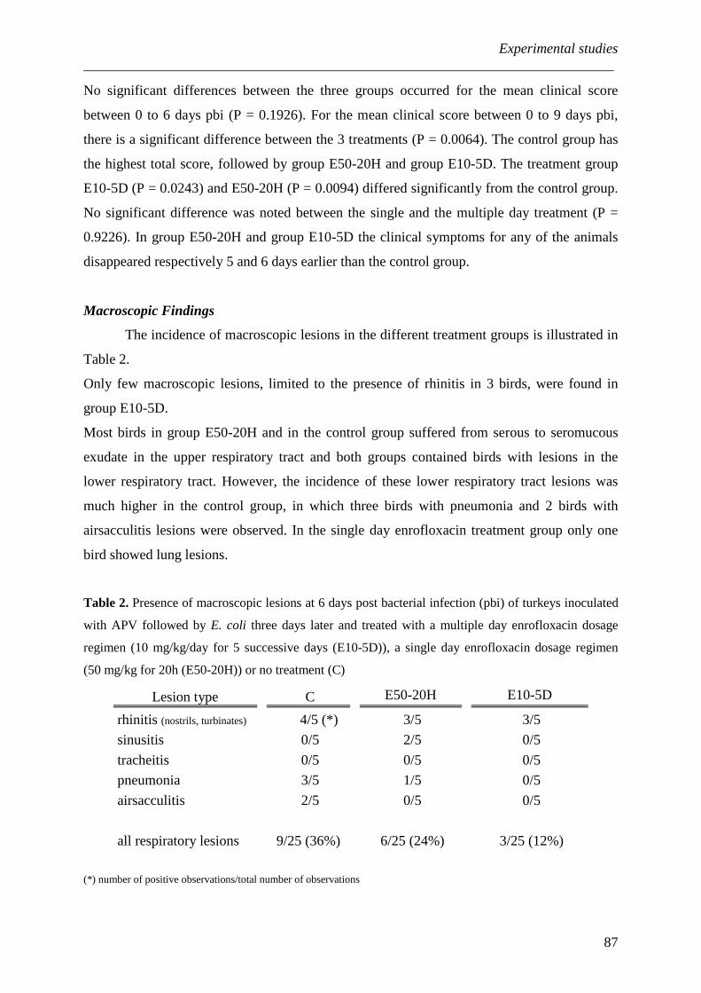
Experimental studies ___________________________________________________________________________
87
No significant differences between the three groups occurred for the mean clinical score
between 0 to 6 days pbi (P = 0.1926). For the mean clinical score between 0 to 9 days pbi,
there is a significant difference between the 3 treatments (P = 0.0064). The control group has
the highest total score, followed by group E50-20H and group E10-5D. The treatment group
E10-5D (P = 0.0243) and E50-20H (P = 0.0094) differed significantly from the control group.
No significant difference was noted between the single and the multiple day treatment (P =
0.9226). In group E50-20H and group E10-5D the clinical symptoms for any of the animals
disappeared respectively 5 and 6 days earlier than the control group.
Macroscopic Findings
The incidence of macroscopic lesions in the different treatment groups is illustrated in
Table 2.
Only few macroscopic lesions, limited to the presence of rhinitis in 3 birds, were found in
group E10-5D.
Most birds in group E50-20H and in the control group suffered from serous to seromucous
exudate in the upper respiratory tract and both groups contained birds with lesions in the
lower respiratory tract. However, the incidence of these lower respiratory tract lesions was
much higher in the control group, in which three birds with pneumonia and 2 birds with
airsacculitis lesions were observed. In the single day enrofloxacin treatment group only one
bird showed lung lesions.
Table 2. Presence of macroscopic lesions at 6 days post bacterial infection (pbi) of turkeys inoculated
with APV followed by E. coli three days later and treated with a multiple day enrofloxacin dosage
regimen (10 mg/kg/day for 5 successive days (E10-5D)), a single day enrofloxacin dosage regimen
(50 mg/kg for 20h (E50-20H)) or no treatment (C)
Lesion type C E50-20H E10-5D
rhinitis (nostrils, turbinates) 4/5 (*) 3/5 3/5 sinusitis 0/5 2/5 0/5 tracheitis 0/5 0/5 0/5 pneumonia 3/5 1/5 0/5 airsacculitis 2/5 0/5 0/5 all respiratory lesions 9/25 (36%) 6/25 (24%) 3/25 (12%)
(*) number of positive observations/total number of observations

Experimental studies ___________________________________________________________________________
88
Necropsy at 15 and 16 days pbi of the remaining birds from the control group showed one
bird with airsacculitis and another bird with rhinitis and pneumonia. No lesions were found in
any turkey of the enrofloxacin treatment groups.
Virological Titration of Tracheal Swabs
Virological titration of tracheal swabs collected at 2 days post viral infection showed
that APV was recovered from every bird. Mean titers were 4.6 log10CD50/ml, 4.9
log10CD50/ml and 4.9 log10CD50/ml for group 10E-5D, group 50E-20H and the control group
respectively.
Bacteriological Titration of Tracheal Swabs
The results of the E. coli titrations of the tracheal swabs are shown in Figure 2.
As from 1 day after bacterial infection, E. coli titers in the control group started to increase,
resulting in maximal titers of 4.32 log10 cfu/ml at 6 days pbi after which bacterial numbers
decreased to 0 at 15 days pbi.
After the first treatment day, i.e. at 2 days pbi, no E. coli was isolated out of any trachea in the
five day enrofloxacin treatment group for the remaining course of the experiment. The single
day enrofloxacin treatment highly reduced E. coli multiplication in the tracheae but was not
able to obtain a permanent clearance of the infection. E. coli titers increased, starting from 4
days pbi with maximal titers of 1.20 log10 cfu/ml at 6 days pbi, a significantly lower number
compared to that of the control group.
There is a significant difference between the three treatments (P < 0.0001) for the mean E.
coli titers between 0 to 6 days pbi. The two treatment groups differ significantly from the
control group (P < 0.0001).
There is also a significant difference between the three treatments (P < 0.0001) for the mean
E. coli titer between 0 to 9 days pbi. The two treatment groups differ significantly from the
control group (P < 0.0001). Furthermore, the five day treated group had significantly lower E.
coli titers than the single day treated group (P = 0.0053).
Titration of the tracheal swabs on enrofloxacin supplemented agar plates (i.e. 0.25 µg/ml agar
and 2 µg/ml agar) did not show any growth of less susceptible or resistant E. coli isolates at
any time during the experiment.

Experimental studies ___________________________________________________________________________
89
0.0
0.5
1.0
1.5
2.0
2.5
3.0
3.5
4.0
4.5
5.0
1 2 3 4 5 6 7 8 9 10 11 12 13 14 15 16
Days post E. coli inoculation
log10 cfu/ml
Figure 2. Mean E. coli titers in trachea mucus collected at different time points from turkeys,
inoculated with APV followed by E. coli three days later, treated with 10 mg/kg enrofloxacin for 5
days (group E10-5D x) or with 50 mg/kg enrofloxacin for 20 hours (group E50-20H ◊). Group C (●)
was enclosed as an untreated positive control group. Enrofloxacin was administered in the drinking
water, 24h after E. coli inoculation.
Bacteriological Examination of Tissue Suspensions and Swabs
E. coli was never isolated from any of the organs of the birds from group E10-5D.
For the control group and group E50-20H, the various mean E. coli titers in the organs at 6
days pbi are shown in Table 3.
Significant differences between treatments were observed for E. coli titers from the turbinates,
and tracheas. For these organs, the control group showed the highest mean scores, followed
by group E50-20H and group E10-5D (lowest mean score). For the turbinates, a pairwise
significant comparison was noted after Bonferroni correction between the control group and
E10-5D. No pairwise significant differences were noted for E. coli titers of the trachea and no
significant difference between treatments was noted for the titers of the lungs.
No E. coli was isolated from the air sacs, pericardium or liver in any bird.
During the final necropsy, E. coli was discovered in only one lung in a turkey from group
E50-20H. In the control group, E. coli was isolated 3 times from turbinates, 1 time out from
airsacs and one time from a trachea. No E. coli was isolated from any turkey of group E10-
↑ First treatment day

Experimental studies ___________________________________________________________________________
90
5D. No significant difference between treatments was noted for the presence of E. coli in the
organs during the final necropsy.
Tab
le 3
. Clin
ical
sco
res
and
isol
atio
n of
E. c
oli o
ut o
f tra
chea
l sw
abs
and
orga
ns (
mea
n va
lue
(sta
ndar
d er
ror)
) of
turk
eys
inoc
ulat
ed w
ith A
PV
follo
wed
by
E. c
oli t
hree
day
s la
ter,
and
treat
ed w
ith a
mul
tiple
day
enr
oflo
xaci
n do
sage
sch
eme
(10
mg/
kg/d
ay fo
r 5 s
ucce
ssiv
e da
ys (E
10-5
D))
,
a si
ngle
day
enr
oflo
xaci
n do
sage
sche
me
(50
mg/
kg fo
r 20h
(E50
-20H
)) o
r no
treat
men
t (C
)
Clin
ical
Sco
res *
Isol
atio
n of
E. c
oli o
ut o
f tra
chea
l sw
abs*
Isol
atio
n of
E. c
oli
at 6
day
s pbi
(lo
g 10
cfu/
ml t
issu
e su
spen
sion
)
M
ean
clin
ical
scor
es
Lo
g 10 c
fu/m
l
Turb
inat
es
Trac
hea
Lung
s
Gro
up
C
1.92
(0.9
2)
2.63
(1.5
0)
7.2
(0.8
) a 3.
7 (2
.1) a
1.
7 (2
.4) a
E50-
20H
1.
19 (1
.14)
a 0.
81 (0
.67)
1.
5 (3
.4) a
b 0.
4 (0
.9) a
0.
00 (0
.00)
a
E10-
5D
1.37
(1.0
1) a
0.22
(0.5
4)
0.0
(0.0
0) b
0.00
(0.0
0) a
0.00
(0.0
0) a
a tre
atm
ents
shar
ing
a le
tter d
iffer
not
sign
ifica
ntly
from
eac
h ot
her a
t the
5%
glo
bal s
igni
fican
ce le
vel
*
from
0 to
9 d
ays p
ost b
acte
rial i
nocu
latio
n (p
bi)

Experimental studies ___________________________________________________________________________
91
Weight
The mean weight gains measured in the after treatment period (i.e. from 1 day before
E. coli infection until 15 or 16 days pbi) in the different turkey groups are 925 g for the
control group, 968 g for group E50-20H and 1038 g for group E10-5D. Although no
significant differences for weight gain are observed between the different groups (P = 0.4749)
during the after-treatment period, there’s a tendency, showing higher weight gains for longer
treatment times (group E10-5D>group E50-20H> control group).
Discussion
The five day drinking water medication with enrofloxacin at a dose of 10 mg/kg body
weight was the most successful in reducing the severity and incidence of the clinical signs and
macroscopic lesions. E. coli tracheal and organ colonization were limited to the highest
extent. Less pronounced, although significant, was the outcome of the single day treatment.
As enrofloxacin exerts a concentration-depending activity (Prescott and Walker, 2000), it was
hypothesized that higher plasma and respiratory tissue concentrations, due to a higher dosage,
could succeed in a better killing of the E. coli bacteria. However, the single day treatment
proved not as efficacious as the five day treatment regimen of 10 mg enrofloxacin/kg body
weight in reducing E. coli multiplication in the respiratory organs. In contrast to the five day
treatment, single day treatment did not achieve elimination of E. coli from the respiratory tract
and the remaining bacteria re-emerged. This indicates that spread of the pathogen to
susceptible animals could not be avoided using the single day treatment. The duration of the
treatment at the appropriate dosage seems therefore as important for clinical efficacy as the
size of the dose. The results obtained in this study, comparing a single and multiple treatment
regimen, are highly similar to a previously performed study using similar enrofloxacin
treatment regimens in turkeys infected experimentally with APV and O. rhinotracheale
(Garmyn et al., in press). Therefore, a single day dosage regimen is not a useful valuable
alternative to the labelled dosage regimen in treating E. coli associated respiratory disease in
turkeys and a five day enrofloxacin treatment regimen seems recommended.
Avian pathogenic E. coli are frequently found to be resistant to commonly used antibacterial
agents as ampicillin, amoxycillin, tetracyclines, trimethoprim+sulphonamide and flumequine.
Also, resistance to enrofloxacin is commonly encountered (Vandemaele et al., 2002).
Acquired quinolone resistance is the consequence of stepwise mutations in the gyr and par
genes. A first step mutation leads to reduced susceptibility after which successive mutations
Experimental studies ___________________________________________________________________________
92
possibly result in resistance (Barnard and Maxwell, 2001; Hooper, 2001). In this study,
however, bacterial clones with reduced susceptibility or resistance were not detected in any of
the enrofloxacin treated groups. Reduced susceptibility or resistance to enrofloxacin also did
not occur when similar dosing regimens were tested previously on turkeys experimentally
infected with O. rhinotracheale (Garmyn et al., in press). This supports pharmacokinetic
findings (Dimitrova et al., 2006; Fraatz et al., 2006) that, when enrofloxacin is used according
labelled directions, the risk of selection for bacterial resistance is minimized.
In conclusion, both the multiple and the single day enrofloxacin treatment regimen reduced E.
coli multiplication in the respiratory tract tissues (turbinates, trachea and lung), but the five
day treatment with 10 mg enrofloxacin/kg in turkeys provided the best results by shortening
the course of clinical disease, by eliminating E. coli from the respiratory tract without re-
multiplication, and by reducing macroscopic lesions. The efficacy of the single day treatment
did not equal that of the 5 day treatment, possibly by not eliminating E. coli from the
respiratory organs, which made it possible for the remaining bacteria to re-emerge in those
organs.
None of the used treatment schemes promoted the selection of bacterial clones with reduced
susceptibility or resistance.
Acknowledgements
This work was supported by a grant from Bayer Animal Health GmbH. The authors would
like to express their appreciation to Marleen Foubert and Veerle Flama for their skilled
technical assistance. Furthermore, we would like to thank Barend Van Dam (BioChek) for the
supply of the ELISA kits.
References
Barnard, F. M., and A. Maxwell. 2001. Interaction between DNA gyrase and quinolones:
effects of alanine mutations at GyrA subunits residues Ser83 and Asp87.
Antimicrobial Agents and Chemotherapy, 45, 1994-2000.
Barnes, H. J., K. N. Lisa, and J. P. Vaillancourt. 2008. Colibacillosis. Pages 691-737 in
Diseases of poultry 12th edn. Y. M. Saif, H. J. Barnes, A. M. Fadly, J. R. Glisson, L. R.
Mc Dougald, L. K. Nolan and D. E. Swayne. Ames, Iowa.

Experimental studies ___________________________________________________________________________
93
Dimitrova, D. J., L. D. Lashev, St. G. Yanev, and V. T. Pandova. 2006. Pharmacokinetics of
enrofloxacin and its metabolite ciprofloxacin in male and female turkeys following
intravenous and oral administration. Veterinary Research Communications, 30, 415-
422.
Dho-Moulin, M., and J. M. Fairbrother. 1999. Avian pathogenic Escherichia coli (APEC).
Veterinary Research, 30, 299-316.
Fraatz, R., R. Froyman, and R. Krebber. 2006. Pharmacokinetics of enrofloxacin and
lorfenicol in turkeys during continuous drinking water medication. Journal of
Veterinary Pharmacology and Therapeutics, 29(Suppl. 1), 267.
Garmyn, A., A. Martel, R. Froyman, H. Nauwynck, L. Duchateau, F. Haesebrouck, and F.
Pasmans. 2009. Efficacy of four enrofloxacin treatment regimens against experimental
infection in turkey poults with avian pneumovirus and Ornithobacterium
rhinotracheale. Avian Pathology, in press.
Gautrais, B., and D. Copeland. 1997. Use of enrofloxacin in turkeys; a worldwide experience.
Proceedings of the 46th Western Poultry Disease Conference, Sacramento, California,
USA, page 79.
Ginns, C. A., G. F. Browning, M. L. Benham, and K. G. Whithear. 1998. Development and
application of an aerosol challenge method for reproduction of avian collibacilosis.
Avian Pathology, 2, 505-511.
Hafez, H. M., J. Emele, and W. Kruse. 1992. Die Pasteurella multocida-Infektion bei den
Puten: Literaturübersicht und praktische Erfahrungen in Putenbestanden. Archiv für
Geflügelkunde, 56, 45-52.
Hooper, D. C. 2001. Emerging mechanisms of fluoroquinolone resistance. Emerging
Infectious Diseases, 7, 337-341.
Jakob, H. P., R. Morgenstern, P. Albicker, and R. K. Hoop. 1998. Reasons for condemnation
of slaughtered broilers from two large Swiss producers. Schweizer Archiv für
Tierheilkunde, 140, 60-64.
Jirjis, F. F., S. L. Noll, D. A. Halvorson, K. V. Nagaraja, F. Martin, and D. P. Shaw. 2004.
Effects of bacterial coinfection on the pathogenesis of avian pneumovirus infection in
turkeys. Avian Diseases, 48, 34-49.
Krebber, R. 2003. Analytical method for the determination of pradofloxacin in serum and
urine by turbulent flow chromatography/tandem mass spectrometry. Journal of
Veterinary Pharmacology and Therapeutics, 26(Suppl. 1):102-103.
Marien, M., A. Decostere A., A. Martel, K. Chiers, R. Froyman, and H. Nauwynck. 2005.

Experimental studies ___________________________________________________________________________
94
Synergy between avian pneumovirus and Ornithobacterium rhinotracheale in turkeys.
Avian Pathology, 34, 204-211.
Marien M., H. Nauwynck, L. Duchateau, A. Martel, K. Chiers, L. Devriese, R. Froyman, and
A. Decostere. 2006. Comparison of the efficacy of four antimicrobial treatment
schemes against experimental Ornithobacterium rhinotracheale infection in turkey
poults pre-infected with avian pneumovirus. Avian Pathology, 35, 230-237.
Marien, M., A. Decostere, L. Duchateau, K. Chiers, R. Froyman, and H. Nauwynck. 2007.
Efficacy of enrofloxacin, florfenicol and amoxicillin against Ornithobacterium
rhinotracheale and Escherichia coli O2:K1 dual infection in turkeys following APV
priming. Veterinary Microbiology, 121, 94-104.
Martinez, M., P. McDermott, and R. Walker. 2006. Pharmacology of the fluoroquinolones: a
perspective for the use in domestic animals. Veterinary Journal, 172, 10-28.
Prescott, J. F., and R. D. Walker. 2000. Principles of antimicrobial treatment. Pages 93-96 in
Antimicrobial Therapy in Veterinary Medicine, 3rd edn. J. F. Prescott, J. D. Baggot,
and R. D. Walker. Ames, Iowa.
Russel, S.M. 2003. The effect of airsacculitis on bird weights, uniformity, fecal
contamination, processing errors, and populations of Campylobacter spp. and
Escherichia coli. Poultry Science, 82,1326-1331.
Turpin, E. A., L. E. Perkins, and D. E. Swayne. 2002. Experimental infection of turkeys with
avian pneumovirus and either Newcastle disease virus or Escherichia coli. Avian
Diseases, 46, 412-422.
Vandemaele, F., M. Vereecken, J. Derijcke, B. M. Goddeeris. 2002. Incidence and antibiotic r
resistance of pathogenic Escherichia coli among poultry in Belgium. Veterinary
Record, 151, 355-356.
Van de Zande, S., H. Nauwynck, D. Cavanagh, and M. Pensaert. 1998. Infections and
reinfections with avian pneumovirus subtype A and B on Belgian turkey farms and
relations to respiratory problems. Zentralblatt Veterinarmedizin, 45, 621-626.
Van de Zande, S., H. Nauwynck, and M. Pensaert. 2001. The clinical, pathological and
microbiological outcome of an Escherichia coli O2:K1 infection in avian pneumovirus
infection in turkeys. Veterinary Microbiology, 81, 353-365.
Yogaratnam, V. 1995. Analysis of the causes of high rates of carcase rejection at a poultry
processing plant. Veterinary Record, 137, 215-217.

Experimental studies ___________________________________________________________________________
95

Experimental studies ___________________________________________________________________________
96
Experimental studies ___________________________________________________________________________
97
3.3. THE EFFECT OF REDUCED TREATMENT TIME AND DOSAGE OF
ENROFLOXACIN ON THE COURSE OF RESPIRATORY DISEASE CAUSED BY
AVIAN METAPNEUMOVIRUS AND ORNITHOBACTERIUM RHINOTRACHEALE
Garmyn, A., Martel, A., Froyman, R., Ludwig, C., Nauwynck, H., Haesebrouck, F. and
Pasmans, F.
Poultry Science (2009), 88, 2315-2323.
___________________________________________________________________________

Experimental studies ___________________________________________________________________________
98

Experimental studies ___________________________________________________________________________
99
Summary
A dose titration and reduced duration medication study were performed to evaluate the
current enrofloxacin treatment schedule in growing turkeys experimentally infected with
avian metapneumovirus (APV) and Ornithobacterium rhinotracheale (O. rhinotracheale).
Experimental groups of 17 four-week-old turkeys were first infected with avian
metapneumovirus (APV) and three days later with O. rhinotracheale. Enrofloxacin treatment
in the drinking water was started 24 h after O. rhinotracheale inoculation. In the dose titration
study enrofloxacin doses of 5, 10 and 20 mg / kg body weight (BW) were administered for 5
successive days. In the reduced duration medication study, following enrofloxacin regimens
were compared: 25 mg / kg BW / day on day 0 and day 2, 15 mg / kg BW / day on days 0, 2
and 4 and 10 mg / kg BW for 5 successive days. In both studies, all enrofloxacin treatments
were equally efficacious, i.e. equally capable of shortening the course of clinical disease,
eliminating O. rhinotracheale from the respiratory tract and reducing gross lesions. O.
rhinotracheale bacteria were not recovered from any of the animals on enrofloxacin
supplemented media, indicating that none of the used treatment regimens promoted the
selection of bacterial clones with reduced susceptibility or resistance to this antimicrobial
agent. In conclusion, none of the alternative enrofloxacin treatment regimens yielded better
results than the current prescribed treatment (i.e. 10 mg/kg BW for 5 successive days) of O.
rhinotracheale infections in turkeys. However, the reduced duration of application would
offer a less time-consuming and equally effective alternative.

Experimental studies ___________________________________________________________________________
100
Introduction
Respiratory disease induced by various viral and/or bacterial pathogens often affects
turkeys during the rearing period causing severe economic losses due to growth retardation,
increased feed conversion rates, increased mortality rates, higher condemnation rates at
slaughter and high medication costs (van Empel and Hafez, 1999).
Ornithobacterium rhinotracheale (O. rhinotracheale) plays a significant role in the aetiology
of the respiratory disease complex (van Empel et al., 1996; Jirjis et al., 2004). Although
indications of primary pathogenicity exist (Van Veen et al., 2000; Sprenger et al., 1998), the
organism is mostly regarded as a facultative pathogen, aggravating the symptoms in the
respiratory tract caused by viruses such as avian metapneumovirus (genus Metapneumovirus)
(APV) (Van Empel et al., 1996; Jirjis et al., 2004; Marien et al., 2005, 2006). This dual APV/
O. rhinotracheale infection frequently occurs in the field (De Rosa et al., 1996; El-Sukhon et
al., 2002).
Pursuit of optimal environmental conditions such as good hygiene, adequate ventilation, low
ammonia levels, optimal relative humidity levels and the prevention of simultaneous
infections may reduce respiratory disease caused by O. rhinotracheale (van Empel and Hafez,
1999). In practice, O. rhinotracheale infections are primarily dealt with using antimicrobials
in the drinking water. Enrofloxacin, a fluoroquinolone antimicrobial compound, is in Europe
commonly used to cure respiratory infections in turkeys and has proven its efficacy against O.
rhinotracheale infections, when administered in the drinking water for five successive days at
a dose of 10 mg/kg body weight (BW), which is the labelled dosage (Marien et al., 2006;
Marien et al., 2007). This treatment regimen, however, is considered as cumbersome and time
consuming by many farmers since the medicated drinking water has to be prepared each day
during five days. This often hinders completion of the full treatment regimen. Other
controlled clinical studies, addressing enrofloxacin efficacy against O. rhinotracheale
infections, have not been reported. In addition, acquired resistance or reduced susceptibility of
O. rhinotracheale against enrofloxacin is commonly encountered (Devriese et al., 1995,
Malik et al., 2003, Soriano et al., 2003, Van Veen et al., 2000) and more specific data are
required to search for a therapeutic regimen that minimizes the risk of selecting antimicrobial
resistant bacteria. In the present study, the efficacy of alternative enrofloxacin treatment
regimens against O. rhinotracheale infections in experimentally infected turkeys was
assessed. First the minimum effective dose of enrofloxacin in animals treated during five days
was determined. In a second study, the efficacy of drinking water treatment with high doses of
enrofloxacin, administered over a shorter period of time was determined.

Experimental studies ___________________________________________________________________________
101
In both experiments, the selection of O. rhinotracheale clones with reduced susceptibility or
resistance was also assessed.
Materials and Methods
Turkeys
Commercial turkey poults (Moorgut Kartzfehn, von Kameke GmbH & Co, Bösel,
Germany) were used in this study. The birds were housed on litter in separate isolation rooms
with HEPA-filtered air, water and feed were available ad libitum, and received 20 hours of
light per day. Before bacterial inoculation, tracheal swabs from all birds were examined for
the presence of O. rhinotracheale and determined to be negative.
Virus
The APV strain A/T6/96 (subtype A) was used. The strain was isolated during a
respiratory outbreak on a Belgian turkey farm (Van de Zande et al., 1998). The virus stock
had a titre of 105.5 50% ciliostatic dose (CD50)/ml after the third passage in tracheal organ
cultures.
Bacteria
The O. rhinotracheale type strain LMG 9086T, originally isolated from a turkey with a
respiratory tract infection, was used. The strain was serotyped as type A in an agar gel
precipitation test (Hafez and Sting, 1999), kindly performed by Professor H. M. Hafez
(Institute of Poultry Diseases, Free University of Berlin, Germany). The minimum inhibitory
concentration (MIC) of enrofloxacin for the strain was determined at < 0.03 µg/ml and the
strain was stored at -70°C. The organism was cultured for 48 h at 37°C on Columbia agar
(Oxoid LTD, Basingstoke, Hampshire, England) with 5% sheep blood in a 5% CO2
atmosphere. The O. rhinotracheale bacteria were transferred into brain heart infusion (BHI)
broth (Oxoid) for 24 h at 37°C with agitation. The bacterial challenge inoculum was prepared
by washing the cultured bacteria twice in phosphate-buffered saline (PBS) followed each time
by five minutes of centrifugation at 3,000 rpm at 4°C. The resulting pellet was resuspended in
PBS to obtain a final concentration of 108 colony-forming units (cfu)/ml. Confirmation of the
number of cfu/ml was done by inoculating ten-fold dilutions in PBS on sheep blood agar and
counting the number of colonies.

Experimental studies ___________________________________________________________________________
102
Experimental Design
In a first trial (dose titration study), 85 turkeys were randomly divided into 5 groups
and inoculated with 104.5 CD50 of APV at 25 days of age. All groups, except group NC, were
inoculated with 108.5 cfu of O. rhinotracheale, 3 days later. Group NC was enclosed as an
untreated negative control group. At 24 h after O. rhinotracheale inoculation, 3 groups
received antimicrobial treatment by medicating the drinking water during 5 successive days.
The first group received enrofloxacin at 5 mg/kg (group E5-5D), the second group at 10
mg/kg (group E10-5D) and the third group at 20 mg/kg (group E20-5D).
In a second trial (reduced duration medication study) 68 turkeys were randomly divided into 4
groups of 17 animals. At 20 days of age the birds of all groups were inoculated with 104.4
CD50 of APV and 3 days later with 108.8 cfu of O. rhinotracheale. Three groups received
enrofloxacin treatment by medicating the drinking water starting at 24 h after O.
rhinotracheale inoculation. Group E10-5D was treated at a dose of 10 mg / kg BW daily for 5
successive days. Group E25-2D was treated at a dose of 25 mg / kg BW / day on day 0 and
day 2 and group E15-3D at a dose of 15 mg / kg BW / day on days 0, 2 and 4.
In both trials group PC was included as an untreated positive control group. Viral and
bacterial inoculation occurred by dividing a total of 250 µl of inoculum equally over the
nostrils and eyes.
In every group, water uptake was measured on a daily basis from three days before until the
end of the treatment periods. Additionally, all animals were weighed before O. rhinotracheale
inoculation. On the basis of these data, the antimicrobial concentrations to be administered in
the water and the actually received dosages were calculated. Water samples from the
medicated drinking water were collected daily to determine the actual amount of enrofloxacin
using turbulent flow chromatography/tandem mass spectrometry (Krebber, 2003).
All birds were examined for clinical signs on a daily basis throughout the experiment until
they were sacrificed. The clinical signs of the upper respiratory tract (nasal exudates, swollen
sinuses, frothy eyes) were scored as described in Van de Zande et al. (2001). The clinical
condition of each bird was assigned a score from 0 (absence of clinical signs) to 6 (death).
Tracheal swabs were taken from all birds two days post viral inoculation (pvi) for APV
enumeration and at regular time points post bacterial inoculation (pbi) for titration of O.
rhinotracheale. The tracheal swabs were collected using cotton-tipped aluminium shafted
swabs (Copan Diagnostics Inc., Corona, USA) and placed in transport medium. The latter
consisted of 1 ml PBS for bacteriological examination which was supplemented with 10%

Experimental studies ___________________________________________________________________________
103
fetal calf serum (Gibco, Invitrogen Corporation, Merelbeke, Belgium), penicillin (1000 U/ml)
(Biopharma, Rome, Italy) and kanamycin (0.5 mg/ml) (Gibco) for virus enumeration.
Five birds of each group were sacrificed at 6 days pbi. The birds were necropsied and
examined for the presence of gross lesions. Samples of the turbinates, trachea and lungs were
collected and a 10% tissue suspension in PBS was prepared for O. rhinotracheale
examination. Air sacs, pericardium and liver were sampled with cotton swabs for O.
rhinotracheale re-isolation.
Equal numbers of the remaining birds of each group were necropsied and examined for the
presence of gross lesions at 15 and 16 days pbi (trial 1) or 14 and 15 days pbi (trial 2).
Turbinates, trachea, lungs, air sacs, liver and pericardium were sampled with cotton swabs for
O. rhinotracheale re-isolation.
All samples were processed immediately after collection as described below.
The birds were weighed the day before the APV inoculation, the day before the O.
rhinotracheale inoculation and subsequently the final day of the experiment.
These experiments were approved by the Ethical Committee of the Faculty of Veterinary
Medicine, Ghent University.
Virological Enumeration and Bacteriological Titration of Tracheal Swabs
The CD50 per ml of APV and the cfu per ml of O. rhinotracheale were determined,
using procedures described in Marien et al. (2005). In addition, in order to detect O.
rhinotracheale isolates with reduced susceptibility to enrofloxacin, samples were also
inoculated on sheep blood agar supplemented with 5 µg/ml gentamicin, 5 µg/ml polymyxin
and 0.25 µg or 2 µg enrofloxacin/ml.
Bacteriological Examination of Tissue Suspensions and Swabs
From samples of the turbinates, trachea, and lungs, the cfu of O. rhinotracheale was
determined in duplicate by incubating undiluted tissue suspensions and tenfold serial dilutions
of tissue suspensions on Columbia Blood Agar supplemented with 5 µg/ml gentamicin and 5
µg/ml polymyxin. After 24-48 h of incubation at 37°C in a 5% CO2 atmosphere, viable counts
were performed and the number of cfu/ml tissue suspension calculated.
The swabs taken from the air sacs, pericardium and liver from the 25 birds sacrificed at 6 days
pbi and the swabs from the turbinates, trachea, lungs, air sacs, pericardium and liver from the
birds sacrificed at 15 and 16 days pbi (trial 1) or at 14 and 15 days pbi (trial 2) were
inoculated on Columbia Blood Agar supplemented with 5 µg/ml gentamicin and 5 µg/ml
Experimental studies ___________________________________________________________________________
104
polymyxin. After 24-48 h of incubation at 37°C in a 5% CO2 atmosphere, the agar was
examined for the presence of O. rhinotracheale colonies.
Statistical Analyses
Tracheal titres of O. rhinotracheale were analysed for all birds using the
measurements between 0 and 6 days pbi and subsequently from the birds that were not
euthanized between 0 and 9 days pbi. The clinical scores and weight were analysed only for
animals that were not euthanized at 6 days pbi.
The average over time for the clinical scoring and tracheal swabbing was analysed using a
fixed-effects model with the treatments group as fixed effect.
Weight gain was compared at the end of the trial using a fixed-effects model.
O. rhinotracheale titres from the turbinates, trachea and lung of the birds, euthanized at 6
days pbi were compared between the five treatment groups with the Kruskall-Wallis test,
whereas the presence of O. rhinotracheale in the airsacs, liver and pericardium of the animals,
euthanized at 6 days pbi and the presence of O. rhinotracheale in the turbinates, trachea,
lungs, airsacs, liver and pericardium of the birds euthanized 15 and 16 days pbi, were
compared between the five treatments with the Fisher’s exact test. For the presence of O.
rhinotracheale in the turbinates at final necropsy also the Chi-Square test was used.
All tests were performed at a global 5% significance level and the pairwise comparisons were
tested at a significance level adjusted using Bonferroni’s multiple comparisons technique
(Agresti, 2002).
Results
Antimicrobial Uptake
Trial 1. Dose Titration Study. The mean actual daily dose for the total medication
period was 5.36 mg/kg (range 3.9-6.1) for group E5-5D, 10.16 mg/kg (range 9.6-11.7) for
group E10-5D mg/kg and 20.8 mg/kg (range 18.1-25.8) for group E20-5D.
Trial 2. Reduced Duration Medication Study. The mean actual daily dose for the total
medication period was 9.8 mg/kg (range 9.0-10.5) for group E10-5D, 15.5 mg/kg (range 15.0-
16.0) for group E15-3D mg/kg and 24.7 (range 22.0-27.4) for group E25-2D.

Experimental studies ___________________________________________________________________________
105
Clinical Signs
Trial 1. Dose Titration Study. No animals died due to the APV/O. rhinotracheale
infections. The mean clinical scores calculated for each group are presented in Figure 1.
Compared to the positive control group, clinical signs were significantly reduced as well as
severity and duration in every enrofloxacin treatment group. The negative control group, only
inoculated with APV, had lower clinical scores compared to all other infected groups at all
time points.
Data presented in Table 1 show the area under the curve (AUC) scores from 0 to 9 days pbi.
The treated groups had significantly lower AUC scores than the positive control (P < 0.037).
When the three enrofloxacin treatments were compared with each other, no significant
difference was noted (P > 0.349). All treatments differed significantly from the negative
control which had higher clinical scores (P < 0.024).
0.0
0.5
1.0
1.5
2.0
2.5
3.0
3.5
4.0
-2 -1 0 1 2 3 4 5 6 7 8 9 10 11 12 13 14 15 16 17
Days post O. rhinotracheale inoculation
Clinical score
Figure 1. Mean clinical scores in turkeys, inoculated with APV followed by O. rhinotracheale three
days later, and treated for 5 successive days with 5 mg/kg/day enrofloxacin (group E5-5D ▲), 10
mg/kg/day enrofloxacin (group E10-5D ◊) or 20 mg/kg/day enrofloxacin (group E20-5D X). The
positive control group (group PC ■), infected with APV and O. rhinotracheale, and the negative
control group (group NC ●), infected with APV remained untreated.
Trial 2. Reduced Duration Medication Study. One turkey died in group E25-2D four
days after the final treatment day, but according to necropsy and bacteriological investigation,
↑ First treatment day

Experimental studies ___________________________________________________________________________
106
this was due to an Enterococcus faecalis infection. As no relation to bacterial inoculation or
study medication was encountered, this turkey was excluded from all statistical analyses.
The mean clinical scores calculated for each group are presented in Figure 2.
Compared to the positive control group clinical signs in the treated groups were less
pronounced (group E10-5D and E25-2D) or the higher scores reduced over time (group E15-
3D). In addition, clinical signs in the treated groups disappeared two days (group E15-3D and
group E10-5D) or one day (group E25-2D) earlier, than the clinical scores in the positive
control group.
From 7 days pbi onwards, one bird from group E25-2D suffered from an airsac rupture. Since
no bacterial organism could be isolated out of the airsacs of this turkey during final necropsy,
this was probably due to trauma.
Data presented in Table 2 show the AUC scores from 0 to 9 days pbi. Comparisons between
pairs of O. rhinotracheale infected groups revealed significant differences between group
E10-5D and the positive control group (P = 0.0085) and also between group E15-3D and the
positive control group (P = 0.0244).
0.0
0.5
1.0
1.5
2.0
2.5
3.0
3.5
-2 -1 0 1 2 3 4 5 6 7 8 9 10 11 12 13 14 Days post O. rhinotracheale inoculation
Clinical score
Figure 2. Mean clinical scores in turkeys, inoculated with APV followed by O. rhinotracheale three
days later, and treated with 10 mg/kg enrofloxacin for 5 days (group E10-5D ▲), with 25 mg/kg
enrofloxacin for 2 days alternated with a non treating day (group E25-2D ◊) and with 15 mg/kg
enrofloxacin for 3 days alternated each time by a non treating day (group E15-3D X). The positive
control group (group PC ■) remained untreated.
↑ First treatment day

Experimental studies ___________________________________________________________________________
107
Tab
le 1
. Obs
erva
tions
of g
ross
lesi
ons a
t 6 d
ays p
ost b
acte
rial i
nfec
tion
and
at 1
5 or
at 1
6 da
ys p
ost b
acte
rial i
nfec
tion
of tu
rkey
s ino
cula
ted
with
APV
follo
wed
by
O. r
hino
trac
heal
e th
ree
days
late
r and
trea
ted
with
diff
eren
t enr
oflo
xaci
n re
gim
ens:
5 m
g/kg
for 5
succ
essi
ve d
ays (
E5-5
D),
10
mg/
kg fo
r 5 su
cces
sive
day
s (E1
0-5D
) and
20
mg/
kg fo
r 5 su
cces
sive
day
s (E2
0-5D
). Th
e po
sitiv
e co
ntro
l gro
up (P
C),
infe
cted
with
APV
and
O.
rhin
otra
chea
le, a
nd th
e ne
gativ
e co
ntro
l gro
up (N
C),
infe
cted
with
APV
rem
aine
d un
treat
ed.
Nec
rops
y da
ys p
bi
Le
sion
type
N
C
PC
E5
-5D
E
10-5
D
E20-
5D
rhin
itis (
nost
rils,
conc
hae)
1
/5 (*
)
5/5
5/5
3/5
1/5
si
nusi
tis
3
/5
5
/5
2
/5
4
/5
3
/5
6
trach
eitis
0/5
0/5
1/5
0/5
0/5
pn
eum
onia
0/5
0/5
0/5
0/5
0/5
ai
rsac
culit
is
0
/5
2
/5
0
/5
0
/5
0
/5
A
ll le
sion
s
4/25
(16%
)
12/2
5 (4
8%)
8
/25
(32%
)
7/2
5 (2
8%)
4
/25
(16%
)
rh
initi
s (no
stril
s,con
chae
)
0/1
2
3/1
2
1/1
2
0/1
2
0/1
2
si
nusi
tis
0
/12
2
/12
0
/12
0
/12
0
/12
15 o
r 16
tra
chei
tis
0
/12
0
/12
0
/12
0
/12
0
/12
pneu
mon
ia
0
/12
0
/12
0
/12
0
/12
0
/12
airs
accu
litis
0/1
2
1/1
2
0/1
2
0/1
2
0/1
2
All
lesi
ons
0
/60
(0%
)
6/6
0 (1
0%)
1
/60
(2%
)
0/6
0 (0
%)
0
/60
(0%
)
(*
) num
ber o
f pos
itive
obs
erva
tions
/tota
l num
ber o
f obs
erva
tions

Experimental studies ___________________________________________________________________________
108
Gross Findings and Lesion Observations
Trial 1. Dose Titration Study. The observations of gross lesions in the organs of the
birds necropsied at 6 days pbi and 15 or 16 days pbi are shown in Table 1. Although no
significant statistical differences between the different groups were noted, the highest
percentage (48%) of clinically affected organs was seen in the positive control group. This
group also showed the most severe lesions. Each turkey in the positive control group showed
mucoserous exudate in the upper respiratory tract (nostrils, turbinates and sinuses) and two
birds also suffered from airsacculitis. In the other groups, lesions were limited to the upper
respiratory tract. When the three enrofloxacin treatment groups were compared, fewer birds
with lesions were seen in group E20-5D (16%) than in group E5-5D (32%) and group E10-5D
(28%). These latter groups also showed more mucous exudate in the upper respiratory tract
while the exudate found in birds from group E20-5D was always clear. Comparing E20-5D
with the negative control group (which was only infected with APV) equal percentages of
organs affected were found (16%) and gross lesions were relatively similar.
During final necropsy at 15 and 16 days pbi, three birds from the positive control group and
one bird from group E5-5D suffered from serous exudate in the upper respiratory tract. One
bird from the positive control group also showed condensed airsacs. No statistically
significant difference between the groups could be noted.
Trial 2. Reduced Duration Medication Study. The observations of gross lesions in the
organs of the birds necropsied at 6 days pbi and 14 or 15 days pbi are shown in Table 2.
Most severe lesions and highest percentage of affected organs (60%) were seen in the positive
control group. Each turkey in this group showed mucous exudate in the upper respiratory tract
(nostrils, turbinates and sinuses). Four birds also suffered from pneumonia and one from
airsacculitis. In the treated turkey groups, not every bird was affected. One turkey from group
E15-3D and 10E-5D and two turkeys from E25-2D did not show any gross lesions. Lesions in
the affected birds (serous to mucous exudate) were limited to the upper respiratory tract,
except for one bird from group E10-5D which showed hyperaemic air sacs. Observations of
gross lesions in the three enrofloxacin treated groups were highly similar.
Only a statistical difference was noticed for pneumonia (P = 0.0026 related to the chi-square
test). Pairwise comparisons with the Fisher’s exact test for each pair of experimental groups
resulted in significant differences (P = 0.0476) for each comparison of an enrofloxacin
treatment group to the positive control group.
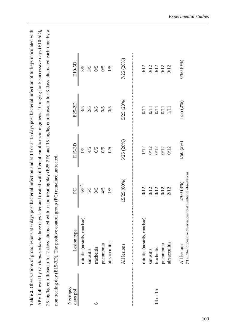
Experimental studies ___________________________________________________________________________
109
Tab
le 2
. Obs
erva
tions
of g
ross
lesi
ons a
t 6 d
ays p
ost b
acte
rial i
nfec
tion
and
at 1
4 or
at 1
5 da
ys p
ost b
acte
rial i
nfec
tion
of tu
rkey
s ino
cula
ted
with
APV
follo
wed
by
O. r
hino
trac
heal
e th
ree
days
late
r and
trea
ted
with
diff
eren
t enr
oflo
xaci
n re
gim
ens:
10
mg/
kg fo
r 5 s
ucce
ssiv
e da
ys (E
10-5
D),
25 m
g/kg
enr
oflo
xaci
n fo
r 2 d
ays
alte
rnat
ed w
ith a
non
trea
ting
day
(E25
-2D
) and
15
mg/
kg e
nrof
loxa
cin
for 3
day
s al
tern
ated
eac
h tim
e by
a
non
treat
ing
day
(E15
-3D
). Th
e po
sitiv
e co
ntro
l gro
up (P
C) r
emai
ned
untre
ated
.
Nec
rops
y
days
pbi
Lesi
on ty
pe
PC
E1
5-3D
E2
5-2D
E1
0-5D
rhin
itis (
nost
rils,
conc
hae)
5
/5(*
)
1/5
3
/5
3
/5
sinu
sitis
5/5
4/5
2/5
3/5
6
tra
chei
tis
0
/5
0
/5
0
/5
0
/5
pneu
mon
ia
4
/5
0
/5
0
/5
0
/5
airs
accu
litis
1/5
0/5
0/5
1/5
All
lesi
ons
1
5/25
(60%
)
5/2
5 (2
0%)
5
/25
(20%
)
7/2
5 (2
8%)
rhin
itis (
nost
rils,
conc
hae)
0/12
1/12
0/11
0/12
si
nusi
tis
0/
12
0/
12
0/
11
0/
12
14 o
r 15
tra
chei
tis
0/
12
0/
12
0/
11
0/
12
pneu
mon
ia
0/
12
0/
12
0/
11
0/
12
airs
accu
litis
2/12
0/12
1/11
0/12
All
lesi
ons
2/
60 (3
%)
1/
60 (2
%)
1/
55 (2
%)
0/
60 (0
%)
(*) n
umbe
r of p
ositi
ve o
bser
vatio
ns/to
tal n
umbe
r of o
bser
vatio
ns

Experimental studies ___________________________________________________________________________
110
During final necropsy at 14 and 15 days pbi, several birds in every turkey group showed
congested lungs or an injected pericardium, a side effect of T61 euthanasia. No statistically
significant results regarding the observations of lesions at the final necropsies could be
observed between the different turkey groups.
Virological Titration from Tracheal Swabs
Virological titration of tracheal swabs collected at two days post viral infection,
showed that APV was recovered from every bird.
Trial 1. Dose Titration Study. Mean titres were 104.5 CD50/ml, 104.5 CD50/ml, 104.3
CD50/ml, 104.5 CD50/ml and 104.3 CD50/ml for group E5-5D, group E10-5D, group E20-5D,
negative control group and the positive control group respectively.
Trial 2. Reduced Duration Medication Study. Mean titres were 104.3 CD50/ml, 104.5
CD50/ml, 104.3 CD50/ml and 104.3 CD50/ml for group E10-5D, group E15-3D, group E25-2D
and the positive control group respectively.
Bacteriological Titration of Tracheal Swabs
Trial 1. Dose Titration Study. The results of the O. rhinotracheale counts of the
tracheal swabs are shown in Figure 3.
O. rhinotracheale was not isolated at any time during the experiment in the negative
control group, which was only infected with APV.
In contrast with the non-treated positive control group, O. rhinotracheale was never isolated
out of any trachea in any enrofloxacin treatment group after the first treatment day, with the
exception of group E10-5D, in which O. rhinotracheale was re-isolated occasionally out of
the trachea of two birds, starting from five days after the last treatment day (10 days pbi), but
only at low numbers and without any marked clinical symptoms.
Using the AUC in the fixed-effects model, all enrofloxacin treatments differed significantly
from the positive control with lower O. rhinotracheale counts (P < 0.001). There was no
significant difference between the enrofloxacin treatment regimens (P > 0.18) (Table 3).

Experimental studies ___________________________________________________________________________
111
0.0
1.0
2.0
3.0
4.0
5.0
6.0
1 2 3 4 5 6 7 8 9 10 11 12 13 14 15 16 17 Days post O. rhinotracheale inoculation
log10 cfu/ml
Figure 3. Mean cfu/ml of O. rhinotracheale in tracheal swabs collected from turkeys, inoculated with
APV followed by O. rhinotracheale three days later and treated for 5 successive days with 5
mg/kg/day enrofloxacin (group E5-5D ▲), 10 mg/kg/day enrofloxacin (group E10 -5D ◊) or 20
mg/kg/day enrofloxacin (group E20-5D X). The positive control group (group PC ■), infected with
APV and O. rhinotracheale, and the negative control group (group NC ●), infected with APV
remained untreated.
Trial 2. Reduced Duration Medication Study. The results of the O. rhinotracheale
counts of the tracheal swabs are shown in Figure 4.
In contrast with the positive control group, O. rhinotracheale was never isolated out of a
trachea in any enrofloxacin treatment group after the first treatment day in group E10-5D and
group E25-2D or after the second treatment day in group E15-3D.
Using the AUC in a fixed effects-model, all enrofloxacin treatment regimens differed
significantly from the positive control group (P < 0.0001). There was no significant difference
between the enrofloxacin treatment regimens (P > 0.05) (Table 4).
Tracheal swabs were plated on enrofloxacin supplemented agar plates (i.e. 0.25 µg/ml agar
and 2 µg/ml agar) and did not show any growth of less susceptible or resistant O.
rhinotracheale isolates at any time during both experiments.
↑ First treatment day

Experimental studies ___________________________________________________________________________
112
0.0
1.0
2.0
3.0
4.0
5.0
6.0
1 2 3 4 5 6 7 8 9 10 11 12 13 14 15 16 17 Days post O. rhinotracheale inoculation
log10 cfu/ml
Figure 4. Mean cfu/ml of O. rhinotracheale in tracheal swabs collected from turkeys, inoculated with
APV followed by O. rhinotracheale three days later and treated with 10 mg/kg enrofloxacin for 5 days
(group E10-5D ▲), with 25 mg/kg enrofloxacin for 2 days alternated with a non treating day (group
E25-2D ◊) and 15 mg/kg enrofloxacin for 3 days alternated each time by a non treating day (group
E15-3D X). The positive control group (group PC ■) remained untreated.
Bacteriological Examination of Tissue Suspensions and Swabs
Trial 1. Dose Titration Study. The mean number of cfu/ml of O. rhinotracheale in
the organs of the birds necropsied at 6 days pbi are shown in Table 3.
During necropsy at 6 days pbi, O. rhinotracheale was only isolated from the respiratory
organs (turbinates, trachea, lungs and airsacs) of the positive control group.
Statistically significant differences were observed for comparison between the enrofloxacin
treatments and the positive control with lower O. rhinotracheale cfu counts (for air sacs, P <
0.035; for lungs, P < 0.001; for tracheas, P < 0.001 and for turbinates P < 0.001) in the treated
groups. There was no statistically significant difference between the enrofloxacin treatment
regimens.
↑ First treatment day

Experimental studies ___________________________________________________________________________
113
Tab
le 3
. Clin
ical
scor
es a
nd is
olat
ion
of O
. rhi
notr
ache
ale
out o
f tra
chea
l sw
abs a
nd o
rgan
s (m
ean
valu
e +
stan
dard
err
or) o
f tur
keys
inoc
ulat
ed
with
APV
follo
wed
by
O. r
hino
trac
heal
e th
ree
days
late
r, an
d tre
ated
with
diff
eren
t enr
oflo
xaci
n re
gim
ens:
5 m
g/kg
for 5
succ
essi
ve d
ays (
E5-
5D),
10 m
g/kg
for 5
succ
essi
ve d
ays (
E10-
5D) a
nd 2
0 m
g/kg
for 5
succ
essi
ve d
ays (
E20-
5D).
The
posi
tive
cont
rol g
roup
(PC
), in
fect
ed w
ith
APV
and
O. r
hino
trac
heal
e, a
nd th
e ne
gativ
e co
ntro
l gro
up (N
C),
infe
cted
with
APV
rem
aine
d un
treat
ed.
Clin
ical
Sco
res *
Isol
atio
n of
O. r
hino
trac
heal
e ou
t of
trach
eal s
wab
s**
Is
olat
ion
of O
. rhi
notr
ache
ale
at 6
day
s pbi
(lo
g 10
cfu/
g tis
sue)
M
ean
AU
C
M
ean
AU
C
Turb
inat
es
Tr
ache
a
Lung
s
G
roup
NC
9.
3 +
5.3 c
0.0
+ 0.
0 b
0.0
+ 0.
0 b
0.00
+ 0
.0 b
0.00
+ 0
.0 b
PC
22.3
+ 6
.0 a
25.0
+ 4
.4 a
6.8
+ 0.
5 a
6.6
+ 0.
5 a
4.3
+ 0.
3 a
E5-5
D
16.1
+ 6
.9 b
0.6
+ 0.
5 b
0.0
+ 0.
0 b
0.00
+ 0
.0 b
0.00
+ 0
.0 b
E10-
5D
15.0
+ 4
.6 b
0.9
+ 0.
5 b
0.0
+ 0.
0 b
0.00
+ 0
.0 b
0.00
+ 0
.0 b
E20-
5D
17.1
+ 6
.8 b
0.7
+ 0.
6 b
0.0
+ 0.
0 b
0.00
+ 0
.0 b
0.00
+ 0
.0 b
a tre
atm
ents
with
diff
eren
t sup
ersc
ripts
diff
er si
gnifi
cant
ly fr
om e
ach
othe
r at t
he 5
% g
loba
l sig
nific
ance
leve
l *
from
0 to
9 d
ays p
ost b
acte
rial i
nocu
latio
n (d
ays p
bi),
** fr
om 0
to 8
day
s pbi
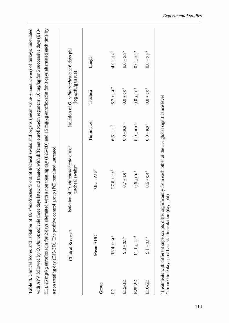
Experimental studies ___________________________________________________________________________
114
Tab
le 4
. Clin
ical
sco
res
and
isol
atio
n of
O. r
hino
trac
heal
e ou
t of
trach
eal s
wab
s an
d or
gans
(m
ean
valu
e +
stan
dard
err
or)
of tu
rkey
s in
ocul
ated
with
APV
follo
wed
by
O. r
hino
trac
heal
e th
ree
days
late
r, an
d tre
ated
with
diff
eren
t enr
oflo
xaci
n re
gim
ens:
10
mg/
kg fo
r 5 su
cces
sive
day
s (E1
0-
5D),
25 m
g/kg
enr
oflo
xaci
n fo
r 2 d
ays
alte
rnat
ed w
ith a
non
trea
ting
day
(E25
-2D
) and
15
mg/
kg e
nrof
loxa
cin
for 3
day
s al
tern
ated
eac
h tim
e by
a no
n tre
atin
g da
y (E
15-3
D).
The
posi
tive
cont
rol g
roup
(PC
) rem
aine
d un
treat
ed.
Clin
ical
Sco
res *
Isol
atio
n of
O. r
hino
trac
heal
e ou
t of
trach
eal s
wab
s*
Is
olat
ion
of O
. rhi
notr
ache
ale
at 6
day
s pbi
(lo
g 10
cfu/
g tis
sue)
M
ean
AU
C
M
ean
AU
C
Turb
inat
es
Tr
ache
a
Lung
s
G
roup
PC
13.4
+ 5
.4 a
27.6
+ 5
.3 a
6.6
+ 1.
1a 6.
7 +
0.4
a 4.
0 +
0.2
a
E15-
3D
9.8
+ 3.
1 b
0.7
+ 1.
0 b
0.0
+ 0.
0 b
0.0
+ 0.
0 b
0.0
+ 0.
0 b
E25-
2D
11.1
+ 3
.3 a
b 0.
6 +
0.6
b 0.
0 +
0.0
b 0.
0 +
0.0
b 0.
0 +
0.0
b
E10-
5D
9.1
+ 3.
1 b
0.6
+ 0.
4 b
0.0
+ 0.
0 b
0.0
+ 0.
0 b
0.0
+ 0.
0 b
a tre
atm
ents
with
diff
eren
t sup
ersc
ripts
diff
er si
gnifi
cant
ly fr
om e
ach
othe
r at t
he 5
% g
loba
l sig
nific
ance
leve
l *
from
0 to
9 d
ays p
ost b
acte
rial i
nocu
latio
n (d
ays p
bi)

Experimental studies ___________________________________________________________________________
115
During necropsy days 15 and 16 days pbi, one turkey belonging to group 10E-5D, showed
presence of O. rhinotracheale in the turbinates and two turkeys from the same groups in the
turbinates and in the trachea. These were the same birds in which O. rhinotracheale was re-
isolated from the tracheal swabs, five days after the final treatment day. O. rhinotracheale
was not isolated from any other organ in any other bird. In the trachea, the difference between
treatments was not statistically significant (P > 0.1). In the turbinates, the Chi-Square test
concluded in significant differences between treatments (P = 0.022) while the Fisher exact test
was statistically borderline (P = 0.051).
Trial 2. Reduced Duration Medication Study. The mean number of cfu/ml of O.
rhinotracheale in the organs at 6 days pbi are shown in Table 4.
During first necropsy 6 days pbi, O. rhinotracheale was also only isolated out of the
respiratory organs (turbinates, trachea, lungs and airsacs) from the positive control group.
Statistically significant differences regarding the O. rhinotracheale titres in the turbinates,
trachea and lungs were observed for all comparisons of the three enrofloxacin treatment
groups to the positive control group (for every comparison P = 0.0079). For these organs
(turbinates, trachea and lungs), no statistical differences were observed between the three
enrofloxacin treatment regimens. Also no statistical differences were observed between the
four experimental groups regarding the presence of O. rhinotracheale in the airsacs, pericard
and liver.
During final necropsy, O. rhinotracheale could not be isolated out of any organ.
Weight
During both trials, no global treatment effect on weight gain could be observed. In trial
1 the positive control group showed the lowest weight gain and there was one single
significant pairwise difference for the 81 g higher weight gain observed in E10-5D compared
to the positive control group (P = 0.0495). In trial 2 group E15-3D showed the highest weight
gain. However, again this difference was not statistically significant
Discussion
The infection model used in these experiments was based on a model developed by
Marien et al. (2005) for the reproduction of infectious respiratory disease in turkeys. Since the
results obtained in the control groups in both experiments are very similar to their findings,
the infection model appears to be highly reproducible.

Experimental studies ___________________________________________________________________________
116
Regarding the clinical symptoms in the dose titration study (trial 1), the negative control
group scored statistically significant better than the other groups. This was expected since the
negative control group was only infected with APV. It is generally acknowledged that O.
rhinotracheale exerts a synergistic effect on APV infections in turkeys by aggravating the
symptoms encountered in the respiratory tract (Van Empel et al., 1996; Jirjis et al., 2004,
Marien et al., 2005). Looking at the clinical scores and the respiratory lesions observed during
necropsy in the positive and negative control group, this experiment (trial 1) confirms these
findings.
Taking in consideration that, compared to the negative control group, the course of clinical
disease in the enrofloxacin treated groups in the dose titration study (trial 1) was only
prolonged for a day and that no significant differences could be noted for the gross lesions
encountered during necropsy, it can be suggested that the viral multiplication of APV had a
major impact on the overt respiratory symptoms in the three enrofloxacin treated groups. In
the APV/ O. rhinotracheale infected, untreated birds, however, clinical disease was more
severe. Thus, the multiple day enrofloxacin treatments were able to eliminate most of the O.
rhinotracheale associated aggravation of the APV infection.
Statistical analysis confirms these findings as each enrofloxacin treatment regimen, used in
trial 1, showed statistically significant better results for most parameters examined, compared
to the APV/O. rhinotracheale infected, untreated positive control group (i.e. clinical scoring,
O. rhinotracheale titres from tracheal swabs and O. rhinotracheale titres from organs 6 days
pbi). Comparable results were observed in the reduced duration medication study (trial 2).
Here also with every enrofloxacin treatment statistically significant better results were
obtained regarding clinical score (with the exception of the group treated with 25 mg/kg for
two days, although capable of limiting the clinical symptoms and shortening the course of
respiratory disease), the O. rhinotracheale titres in the tracheas, the O. rhinotracheale counts
in the organs and the observations of pneumonia at necropsy 6 days pbi, compared to the
positive control group. As no adverse reactions were observed the reduced duration
medication was apparently well tolerated. However the alternative medication regimens with
higher daily doses at fewer medication days could affect the drug depletion period and
subsequent withdrawal time.
Both in the dose titration study and in the reduced duration medication study no statistical
differences could be noted between the different enrofloxacin treatments. Every enrofloxacin
regimen resulted, in both trials, in an equal reduction of clinical signs and equal shortening of
the course of respiratory disease. Moreover, all regimens were able to eliminate the O.

Experimental studies ___________________________________________________________________________
117
rhinotracheale infection from the diseased turkeys since no O. rhinotracheale bacteria could
be isolated out of any trachea or organ after the first treatment day. Such clearance of O.
rhinotracheale could not be obtained after the administration of a single day dose of 50 mg/kg
which resulted in the re-emergence of the bacterium after treatment (Garmyn et al., 2009).
Re-emergence was not observed when the 50 mg/kg dose was split over a two or three day
period.
Overall, for the treatment of O. rhinotracheale infections with an enrofloxacin sensitive
strain, a five day treatment regimen of 20 mg enrofloxacin/kg was not shown to be more
efficacious than the labelled dosage (10 mg enrofloxacin/kg daily for 5 days) and a five day
treatment regimen of 5 mg enrofloxacin/kg was found equally efficient as the labelled dosage.
Whether under field circumstances, with frequent O. rhinotracheale/ E. coli mixed infections
involved in turkey respiratory disease, clinical disease could be controlled with the 5 mg/kg
regimen remains to be determined. The labelled dosage for enrofloxacin for respiratory
colibacillosis is 10 mg/kg BW. Also reduced duration medication could be a valuable
alternative to the labelled dosage against O. rhinotracheale infections in turkeys, providing
that the total administered dose (50 mg/kg) is complied with i.e. 2 times 25 mg/kg or 3 times
15 mg/kg, alternated each time by a day without treatment. This would improve user
convenience by reducing the workload for the farmer who will only have to prepare
medicated water for two or three days instead of five. In a previous study performed by
Marien et al. (2006) an enrofloxacin drinking water treatment of 3 or 5 continuous days
showed to be equally effective. The results obtained in this study suggest that every other day
treatment with enrofloxacin would seem to be of value. Hence, it is demonstrated that when
enrofloxacin is used in the drinking water, a flexible adaptation of the duration of treatment,
depending on the evolution of the respiratory disease in the stable, is possible.
Five days after the final enrofloxacin administration in the dose titration study (trial 1), low
titres of O. rhinotracheale were isolated out of two birds from the group receiving 10 mg
enrofloxacin/kg daily for 5 days. This tracheal re-colonization, several days after the final
treatment day, was not seen in the lower and higher dose group, and is expected not to be dose
dependent. It is reported that O. rhinotracheale can survive for several days in the
environment (Lopes et al., 2002). Therefore this re-excretion or re-infection, which was not
accompanied by clinical relapse, is likely just incidental.
Acquired resistance to enrofloxacin is commonly encountered in O. rhinotracheale (Devriese
et al., 1995; Malik et al., 2003) and is the consequence of stepwise mutations in the gyr and
par genes (Barnard and Maxwell, 2001; Hooper, 2001). In these studies, however, O.

Experimental studies ___________________________________________________________________________
118
rhinotracheale clones with reduced susceptibility or resistance were not detected in any of the
enrofloxacin treatment groups.
In conclusion, the enrofloxacin treatments tested in the dose titration study (5, 10 or 20 mg
enrofloxacin/kg, daily for 5 days) and the reduced duration medication study (2 x 25 mg
enrofloxacin/kg, 3 x 15 mg enrofloxacin/kg, alternated each time by a day without treatment
and 10 mg enrofloxacin/kg, daily for 5 days) were equally capable of shortening the course of
clinical disease, eliminating O. rhinotracheale from the respiratory tract and reducing gross
lesions. Since little differences could be observed for the clinical scores and the gross lesions,
when the enrofloxacin treatment groups and the negative control group were compared, the
viral multiplication of APV had a major impact on the overt respiratory disease symptoms in
all experimental groups. However, all multiple day enrofloxacin treatments were able to
eliminate most of the O. rhinotracheale associated aggravation of the APV infection, which
resulted in more severe and long-lasting respiratory symptoms in the non treated positive
control group.
As no O. rhinotracheale bacteria grew on the enrofloxacin supplemented media, none of the
used treatment regimens promoted the selection of bacterial clones with reduced susceptibility
or resistance.
Acknowledgments
This work was supported by a grant from Bayer Animal Health GmbH. The authors
would like to express their appreciation to Sofie De Bruyckere, Veerle Flama, Marleen
Foubert and Nathalie Van Rysselberghe for their skilled technical assistance. Furthermore, we
would like to thank Mr. B. Van Dam (BioChek) for the supply of the ELISA kits.
References
Agresti, A. 2002. Categorical Data Analysis 2nd ed. John Wiley & Sons, Inc., Hoboken, New
Jersey. p. 514.
Barnard, F. M., and A. Maxwell. 2001. Interaction between DNA gyrase and quinolones:
effects of alanine mutations at GyrA subunits residues Ser83 and Asp87.
Antimicrobial Agents and Chemotherapy, 45,1994-2000.

Experimental studies ___________________________________________________________________________
119
De Rosa, M., R. Droual, R. P. Chin, H. L. Shivaprasad, and R. L. Walker. 1996.
Ornithobacterium rhinotracheale infection in turkey breeders. Avian Diseases, 4 0,
865-874.
Devriese, L. A., J. Hommez, P. Vandamme, K. Kersters, and F. Haesebrouck. 1995. In vitro
antibiotic sensitivity of Ornithobacterium rhinotracheale strains from poultry and wild
bird. Veterinary Record, 137, 435-436.
El-Sukhon, S. N., A. Musa, and M. Al-Attar. 2002. Studies on the bacterial etiology of
airsacculitis of broilers in Northern and Middle Jordan with special reference to
Escherichia coli, Ornithobacterium rhinotracheale, and Bordetella avium. Avian
Diseases, 46, 605-612.
Garmyn, A., A. Martel, R. Froyman, H. Nauwynck, L. Duchateau, F. Haesebrouck, and F.
Pasmans. 2009. Efficacy of four enrofloxacin treatment regimens against experimental
infection in turkey poults with avian pneumovirus and Ornithobacterium
rhinotracheale. Avian Pathology, in press.
Hafez, H. M., and R. Sting. 1999. Investigations on different Ornithobacterium
rhinotracheale “ORT” isolates. Avian Diseases, 34,1-7.
Hooper, D. C. 2001. Emerging mechanisms of fluoroquinolone resistance. Emerging
Infectious Diseases, 7, 337-341.
Jirjis, F. F., S. L. Noll, D. A. Halvorson, K. V. Nagaraja, F. Martin, and D. P. Shaw. 2004.
Effects of bacterial coinfection on the pathogenesis of avian pneumovirus infection in
turkeys. Avian Diseases, 8, 34-49.
Krebber, R. 2003. Analytical method for the determination of pradofloxacin in serum and u
urine by turbulent flow chromatography/tandem mass spectrometry. Journal of
Veterinary Pharmacology and Therapeutics, 26(Suppl. 1), 102-103.
Lopes, V.C., B. Velayudhan, D. A. Halvorson, and K. V. Nagaraja. 2002. Survival of
dfdfffffOrnithobacterium rhinotracheale in poultry litter. Avian Diseases, 46, 1011-1014.
Malik, Y. S., K. Olsen, K. Kumar, and S. M. Goyal. 2003. In vitro antibiotic resistance
sdssdfprofiles of Ornithobacterium rhinotracheale strains from Minnesota turkeys during
sdfdg1996-2002. Avian Diseases,47, 588-593.
Marien, M., A. Decostere, A. Martel, K. Chiers, R. Froyman, and H. Nauwynck. 2005.
Synergy between avian pneumovirus and Ornithobacterium rhinotracheale in turkeys.
Avian Pathology, 34, 204-211.
Marien, M., H. Nauwynck, L. Duchateau, A. Martel, K. Chiers, L. Devriese, R. Froyman, and
Decostere A. 2006. Comparison of the efficacy of four antimicrobial treatment

Experimental studies ___________________________________________________________________________
120
schemes against experimental Ornithobacterium rhinotracheale infection in turkey
poults pre-infected with avian pneumovirus. Avian Pathology 35:230-237.
Marien, M., A. Decostere, L. Duchateau, K. Chiers, R. Froyman, and H. Nauwynck. 2007.
Efficacy of enrofloxacin, florfenicol and amoxicillin against Ornithobacterium
rhinotracheale and Escherichia coli O2:K1 dual infection in turkeys following APV
priming. Veterinary Microbiology, 121, 94-104.
Soriano, V.E., N. A. Vera, C. R. Salado, R. P. Fernández, and P. J. Blackall. 2003. In vitro
susceptibility of Ornithobacterium rhinotracheale to several antimicrobial drugs.
Avian Diseases, 47, 476-480.
Sprenger, S. J., A. Back, D. P. Shaw, K. V. Nagaraja, D. C. Roepke, and D. A. Halvorson.
1998. Ornithobacterium rhinotracheale infection in turkeys: experimental
reproduction of the disease. Avian Diseases, 42,154-161.
Van de Zande, S., H. Nauwynck, D. Cavanagh, and M. Pensaert. 1998. Infections and
reinfections with avian pneumovirus subtype A and B on Belgian turkey farms and
relations to respiratory problems. Zentralblatt Veterinarmedizin, 45, 621-626.
Van de Zande, S., H. Nauwynck, and M. Pensaert. 2001. The clinical, pathological and
microbiological outcome of an Escherichia coli O2:K1 infection in avian pneumovirus
infection in turkeys. Veterinary Microbiology, 81, 353-365.
Van Empel, P., H. van den Bosch, D. Goovaerts, and P. Storm. 1996. Experimental infection
in turkeys and chickens with Ornithobacterium rhinotracheale. Avian Diseases, 40,
858-864.
Van Empel, P., and M. Hafez. 1999. Ornithobacterium rhinotracheale: a review. Avian
Pathology, 28, 217-227.
Van Veen, L., C. P. van Empel, and T. Fabria. 2000. Ornithobacterium rhinotracheale, a
primary pathogen in broilers. Avian Diseases, 44, 896-900.
Van Veen, L., E. Hartman, and T. Fabri. 2001. In vitro antibiotic sensitivity of strains of
Ornithobacterium rhinotracheale isolated in The Netherlands between 1996 and 1999.
Veterinary Record, 149, 611-613.

Experimental studies ___________________________________________________________________________
121

122

General Discussion ___________________________________________________________________________
123
CHAPTER 4: GENERAL DISCUSSION
___________________________________________________________________________

General Discussion ___________________________________________________________________________
124
GENERAL DISCUSSION
Antimicrobial use in poultry production: a hot and controversial topic
Antimicrobial use in the poultry industry is currently under scrutiny. Emerging
antimicrobial resistance is becoming an increasing problem in most bacterial species. For O.
rhinotracheale reduced susceptibility or resistance have been reported against amoxicillin,
ampicillin, doxycycline, enrofloxacin, flumequine, gentamicin, lincomycin, trimethoprim-
sulfonamide, tetracycline and tylosin (Van Empel and Hafez, 1999; Devriese et al., 2001; van
Veen et al., 2001; Malik et al., 2003; Soriano et al., 2003). Avian pathogenic E. coli are
frequently (multi-)resistant to tetracyclines, sulfonamides, ampicillin, and streptomycin
(Barnes et al., 2008), but also for other antimicrobials commonly used in the poultry industry
reports of reduced susceptibility or resistance exist (Cormican et al., 2001; Altekruse et al.,
2002; Barnes et al., 2008). For both bacterial species susceptibility may depend on the regime
used by the poultry industry in various geographical locations (Barnes et al., 2008).
Concerns are also growing about antimicrobial resistance in bacteria from food animals, and
their impact on human health. These resistant strains may be zoonotic, but also resistant
commensals pose a hazard, since they can transfer their resistance genes to human pathogens.
Heated discussions regarding the degree of risk this hazard poses, have been and are still
being held between those who are very concerned about the risk (Witte, 1998) and those who
believe that the evidence for a substantial risk remains sparse (Phillips et al., 2004).
Many risk assessments have been conducted, but in many cases the results were impended by
shortage of suitable data sets (Snary et al., 2004). In addition, a number of countries have
incorporated surveillance programs, e.g. National Antimicrobial Resistance Monitoring
System (NARMS), European Antimicrobial Susceptibility Surveillance in Animals (EASSA)
or Danish Integrated Antimicrobial Resistance Monitoring and Research Programme
(DANMAP), but these monitoring programs do not yet provide adequate information
regarding the risk of antimicrobial resistance, which is therapeutic failure due to a specific
drug resistance in humans (Mathew, 2007). Although no definite conclusion has come from
the several meetings designed to discuss the question of antimicrobial resistance and the use
of antibiotics in poultry, the controversy has led to legislative changes affecting the
availability of antimicrobials approved for use in poultry. From September 2005, the US Food
and Drug Administration banned the use of enrofloxacin in poultry farms in the USA (not in
Europe) as it was expected to jeopardize human health through the emergence of
General Discussion ___________________________________________________________________________
125
fluoroquinolone-resistant strains of Campylobacter in enrofloxacin medicated poultry (FDA,
2005). Since January 2006, the water -or feed- based administration of antimicrobials to
animals as growth promoters has been completely banned in the European Community as
such use has been shown to contribute to the increased prevalence of antimicrobial-resistant
bacteria of human significance (e.g. enterococci). It is certainly true that the widespread
practice of mass dosing antimicrobials in the drinking water, using sub-therapeutic levels
promotes the emergence of resistance (Iovine and Blaser, 2005; Johnsen et al., 2009). It is
also true that removal of the selective pressure by abolishing the use of a certain antimicrobial
can reduce the frequency of resistance (Han et al., 2009; Johnsen et al., 2009). However,
complete reversal of acquired drug resistance in bacterial populations that were no longer
exposed to a specific antimicrobial drug has never been reported (Sepala et al., 1997; Enne et
al., 2001; Sorum et al., 2006; Price et al., 2007) and any use of antimicrobial drug may
eventually lead to resistance. Redundant use of antimicrobials should certainly be avoided and
the use of antimicrobials should certainly not be a crowning piece of disease control
programmes. Yet, a ban of the use of an antimicrobial product has implications for animal
welfare by limiting efficacious drugs to treat disease. Therapeutic applications of
antimicrobials are vital for profitable and livestock production, essential for animal welfare
and minimizing economic losses (McMullin, 2004). To retain the efficacy of antimicrobial
drugs currently available for the control of bacterial infections, prescription and
administration of antimicrobial agents should be undertaken with discretion, supported by an
accurate diagnosis and a careful choice of the respective substance(s) (Schwarz et al., 2001).
Efficacy of the labelled enrofloxacin regimen
The principal goal of therapeutic antimicrobial use in poultry is maximum eradication
of the pathogen as quickly as possible with minimal adverse effects on the recipient and
prevention of the emergence of resistant organisms (Tanner et al., 2003).
Currently, enrofloxacin is the most potent antimicrobial drug so far used for respiratory
infections in poultry medicine. From all fluoroquinolone antimicrobials which are approved
for poultry use, enrofloxacin shows the highest in vitro activities against avian respiratory
pathogens (Pirro, 1997; Norcia et al., 1999, Fraatz et al., 2002; Himmler et al., 2002; Miller
et al., 2005). Administered in the drinking water at 10 mg/kg for 5 successive days (the
currently labelled dose) enrofloxacin proved efficacious against any bacteria playing a
significant role in turkey respiratory disease (Braunius, 1987; Behr et al., 1988; Hafez et al.,
1992; Cargill, 1995; Gautrais and Copeland, 1997; Marien et al., 2006). During this research,

General Discussion ___________________________________________________________________________
126
the efficacy of this treatment against respiratory disease in turkeys caused by APV/O.
rhinotracheale or APV/E. coli infections has been endorsed. In every study, E coli or O.
rhinotracheale was rapidly eradicated from the respiratory tract. Already one day after onset
of treatment, neither E. coli nor O. rhinotracheale could be isolated out of the trachea. When
turkeys were necropsied immediately after the final treatment day, the respective pathogens
were never cultured from the internal organs. The course of respiratory disease was shortened
with several days and clinical signs and gross lesions were less severe than those observed in
non-treated, E. coli or O. rhinotracheale infected control groups. It might even be assumed
that most of the clinical signs observed originated from the APV inoculation, which was used
to trigger the bacterial infections.
Considering PK/PD parameters, the clinical and bacterial outcome of this treatment regimen
was expected. Fraatz et al. (2006) observed that after continuous drinking medication at a
dose of 10 mg/kg body weight in turkeys, steady state serum concentrations of enrofloxacin
ranged between 0.32-0.45 µg/ml. In the respiratory tract, concentrations were even 2 times
higher. These concentrations exceed MIC90 values for E. coli (0.06 µg/ml) by approximately
four log2 and for O. rhinotracheale (0.25 µg/ml) by two steps (Fraatz et al., 2006). A single
oral dose PK study with 10 mg/kg BW enrofloxacin resulted in mean Cmax values of 1.89
µg/ml and mean AUC0→24 values of 18.1 h.g/ml (Fraatz, 2006, data on file). Dimitrova et al.
(2007) observed Cmax/MIC ratios from 161.23 h to 12.90 h and AUC0→24/MIC ratios from
2153.44 h to 137.82 h for clinically significant micro-organisms whose MIC varied from
0.008 µg/ ml to 0.125 µg/ml (10 mg/kg BW). Although in the latter study, enrofloxacin was
given in a pulsed manner, continuous drinking water treatment is believed to reach similar
AUC levels (Stegeman, 1995). Based on the above, a treatment regimen of 10 mg/kg
enrofloxacin for 5 continuous days against strains of O. rhinotracheale and E. coli with MIC
of 0.03 µg/ml, should be sufficient to reach Cmax/MIC ratios >10 and AUC0→24/MIC ratios
>100-125. These ratios are associated with maximal clinical and microbiological effects in
Gram-negative bacteria (Blaser et al., 1987; Madaras-Kelly et al., 1996, Frimodt-Moller,
2002).
The dosage of enrofloxacin: time for change?
The dose titration study, administering 5 mg enrofloxacin/kg BW instead of 10 mg/kg
BW, continuously in the drinking water for five successive days proved equally effective as
the enrofloxacin treatment of 10 mg/kg BW for five continuous days against susceptible O.
rhinotracheale infections in turkeys (MIC 0.03 µg/ml). Here also, efficacy could have been

General Discussion ___________________________________________________________________________
127
predicted. A pharmacokinetic study performed in chickens by Reinhardt et al. (2005) in which
enrofloxacin concentrations in serum samples were determined after administration of
increasing doses of enrofloxacin against Mycoplasma gallisepticum, showed that serum
concentrations of enrofloxacin are directly proportional to the rate of absorption, which, in
turn, is dependent on the concentration at the absorption site. In theory, based on previous
Cmax/MIC ratios and AUC0→24/MIC ratios, even half the concentration of enrofloxacin in the
plasma and respiratory tissues are considered adequate for clinical and microbiological
effectiveness against susceptible O. rhinotracheale isolates. In the field usually mixed
infections occur and first step mutants may be involved in the infection. Still, the efficacy of
the 5 mg enrofloxacin/kg BW treatment has not been established against other bacterial
respiratory pathogens or less susceptible O. rhinotracheale isolates. Hence, it would be
advisable to use the 10 mg/kg treatment in respiratory disease outbreaks in the field. Despite
their efficacy, previous treatment regimens are considered as cumbersome and time
consuming by poultry breeders. Since enrofloxacin is a photosensitive molecule (Sumano et
al., 2004) the medicated drinking water has to be prepared each day during five days. And,
because of the resistance problem associated with this class of antimicrobials, the use of
enrofloxacin and other fluoroquinolones in the poultry industry is currently under scrutiny.
Although during this entire research period, less susceptible or resistant E. coli or O.
rhinotracheale isolates were never encountered after enrofloxacin treatments of 10 mg/kg BW
administered for 5 successive days, other investigators did (Barrow et al., 1998, Marien et al.,
2006a). Isolates with reduced susceptibility or even resistance to fluoroquinolone have also
been reported in Campylobacter sp. (McDermott et al., 2002, Luo et al., 2003, Van Boven et
al., 2003) and Salmonella sp. (Barrow et al., 1998; Randall et al., 2005; 2006), even after
single administration of the labelled regimen.
Previous dosing strategies have been designed according PK/PD parameters based on MICs.
MIC values, however, refer to drug susceptible populations and are therefore not relevant to
the prevention of growth of resistance mutants (Firsov et al., 2003; Drlica and Zhao, 2007). In
wild type cells, spontaneous mutations in DNA arise at frequencies varying from one in 106 to
one in 109. For MIC measurements, inocula only contain 104 cfu (Andrews, 2001). In such
relatively small bacterial populations, the occurrence of mutant bacterial isolates is unlikely.
However, in diseased animals bacterial populations are more likely the size of 107 and 108 cfu.
Therefore, at the site of infection, bacterial variants with reduced drug susceptibility to
enrofloxacin (first-step mutants) will be most likely present (Pourkbahsh et al., 1997; Sanders,
2001; Drlica and Malik, 2003; Wetzstein, 2005). Recently the concept of the mutant selection

General Discussion ___________________________________________________________________________
128
window hypothesis (Zhao and Drlica, 2008) has found quite some support. The upper
boundary of the window, called the mutant prevention concentration (MPC) defines a drug
threshold at which visible re-growth from a bacterial population compromising 109 to 1010 cfu
is completely inhibited. Therefore, the heterogenity in sensitivity of the bacterial population at
the infection site, which remains undetected in MIC testing, is revealed in MPC curves
(Wetzstein, 2005). When a drug falls below the MPC into the so called mutant selection
window (of which the lower limit can be approximated by MIC) selection pressure exerted by
fluoroquinolone treatment will enrich mutant subpopulations (first step mutants) (Sanders,
2001; Drlica and Malik, 2003; Blondeau et al., 2004). On the other hand, maintaining drug
concentrations above the selection window throughout therapy should severely restrict the
acquisition of drug resistance. To grow above the MPC, a bacterial isolate would require > 2
concurrent mutations. Since this happens at frequencies of one in 10-14 cells, this would be
very unlikely (Blondeau et al., 2004). MIC is said to define the sensitivity of the first target.
MPC on the other hand defines the sensitivity of the secondary fluoroquinole targets
(Wetzstein, 2005).
Resistance to fluoroquinolones arises almost exclusively by de novo mutations and is the
consequence of gradual accumulation of mutations in the target genes. A first step mutation
usually only leads to reduced susceptibility which is regarded as clinically irrelevant (Hooper,
2001; Andriole, 2003). However, when such less susceptible isolates survive in the
environment, the MIC of bacterial populations may increase stepwise. Eventually, only 1
additional mutation will be needed before resistance occurs (Schentag et al., 2003). Therefore
the selection of less susceptible isolates should be avoided by maintaining drug concentrations
above the MPC throughout therapy. To determine whether MPC can be clinically applied, it
must be measured for relevant bacterial populations. To be useful, the MPC should be within
the concentration limits that can be safely reached in patients.
The possibility has been raised that MPC might be a fixed multiple of MIC (Sanders, 2001). If
so, MPC could be determined from MIC measurements. However MPC and MICs correlate
poorly (Drlica et al., 2006). For enrofloxacin, MPC’s have only been reported for a few
pathogens (Etienne et al., 2004; Wetzstein, 2005). For wildtype E. coli populations (MIC
0.03), wildtype E. coli WT (MIC 0.06) and E. coli ATCC 25922 (MIC 0.03), the MPC for
enrofloxacin were respectively 0.3-0.35 µg/ml, 0.4-0.5 µg/ml, and 0.15-0.175 µg/ml
(Wetzstein, 2005). Since values for O. rhinotracheale were not described, the provisional
MPC of the O. rhinotracheale strain (MIC 0.03 µg/ml) was determined following Blondeau
(2001). For this, starter cultures were spread on blood agar plates and incubated 48h at 37°C

General Discussion ___________________________________________________________________________
129
in 5% CO2. Bacterial cells were then transferred from the plates to 500 ml BHI broth (Oxoid)
followed by overnight incubation at 37°C in 5% CO2. After incubation, cultures were
estimated to have concentrations of > 3 x 10⁸ cfu/ml by turbidity measurements. Cultures
were concentrated by centrifugation at 5.000 x g for 10 min and resuspended in 3 ml of PBS.
Aliquots of 200 µl, containing > 1010cfu, were applied to Muller Hinton II agar (Oxoid)
dilution plates containing 5% defibrinated horse blood. Agar dilution plates were freshly
prepared by incorporating enrofloxacin at thirteen concentrations and dilutions of
enrofloxacin (1x to 128x MIC). The fully susceptible LMG ORT strain was included as
control. Inoculated plates were incubated for 48h at 37°C in 5%CO2 and then screened for
growth. All plates were re-incubated for an additional 24h and re-examined. MPC was
recorded as the lowest antibiotic concentration that allowed no growth. The provisional MPC
of O. rhinotracheale measured 2 µg/ml. Since the provisional MPC overestimates MPC by
about twofold (Blondeau et al. 2001), the MPC value of this O. rhinotracheale strain is 1
µg/ml. Determinations were made in duplicate, and the results were identical. After
continuous drinking medication at a dose of 10 mg/kg body weight in turkeys, steady state
serum concentrations of enrofloxacin only ranged between 0.32-0.45 µg/ml (Fraatz et al.,
2006). According to the concept of the mutant selection window hypothesis the labelled
treatment should not prevent the selection of first step mutations in the O. rhinotracheale
strains used in this research since enrofloxacin concentrations are too low to exceed O.
rhinotracheales MPC. Therefore, the treatment regimen in which enrofloxacin was
administered at 20 mg/kg BW for 5 successive days, probably better reduces the selection of
less susceptible or resistant microbial subpopulations of O. rhinotracheale. The increased
(doubled) steady-state concentrations in serum and tissues should offer improved conditions
maintaining drug concentrations above the selection window throughout therapy. Application
of this dosing strategy is, however, not completely straightforward. A five day treatment at 20
mg/kg BW/daily proved only to be equally efficacious as the current labelled dose against O.
rhinotracheale infections. Improved clinical effect or better bacterial eradication was not
noticed. This suggests that drug concentrations in the body were probably higher than
necessary for clinical cure against susceptible O. rhinotracheale infections. On the other hand,
increasing the daily drug dose also means an increase of the treatment cost. In addition,
treatment would be still as labour-intensive as the “traditional” labelled dosage regimen.

General Discussion ___________________________________________________________________________
130
Rethinking the treatment duration for improved treatment efficacy
To deal with these inconveniences, it was hypothesized that reduced duration therapies
could offer a solution. For drugs like enrofloxacin with concentration dependent killing
activity higher peak drug concentration in serum and tissues ensure better bacterial
eradication and clinical effect, reducing the time needed to treat (Frimodt-Moller, 2002).
Therefore administering higher doses of fluoroquinolones might shorten the course of
treatment while efficacy is maintained and in the meanwhile the development of resistance in
bacteria is reduced (Drusano et al., 1993; Forrest et al., 1993; Meinen et al., 1995, Scaglione
et al., 2003, Randall et al., 2006). However, observations made during this research presume
that, for an optimal antimicrobial effect, dosing strategies do not only seem to confer
appropriate drug concentrations but also an appropriate time of exposure for the target
pathogen. Although it has been frequently stated that the antibacterial effects of enrofloxacin
at the clinical level are more dependent upon a higher peak in serum concentrations than an
extended residence time (Puyt, 1995; Brown, 1996, Riviere, 1999; Sumano and Gutierrez,
2000) the exposure time of enrofloxacin achieved after single day dosing for the treatment of
APV/E. coli and APV/O. rhinotracheale infections was found too short.
Providing the same total dose of 50 mg enrofloxacin /kg BW administered in 5, 10 or 20h
proved not to be equally efficacious as the labelled regimen of 10 mg/kg BW for 5 successive
days. Although respiratory disease was reduced in time and severity, the clinical and
antimicrobial effects of the single day regimens did not match those of the labelled treatment
regimen. Moreover, O. rhinotracheale and E. coli were not completely eradicated from the
turkey’s respiratory tract, since the pathogens rapidly re-emerged after treatment. Since it is
implicit for an optimal dosing strategy that the offending pathogen is eradicated (Tanner,
2003; McKellar et al, 2004) single day treatment regimens are not considered a valuable
alternative to the labelled dose. Deplorable, since Randall et al. (2006) reported that a single
dose of 50 mg enrofloxacin/kg BW in chickens experimentally infected with Salmonella
enterica might evidence for a decreased selection of less susceptible isolates.
The higher Cmax achieved with single day dosing, hypothesized to be advantageous compared
to the continuous dosing, did not result in improved efficacy against E. coli and O.
rhinotracheale infections in turkeys. Previously, studies comparing pulse enrofloxacin dosing
(i.e. administration of the daily dosis enrofloxacin in two to four hour period) and continuous
enrofloxacin dosing (administering enrofloxacin in the drinking water over a 24-h period)
came to similar observations. In these studies also, higher peak concentrations associated with
General Discussion ___________________________________________________________________________
131
pulse dosing were not linked to improved clinical efficacy (Stegeman, 1995; Charleston,
1998; Froyman and Cooper, 2003).
Drug exposure time proved appropriate when the total dose of 50 mg/kg dose was split over
two or three days i.e. 2 times 25 mg/kg or 3 times 15 mg/kg, alternated each time by a day
without treatment. Both reduced duration medications proved equally efficacious as the
labelled regimen in reducing clinical signs, shortening the course of respiratory disease ànd
eradicating the target pathogen, e.g. O. rhinotracheale. Comparable dosing regimens, splitting
the total dose of 50 mg fluoroquinolone/kg BW over 2 or 3 days, tested in humans and
chickens against Salmonella infections proved also equally efficacious as a 5 day treatment at
10 mg/kg (Hien et al., 1995; Randall et al., 2006). Although only equal and not improved
clinical and antimicrobial efficacies were observed - compared to the current labelled
treatment regimen - it is believed that these reduced duration medication regimens might have
additional advantages. Since the course of treatment is shortened, medicated drinking water
will only have to be prepared two or three times instead of five, which is less-time consuming
for the turkey breeder. In addition, a regimen of 2 x 25 mg enrofloxacin/kg BW alternated
with a non treating day could result overall in a shorter withholding time. Furthermore, the
higher peak plasma and respiratory tissue concentrations, resulting from the higher dosages
might offer better conditions for restricting selection of less susceptible isolates than the
current treatment schedule. This is evidenced by Randall et al. (2006), who observed less
isolates of Salmonella enterica with reduced susceptibility after a 2-day treatment at a dose of
25 mg/kg than after a five day treatment of 10 mg/kg. Therefore, reducing the treatment
period against O. rhinotracheale infections in turkeys might reduce the rate at which less
susceptible O. rhinotracheale isolates appear. It is also important to emphasize that the total
enrofloxacin dose, compared to current labelled doses, administered was unaltered. Hence, the
medication cost is not increased. Therefore, reduced duration medications providing 2 x 25
mg enrofloxacin/kg or 3 x 15 mg enrofloxacin/kg, alternated each time by a day without
treatment might be considered as valuable dosing regimens against O. rhinotracheale
infections in turkeys.
Considerations regarding the safety of the modified regimens, their traits to reduce the
emergence of resistance and the used challenge model
Enrofloxacin is regarded as a safe antimicrobial (Anonymous, 2003). In poultry, side-
effects have never been observed after administering the registered treatment dose. However,
when (daily) antimicrobial drug doses are increased, the risk exists that these higher drug

General Discussion ___________________________________________________________________________
132
concentrations might induce toxic effects. Toxic signs sometimes associated with very high
enrofloxacin doses in poultry are gastrointestinal disturbances and in one single case
arthropathies caused by cartilage lesions have been observed in the past (Walker, 2000).
Nevertheless, such symptoms or other adverse events were not observed in any turkey during
the studies performed for this research whenever (daily) increased enrofloxacin doses (higher
than the labelled 10 mg/kg BW) were administered. Therefore, every treatment regimen tested
may be considered safe.
During treatment every enrofloxacin regimen was checked for selection of less-
susceptible or enrofloxacin resistant O. rhinotracheale or E. coli isolates. Since neither the
current labelled dosage nor any modified regimen ever promoted the selection of such less
susceptible or resistant isolates, it is difficult to conclude if the presumed improved
pharmacokinetics and pharmacodynamics of certain modified enrofloxacin regimens (20
mg/kg for five continuous days and the alternative day- reduced duration medications) really
reduce emergence of less susceptible or resistant strains than the currently labelled dosis.
Applying these regimens in field studies enclosing larger number of birds could result in more
conclusive evidence.
When the efficacies of different treatment regimens against a specific disease are to be
compared, it is of extreme importance to use a highly reproducible challenge model. If not, it
would not be clear whether the differences in clinical and antimicrobial outcome between
different treatment groups are due to the impact of the treatment in se or whether they are the
result of external factors. This holds true, especially for reproducing a disease as complex as
respiratory disease in poultry of which the course and the severity of clinical signs can be
influenced by many different environmental factors (Kleven, 2008). Only very recently,
Marien (2008) succeeded in the development of such infection model that can reproduce
respiratory disease caused by respiratory pathogens currently considered significant in the
turkey industry (i.e. APV, E. coli, O. rhinotracheale, and Mycoplasma). Marien et al. (2006)
used this infection model to compare the efficacy of different classes of antimicrobials.
Present research shows that the model also proves extremely valuable to compare efficacies of
different treatment regimens, since in the currently performed O. rhinotracheale trials
equivalent results are observed when the clinical scores (Figure 1) and the tracheal titres
(Figure 2) of the different positive control groups are compared to each other.

General Discussion ___________________________________________________________________________
133
0.00
1.00
2.00
3.00
4.00
-4 0 4 8 12 16
days post O. rhinotracheale inoculation
clinical score
single day treatment
dose titration study
reduced duration medication study
Figure 1. Mean clinical scores in turkeys belonging to the positive control group (i.e. inoculated with
APV and three days later with O. rhinotracheale without successive enrofloxacin treatment) during
the single day treatment study (♦), the dose titration study (■) and the reduced duration medication
study (▲).
0
1
2
3
4
5
6
0 2 4 6 8 10 12 14 16
days post O. rhinotracheale inoculation
log10 cfu/ml
single day treatmentdose titration studyreduced duration medication
Figure 2. Mean tracheal titres in turkeys belonging to the positive control group (i.e. inoculated with
APV and three days later with O. rhinotracheale without successive enrofloxacin treatment) during
the single day treatment study (♦), the dose titration study (■) and the reduced duration medication
study (▲).

General Discussion ___________________________________________________________________________
134
Conclusion and recommendations for further research
Overall, the current labelled treatment regimen proves very efficacious against
respiratory disease caused by susceptible O. rhinotracheale and E. coli strains and did not
promote the selection of less susceptible or resistant isolates in any study performed during
this research. Although not clinically more efficient, certain modified regimens tested, e.g. the
alternative day-reduced duration medications could offer a valuable alternative.
However, further work is needed to ascertain how effective these modified treatment
regimens are against other bacterial respiratory pathogens not included in this research or
mixed bacterial respiratory infections. It would also be interesting to look at the effect of these
higher enrofloxacin treatment levels on the gut microbiota, since the selection of resistant
mutants during antimicrobial treatment may not only take place at the site of infection but at
any bacterial population resident in the body. Furthermore, since evidence exists that less
susceptible enrofloxacin isolates are not as clinically irrelevant as primarily thought, it would
be useful to test the efficacy of labelled dosage and modified regimens against first step
mutants of O. rhinotracheale or E. coli.

General Discussion ___________________________________________________________________________
135
References
Altekruse, S.F., Elvinger, F., Lee, K.Y., Tollefson, L.K., Pierson, E.W.; Eifert, J., and
Sriranganathan, N. (2002). Antimicrobial susceptibilities of Escherichia coli strains
from a turkey operation. Journal of the American Veterinary Medical Association,
221, 411-416.
Andrews, J.A. (2001). Determination of minimum inhibitory concentrations. Journal of
Veterinary Chemotherapy, 48, (Suppl.1), 5-16.
Andriole, V.T. (2003). Quinolones. In: Finch, R.G., Greenwood, D., Norrby, S.R., and
Withley, R.W. (ed.), Antibiotic and chemotherapy, 8th edition. Churchill Livingstone,
London, England, p. 349-373.
Anonymous (2003). USP-veterinary pharmacoceutical monographs,
fluoroquinolones/veterinary-systemic. Journal of Veterinary Pharmacology and
Therapeutics, 26, (Suppl. 2), 87-108
Barnes, H.J., Nolan L.K., and Vaillancourt J.-P. (2008). Colibacillosis. In: Saif, Y.M, Fadly,
A.M., Glisson, J.R., Mcdougald, L.R., Nolan, L.K., and Swayne, D.E. (ed.). Diseases
of Poultry, 12th edition. Iowa State Press, Iowa, USA, pp.691-737.
Barrow, P.A., Lovell, M.A., Szmolleny, G., and Murphy, C.K. (1998). Effect of enrofloxacin
administration on excretion of Salmonella enteritidis by experimentally infected
chickens and on quinolone resistance of their Escherichia coli flora. Avian Pathology.
27, 586-590.
Behr, K.P., Friedrichs, M., Hinz, K.-H., Lüders, and Siegmann, H. (1988). Klinische
Erfahrungen mit dem Chemotherapeutikum Enrofloxacin in Hühner- und
Putenherden. Tierärztl Umschau, 43, 507-515.
Blaser, J., Stone, B.B., Groner, M.C., and Zinner, S.H. (1987). Comparative study with
enoxacin and netilmicin in a pharmacodynamic model to determine importance of
ratio of antibiotic peak concentration to MIC for bactericidal activity and emergence
of resistance. Antimicrobial Agents and Chemotherapy
Blondeau, J.M., Hansen, G., Metzler, K., and Hedlin, P. (2004). The role of PK/PD
parameters to avoid selection and increase of resistance: mutant prevention
concentration. Journal of Chemotherapy, 16, suppl.3, 1-19.
, 31, 1054-1060.
Blondeau, J.M., Zhao, X., Hansen, G., and Drlica, K. (2001). Mutant prevention concentration
of fluoroquinolones for clinical isoaltes of Streptococcus pneumoniae. Antimicrobial
agents and Chemotherapy, 45, 433-438.
General Discussion ___________________________________________________________________________
136
Braunius, W.W. (1987). Effect van Baytrill (Bay Vp 2674) op jonge kalkoenen lijdende aan
luchtweginfecties. Tijdschrift Diergeneeskunde 112, 531-533.
Brown, S.A. (1996). Fluoroquinolones in animal health. Journal of Veterinary Pharmacology
and Therapeutics, 19, 1-14.
Cargill, P.W. (1995). Amoxicillin and enrofloxacin efficacy comparison in turkey poults. 1st
Bayer European Poultry Symposium, Leverkusen, Germany, 47-51.
Charleston, B., Gate, J.J., Aitken, I.A., Stephan, B., and Froyman, R. (1998). Comparison of
the efficacies of three antimicrobial agents, given as a continuous or pulsed-water
medication, against Escherichia coli infections in chickens. Antimicrobial Agents and
Chemotherapy, 42, 83-87.
Cormican, M., Buckley, V., Corbett-Feeney, G., and Sheridan, F. (2001). Antimicrobial
resistance in Escherichia coli isolates from turkey hens in Ireland. Journal of
Antimicrobial Chemotherapy, 48, 587-588.
DANMAP 2001–07, Use of antimicrobial agents and occurrence of antimicrobial resistance
in bacteria from food animals, foods, and humans in Denmark
http://www.danmap.org/ (accessed April 22, 2009).
Devriese, L.A., De Herdt, P., and Heasebrouck, F. (2001). Antibiotic sensitivity and
resistance in Ornithobacterium rhinotracheale strains from Belgian broiler chickens.
Avian Pathology, 30, 197-200.
Dimitrova, D.J., Lashev, L.D., Yanev, S.G., and Pandova, B. (2007). Pharmacokinetics of
enrofloxacin in turkeys. Research in Veterinary Science, 82, 392-397.
Drlica, K., and Malik, M. (2003). Fluoroquinolones: action and resistance. Current Topics in
Medicinal Chemistry, 3, 249-282.
Drlica, K., and Zhao, X. (2007). Mutant selection window hypothesis updated. Clinical
Infectious Diseases, 44, 681-688.
Drlica, K., Zhao, X., Blondeau, J.M., and Hesje, C. (2006). Low correlation between MIC
and mutant prevention concentration. Antimicrobial Agents and Chemotherapy, 50,
403-404.
Drusano, G.L., Johnson, D.E., Rosen, M., and Standiford, H.C. (1993). Pharmacodynamics of
a fluoroquinolone antimicrobial agent in a neutropenic rat model of Pseudomonas
sepsis. Antimicrobial Agents and Chemotherapy, 37, 483-490.
Enne, V.L., Livermore, D.M., Stephens, P., and Hall, L.M. (2001). Persistence of
sulphonamide resistance in Escherichia coli in the UK despite national prescribing
restriction. Lancet, 357, 1325-1328.

General Discussion ___________________________________________________________________________
137
FDA Food and Drug Administration. Final decision of the commissioner: withdrawal of
approval of the new animal drug application for enrofloxacin in poultry, Rockville,
MD, 2005
Firsov, A.A., Vostrov, S.N., Lubenko, I.Y. et al., (2004). ABT492 and levofloxacin:
comparison if their pharmacodynamics and their abilities to prevent the selection of
resistant Staphylococcus aureus in an in vitro dynamic model. Journal of
antimicrobial Chemotherapy, 58, 1185-1192.
Forrest, A., Nix, D.E., Ballow, C.H., Goss, T.F., Birmingham, M.C., and Schentag, J.J.
(1993). Pharmacodynamics of intravenous ciprofloxacin in seriously ill patients.
Antimicrobial Agents and Chemotherapy, 37, 1073-1081.
Fraatz, K., Heinen, E., and Stein, C. (2002). Efficacy of old and new generation quinolones in
a rapid avian colibacillosis screening model, using Baytril as a benchmark. 3rd Bayer
European Poultry Symposium, Leipzig, Germany, 27-31.
Fraatz, K., Froyman, R., and Krebber, R. (2006). Pharmacokinetics of enrofloxacin and
florfenicol in turkeys during continuous drinking water medication. Journal of
Veterinary Pharmacology and Therapeutics, 29 (suppl. 1), 239-301.
Frimodt-Moller, N. (2002). How predictive is PK/PD for antibacterial agents? International
Journal of Antimicrobial Agents, 19, 333-339.
Froyman, R., and Cooper, J. (2003). Assessment of the efficacy of fluoroquinolones and the
other antimicrobials against respiratory colibacillosis and septicaemia in chickens
under standardized challenge conditions. In: Proceedings of the XIII Congress of the
WVPA (p. 84), Denver, USA.
Gautrais, B., and Copeland, D. (1996). Use of enrofloxacin in turkeys; a worldwide e
experience. 46th Western Poultry Disease Conference, Sacramento, California, USA,
79.
Hafez, H.M., Emele, J., and Kruse, W. (1992). Die Pasteurella multocida-Infektion bei den
Puten: Literaturübersicht und praktische Erfahrungen in Putenbestanden. Archiv für
Geflügelkunde, 56, 45-52.
Han, F., Lestari, S.I., Pu, S., and Ge, B. (2009). Prevalence and antimicrobial resistance
among Campylobacter spp. in Louisiana retail chickens after the enrofloxacin ban.
Foodborne Pathogens and Disease, 6,163-71.
Hien, T.T., Bethell, D., Hoa, N.T. et al. (1995). Short courses of ofloxacin for treatment of
multidrug-resistant typhoid. Clinical Infectious Diseases, 20, 917-923.
General Discussion ___________________________________________________________________________
138
Himmler, T., Hallenbach, W., Marhold, A., Pirro, F., Wetzstein, H.-G., and Bartel, S. (2002).
Synthesis and in vitro activity of pradofloxacin, a novel 8-cyanofluoroquinolone. 42nd
Annual Interscience Conference of Antimicrobial Agents and Chemotherapy, poster F-
566.
Hooper, D.C. (2001). Emerging mechanisms of fluoroquinolone resistance. Emerging
Infectious Diseases, 7, 337-341.
Johnsen, P.J., Townsend, J.P., Bohn, T., Simonson, G.S., Sundsfjord, A., and Nielsen, K.M.,
(2009). Factors affecting the reversal of antimicrobial-drug. The Lancet Infectious
Disease, 9, 357-367.
Kleven, S.H. (2008). Multicausal Respiratory Diseases. In: Saif, Y.M, Fadly, A.M., Glisson,
J.R., Mcdougald, L.R., Nolan, L.K. and Swayne, D.E. (ed.). Diseases of Poultry, 12th
edition. Iowa State Press, Iowa, USA, pp.765-774.
Luo, N., Sahin, O., Lin, J., Michel, L.O., and Zhang, Q.(2003). In vivo selection of
Campylobacter isolates with high levels of fluoroquinolone resistance associated with
gyrA mutations and the function of the CmeABC efflux pump. Antimicrobial Agents
and Chemotherapy,
Madaras-Kelly, K.J., Ostergaard, B.E., Hovde, L.B., and Rotschafer, J.C. (1996) Twenty-
four-hour area under the concentration-time curve/MIC ratio as a generic predictor of
fluoroquinolone antimicrobial effect by using three strains of Pseudomonas
aeruginosa and an in vitro pharmacodynamic model.
47, 390-394
Antimicrobial Agents and
Chemotherapy,
Marien, M. (2007). Mixed respiratory infections in turkeys, with emphasis on avian
metapneumovirus, Ornithobacterium rhinotracheale, Escherichia coli and
Mycoplasma gallisepticum. PhD thesis, University of Ghent, Belgium.
40, 627-632.
Marien, M., Decostere, A., Nauwynck, H., Froyman, R., Devriese, L., and Haesebrouck, F.
(2006a). In vivo selection of reduced enrofloxacin susceptibility in Ornithobacterium
rhinotracheale and its resistance-related mutations in gyrA. Microbial Drug
Resistance, 12, 140-144.
Marien, M., Nauwynck, H., Duchateau, L., Martel, A., Chiers, K., Devriese, L., Froyman, R.,
and Decostere, A. (2006b). Comparison of the efficacy of four antimicrobial treatment
schemes against experimental Ornithobacterium rhinotracheale infection in turkey
poults pre-infected with avian pneumovirus. Avian Pathology, 35, 230-237.

General Discussion ___________________________________________________________________________
139
Malik, Y.S., Olsen, K., Kumar, K., and Goyal, S.M. (2003). In vitro antibiotic resistance
profiles of Ornithobacterium rhinotracheale strains from Minnesota turkeys during
1996-2002. Avian Diseases, 47, 588-593.
Mathew, A.G., Cissell, R., and Liamthong, S. (2007). Antibiotic resistance in bacteria
associated with food animals: a unites states perspective of livestock production.
Foodborne Pathogens and Disease, 4, 115-133.
McDermott, P.F., Bodeis, S.M., English, L.L., White, D.G., Walker, R.D., Zhao, S., Simjee,
S., and Wagner, D.D. (2002). Ciprofloxacin resistance in Campylobacter jejuni e
evolves rapidly in chickens treated with fluoroquinolones. The Journal of Infectious
Diseases, 185, 837-840.
McMullin, P.F. (2004). Poultry production and diminishing availability of pharmaceuticals:
what does the practicing poultry veterinarian need? In: Book of abstracts, XXII
World’s Poultry Congress, p.93.
Meinen, J.B., McClure, J.T., and Rosin, E. (1995). Pharmacokinetics of enrofloxacin in
clinically normal daos and mice and drug pharmacodynamics in neutropenic mice with
Escherichia coli and staphylococcal infections. American Journal of Veterinary
Research, 56, 1219-1224.
Miller, R.A., Walker, R.D., Carson, J., Coles, M., Coyne, R., Dalsgaard, I., Gieseker, C., Hsu,
H.M., Mathers, J.J., Papapetropoulou, M., Petty, B., Teitzel, C., and Reimschuessel, R.
(2005). Standardization of a broth microdilution susceptibility method to determine m
minimum inhibitory concentrations of aquatic bacteria. Diseases of Aquatic
Organisms, 64, 211-222.
Norcia, L.J., Silvia, A.M., and Hayashi, S.F. (1999). Studies on time-kill kinetics of different
classes of antibiotics against veterinary pathogenic bacteria including Pasteurella,
Actinobacillus, and Escherichia coli. Journal of Antibiotics (Tokyo), 52, 52-60.
Phillips, I., Casewell, M., Cox, T., De Groot, B., Friis, C., Jones, R., Nightingale, C., Preston
R., and Waddell, J. (2004). Does the use of antibiotics in food animals pose a risk to
human health? A critical review of published data. Journal of Antimicrobial
Chemotherapy, 53, 28-52.
Pirro, F., Edingloh, M., and Schmeer, N. (1999). Bactericidal and inhibitory activity of e
enrofloxacin and other fluoroquinolones in small animal pathogens. Suppl
Compendium of Continuing Education in Pract Vet, 21 (12M): 19-25.

General Discussion ___________________________________________________________________________
140
Price, L.B, Lakey, L.G., Vailes, R., and Silbergeld, E. (2007). The persistence of
fluoroquinolone-resistant Campylobacter in poultry production. Environmental Health
Perspectives, 115, 1035-1039.
Puyt, J.D. (1995). Antibiotic therapy in poultry production. Bulletin des Groupements
Techniques Vétérinaires, 5, 17-110.
Randall, L.P., Cooles, S.W., Coldham, N.C., Stapleton, K.S., Piddock, L.J., and Woodward,
M.J. (2006). Modification of enrofloxacin treatment regimens for poultry
experimentally infected with Salmonella enterica Serovar Typhimurium DT104 to
minimize selection of resistance. Antimicrobial Agents and Chemotherapy, 50, 4030-
4037.
Randall, L.P., Cooles, S.W., Piddock L.J. and Woodward, M.J. (2004). Mutant prevention
concentration of ciprofloxacin and enrofloxacin for Salmonella enterica. Journal of
Antimicrobial Chemotherapy, 54, 688-691.
Randall, L.P., Eaves, D.J., Cooles, S.W., Ricci, V., Buckley, A., Woodward, M.J. and
Piddock, L.J. (2005). Fluoroquinolone treatment of experimental Salmonella enterica
serovar Typhimurium DT104 infections in chickens selects for both gyrA mutations
and changes in efflux pump gene expression. Journal of Antimicrobial Chemotherapy,
56, 297-306.
Reinhardt, A.K., Gautier-Bouchardon, A.V., Gicquel-Bruneau, M., Kobisch, M., and Kempf,
I. (2005). Persistence of Mycoplasma gallisepticum in chickens after treatment with
enrofloxacin without development of resistance. Veterinary Microbiology, 106, 129-
137.
Riviere, J.E. (1999). Comparative Pharmacokinetics. Principes, Techniques, and
Applications. Ames, Iowa State University Press.
Sanders, C. (2001). Mechanisms responsible for cross-resistance and dichotomous resistance
among the quinolones. Clinical Infectious Diseases, 32, S1-8.
Scaglione, F., Mouton, J.W., Mattina, R., and Fraschini, F. (2003). Pharmacodynamics of l
levofloxacin and ciprofloxacin in a murine pneumonia model: peak concentration/MIC
versus area under the curve/MIC ratios. Antimicrobial Agents and Chemotherapy, 47,
2749-2755.
Schentag, J.J., Meagher, A.K., and Forrest, A. (2003). Fluoroquinolone AUIC break points
and the link to bacterial killing rates. Part 1: In vitro and animal models. Infectious
Diseases, 37, 1287-1298.

General Discussion ___________________________________________________________________________
141
Snary, E.L., Kelly, L.A., Davison, H.C., Teale, C.J., and Wooldridge, M. (2004).
Antimicrobial resistance: a microbial risk assessment perspective. Journal of
Antimicrobial Chemotherapy, 53, 906-917.
Soriano, V.E., Vera, N.A., Salado, C.R., Fernandez, R.P., and Blackall, P.J. (2003). In vitro
susceptibility of Ornithobacterium rhinotracheale to several antimicrobial drugs.
Avian Diseases, 47, 476-480.
Sorum, M., Johnson, P.J., Aasnes, B. et al. (2006). Prevalence, persistence, and molecular
characterization of glycopeptide-resistant enterococci in Norwegian poultry and
poultry farmers 3 to 8 years after the ban on avoparcin. Applied and Environmental
Microbiology, 72, 516-521.
Stegeman, M. (1995). Comparative pharmacokinetic studies of pulse and continuous dosing.
In: Proceedings of the International Poultry Symposium (Bayer) (p. 37-41).
Leverkusen, Germany.
Sumano, L.H., and Gutierrez, O.L. (2000). Bases farmacologicas del uso de la enrofloxacina
en la aviculture en Mexico. Veterinaria Mexico, 31, 56-61.
Sumano, L.H., Gutierrez, O.L., Aguilera, R., Rosiles, M.R., Bernard, B.M., and Garcia, M.J.
(2004). Influence of hard water on the bioavailability of enrofloxacin in broilers.
Poultry Science, 83, 726-731.
Tanner, A.C. (2000). Antimicrobial drug use in poultry. In: Prescott, J.F., Baggot J.D., and
Walker R.D. (ed.). Antimicrobial Therapy in Veterinary Medicine, 3rd edition. Ames,
Iowa. pp. 637-655.
Van Boven, M.., Veldman, K.T., de Jong, M.C., Mevius, D.J. (2003). Rapid selection of
quinolone resistance in Campylobacter jejuni but not in Escherichia coli in
individually housed broilers. Journal of Antimicrobial Chemotherapy, 52, 719-23.
Van Empel, P.C.M, and Hafez, H.M. (1999). Ornithobacterium rhinotracheale: a review.
Avian Pathology, 28, 217-227.
Van Veen, L., Hartman, E., and Fabri, T. (2001). In vitro antibiotic sensitivity of strains of
Ornithobacterium rhinotracheale isolated in the Netherlands between 1996 and 1999.
The Veterinary Record, 149, 611-613.
Walker, R.D. (2000). Fluoroquinolones. In: Prescott, J.F., Baggot, J.D., and Walker, R.D.
(ed.) Antimicrobial Therapy in Veterinary Medicine, 3rd edition. Ames, Iowa, pp. 315-
338.
General Discussion ___________________________________________________________________________
142
Wetzstein, H.-G. (2005). Potential significance of the mutant prevention concentration of
Baytril in the treatment of avian colibacillosis. In: Proceedings of the 4th International
Poultry Symposium (Bayer) (p. 48-56). Istanbul, Turkey.
Witte, W., 1998. Medical consequences of antibiotic use in agriculture. Science, 279, 996-
997.
Zhao, K., and Drlica, K. (2008). A unified anti-mutant dosing strategy. Journal of
Antimicrobial Chemotherapy, 62, 434-436.

143

144

Summary ___________________________________________________________________________
145
SUMMARY ___________________________________________________________________________

Summary ___________________________________________________________________________
146
SUMMARY
Respiratory disease on turkey farms world-wide leads to economic losses due to
growth retardation, increased feed conversion rates, increased mortality rates, higher
condemnation rates at slaughter and high medication costs. Bacterial pathogens which play a
significant role in the respiratory disease complex are O. rhinotracheale and E. coli.
To treat respiratory infections in turkeys, drinking water treatment with enrofloxacin is
widely used. The current treatment regimen advises a five day treatment at 10 mg/kg body
weight. Although this labelled dosage proves very efficacious, the regimen is regarded as
cumbersome for many farmers, since the medicated drinking water has to be prepared freshly
every day for five days. Besides, in vivo emergence of diminished susceptibility or even
resistance to fluoroquinolones is commonly encountered in many bacterial species from
poultry. Several dosage strategies have been developed for the fluoroquinolone antimicrobials
which may increase their efficacy and may reduce the selection pressure for resistance.
Nevertheless, controlled clinical studies which address their efficacy against O.
rhinotracheale and Escherichia coli infection have not been reported.
The general introduction starts with a survey on the characteristics of enrofloxacin.
The relationship between its structure and activity, the physicochemical properties of the
molecule, the mechanism of enrofloxacin action, its pharmacokinetic and pharmacodynamic
parameters, related toxic effects and drug interactions, aspects of fluoroquinolone resistance
and dosing strategies are the themes that have been reviewed.
Next a review of the literature is given on O. rhinotracheale infections in turkeys. Properties
described are the general characteristics of O. rhinotracheale, the pathogenesis,
epidemiology, clinical signs and lesions, diagnosis, preventive measures and treatment. The
last part of the general introduction deals with APEC infections in turkeys. The briefy
reviewed are the aspects of respiratory colibacillosis in turkeys, the virulence associated
factors and diagnosis, treatment and prevention of APEC infections.
The scientific aim of this research was to optimize enrofloxacin dosage regimens
against respiratory disease caused by avian metapneumovirus (APV)/O. rhinotracheale and
APV/E. coli infections in turkeys by modifying the duration of the treatments and/or the daily
administered enrofloxacin concentrations.

Summary ___________________________________________________________________________
147
In a first experimental study, three single day enrofloxacin treatment regimens
providing 50 mg/kg body weight during a 5, 10 or 20 hour period were compared to the
advised multiple day treatment regimen with 10 mg/kg body weight for 5 successive days in
turkeys experimentally inoculated with APV and O. rhinotracheale. Although initially all
dosage regimens cleared O. rhinotracheale from the trachea, four days after onset of
treatment, O. rhinotracheale bacteria were re-excreted in the single day regimens but without
clinical relapse. The five day treatment with 10 mg enrofloxacin/kg in turkeys provided the
best results for the treatment of an O. rhinotracheale infection in growing turkeys by
shortening the course and reducing the severity of clinical disease and by eliminating O.
rhinotracheale from the respiratory tract without re-emergence. None of the treatment
regimens promoted the selection of bacterial clones with reduced susceptibility or resistance.
In a second study, it was assessed whether a single day treatment schedule with 50
mg/kg body weight was clinically equivalent to the advised multiple day treatment schedule
with 10 mg/kg body weight for five days against turkeys experimentally inoculated with APV
and three days later with E. coli. Both the multiple and the single day enrofloxacin treatment
regimens reduced E. coli multiplication in the respiratory tract tissues (turbinates, trachea and
lung) but the five day treatment with 10 mg enrofloxacin/kg in turkeys provided the best
results by shortening the course of clinical disease, by eliminating E. coli from the respiratory
tract without re-emergence, and by reducing macroscopic lesions. None of the treatment
regimens promoted the selection of bacterial clones with reduced susceptibility or resistance.
A third experimental study was a dose titration study. Growing turkeys were first
infected with APV and three days later with O. rhinotracheale. Twenty four h after O.
rhinotracheale inoculation, enrofloxacin doses of 5, 10 or 20 mg/kg body weight were
administered for 5 successive days. All enrofloxacin treatments were equally efficacious, i.e.
equally capable of shortening the course of clinical disease, eliminating O. rhinotracheale
from the respiratory tract and reducing gross lesions. None of the used treatment regimens
promoted the selection of bacterial clones with reduced susceptibility or resistance to
enrofloxacin.
Finally, a reduced duration medication study was performed. To evaluate the current
enrofloxacin treatment schedule in growing turkeys experimentally infected with APV and O.
rhinotracheale, the following enrofloxacin regimens providing the same total administered
dose (50 mg / kg BW) were compared: 25 mg / kg BW / day on day 0 and day 2 or15 mg / kg
BW / day on days 0, 2 and 4 and 10 mg / kg BW for 5 successive days. Again, all
enrofloxacin treatments proved equally efficacious, i.e. equally capable of shortening the

Summary ___________________________________________________________________________
148
course of clinical disease, eliminating O. rhinotracheale from the respiratory tract and
reducing gross lesions. None of the used treatment regimens promoted the selection of
bacterial clones with reduced susceptibility or resistance to enrofloxacin.
Based on the results in the experimental studies performed during this research, the
current labelled treatment regimen proved very efficacious against respiratory disease caused
by susceptible O. rhinotracheale and E. coli strains. The efficacy of the single day treatments
against O. rhinotracheale and E. coli infections did not equal that of the five day treatment,
possibly by not eliminating the respiratory pathogens from the organs, which made it possible
for the remaining bacteria to re-emerge. The regimens tested during the dose titration and
reduced duration studies proved equally effective against susceptible O. rhinotracheale
infections. Therefore, depending on the evolution of the respiratory disease in the stable, a
flexible adaptation of the duration of enrofloxacin drinking water treatment is possible. The
reduced duration applications providing 2 times 25 mg/kg or 3 times 15 mg/kg enrofloxacin,
alternated each time by a day without treatment, could offer less time-consuming and equally
effective alternatives to the currently labelled treatment against O. rhinotracheale infections
in turkeys. Neither the current labelled dosage nor any modified regimen ever promoted the
selection of less susceptible or resistant isolates.

Samenvatting ___________________________________________________________________________
149
SAMENVATTING
___________________________________________________________________________

Samenvatting ___________________________________________________________________________
150
SAMENVATTING In de kalkoenindustrie zijn luchtweginfecties wereldwijd een vaak voorkomend
probleem met zware economische verliezen als gevolg. De verliezen zijn voornamelijk te
wijten aan verhoogde voederconversie, vermindere groei, verhoogde sterftepercentages, een
hoger aantal karkasafkeuringen bij verwerking en hogere medicatie kosten. Twee bacteriële
ziektekiemen die vaak geassocieerd worden met ademhalingsproblemen bij kalkoenen zijn
Ornithobacterium rhinotracheale en Escherichia coli.
Enrofloxacine is een antimicrobieel product, behorend tot de klasse van de fluoroquinolones,
dat vaak wordt voorgeschreven ter behandeling van zulke ademhalingsinfecties in kalkoenen.
Het huidig behandelingsschema adviseert een dosis van 10 mg enrofloxacine/kg
lichaamsgewicht, toe te dienen via het drinkwater gedurende 5 opeenvolgende dagen (label
dosering). Ondanks zijn bewezen doeltreffendheid, vinden veel kalkoenhouders dit
behandelingsschema omslachtig omdat het gemedicineerd drinkwater elke dag -vijf dagen
lang- opnieuw moet worden aangemaakt. Ook worden er in vivo uit pluimvee regelmatig
bacteriën geïsoleerd die een verminderde gevoeligheid of zelfs resistentie tegenover
fluoroquinolones vertonen. Recent zijn er echter verscheidene dosisstrategieën ontwikkeld
die de werkzaamheid van deze klasse antimicrobiële middelen zouden kunnen verhogen en
daarnaast de selectie van resistente kiemen zouden verminderen. De efficaciteit van deze
strategieën zijn echter nog nooit uitgetest op O. rhinotracheale en E. coli infecties.
In de algemene introductie van dit werk wordt eerst enrofloxacine onder de loep
genomen. De structuur, de fysicochemische eigenschappen, het werkingsmechanisme van de
molecule, de farmacodynamische en farmacokinetische eigenschappen, de toxische effecten,
de interacties met andere preparaten, resistentievorming en de ontwikkelde dosisstrategieën
van het antimicrobieel product worden besproken.
Vervolgens wordt er een literatuuroverzicht gegeven over O. rhinotracheale infecties in
kalkoenen. De algemene kenmerken, de pathogenese, de epidemiologie, klinische symptomen
en klinische letsels, diagnose, de behandeling van O. rhinotracheale infecties en preventieve
maatregelen komen aan bod. Tenslotte volgt er een stukje over Aviaire Pathogenische E. coli
(APEC) infecties in kalkoenen, met daarin de volgende onderwerpen: respiratoire
colibacillose, de geassocieerde virulentie factoren en de diagnose, de behandeling en de
preventie van APEC infecties.

Samenvatting ___________________________________________________________________________
151
Het doel van dit onderzoek was om het huidige behandelingsschema van enrofloxacin
dat gebruikt wordt ter behandeling van APV/O. rhinotracheale and APV/E.coli infecties te
optimaliseren door de behandelingsduur en/of de dagelijks toegediende dosis van
enrofloxacine aan te passen.
In een eerste studie werd de efficaciteit van eendaagse enrofloxacine behandelingen
vergeleken met die van het huidig geregistreerde meerdaagse behandelingsschema. Na
inoculatie met APV en O. rhinotracheale werden drie groepen kalkoenen gedurende 5, 10 of
20 u behandeld met een dosis van 50 mg enrofloxacine/kg lichaamsgewicht. Een vierde groep
kreeg gedurende 5 opeenvolgende dagen 10 mg/kg enrofloxacine toegediend. Alle
behandelingen slaagden erin O. rhinotracheale uit de trachea te verwijderen, maar enkele
dagen na start van de medicatie werd na de eendags-therapieën O. rhinotracheale opnieuw in
de trachea aangetroffen. Van de vier behandelingen, slaagde de huidig geregistreerde
behandeling er het beste in het ziekteverloop in te korten en het ziektebeeld te verzachten
zonder re-excretie van O. rhinotracheale. Tijdens de studie werden geen O. rhinotracheale
isolaten geïsoleerd met verminderde gevoeligheid of resistentieontwikkeling tegenover
enrofloxacine.
In een tweede studie werd onderzocht of één dag behandelen aan een dosis van 50 mg
enrofloxacine/kg lichaamsgewicht even werkzaam is als de huidig geregistreerde behandeling
van 10 mg enrofloxacine/kg gedurende 5 opeenvolgende dagen voor de behandeling van
APV/E. coli infecties in kalkoenen. Beide behandelingen reduceerden de E. coli
vermenigvuldiging in de luchtwegen, maar de geregistreerde behandeling slaagde er het best
in E. coli uit de kalkoenen te elimineren, de ziekteduur te verkorten, en de symptomen en
letsels te beperken. Tijdens de studie werden geen O. rhinotracheale isolaten geïsoleerd met
verminderde gevoeligheid of resistentieontwikkeling tegenover enrofloxacine.
Vervolgens werd een dosis-titratie studie uitgevoerd. Na inoculatie met APV en O.
rhinotracheale werden kalkoenen 5 dagen na elkaar behandeld met 5, 10 of 20 mg
enrofloxacine/kg lichaamsgewicht/dag. Alle behandelingen bleken even effectief in het
elimineren van O. rhinotracheale uit de luchtwegen, het verkorten van de ziekteduur, en het
verminderen van klinische symptomen en letsels. Tijdens de studie werden geen O.
rhinotracheale isolaten geïsoleerd met verminderde gevoeligheid of resistentieontwikkeling
tegenover enrofloxacine.
Tenslotte werd een studie uitgevoerd waarin het huidige geregistreerde
behandelingsschema van enrofloxacine (10 mg/kg gedurende 5 opeenvolgende dagen) werd
Samenvatting ___________________________________________________________________________
152
vergeleken met therapieën waarbij op alternerende dagen werd behandeld met behoud van de
totale dosis: 25 mg/kg/dag op dag 0 en dag 2 of 15 mg/kg/dag op dag 0, 2 en 4. Ook in deze
studie bleken alle behandelingen even effectief in het elimineren van O. rhinotracheale uit de
luchtwegen, het verkorten van de ziekteduur, en het verminderen van klinische symptomen en
letsels. Tijdens de studie werden geen O. rhinotracheale isolaten geïsoleerd met verminderde
gevoeligheid of resistentieontwikkeling tegenover enrofloxacine.
Uit de resultaten van de uitgevoerde experimenten kunnen we besluiten dat het huidige
geregistreerde behandelingschema van enrofloxacine (10 mg/kg gedurende 5 opeenvolgende
dagen) zeer effectief is tegen ademhalingsinfecties veroorzaakt door APV/O. rhinotracheale
en APV/E. coli. Behandelingsschema’s waarin de totale enrofloxacine dosis (50 mg) in
éénzelfde dag wordt toegediend bleken geen goed alternatief, aangezien zowel O.
rhinotracheale als E. coli niet volledig werden geëlimieerd en na enkele dagen opnieuw
konden vermenigvuldigen in de luchtwegen van de kalkoenen. De behandelingsschema’s die
werden getest tijdens de dosis titratiestudies en de medicaties waarbij op alternerende dagen
werd behandeld bleken even effectief als het huidig geregistreerde regime. Dit wijst erop dat,
afhankelijk van het verloop van de ziekte in de stal, drinkwater therapie met enrofloxacine
flexibel kan worden aangepast. Bovendien leveren de therapieën waarbij 2 keer 25 mg/kg of 3
keer 15 mg/kg enrofloxacine wordt toegediend een waardevol alternatief voor het
geregistreerde regime, aangezien maar 2 of 3 dagen moet behandeld worden in plaats van 5.

Curriculum vitae ___________________________________________________________________________
153
CURRICULUM VITAE
___________________________________________________________________________
Curriculum vitae ___________________________________________________________________________
154
CURRICULUM VITAE
An Garmyn werd op 29 juni 1980 geboren te Antwerpen. Na het beëindigen van haar
studies secundair onderwijs, richting Latijn-Wetenschappen, aan het sint-Michiels college te
Brasschaat, startte ze in 1998 met de studies Diergeneeskunde aan de Universiteit van Gent.
In 2006 studeerde ze met onderscheiding af als dierenarts.
Vanaf september 2006 trad zij in dienst als wetenschappelijk medewerker bij de
vakgroep Pathologie, Bacteriologie en Pluimveeziekten. Gedurende drie jaar werkte zij aan
een onderzoeksproject “Enrofloxacine medicaties ter behandeling van respiratoire infecties bij
kalkoenen”, dat gesteund werd door Bayer HealthCare, Animal Health.
An Garmyn is auteur van meerdere publicaties in internationale tijdschriften.

Publications ___________________________________________________________________________
155
PUBLICATIONS
___________________________________________________________________________

Publications ___________________________________________________________________________
156
PUBLICATIONS
Garmyn, A., Martel, A., Froyman, R., Nauwynck, H., Duchateau, L., Haesebrouck, F. and
Pasmans, F. (2009) Efficacy of four enrofloxacin treatment regimens against experimental
infection in turkey poults with avian pneumovirus and Ornithobacterium rhinotracheale.
Avian Pathology, 38, 287-292.
Garmyn, A., Martel, A., Froyman, R., Nauwynck, H., Duchateau, L., Haesebrouck, F. and
Pasmans, F. (2009) Effect of multiple and single day enrofloxacin medications against dual
experimental infection with avian pneumovirus and Escherichia coli in turkeys. Poultry
science, 88, 2093-2100.
Garmyn, A., Martel, A., Froyman, R., Ludwig, C., Nauwynck, H., Haesebrouck, F. and
Pasmans, F. (2009). The effect of reduced treatment time and dosage of enrofloxacin on the
course of respiratory disease caused by avian pneumovirus and Ornithobacterium
rhinotracheale. Poultry Science, 88, 2315-2323.

Dankwoord ___________________________________________________________________________
157
DANKWOORD
___________________________________________________________________________

Dankwoord ___________________________________________________________________________
158
DANKWOORD
Tenslotte tijd voor de laatste paragrafen van dit proefschrift. Een woordje van dank aan
iedereen die een bijdrage heeft geleverd aan dit werk.
Tijdens mijn eerste stapjes in het onderzoek werd ik begeleid door Professor
Decostere. Ik wil haar bedanken voor haar enthousiaste ontvangst en haar hulpvaardigheid
tijdens mijn eerste maanden op het labo. Aangezien zij echter andere horizonten ging
verkennen, werd haar taak als promotor overgenomen door Professor Martel en Professor
Pasmans. An en Frank, bedankt voor jullie steun, raad en daad tijdens deze drie jaar. Jullie
gaven mij de zelfstandigheid en het vertrouwen om zelf mijn zaken te regelen, maar wanneer
ik met vragen zat of problemen had, stond jullie deur steeds voor mij open. Hopelijk kan onze
samenwerking nog een tijdje langer doorlopen. Ook fijn dat jullie de drempel tussen jullie en
je doctoraatsstudentjes laag houden. Ik denk dat er maar weinig met hun “bazen” een pint
kunnen gaan pakken in Gent of op survival weekend gaan in de Ardennen. Professor
Haesebrouck bedankt om mij de kans te geven te mogen starten aan de dienst bacteriologie.
Ook voor het steeds kritische nalezen van al mijn geschreven werk.
Dit project werd mogelijk gemaakt met de financiële steun van Bayer Healthcare
GmbH. Daarom ook aan hen mijn dankbetuigingen.
Robrecht Froyman was de eerst twee jaar van mijn onderzoek de vertegenwoordiger
van de firma. Alvorens onze eerste ontmoeting had Maja mij gewaarschuwd dat je een streng
voorkomen had, maar dat schijn bedriegt…en dat is dan ook gebleken. In het begin hadden
we het een beetje lastig met de administratieve toepassingen van de GCP guidelines, maar
uiteindelijk hebben we QM toch tevreden kunnen stellen. Dank je voor je input, je
correctheid, je hulp en kritisch nalezen van mijn artikelen. In mijn laatste jaar werd jouw taak
overgenomen door Dr. Carolin Ludwig. Carolin, bedankt voor de leuke samenwerking, je
blijk van appreciatie voor mijn werk en de leuke persoonlijke toetsen in je e-mails. Moest je
nog eens in Gent terecht komen, ben ik altijd bereid om eens persoonlijk gids te spelen in
plaats van tips op papier te geven.
Ook mag ik zeker Luc Jansegers niet vergeten. Aangezien mijn experimenten
verliepen volgens GCP condities moest er een monitor worden aangesteld. Toen ik dit hoorde,
dacht ik: “Oh nee, ene die constant op mijn vingers komt zien!” Maar achteraf bleek dat
allemaal goed mee te vallen. Je bent een joviale man. Ik heb deze drie jaar graag met je
Dankwoord ___________________________________________________________________________
159
samengewerkt en ook jij bedankt voor je kritische input tijdens het schrijven van de vele
protocols en study reports.
Speciaal wil ik mijn projectvoorgangster bedanken, Dr. Maja Marien. Hoewel je al bij
Alpharma aan de slag was, heb je toch nog tijd weten uit te trekken om mij wegwijs te maken
in het labo en de praktische zaken van dit onderzoek en later kon ik steeds met mijn
praktische vragen bij jou terecht. Ook je achtergelaten documentatiemappen zijn voor mij van
onschatbare waarde geweest.
Voor publicatie werd dit proefschrift nagelezen door mijn begeleidingscommissie,
bestaande uit Dr. R. Froyman en Prof. Dr. H. Nauwynck en de leden van de
examencommissie, zijnde Prof. Dr. F. Gasthuys, Prof. Dr. L. De Zutter, Prof. Dr. P. De
Backer, Prof. Dr. L. Duchateau en Dr. M. Marien. Dank, voor jullie tijd en kritische
opmerkingen.
In vivo proeven zijn hard labeur en aangezien ik als enige aangesteld was op dit
project, had ik vaak hulp nodig van helpende handen. Hulp die ik vanuit verschillende hoeken
met veel bereidwilligheid heb gekregen. Ik denk dat de meesten wel eens een handje hebben
toegestoken -iedereen bedankt daarvoor!- maar ik wil toch mijn speciale appreciatie uiten aan
Ellen Dewaele, Marleen Foubert en Veerle Flama voor het vele helpen bij het swabben en
uitvoeren van de autopsies en aan mijn dispensers Nathalie Van Rysselberghe en Sofie De
Bruyckere.
Een universitaire werkplek is altijd een beetje een komen en gaan van mensen. Tijdens
mijn onderzoeksperiode heb ik dan ook met verschillende mensen “den bureau” gedeeld. Bij
mijn start zat ik bij Liesbeth en Hilde. Dr. Liesbeth vertrok naar Janssens en Hilde ging op
zwangerschapsverlof. Lieven nam even de leeggekomen plek in tot Hildes terugkeer. Dr.
Hilde vertrok naar Alcon en David verscheen in de plaats samen met Melanie, toen nog
laatstejaarsstudente. En dan hadden we ook nog de buitenlandse gasten: Bill de Griek en
Vietnamese Dung. Met al mijn bureaugenootjes heb ik goed kunnen opschieten en ik denk zij
ook wel met mij. Bedankt voor de leuke tijd samen!
De dienst bacteriologie is een leuke werkplek, waar tussen het serieuze werken door
ten gepaste tijde plezier kan worden gemaakt. Daarom ook aan al de andere collega’s van de
bacterio en pluimvee van nu en weleer: Tom, Lies, Lieve, Urszula, Sandrine, Jo, Gunter,

Dankwoord ___________________________________________________________________________
160
Katleen, Marc, Kim, Anouk, Leen, Miet, Venessa, Sofie, Renzo, Filip, Koen, Annemieke,
Pascale, Els, Dries, Kathleen, Jana, Elin, Bram, Alexander, Hanne, Anja, Serge, Arlette,
Rosalie, Roseline, Ruth en Sofie. Bedankt voor jullie collegialiteit en vriendschap. (BTW,
moest ik iemand in het lijstje vergeten zijn…mijn oprechte verontschuldigingen hiervoor! Het
was niet express, maar we zijn dan ook met zoveel!)
Ook nog een bedankinkje aan enkele mensen van de dienst virologie. Aan Carine, om
mij in te werken in de trachearingetjes en virustitraties. En ook aan Fernand en Geert om erin
te slagen de rotor, ondanks zijn vergevorderde leeftijd, te laten blijven draaien en ook te
helpen bij de installatie van de isolatoren tijdens mijn eerste dierproef.
Ook mijn appreciatie aan Magda. Bedankt voor al het werk dat je verzet in de stallen.
Meteen neem ik deze gelegenheid ter harte om eens mijn vrienden en familie te bedanken, om
te zijn wie ze zijn.
Aan “the girls”: meisjes, als wij uitgaan is het nooit saai! Dank jullie voor de vele
leute, zowel met als zonder drama, die we al beleefd hebben en hopelijk nog vaak zullen
beleven. Met Bregje kreeg ik er ineens ook een boel andere vriendjes bij. Allez, het is te
zeggen vriendinnetjes en Jef. Windeke clan, ook jullie bedankt voor de vele leuke momenten
die we al beleefd hebben.
Papa, jij en mama hebben ons altijd de vrijheid gegeven om zelf onze weg te kiezen in
het leven en in de keuzes die we maakten werden we altijd gesteund. We werden niet
betutteld, maar we hebben nooit iets tekort gehad en als er iets was stonden jullie en sta je nog
altijd klaar voor ons. Dank je hiervoor. De voorbije jaren zijn niet altijd even gemakkelijk
voor je geweest, maar tegenwoordig is er weer een lichtpunt in je leven. Je straalt en dat gun
ik je van harte. Mappi, bedankt om papa weer te laten stralen.
Mama, ik mis je nog steeds elke dag. Er zijn zo van die speciale momenten in iemands
leven die je graag wilt delen met de mensen die je graag ziet. Op zulke momenten, zoals
vandaag, mis ik je des te meer. Bedankt voor alles wat je ooit voor me gedaan hebt en ooit
zien we elkaar weer.
Broertjes en zusje, het spreekwoord zegt: “Uw vrienden kun je kiezen, uw familie
niet”. Moest ik kunnen kiezen, ik zou jullie voor geen geld van de wereld willen inruilen.
Weet dat jullie altijd op mij kunnen rekenen! Schoonzusjes en schoonbroertje, kindjes, jullie
zie ik ook allemaal graag.

Dankwoord ___________________________________________________________________________
161
Chris en Theo, vanaf onze eerste ontmoeting ben ik door jullie steeds warm onthaald
geweest. Dank voor jullie gastvrijheid, liefde en steun. Ook Lemke, Simon en kleine Arend,
bedankt voor de vriendschap.
Bregje, mijn liefste schatje. De reden dat ik hier vandaag op deze plek sta is
grotendeels te danken aan jou. Moest jij er niet geweest zijn, stond ik nu waarschijnlijk ergens
pinten te tappen aan één of andere toog, ofzo…. Ik hou van jou, oprechter elke dag, versteld
van het zalige gevoel dat je me geeft telkens weer als we bij elkaar zijn. Bedankt voor je
steun, je liefde en om er steeds weer te zijn voor mij…elke dag opnieuw. Je bent mijn alles.


