
469S E C T I O N 1 5 • N O N - H O D G K I N ’ S LY M P H O M A ( N H L )
15. Non-Hodgkin’s Lymphoma(NHL)
IntroductionIn Sweden, 1 342 cases of non-Hodgkin’s lymphoma’s (NHL), excluding
chronic lymphatic leukemia were diagnosed in 2000, corresponding to
3 per cent of all new malignant tumour diagnoses [9]. NHL is generally
a disease of older adults and increases in incidence with advancing age
and somewhat more than 50 per cent of the patients are above 70 years
at diagnosis (from Cancer incidence in Sweden 1995–99).
NHL comprises a very heterogeneous group of tumours and the classi-
fication systems are in a continuous evolution. In the previous report
(SBU-report 129/2, 1996) the Kiel classification was used. In 1994 the
REAL classification (Revised European-American Classification of
Lymphoid Neoplasms) was presented [27]. This classification considers
not only morphological and biological observations but also immunological
and genetic findings as well as clinical observations to identify specific
disease. Recently a modification of the REAL classification, the WHO
(World Health Organization) classification, has been introduced [28].
In the studies below with long follow-up periods older classification
systems were used. However, in this report a clinical grouping in indolent
and aggressive lymphomas1) according to Hiddemann et al has been
used [29].
Treatment strategies depend upon the subtype of the disease, localiza-
tion and stage. The Cotswold2) staging classification is widely used for
staging of NHL.
In 1993 the International Non-Hodgkin’s Lymphoma Prognostic
Factors Project analyzed data on more than 2 000 patients from 16
institutions and presented the so-called International Prognostic Index
(IPI)3).
1) 2) 3) The footnotes are explained on the following pages.

R A D I OT H E R A P Y F O R C A N C E R I N S W E D E N470
1) Proposed Clinical Schema for Malignancies of the Lymphoid System:
B-Cell Lineage T-Cell Lineage
I. Indolent lymphomas (low risk) I. Indolent lymphomas (low risk)
Chronic lympocytic leukaemia/small Large granular lymphocytic leukaemia, lymphocytic lymphoma* T and NK cell types***Lympoplasmacytic lymphoma/immuno- Mycosis fungoides/Sézary syndromecytoma**/Waldenstrom’s macroglobulinemia Smoldering and chronic adult T-cell Hairy cell leukaemia leukaemia/lymphoma (HTLV1**)***Splenic marginal zone lymphomaMarginal zone B-cell lymphoma– Extranodal (MALT-B-cell lymphoma)– Nodal (monocytoid)
Follicle center lymphoma/follicular, (small cell)-grade IFollicle center lymphoma/follicular, (mixed small and large cell)-grade II
II. Aggressive lymphomas (intermediate risk) II. Aggressive lymphomas (intermediate risk)
Prolymphocytic leukaemia*** Prolymphocytic leukaemia***Plasmacytoma/multiple myeloma Peripheral T-cell lymphoma, Mantle cell lymphoma unspecified***Follicle center lymphoma/follicular, Angioimmunoblastic lymphoma***(large cell)-grade III Angiocentric lymphoma***Diffuse large B-cell lymphoma (includes Intestinal T-cell lymphoma***immunoblastic & diffuse large & Anaplastic large cell lymphoma centroblastic lymphoma) (T- and null cell type)Primary mediastinal (thymic)large B-celllymphomaHigh-grade B-cell lymphoma, Burkitt-like***
III. Very aggressive lymphomas (high risk) III. Very aggressive lymphomas (high risk)
Precursor B-lymphoblastic lymphoma/ Precursor B-lymphoblastic leukaemia lymphoma/leukaemiaBurkitt’s lympoma/B-cell acute leukaemia Adult T-cell lymphoma/leukaemiaPlasma cell leukaemia
IV. Hodgkin’s disease
* Includes B-CLL with plasmacellular differentiation (equivalent to lymhoplasma-cytoid lymphoma of the KIEL system).
** Note that the term was changed in the REAL clissification from lymphoplasmacytoidto lymphoplasmacytic lymphoma to avoid confusion with the Kiel term lympho-plasmacytoid which represents B-CLL with plasmacellular differentiation
*** Provisional clinical grouping

471S E C T I O N 1 5 • N O N - H O D G K I N ’ S LY M P H O M A ( N H L )
2) The Cotswold Staging Classification:
Stage I= Involvement of single lymph node region or lymphoid structure
(eg. spleen, thymus, Waldeyer’s ring).
Stage II= Involvement of two or more lymph node regions on the same side of the
diaphragm (the mediastinum is a single site, hilar lymph nodes are lateralized).
The number of anatomical sites should be indicated by a suffix (eg, II3).
Stage III= Involvement of lymph node regions or structures on both sides of the
diaphragm
III1: with or without splenic hilar, coeliac, or portal nodes
III2: with paraaortic, iliac, mesenteric nodes.
Stage IV= Involvement of extranodal site(s) beyond that designated “E”
A= No symptoms.
B= Fever, drenching sweats, weight loss.
X= Bulky disease (>1/3 widening of mediastinum, >10 cm maximum dimension
of nodal mass).
E= Involvement of extra lymphatic tissue, contiguous or proximal to know nodal site.
A single extralymphatic site as the only site of disease is classified IE.
3) The International Prognostic Index (IPI)
Patients of all ages Patients ≤60 years of age
Age (≤60 years vs >60 years) LDH (≤normal vs >normal)
LDH (≤normal vs >normal) Performance status (0–1 vs 2–4)
Performance status (0–1 vs 2–4) Stage (I/II vs III/IV)
Stage (I/II vs III/IV)
Extranodal involvement (≤1 site vs >1 site)
The number of IPI factors effectively predict prognosis and is now used
for almost all subtypes of NHL [64], but it is sparsely used in the
publications reviewed in this report.
Summary of the earlier report, SBU 129/2The synthesis of the literature on radiotherapy for non-Hodgkin’s lym-
phoma was based on 158 scientific articles, including 16 randomized studies,
18 prospective studies, and 90 retrospective studies. These studies involved
14 137 patients. The report covered the literature until 1993 and it also
included seven articles of later date.

R A D I OT H E R A P Y F O R C A N C E R I N S W E D E N472
Conclusions• Non-Hodgkin’s lymphomas are highly radiosensitive, and local
recurrence following radiotherapy is unusual.
• Radiotherapy probably cures approximately 50 per cent of both low-
grade and high-grade malignant NHL at stage I. Involved field treat-
ment is apparently sufficient, however, higher doses are required for
high-grade malignant lymphomas.
• Chemotherapy is recommended for stage II. Consolidation radio-
therapy after chemotherapy may increase the number of complete
remissions. The value of adjuvant radiotherapy has not been confirmed.
• Radiotherapy plays a limited role at stages III and IV.
• Radiotherapy is clearly indicated for extranodal localized disease in
the skin and in the orbit.
• It is important to identify groups and subgroups in which radio-
therapy alone is sufficient, i.e., the risk for distant recurrence is small.
MALT lymphoma belongs to this group.
• Radiotherapy is often valuable in palliative situations.
DiscussionIn stage I, limited radiotherapy leads to local control in over 90 per cent
of the patients with both indolent and aggressive lymphomas, with a long-
term survival of about 50 per cent. In indolent lymphomas no studies
showed improved results with the addition of chemotherapy, while some
studies suggested an extended relapse-free survival with the addition of
chemotherapy in aggressive lymphomas. Combination chemotherapy was
dominant for treatment of aggressive lymphomas of more advanced stages.
Consolidation radiotherapy might increase the number of complete
remissions but no effect on survival was proven. Consequently irradiation
had little value in advanced stages. Radiotherapy had a well-documented
place in certain extranodal lymphomas e.g., indolent orbital and localized
thyroid lymphomas as well as for cutaneous lymphomas in special situa-
tions. Radiotherapy could also be an important tool in palliative situations.
473S E C T I O N 1 5 • N O N - H O D G K I N ’ S LY M P H O M A ( N H L )
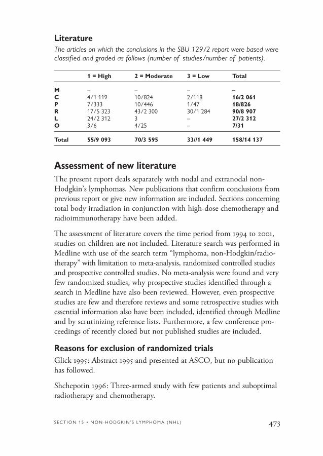
LiteratureThe articles on which the conclusions in the SBU 129/2 report were based wereclassified and graded as follows (number of studies/number of patients).
1 = High 2 = Moderate 3 = Low Total
M – – – –C 4/1 119 10/824 2/118 16/2 061P 7/333 10/446 1/47 18/826R 17/5 323 43/2 300 30/1 284 90/8 907L 24/2 312 3 – 27/2 312O 3/6 4/25 – 7/31
Total 55/9 093 70/3 595 33//1 449 158/14 137
Assessment of new literatureThe present report deals separately with nodal and extranodal non-
Hodgkin’s lymphomas. New publications that confirm conclusions from
previous report or give new information are included. Sections concerning
total body irradiation in conjunction with high-dose chemotherapy and
radioimmunotherapy have been added.
The assessment of literature covers the time period from 1994 to 2001,
studies on children are not included. Literature search was performed in
Medline with use of the search term “lymphoma, non-Hodgkin/radio-
therapy” with limitation to meta-analysis, randomized controlled studies
and prospective controlled studies. No meta-analysis were found and very
few randomized studies, why prospective studies identified through a
search in Medline have also been reviewed. However, even prospective
studies are few and therefore reviews and some retrospective studies with
essential information also have been included, identified through Medline
and by scrutinizing reference lists. Furthermore, a few conference pro-
ceedings of recently closed but not published studies are included.
Reasons for exclusion of randomized trialsGlick 1995: Abstract 1995 and presented at ASCO, but no publication
has followed.
Shchepotin 1996: Three-armed study with few patients and suboptimal
radiotherapy and chemotherapy.

R A D I OT H E R A P Y F O R C A N C E R I N S W E D E N474
Overview of new studies
Nodal non-Hodgkin’s lymphomas
Indolent non-Hodgkin’s lymphomas
The indolent lymphomas (previously usually designated low-grade
malignant) are quite rare before the age of 30, primarily affecting older
patients in the sixth through eighth decades. These lymphomas often
follow an indolent course with a long survival. Transformation to aggres-
sive lymphoma frequently occurs and spontaneous regression, of longer
or shorter duration may also occur. Most commonly they involve the
lymph nodes and are usually of follicular subtype, but they also have
a predilection for certain extranodal sites, particularly those associated
with mucosa (see below).
Localized disease: stage I–II
Few patients with indolent lymphoma present with localized disease
(10–20 per cent). Indolent lymphomas are very radiosensitive and
radiotherapy has been the mainstay for patients with localized indolent
lymphomas.
Radiotherapy alone
Long-term follow-up data i.e. ≥15 years after radiotherapy are now
emerging in retrospective series. One study reports 33 per cent progression-
free survival for patients in stage I at 15 years [55]. Another study reports
40 per cent relapse free survival at 15 years for patients in stage I and II
with very few relapses 10 years after radiotherapy [43]. British National
Lymphoma Investigation, BNLI, has performed a retrospective analysis
on patients in stage I/IE and report a complete remission rate of 98 per
cent after 35 Gy and a relapse rate at 10 years of 51 per cent with most
of the recurrences occurring within five years [69]. From Florida a 49 per
cent freedom from relapse at 20 years is reported for patients in stage I and
II, the majority of relapses occurred within five years but some recurrences
were seen between five and ten years [32].
Interestingly a report on 26 patients in complete remission after the
surgical biopsy was not treated at all but only followed. After a median
follow-up of 4.6 years 50 per cent of the patients had not relapsed and
475S E C T I O N 1 5 • N O N - H O D G K I N ’ S LY M P H O M A ( N H L )
7/13 recurrences were distant relapses [62]. However, patients with this
presentation are very rare and no controlled randomized trial comparing
radiotherapy with wait and see policy in this situation has been performed.
The optimal radiation dose for indolent lymphomas has not been deter-
mined in any prospective randomized study but since 1997 BNLI performs
a randomized trial comparing 24 Gy with 40–45 Gy in stage I. A German
prospective multicenter study recommends 36–44 Gy to involved lymph
nodes [63]. In a retrospective analysis from Florida no local recurrences
occurred after 30 Gy, suggesting that most indolent lymphomas are
adequately treated with 30 Gy [32]. How the doses were specified was
not given, so different modes of specifications may partly explain the
different recommendations.
Involved field, IF, treatment is for the most part advocated and used
but the definition of IF may vary somewhat among different centers.
Because most recurrences occur outside the irradiated area a German
prospective multicenter study has used extended field (EF) mostly meaning
mantle and paraaortic fields or even more comprehensive radiotherapy
with total central lymphatic irradiation (TCL) meaning irradiation of
Waldeyer’s ring, mantle field and the whole abdomen for patients in
stage I and II. The median follow-up was 68 months and the relapse
rate at seven years was 26 per cent in stage I and 44 per cent in stage II
[63]. Results of TCL to about 100 patients in limited stage III are
published and one of these reports suggests a possible increase of
second malignancies [16].
Two randomized trials were reported in previous report (SBU-report
129/2, 1996) which compared involved field with total nodal irradiation
(TNI) and no difference in therapeutic results were found since then,
no further controlled trials have been published.
Radiotherapy alone versus radiotherapy plus chemotherapy
Earlier randomized trials have failed to demonstrate that the addition of
chemotherapy after radiotherapy is superior to radiation alone (SBU-report
129/2, 1996), which was confirmed in a randomized British trial starting
in the mid-1970s, with a minimum of 11 year follow-up, showing that the
addition of chemotherapy (chlorambucil) after radiotherapy gave no

R A D I OT H E R A P Y F O R C A N C E R I N S W E D E N476
benefit in outcome [33]. A later analysis of the same data but on some-
what fewer patients report 15 years relapse-free survival of 55 per cent for
patients in stage I and 29 per cent for patients in stage II and the local
recurrence rate after radiotherapy was 2 per cent [17].
The literature shows that:
• One third to one half of patients with indolent lymphoma in stage I
seem to be cured by radiotherapy (follow-up more than 15 years).
• The optimal dose is still not defined.
• There is no proof that extended radiotherapy is superior to
involved field.
• There is no evidence that the addition of chemotherapy to radio-
therapy will improve the overall outcome.
Indolent non-Hodgkin’s lymphomas localized disease: stage I–II.
1 = High 2 = Moderate 3 = Low Total
C – – 2/148 2/148P – 1/100 1/26 2/126R 1/208 2/249 2/61 5/518
Total 1/208 3/349 5/235 9/792
Advanced disease: stages III–IV
The majority of patients with indolent lymphomas present with advanced
disease mainly due to bone marrow involvement. The optimal treatment
strategy is still controversial. Radiotherapy plays a very limited role in
advanced stages as concluded in the previous report (SBU-report 129/2,
1996). Since then reports of significance concerning radiotherapy in
advanced stages have not been published. However, in a retrospective
study, low dose radiation (4 Gy in 2 fractions) proved efficient with
long-lasting effects (more than 50 per cent freedom from local progres-
sion at 2 years) [23].

477S E C T I O N 1 5 • N O N - H O D G K I N ’ S LY M P H O M A ( N H L )
Aggressive non-Hodgkin’s lymphomas
Aggressive NHL (previously designated high-grade malignant) comprise
a diverse group of diseases with varying presentations, natural histories
and responses to therapy. They can occur at any age but they are in
general diseases of middle-aged and older adults.
Localized disease: stage I–II
About one third of aggressive NHL present as localized disease and
many of them with extranodal location. Until the 1980s the majority of
patients with early-stage aggressive lymphoma were treated with radio-
therapy alone. The literature review in the previous report (SBU-report
129/2, 1996) showed that somewhat more than 50 per cent of patients
in stage I are cured. The relapse-free 5-year survival of similarly treated
clinical stage II patients ranged between 0–35 per cent and chemotherapy
was unanimously recommended. Combination therapy (chemotherapy
followed by radiotherapy) was then not tested in randomized trials, but
the results from retrospective comparisons of combination therapy were
not different from chemotherapy alone, and the value of additional
radiotherapy in localized disease remained to be demonstrated.
Radiotherapy alone
BNLI has performed a retrospective analysis on patients in stage I/IE
and reported a complete remission rate of 84 per cent after 40 Gy and a
relapse rate at 10 years of 32 per cent with no relapse occurring after five
years [69]. In a Swedish retrospective study on patients in stage I/IE the
complete remission rate was 92 per cent after a median radiation dose
of 44 Gy. Twentynine percent of the patients relapsed after a median
follow-up of five years, 4 per cent within the radiation field. The 5-year
survival was comparable for nodal and extranodal lymphomas [54].
The optimal radiation dose for aggressive lymphomas has not been
determined in any prospective randomized study but since 1997 BNLI
has been performing a randomized trial comparing 30 Gy with 40–45
Gy for patients in stage I treated with radiotherapy alone.
A prospective study from Texas with combined modality treatment
found significantly better local control with doses ≥40 Gy than with
lower doses [22].

R A D I OT H E R A P Y F O R C A N C E R I N S W E D E N478
Radiotherapy versus chemotherapy plus radiotherapy
No randomized trial has been published since previous report.
In a Swedish non-randomized study from three institutions the relapse
rate was higher in the radiotherapy group compared with the combined
modality therapy group for patients in stage I/IE (29 per cent vs 15 per
cent, p=0.05) [54]. The median radiation dose was 44 Gy. In another
non-randomized study from four institutions in the Netherlands and
Belgium significantly better progression-free survival at 10 years for
patients in stage I/IE was shown for combined modality therapy compared
with radiotherapy alone (83 per cent vs 47 per cent). The median radia-
tion dose was 40 Gy. In the radiotherapy group approximately 75 per cent
had their first recurrence either at nodal sites on the other side of the
diaphragm or at extranodal sites [68]. A Japanese retrospective survey
from 25 institutions on almost 800 patients in stage I/IE-II/IIE showed
significantly better 5-years event-free survival for patients treated with
chemotherapy (mainly CHOP: cyclophosphamide, doxorubicin, vinc-
ristine, prednisone) followed by radiotherapy to involved field (median
dose 42 Gy) compared with radiotherapy alone (63 per cent vs 47 per cent).
According to the international prognostic index (IPI) 5-years event-free
survival was 76 per cent for patients with 0–1 risk factor, 61 per cent for
patients with two risk factors, and 26 per cent with three or more risk
factors. In this report only about 25 per cent of the patients had only
nodal manifestations but event-free survival was identical for nodal and
extranodal involvement [53].
The optimal radiation dose after chemotherapy has not been determined.
A retrospective study from Florida recommended 30 Gy for patients with
nonbulky tumour (≤6 cm) if complete remission was achieved by chemo-
therapy, otherwise 40–45 Gy was recommended after chemotherapy [32].
Chemotherapy plus radiotherapy versus chemotherapy alone
A randomized trial performed by SWOG (Southwest Oncology Group)
has shown that combined modality therapy with three cycles of CHOP
followed by involved field radiotherapy (40–55 Gy) is significantly superior
to eight cycles CHOP with respect to both progression-free (77 per cent
vs 64 per cent) and overall survival (82 per cent vs 72 per cent) at five

479S E C T I O N 1 5 • N O N - H O D G K I N ’ S LY M P H O M A ( N H L )
years for patients in stage I/IE including bulky disease and nonbulky
stage II/IIE. Furthermore, life-threatening toxicity (mostly grade 4 neu-
tropenia or decreased left ventricular ejection fraction) was less common
(although not significant, p=0.06) in the combined treatment group
than in patients who received eight cycles of CHOP [47]. This trial
began in 1988, and although the international prognostic index (IPI)
was not published until 1993, the authors have retrospectively analysed
the clinical characteristics of the 401 patients according to stage-modified
IPI. Most of the patients (72 per cent) were considered to be in the low
risk group. Patients with 0–1 risk factors had an estimated 5-year pro-
gression free survival of 77 per cent, for patients with two risk factors
60 per cent, and only 34 per cent for patients with three risk factors.
With longer follow-up, median eight years, the actuarial curves for
failure free survival and overall survival of the two treatment groups are
overlapping, due to increased late relapses and deaths in the combined
therapy group [47a].
Chemotherapy plus radiotherapy
Some uncontrolled studies have been published on the efficacy of com-
bined modality therapy with anthracykline based-chemotherapy. A pro-
spective study from Texas with combined modality treatment reported
after a minimum follow-up of five years a relapse-free survival of 81 per
cent in stage I and 59 per cent in stage II [22]. In a French prospective
study with alternating chemotherapy and radiotherapy in stages I–II the
5-year disease-free survival was 77 per cent for patients in stage I and
67 per cent in stage II [50]. In an Italian cooperative study with a 6-week
chemotherapy regimen followed by locoregional radiotherapy (36 Gy)
the 4-year relapse-free survival for stage I and II were 80 per cent and
78 per cent respectively [20].
The literature shows that:
• About half of patients (47–65 per cent) with aggressive non-
Hodgkin’s lymphoma in stage I are cured by radiotherapy alone.
• Randomized and non-randomized studies favour combined modality
treatment with chemotherapy followed by radiotherapy instead of
radiotherapy or chemotherapy alone in localized disease. With com-

R A D I OT H E R A P Y F O R C A N C E R I N S W E D E N480
bined modality treatment the 5-year progression-free survival ranges
between 74–83 per cent in stage I and 59–78 per cent in stage II.
• The international prognostic index (IPI) is a better prognostic
instrument than the traditional staging.
• The optimal radiation dose either for radiation alone or after
chemotherapy has not been established.
Aggressive non-Hodgkin’s lymphomas localized disease: stage I–II.
1 = High 2 = Moderate 3 = Low Total
C 1/401 – – 1/401P – 3/326 – 3/326R 3/1 243 2/509 – 5/1 752
Total 4/1 644 5/835 – 9/2 479
Advanced disease: stages III–IV
In advanced stages of aggressive non-Hodgkin’s lymphoma combination
chemotherapy is the therapy of choice and radiotherapy plays only a
minor if any role in the management of these patients.
Bulky disease has in many studies proved to be an adverse prognostic
factor and consolidating radiotherapy is often recommended to reduce
the incidence of local recurrence and improve survival. But despite radio-
therapy bulky disease seems to remain an unfavourable factor. In a pro-
spective study with combined modality treatment in stages I–II, bulky
disease (≥10 cm) proved to be the only unfavourable prognostic factor
[50]. The adverse prognostic value of tumour bulk (>6 cm) was also
confirmed in a large retrospective survey from Japan where 70 per cent
of the patients had received combined modality therapy [53]. Similar
results were seen in a retrospective series from Florida [32].
Only one randomized trial has been published where patients with
aggressive lymphoma in stage IV with initially bulky disease (≥10 cm)
in complete remission after chemotherapy were randomized to involved
field radiotherapy (40–50 Gy) or observation. The combined modality

481S E C T I O N 1 5 • N O N - H O D G K I N ’ S LY M P H O M A ( N H L )
therapy was significant superior to chemotherapy alone with respect both
to disease free and overall survival [6]. But few patients were included and
the statistical methods used were poor so the result must be interpreted
with caution. However, these results were recently confirmed by a retro-
spective Italian study. Overall survival was significantly superior after
consolidation radiotherapy in patients with advanced aggressive NHL
and initially bulky disease in complete remission after chemotherapy
compared to no radiotherapy [19].
Total body irradiation (TBI)
Total body irradiation means that the entire body is irradiated with
0.1 to 0.15 Gy/fractions two to five times per week, achieving a final
dose of 1.5–3.0 Gy.
In a French pilot study 26 previously untreated patients with indolent
lymphoma in stages I–II were treated by radiotherapy with TBI to 1.5 Gy
followed by involved field to 40 Gy. Twentyfour patients achieved com-
plete remission after TBI, and after a median follow-up of 53 months,
19 patients remained alive and disease-free [58]. These results initiated
an EORTC (European Organization for Research and Treatment of
Cancer) trial comparing radiotherapy to involved fields with TBI plus
involved fields in indolent lymphomas in stages I–II. How these low
doses could control the disease is difficult to understand but there are
experimental data suggesting that the efficacy might be explained by
immune enhancement, induction of apoptosis and hypersensitivity to
low radiation doses [60].
A major concern is that TBI may increase the risk of secondary leukemia.
In a retrospective cohort study on 61 patients with mostly indolent NHL
initially treated with TBI with a median follow-up of about nine years,
five cases of ANLL/MDS (acute non-lymphocytic leukemia, myelodysplastic
syndrome) were observed in patients who subsequently had received either
alkylating agents alone or combined modality treatment. The cumulative
15-year risk of leukemia was 17 per cent and the relative risk 117. A pre-
vious case control study by the same authors on leukemia following
NHL treated by similar chemotherapy regimens but without TBI has
shown a twofold to 13-fold relative risk for the various regimens [67].

R A D I OT H E R A P Y F O R C A N C E R I N S W E D E N482
As most patients with advanced indolent NHL will eventually relapse
and be treated with chemotherapy the approach with initial TBI seems
very hazardous. Recently a small retrospective study with TBI or total
abdominal-pelvic irradiation (TAI) in heavily pretreated patients with
advanced indolent lymphomas was presented. Two daily fractions of
0.75–0.8 Gy were given to a total dose of 20 Gy for TAI and 15 Gy for
TBI, which consisted of two successive half-body irradiations with four
weeks between each of them. Seventyfive per cent of the TBI patients
achieved complete remission and median survival was 43 months. The
TAI patients who were not so heavily pretreated achieved complete
remission in 77 per cent but had a median survival of 78 months [44].
Fractionated TBI (fTBI) in conjunction with high-dose chemotherapy (HDCT) and stem cell rescue
Fractionated TBI to 12 Gy is widely used in combination with high
doses of various cytostatic drugs as conditioning therapy followed by
stem cell rescue in patients with relapsed or refractory lymphoma.
(HDCT in these situations has been reviewed and evaluated in SBU-
report no 155/2, 2001 and its value is not unequivocally proven).
A British study with HDCT and fTBI in patients with indolent lym-
phomas showed prolonged freedom from recurrence but no survival
advantage in comparison with a historical control group with conven-
tional treatment. Twelve per cent of the high-dose treated patients
developed secondary ANLL/MDS and the lack of survival advantage was
probably due to therapy-related deaths from ANLL/MDS [4]. A review
on retrospective or registry data found that the combination of fTBI
and cytostatic drugs was not superior to chemotherapy regimens alone
in aggressive non-Hodgkin’s lymphoma [49]. The conclusion in another
very comprehensive review on conditioning regimens in HDCT was the
lack of convincing evidence that TBI-containing regimens were better
than chemotherapy alone both in indolent and aggressive NHL [5].
In a report 1999 from the European Bone Marrow Transplantation
(EBMT) Lymphoma Registry on about 5 000 transplanted patients from
131 centers the actuarial risk for ANLL/MDS at five years post-transplant
was 3 per cent for patients with NHL and a multivariate analysis demon-
strated fTBI as a risk factor. But these risks for ANLL/MDS may not

483S E C T I O N 1 5 • N O N - H O D G K I N ’ S LY M P H O M A ( N H L )
exceed those for a similar group of patients after standard treatment
and a further survey is underway [48].
The literature shows that:
• Benefit of radiotherapy for bulky disease has not been definitely
confirmed.
• The value of total body irradiation for treatment of non-Hodgkin’s
lymphoma has not been proven.
• Fractionated TBI in conjunction with high-dose chemotherapy has not
been demonstrated to be superior to chemotherapy regimens alone.
Bulky disease and total body irradiation (TBI).
1 = High 2 = Moderate 3 = Low Total
C – – 1/88 1/88P – 1/96 1/26 2/122R 3/5 884 3/440 1/34 7/6 358L 3 – – 3
Total 6/5 884 4/536 3/148 13/6 568
Primary extra nodal non-Hodgkin’s lymphomasPrimary extra nodal lymphomas can arise in almost every organ and the
frequency of each entity is low and thus randomized trials are nearly
impossible to perform and the available literature may not reflect the
optimal therapeutic approach. Frequently extra nodal NHL present as
localized disease and have the potential to be cured by local treatment.
Generally the principles of therapy for primary extranodal lymphomas
are similar to those of localized nodal lymphomas and they are often
included in studies on nodal lymphomas (see above). Indolent localized
lymphomas are mostly treated with radiotherapy alone and aggressive
lymphomas with chemotherapy followed by radiotherapy. Exceptions
to that approach are sometimes made due to certain known traits of
aggressiveness and/or anatomic extent of the disease. Special consideration
must be taken in organs for which curative doses of radiation compromize
function, such as lung and kidney.

R A D I OT H E R A P Y F O R C A N C E R I N S W E D E N484
Primary CNS lymphomas (PCNSL)
Primary CNS lymphoma is usually an aggressive B-cell-lymphoma arising
in the brain tissue as a single or multifocal brain tumour, often involving
the leptomenginges, sometimes also intraocular structures and rarely the
spinal cord parenchyma. It is a rare tumour, despite rising incidence not
only in immunocompromized patients but also in immunocompetent
patients and the rate of increase is greater than for NHL at other sites
[12,45]. However, in a Danish population-based study no increased
incidence of non-AIDS related primary CNS lymphoma was found [34].
The prognosis is dismal.
The previous report (SBU-report 129/2, 1996) concluded that the tradi-
tional role of radiotherapy for CNS lymphoma has been reevaluated due
to the high frequency of local recurrence. The median survival was only
12 to 18 months after radiotherapy and some authors recommended
combined modality treatment with initial chemotherapy followed by
radiotherapy while others believed it was too early to recommend this
combination.
In an attempt to diminish the local recurrence rate a pilot study with
accelerated radiation therapy (50 Gy/25 fractions/13 days) in Toronto
was discontinued due to toxicity and no improved outcome [37].
The introduction of chemotherapy has prolonged survival. High-dose
methotrexate (HD MTX) is the most effective drug in PCNSL. In a
review study, the addition of other drugs at conventional doses has not
improved the outcome compared to high-dose methotrexate alone [18].
However, in a prospective study with combined modality treatment with
multiagent chemotherapy including high-dose methotrexate very long
survival was achieved. The median overall survival was 60 (1–77) months
for all the patients, and for patients below 60 years with a median follow-
up of 50 months the median OS or DFS have not been reached. This may
suggest a positive effect of the other drugs (vincristine, procarbazine
and high-dose cytarabine) [2].
Only one randomized trial has been reported. A British multicenter
trial compared radiation alone with radiation followed by chemo-
therapy (CHOP). No benefit from additional CHOP was observed

485S E C T I O N 1 5 • N O N - H O D G K I N ’ S LY M P H O M A ( N H L )
[46]. This result was not surprising since CHOP therapy is not optimal
in CNS lymphoma because the drugs in this regimen have a poor blood-
brain barrier penetration. Furthermore, the chemotherapy was given after
irradiation when the blood-brain barrier probably was restored. Few
patients were included in the trial, and there was imbalance between
the two arms with respect to age and neurological performance status,
which are known prognostic factors.
In a review of 50 series published, 1980–95 with more than 1 000 immuno-
competent patients with PCNSL, 676 patients could be analyzed with
respect to prognostic factors and therapeutic outcome from different
treatment modalities. Multivariate analysis confirmed the independent
favourable prognostic value of age below 60 years, whole brain radiation
of >40 Gy, HD MTX therapy and intrathecal chemotherapy. The addi-
tion of spinal irradiation failed to improve survival. Patients treated with
combined modality therapy had significantly longer survival than those
treated with either radiotherapy or chemotherapy alone. The impact on
survival of whole brain radiotherapy and the dose to tumour bed in these
patients could not be analyzed because of the heterogeneity of chemo-
therapy regimens and therapy sequence [57].
Prospective studies with chemotherapy including high dose methotrexate
followed by radiotherapy have shown median survival of 33–60 months
and 5-year survival of 22–40 per cent [1,2,52] in comparison to median
survival of 17–21 months and 5-year survival of 26–27 per cent reported
with radiotherapy alone in recent retrospective series [13,36]. All studies
comprise few patients.
A major concern is the development of severe neurotoxicity with neuro-
logical impairment and/or dementia early after combined modality treat-
ment especially in elderly patients [2]. Long-term follow-up has revealed
that after one year nearly 80 per cent of the survivors over the age of
60 at diagnosis, had developed progressive leukoencephalopathy, and
almost 100 per cent within four years of treatment. Only 30 per cent
of younger patients had similar symptoms after a 7.5-year latency [1,14].
Therefore, one author recommends that patients above 60 years who
achieve a complete remission with chemotherapy should not receive
radiation but should be observed closely [14]. It is likely that the

R A D I OT H E R A P Y F O R C A N C E R I N S W E D E N486
neurotoxicity is a sequelae of whole-brain radiation exacerbated by the
toxicity of methotrexate and cytarabine [45].
The literature shows that:
• Since radiotherapy alone induces response of short duration and
appears to predispose to major neurotoxicity its role is questionable
and remains to be determined.
• High-dose methotrexate therapy alone leads to longer survival than
radiotherapy alone.
• The results of combined modality therapy are difficult to interpret
because of different inclusion criteria, heterogeneity of drug regimens
and radiotherapy schedules and the paucity of randomized trials.
• Young patients with a good performance status have a significant
chance of long-term survival (even with radiation therapy only).
• To minimize the risk of neurotoxicity of combined modality treat-
ment it has been proposed to treat patients who obtain a complete
remission, with chemotherapy alone, and delay radiotherapy for
relapses or persistent disease. But the efficacy of this strategy is not
proven because only a few prospective trials with few patients have
assessed the impact on survival and toxicity.
• Until well-established standard therapy is defined, younger patients
are recommended to receive primary chemotherapy including high-
dose methotrexate followed by radiotherapy. This approach has in
prospective and retrospective series shown improved survival compared
with radiotherapy alone. For patients above 60 years one report
recommends deferring radiotherapy until relapse.

487S E C T I O N 1 5 • N O N - H O D G K I N ’ S LY M P H O M A ( N H L )
Primary CNS lymphomas
1 = High 2 = Moderate 3 = Low Total
C – – 1/53 1/53P – 3/108 1/31 4/139R – – 2/111 2/111L 4/676 – – 4/676O 2 – – 2
Total 6/676 3/108 4/195 13/979
Orbital lymphomas
Primary orbital lymphomas include adnexal involvement in retrobulbar
tissues, conjunctivae, eyelids and lacrimal glands. They are generally
indolent lymphomas, often of MALT-type see below. No publications
have changed the conclusions from the previous report (SBU-report
129/2, 1996) that radiotherapy to a low dose is the standard treatment for
localized indolent orbital NHL. Intraocular lymphomas are not included
here because they frequently spread to CNS or are a manifestation of
primary CNS lymphoma and should subsequently be treated as PCNSL.
Testicular lymphomas
Testicular lymphoma is a rare disease with an incidence of 0.26 per
100 000 men [25] which make randomized trials almost impossible.
The conclusions from the previous literature review (SBU-report 129/2,
1996) are on the whole unchanged i.e. full agreement that chemotherapy
should be given initially (after orchiectomy), irradiation to scrotum was
recommended due to high risk of recurrence in the other testicle, and a
high risk of recurrence in the CNS but no consensus of the value of
CNS prophylaxis.
Recently the International Extranodal Lymphoma Study Group (IELSG)
has conducted a retrospective study on 373 men with primary testicular
diffuse large B-cell lymphoma from 23 centers to evaluate patterns of
presentation, treatment, and outcomes [76]. The median age of the
patients was 66 years, and 57 per cent of the patients presented with
stage I, and 22 per cent with stage II. The majority of patients (75 per
cent) were treated with combination chemotherapy, 34 per cent had

R A D I OT H E R A P Y F O R C A N C E R I N S W E D E N488
received prophylactic scrotal irradiation, but only 18 per cent had received
prophylactic intrathecal chemotherapy. The median survival for patients
with stages I–II was 5.8 years with an actuarial 10-year overall survival of
27 per cent. Prophylactic scrotal irradiation was associated with a better
progression-free and overall survival. Without scrotal irradiation the long-
term recurrence risk in the contralateral testis was 40 per cent. Prophylactic
intrathecal chemotherapy was associated with a better progression-free
survival. CNS recurrences occurred up to 10 years after diagnosis and
the actuarial 10-year risk was 35 per cent.
IELSG is now performing a prospective study to assess the efficacy of
prophylactic scrotal RT and intrathecal chemotherapy in addition to
CHOP chemotherapy. However, many CNS failures occur in brain
parenchyma rather than in meninges and intrathecal chemotherapy is
unlikely to prevent these CNS failures [25]. Prophylactic cranial irradiation
might prevent relapses in brain parenchyma but there are few published
data regarding the benefit of prophylactic whole brain radiation and no
ongoing studies.
MALT lymphomas (Mucosa Associated Lymphoid Tissue)
MALT lymphomas recapitulate the features of MALT (Peyer’s patches)
rather than those of lymph nodes. They arise in numerous extranodal
sites (gastrointestinal tract, orbital structures including lacrimal glands
and conjunctivae, salivary glands, Waldeyer’s ring, larynx, thyroid, thymus,
breast, lung, liver, kidney, bladder and dura) and account for about
7 per cent of all NHL [61,65]. Typically, MALT lymphomas arise from
lymphoid tissue that has been acquired as the result of a chronic inflam-
matory, often autoimmune disorders [31]. They tend to remain localized
for a long time and are therefore treated with regional therapy. The out-
come and prognosis for MALT lymphomas are more favourable than for
other extranodal lymphomas. Non-gastrointestinal locations represent
about half of the MALT lymphomas [28].
Lymphomas in the gastrointestinal tract
The gastrointestinal tract is the most common site of extranodal lym-
phomas. A special staging system was designed for these lymphomas at
the Lugano Workshop 1993 (59). The optimal treatment of gastrointestinal
lymphomas is a very controversial issue and depends on the histological
489S E C T I O N 1 5 • N O N - H O D G K I N ’ S LY M P H O M A ( N H L )
type and the stage of the disease. In advanced stages of aggressive
lymphomas chemotherapy is the therapy of choice [74].
Stomach lymphoma
MALT lymphomas constitute the majority of indolent lymphomas of
the stomach and may undergo transformation to aggressive lymphoma.
Usually gastric MALT lymphomas present with stage IE but approximately
20 per cent have spread to the gastric lymph nodes or beyond at the
time of diagnosis [30,31,40]. Gastric MALT lymphomas are in most
cases preceded by Helicobacter pylori-associated (HP) chronic gastritis.
Eradication of H pylori by antibiotic and anti-acid therapy leads to regres-
sion of the lymphoma in approximately 75 per cent of the cases [31].
However, relapse following antibiotics is not uncommon. Only long-term
prospective studies can answer the value eradication of H pylori [51].
The classical approach for management of gastric lymphomas has been
primary surgery [61]. However, treatment recommendations vary widely
in the literature and prospective randomized clinical trials have not been
preformed. An international survey was performed 1996–97 including
19 centers in Europe, United States and Japan. All centers initially used H.
pylori eradication in localized indolent MALT-NHL as monotherapy.
Retreatment after failure varied considerably, radiotherapy alone was the
most common choice followed by chemotherapy alone, but some centers
preferred surgery sometimes combined with radiotherapy or chemotherapy.
In two centers patients were entered in randomized trials with gastric
resection versus radiotherapy or chemotherapy. When H pylori eradication
was not suitable due to histology or stage the preferred treatment also
showed great variation [15]. Radiotherapy alone may be one approach
to treat MALT lymphomas refractory to antibiotic therapy. In a small
prospective study from Memorial Sloan Kettering Cancer Center
(MSKCC) 29 patients with localized indolent MALT lymphoma with-
out prior evidence of H pylori or persistent lymphoma after antibiotic
therapy were irradiated with a low dose (median 30 Gy). All patients
obtained a biopsy-confirmed complete remission [72].
A German prospective multicenter study performed 1992–96 on primary
lymphoma of the stomach found no difference in event free or overall
survival between gastric resection and treatment with comprehensive
R A D I OT H E R A P Y F O R C A N C E R I N S W E D E N490
radiotherapy and/or chemotherapy in stage I or II [70]. In a following
study, ongoing, the radiotherapy target volumes are reduced and H pylori
eradication is part of the protocol.
Small prospective studies with chemotherapy alone or combined with
radiotherapy in primary resectable aggressive gastric lymphomas have
also been performed [26,66] but definitive conclusions are difficult to
draw because the series included few patients and they mixed chemo-
therapy and radiotherapy.
On the other hand there are still advocates for primary gastric resection.
A German-Austrian prospective multicenter study was performed
1993–96. Non-responders to H pylori eradication and patients with
indolent stage IIE underwent gastric resection and depending on the
residual tumour status and risk factors the patients received either radio-
therapy or no further treatment. Patients with high-grade lymphoma
stage IE/IIE received chemotherapy after surgery and in case of incomplete
resection also radiotherapy. The 2-year overall survival for indolent lym-
phomas ranged between 89 to 96 per cent. For high-grade lymphomas,
patients with complete resection or microscopic tumour residuals had
significantly better overall survival (88 per cent for stage IE and 83 per
cent for stage IIE) than those with macroscopic residues (53 per cent).
The authors claim that, except for H pylori-positive indolent lymphoma
stage IE and locally advanced high-grade lymphomas, resection remains
the therapy of choice. However, they also propose a randomized trial
comparing surgery with conservative treatment [21].
In diffuse large cell gastric lymphomas a prospective randomized trial
comparing chemotherapy with chemotherapy plus irradiation is ongoing
(IELSG 4).
The literature shows that:
• Optimal management of gastrointestinal lymphomas is still a very
controversial issue and has not been established in prospective rando-
mized clinical trials. The treatment recommendations vary widely in
the literature.

491S E C T I O N 1 5 • N O N - H O D G K I N ’ S LY M P H O M A ( N H L )
• For indolent gastric MALT lymphomas there is a general agreement
that eradication of H pylori is the first therapeutic option. Whether
the disappearance of H pylori definitely cures the patients is not
known.
• Primary surgery for gastric lymphomas has been the classical approach
but there is now an increasing trend toward stomach-conserving therapy
with radiotherapy and/or chemotherapy.
• The roles of radiotherapy and chemotherapy in gastric lymphomas
have still to be defined.
MALT lymphomas (Mucosa Associated Lymphoid Tissue).
1 = High 2 = Moderate 3 = Low Total
P 3/455 – 2/41 5/496L 3 – – 3O 6/22 – – 6/22
Total 12/477 – 2/41 14/518
Thyroid lymphomas
The majority (~80 per cent) of thyroid lymphomas present with stage I
or II disease. Thyroid lymphoma occurs more frequently in women than
in men and is commonly associated with Hashimoto’s thyroiditis. The
predominant histology is diffuse large B-cell lymphoma but indolent
malignant lymphoma of MALT type occur in about 25 per cent [3,24].
No randomized trials concerning therapy exist. In a retrospective analysis
with most patients treated with radiotherapy the cause specific survival
at five years was significantly better for MALT lymphomas; 90 per cent
compared with 55 per cent if no evidence of MALT lymphoma existed
and nearly all these patients had aggressive lymphomas. These results
indicate that radiotherapy could be a satisfactory treatment in indolent
lymphomas [35]. Patients with aggressive lymphoma managed with
radiotherapy alone have a high frequency of relapse mostly outside the
treated field. A review of retrospective data from different institutions
supports the use of combined modality treatment in these patients [3].

R A D I OT H E R A P Y F O R C A N C E R I N S W E D E N492
The literature shows that:
• Radiotherapy alone is considered appropriate therapy for patients
with indolent thyroid lymphomas in stage I.
• The recommended therapy for localized aggressive thyroid lym-
phomas is combined treatment modality with chemotherapy followed
by radiation.
Head and neck lymphomas
The head and neck area is the second most common site of extranodal
presentation of NHL of different entities. The tonsils are the most
common site followed by nasopharynx, oral cavity, salivary glands,
paranasal sinuses and the base of tongue. The therapy results vary
greatly depending on histology and anatomic site [75].
Lymphoma in Waldeyer’s ring (tonsil, base of the tongue and nasopharynx)
Predominantly, head and neck lymphomas occur in Waldeyer’s ring.
About 70 per cent of primary tonsil NHL are of the diffuse large B-cell
type, MALT-lymphomas are uncommon but other indolent lymphomas
such as follicular lymphomas are quite common. Often there is a relation-
ship to gastrointestinal involvement [75].
One prospective randomized study has been published on NHL in
Waldeyer’s ring. Threehundredsixteen patients with aggressive lymphoma
in stage I were randomized between radiotherapy alone, chemotherapy
alone (CHOP or CHOP like) and radiotherapy followed by chemotherapy.
Failure-free and overall survival at 5 years were significantly better in the
combined modality group compared with either radiotherapy alone or
chemotherapy alone (FFS, 83 per cent vs 48 per cent vs 43 per cent;
OS, 90 per cent vs 58 per cent vs 56 per cent) [7]. In this study radio-
therapy was given before chemotherapy but in reviews chemotherapy
before radiotherapy is advocated [24,73,75].
Lymphoma in salivary glands
The majority of the lymphomas in salivary glands are located in the
parotid, mostly of indolent histology and often associated with Sjögren’s
syndrome [73]. A small randomized trial of 39 patients with stage I or II
493S E C T I O N 1 5 • N O N - H O D G K I N ’ S LY M P H O M A ( N H L )
indolent lymphoma of the parotid glands compared radiotherapy alone
with radiotherapy followed by adjuvant chemotherapy (COP: cyclophos-
phamide, vincristine, prednisone). No significant difference was found
between the treatment groups with an overall 5-years survival of 90–95
per cent. Radiotherapy alone was considered the therapy of choice [8].
Lymphoma in the nasal cavity and paranasal sinuses
Both B-and T-cell lymphomas occur in this site. These lymphomas
appear to be rare in Western countries, where they usually show a B-cell
phenotype. They are relatively common in Asian countries and most of
them have a T/NK-cell phenotype. They frequently spread to the central
nervous system and hemophagocytic syndrome is a common clinical
complication. This category of lymphoma has been referred to in the past
as lethal midline granuloma and, more recently, as angiocentric T/NK-
cell nasal lymphoma. Indolent lymphomas are rare. The treatment results
have been dismal, with 5-year survival of only 12–15 per cent after radio-
therapy alone [73,75]. Neither randomized trials nor prospective studies
concerning optimal therapy exist. Some retrospective analysis suggest
that combination of chemotherapy and radiation may offer the best
chance of long-term disease free and overall survival [38,42], while others
do not find any significant improvement of the prognosis with combined
modality [10,39]. However, the studies showed that immunophenotype,
stage, local extensions and IPI-factors are very important prognostic factors
for the choice of therapy. In reviews combined modality is recommended
[24,73,75].
The literature shows that:
• For aggressive lymphomas in Waldeyer’s ring with limited disease
combined modality with chemotherapy and radiotherapy is the current
recommendation in the literature.
• For localized indolent lymphomas in salivary glands radiotherapy
alone is recommended.
• For lymphomas in the nasal cavity and paranasal sinuses the current
practice is combined modality with chemotherapy and radiotherapy.
R A D I OT H E R A P Y F O R C A N C E R I N S W E D E N494
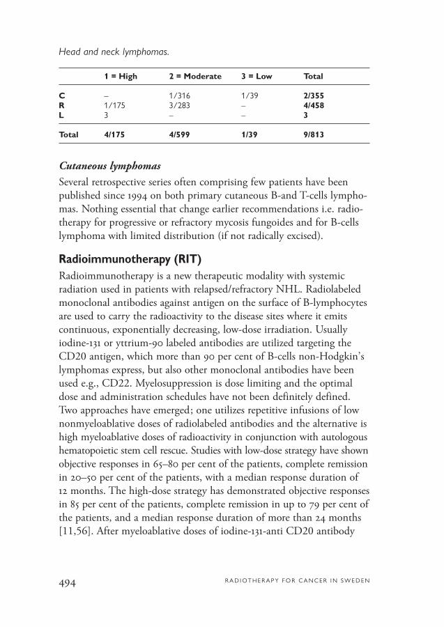
Head and neck lymphomas.
1 = High 2 = Moderate 3 = Low Total
C – 1/316 1/39 2/355R 1/175 3/283 – 4/458L 3 – – 3
Total 4/175 4/599 1/39 9/813
Cutaneous lymphomas
Several retrospective series often comprising few patients have been
published since 1994 on both primary cutaneous B-and T-cells lympho-
mas. Nothing essential that change earlier recommendations i.e. radio-
therapy for progressive or refractory mycosis fungoides and for B-cells
lymphoma with limited distribution (if not radically excised).
Radioimmunotherapy (RIT)Radioimmunotherapy is a new therapeutic modality with systemic
radiation used in patients with relapsed/refractory NHL. Radiolabeled
monoclonal antibodies against antigen on the surface of B-lymphocytes
are used to carry the radioactivity to the disease sites where it emits
continuous, exponentially decreasing, low-dose irradiation. Usually
iodine-131 or yttrium-90 labeled antibodies are utilized targeting the
CD20 antigen, which more than 90 per cent of B-cells non-Hodgkin’s
lymphomas express, but also other monoclonal antibodies have been
used e.g., CD22. Myelosuppression is dose limiting and the optimal
dose and administration schedules have not been definitely defined.
Two approaches have emerged; one utilizes repetitive infusions of low
nonmyeloablative doses of radiolabeled antibodies and the alternative is
high myeloablative doses of radioactivity in conjunction with autologous
hematopoietic stem cell rescue. Studies with low-dose strategy have shown
objective responses in 65–80 per cent of the patients, complete remission
in 20–50 per cent of the patients, with a median response duration of
12 months. The high-dose strategy has demonstrated objective responses
in 85 per cent of the patients, complete remission in up to 79 per cent of
the patients, and a median response duration of more than 24 months
[11,56]. After myeloablative doses of iodine-131-anti CD20 antibody

495S E C T I O N 1 5 • N O N - H O D G K I N ’ S LY M P H O M A ( N H L )
and autologous stem-cell rescue in patients with relapsed NHL treated
with a median of three prior chemotherapy regimens, 12 of 29 patients
continued in unmaintained remission for more than 3 years after treat-
ment. The progression-free survival rate at 4 years was estimated to be
51 per cent for patients with indolent lymphomas and 20 per cent for
those with aggressive lymphomas. Reversible cardiopulmonary toxicity
was dose limiting in this setting [41].
Only one randomized clinical trial with RIT has been performed. One
hundred fortythree patients with relapsed/refractory indolent or trans-
formed NHL were randomized between Zevalin (ibritumomab tiuxetan:
anti-CD20 monoclonal antibody conjugated with 90Y) radioimmuno-
therapy and rituximab (anti-CD20 monoclonal antibody) immuno-
therapy. The overall response rate was 80 per cent in the Zevalin arm vs
56 per cent in the rituximab arm (p=0.002) with a 30 per cent vs 16 per
cent complete remission, respectively [71].
Comparison between different radionuclides or antibodies in clinical
trials has not been performed.
The literature shows that:
• Several phase I and II studies with radioimmunotherapy have
documented promising results by this new therapeutic modality
with systemic radiation.
• A variety of monoclonal antibodies, radionuclides and study designs
have resulted in high response rates with a number of durable complete
and partial remissions with both myeloablative and nonmyeloablative
approaches in patients with recurrent or refractory lymphomas. In some
studies these responses have lasted longer than prior remissions from
previous chemotherapy.
• One randomized clinical trial showed superior therapy results with
radiolabelled antibody compared with the corresponding unlabelled
antibody.
• Future studies are needed to define the role of RIT and which RIT
regimen(s) will be most efficacious in the management of NHL.

R A D I OT H E R A P Y F O R C A N C E R I N S W E D E N496
Radioimmunotherapy.
1 = High 2 = Moderate 3 = Low Total
C – 1/143 1/143P – 1/29 – 1/29L 2 – – 2
Total 2 2/172 4/172
LiteratureThe articles on which the conclusions in this report were based were classifiedand graded as follows (number of studies/number of patients).
1 = High 2 = Moderate 3 = Low Total
C 1/401 2/459 4/328 7/1 188P 3/455 8/563 5/124 16/1 142R 7/8 754 10/1 289 5/206 22/10 249L 17/676 – – 17/676O 10/20 – – 10/20
Total 38/10 306 20/2 311 14/658 72/13 275
Conclusions and Comments
Indolent lymphomas• Data support that one third to one half of patients with indolent
lymphoma in stage I are cured by radiotherapy (follow-up more
than 15 years). ([32]R2, [33]C3, [43]R2, [55]R3, [69]R1).
• Addition of chemotherapy to radiotherapy does not suggest any
improvement of overall outcome. ([33]C3).
Optimal radiation dose is not defined and extended field is not superior
to involved field.
No new reports concerning radiotherapy in advanced stages have appeared
and the statements from SBU report 129/2 1996 are still valid.

497S E C T I O N 1 5 • N O N - H O D G K I N ’ S LY M P H O M A ( N H L )
Aggressive localized lymphomas• Data indicate that one half of patients in stage I is cured by radio-
therapy alone. ([54]R1, [69]R1).
• Although randomized and non-randomized studies favour combined
modality treatment with chemotherapy followed by radiotherapy
instead of radiotherapy or chemotherapy alone in localized disease no
firm conclusions can be drawn. (Pro [20]P2, [22]P2, [47]C1, [50]P2,[53]R1, [54]R1, [68]R2, con [47a]C3).
• Conflicting data have been published on the value of radiotherapy
towards bulky disease and no firm conclusions can be drawn.
(Pro [6 ]C3, [19]R2, con [32]R2, [50]P2, [53]R1).
Optimal dose for radiation alone or after chemotherapy has not been
established.
Total body irradiation (TBI)The value of total body irradiation for treatment of non-Hodgkin’s
lymphoma has not been proven.
• There is no proof that fractionated TBI in conjunction with high-
dose chemotherapy is superior to chemotherapy regimens alone.
([4]R1, [5]L1, [49]L1).
Primary CNS lymphomas• Data show that radiotherapy induces response of short duration and
is associated with major neurotoxicity, especially in elderly patients.
([1]P3, [2]P2, [13]R3, [18]L1, [36]R3, [37]P2, [57]L1).
• High-dose methotrexate therapy seems to lead to longer survival
than radiotherapy alone. No randomized trials are performed.
([1]P3, [2]P2, [18]L1, [52]P2).
• There is fairly good support for primary chemotherapy including
high-dose methotrexate followed by radiotherapy in patients below
60 years. ([1]P3, [2]P2, [18]L1, [57]L1).
R A D I OT H E R A P Y F O R C A N C E R I N S W E D E N498
To minimize the risk of neurotoxicity of combined modality treatment
it has been proposed to use chemotherapy alone and delay radiotherapy
for relapse, especially in patients above 60 years or use it in chemotherapy
resistent disease. Optimal chemotherapy regimen is not defined and the
role of radiotherapy remains to be determined.
Head and neck lymphomas• There is some support for combined modality treatment with chemo-
therapy and radiotherapy for aggressive lymphomas in Waldeyer’s
ring with limited disease. ([7]C2, [24]L1, [73]L1, [75]L1).
• There is sparse data supporting radiotherapy alone in localized indolent
lymphomas in salivary glands. ([8]C3).
Radioimmunotherapy (RIT)Radioimmunotherapy is a new treatment modality with systemic radia-
tion for patients with advanced NHL, where conventional external beam
radiotherapy plays only a minor role.
Several phase I and II studies with RIT have documented promising
results. A variety of monoclonal antibodies, radionuclides and study designs
with both myeloablative and nonmyeloablative approach have resulted
in high response rates in patients with recurrent or refractory NHL.
• One randomized clinical trial is published, showing superior therapy
results with radiolabelled antibody compared with the corresponding
unlabelled antibody. ([71]C2).

499S E C T I O N 1 5 • N O N - H O D G K I N ’ S LY M P H O M A ( N H L )
1. Abrey L E, DeAngelis L M, Yahalom J.Long-term survival in primary CNS lym-phoma. J Clin Oncol 1998;16:859-63(P3/31).
2. Abrey L E, Yahalom J, DeAngelis L M.Treatment of primary CNS lymphoma:The next step. J Clin Oncol 2000;18:3144-50 (P2/52).
3. Ansell S M, Grant C S, Haberman T M.Primary thyroid lymphoma. Semin Oncol1999;26:316-23 (L1).
4. Apostolidis J, Gupta R K, Grenzelias D,Johnson P W M, Pappa V I, Summers K Eet al. High-dose therapy with autologousbone marrow support as consolidation ofremission in follicular lymphoma: long-term clinical and molecular follow-up. J Clin Oncol 2000;18:527-36 (R1/99).
5. Aristei C, Tabilo A. Total-body irradia-tion in the conditioning regimens for auto-logous stem cell transplantation in lympho-proliferative diseases. Oncologist 1999;4:386-97 (L1).
6. Avilés A, Delgado S, Nambo M J,Alatriste S, Diaz-Maqueo J C. Adjuvantradiotherapy to sites of previous bulky disease in patients stage IV diffuse large celllymphoma. Int J Radiat Oncol Biol Phys1994;30:799-803 (C3/88).
7. Avilés A, Delgado S, Ruiz H, de la TorreA, Guzman R, Talavera A. Treatment ofnon-Hodgkin’s lymphoma of Waldeyer’sring: radiotherapy versus chemotherapyversus combined therapy. Oral Oncol Eur JCancer 1996;32B:19-23 (C2/316).
8. Avilés A, Delgado S, Huerta-Guzman J.Marginal zone B cell lymphoma of the
parotid glands: results of a randomized trialcomparing radiotherapy to combined the-rapy. Oral Oncol Eur J Cancer 1996;32B:420-22 (C3/39).
9. Cancer incidence in Sweden 2000.Stockholm: National Board of Health andWelfare, Centre for Epidemiology, TheCancer Registry, 2001.
10. Cheung M M C, Chan J K C, Lau W H, Foo W, Chan P T M, Ng C S et al. Primary non-Hodgkin’s lymphoma of the nose and nasopharynx: clinical features, tumour immunophenotype, and treatment outcome in 113 patients. J Clin Oncol 1998;16:70-7 (R2/113).
11. Corcoran M C, Eary J, Bernstein I,Press O W. Radioimmunotherapy strategiesfor non-Hodgkin’s lymphomas. Ann Oncol1997;8(1):133-8 (L1).
12. Corn B W, Marcus S M, Topham A,Hauck W, Curran Jr W J. Will primarycentral nervous system lymphoma be themost frequent brain tumour diagnosed inthe year 2000? Cancer 1997;79(12):2409-13 (O1).
13. Corry J, Smith J G, Wirth A, QuongG, Hoe Liew K. Primary central nervoussystem lymphoma: age and performancestatus are more important than treatmentmodality. Int J Radiat Oncol Biol Phys1998;41:615-20 (R3/62).
14. DeAngelis L M. Primary central nervous system lymphoma: an update.Educational Book of the 37th AnnualMeeting of the American Society ofClinical Oncology; May 12-15, SanFrancisco 2001;277-80 (L1).
References

R A D I OT H E R A P Y F O R C A N C E R I N S W E D E N500
15. de Jong D, Aleman B M P, Taal B G,Boot H. Controversies and consensus inthe diagnosis, work-up and treatment ofgastric lymphoma: An international survey.Ann Oncol 1999;10:275-80 (O1).
16. De Los Santos J F, Mendenhall N P,Lynch J W. Is comprehensive lymphaticirradiation for low-grade non-Hodgkin’slymphoma curative therapy? Long-termexperience at a single institution. Int JRadiat Oncol Biol Phys 1997;38:3-8 (R3/21).
17. Denham J W, Denham E, Dear K B,Hudson G V. The follicular non-Hodgkin’slymphomas – I. The possibility of cure. EurJ Cancer 1996;32A:470-9 (C3/-).
18. Ferreri A J M, Reni M, Villa E.Therapeutic management of primary cen-tral nervous system lymphoma: Lessonsfrom prospective trials. Ann Oncol 2000;11:927-37 (L1).
19. Ferreri A J M, Dell’Oro S, Reni M,Ceresoli G L, Cozzarini C, Ponzoni M etal. Consolidation radiotherapy to bulky orsemibulky lesions in the management ofstage III-IV diffuse large B cell lymphomas.Oncology 2000;58:219-26 ((R2/94).
20. Freilone R, Botto B, Vitolo U, BertiniM, Audisio E, Calvi R et al. Combinedmodality treatment with a weekly brief chemo-therapy (ACOP-B) followed by locoregionalradiotherapy in localized-stage intermediate-to high-grade non-Hodgkin’s lymphoma.Ann Oncol 1996;7:919-24 ((P2/84).
21. Fischbach W, Dragosics B, Kolve-Goebeler M-E, Ohmann C, Greiner A,Yang Q et al for the German-Austrian gas-trointestinal study group. Primary gastricB-cell lymphoma: results of a prospectivemulticenter study. Gastroenterology2000;119:1191-202 (P1/236).
22. Fuller L M, Krasin M J, Velasquez WS, Allen P K, McLaughlin P, Rodriguez MA et al. Significance of tumour size andradiation dose to local control in stage I-IIIlarge cell lymphoma treated with CHOP-bleo and radiation. Int J Radiat Oncol BiolPhys 1995;31:3-11 (P2/146).
23. Girinsky T, Guillot-Vals D, Koscielny S,Cosset JM, Ganem G, Carde P et al. A highand sustained response rate in refractory orrelapsing low-grade lymphoma masses afterlow-dose radiation: analysis of predictiveparameters of response to treatment. Int JRadiat Oncol Biol Phys 2001;51(1):148-55(R2/48).
24. Gospodarowicz M K, Sutcliffe S B. Theextranodal lymphomas. Semin RadiatOncol 1995;5:281-300 (L1)
25. Gospodarowicz M K, Zucca E. Primarytestis lymphoma: presentation, treatment,patterns of failure, and outcomes. Educa-tional Book of the 37th Annual Meeting ofthe American Society of Clinical Oncology;May 12-15, 2001; San Francisco pp 281-5(L1).
26. Haim N, Leviov M, Ben-Arieh Y,Epelbaum R, Freidin N, Reshef R et al.Intermediate and high-grade gastric non-Hodgkin’s lymphoma: a prospective studyof non-surgical treatment with primarychemotherapy, with or without radiotherapy.Leuk Lymphoma 1995;17:321-6 (P3/24).
27. Harris N L, Jaffe E S, Stein H, Banks P M, Chan J K C, Cleary M L et al. Arevised European-American classification of lymphoid neoplasms: A proposal fromthe International Lymphoma Study Group.Blood 1994;84:1361-92 (O1).
28. Harris N L, Jaffe E S, Diebold J,Flandrin G, Muller-Hermelink H K,

501S E C T I O N 1 5 • N O N - H O D G K I N ’ S LY M P H O M A ( N H L )
Vardiman J. Lymphoma classification –from controversy to consensus: The R.E.A.L.and WHO Classification of lymphoid neo-plasms. Ann Oncol 2000;11:3-10 (O1).
29. Hiddemann W, Longo DL, Coiffier B,Fisher RI, Cabanillas F, Cavalli F et al.Lymphoma classification--the gap betweenbiology and clinical management is closing.Blood. 1996;88(11):4085-9 (01).
30. Isaacson P G. Gastric MALT lympho-ma: From concept to cure. Ann Oncol1999;10:637-45 (O1).
31. Isaacson P G. Extranodal marginal zoneB-cell lymphoma of mucosa-associated lym-phoid tissue type. Educational Book of the37th Annual Meeting of the AmericanSociety of Clinical Oncology; May 12-15,2001; San Francisco pp 273-6 (L1).
32. Kamath S S, Marcus Jr R B, Lynch JW, Mendenhall N P. The impact of radio-therapy dose and other treatment-relatedand clinical factors on in-field control instage I and II non-Hodgkin’s lymphoma.Int J Radiat Oncol Biol Phys 1999;44:563-8 (R2/285).
33. Kelsey S M, Newland A C, VaughanHudson G, Jelliffe A M. A British NationalLymphoma Investigation randomized trialof single agent chlorambucil plus radiothe-rapy versus radiotherapy alone in low grade,localized non-Hodgkin’s lymphoma. MedOncol 1994;11:19-25 (C3/148).
34. Krogh-Jensen M, D’Amore F, JensenMK, Christensen B E, Thorling K,Pedersen M et al. Clinicopathological features, survival and prognostic factors of primary central nervous system lympho-mas: trends in incidence of primary centralnervous system lymphomas and primarymalignant brain tumours in a well-defined
geographical area. Population-based datafrom the Danish Lymphoma Registry,LYFO and the Danish Cancer Registry.Leuk Lymphoma 1995;19:223-33 (O1).
35. Laing R W, Hoskin P, VaughanHudson B, Vaughan Hudson G, HarmerC, Bennett M H et al. The significance of MALT histology in thyroid lymphoma:a review of patients from the BNLI andRoyal Marsden Hospital. Clin Oncol1994;6:300-4 (R2/45).
36. Laperriere N J, Cerezo L, Milosevic MF, Wong C S, Patterson B, Panzarella T.Primary lymphoma of brain: results ofmanagement of a modern cohort with radi-ation therapy. Radiother Oncol 1997;43:247-52 (R3/49).
37. Laperriere N J, Wong C S, MilosevicM F, Whitton A C, Wells W A, PattersonB. Accelerated radiation therapy for prima-ry lymphoma of the brain. RadiotherOncol 1998;47:191-5 (P2/10).
38. Li Y X, Coucke P A, Li J-Y, Gu D-Z,Liu X-F, Zhou L-Q et al. Primary non-Hodgkin´s lymphoma of the nasal cavity.Prognostic significance of paranasal exten-sion and the role of radiotherapy and chemo-therapy. Cancer 1998;83:449-56 (R1/175).
39. Liang R, Todd D, Chan T K, Chiu E,Lie A, Kwong Y L et al. Treatment outco-me and prognostic factors for primary nasallymphoma. J Clin Oncol 1995;13:666-70((R2/100).
40. Liu H, Ruskone-Fourmestraux A,Lavergne-Slove A, Ye H, Molina T,Bouhnik Y et al. Resistance of t(11;18)positive gastric mucosa-associated lym-phoid tissue lymphoma to Helicobacterpylori eradication therapy. Lancet2001;357:39-40 (O1/22).
R A D I OT H E R A P Y F O R C A N C E R I N S W E D E N502
41. Liu S Y, Eary J F, Petersdorf S H,Martin P J, Maloney D G, Appelbaum F Ret al. Follow-up of relapsed B-cell Lymphomapatients treated with iodine-131-labeled anti-CD20 antibody and autologous stem-cellrescue. J Clin Oncol 1998;16:3270-8 (P2/29).
42. Logsdon M D, Ha C S, Kavadi V S,Cabanillas F, Hess M A, Cox J D.Lymphoma of the nasal cavity and parana-sal sinuses. Improved outcome and alteredprognostic factors with combined modalitytherapy. Cancer 1997;80:477-88 (R2/70).
43. Mac Manus M P, Hoppe R T. Is radiot-herapy curative for stage I and II low-gradefollicular lymphoma? Results of a long-term follow-up study of patients treated atStanford University. J Clin Oncol 1996;14:1282-90 (R2/177).
44. Mahé M-A, Bourdin S, Le Pourhiet-LeMevel A, Moreau P, Campion L et al.Salvage extended-field irradiation in follicu-lar non-Hodgkin’s lymphoma after failureof chemotherapy. Int J Radiat Oncol BiolPhys 2000;47:735-8 (R3/34).
45. Maher E A, Fine H A. Primary CNSlymphoma. Semin Oncol 1999;26:346-56(L1).
46. Mead G M, Bleehen N M, Gregor A,Bullimore J, Murell D S; Rampling R P etal. A medical research council randomizedtrial in patients with primary cerebral non-Hodgkin lymphoma. Cancer 2000;89:1359-70 (C3/53).
47. Miller T P, Dahlberg S, Cassady J R,Adelstein D J, Spier C M, Grogan T M etal. Chemotherapy alone compared withchemotherapy plus radiotherapy for locali-zed intermediate- and high-grade non-Hodgkin’s lymphoma. N Engl J Med1998;339: 21-6 (C1/401).
47a. Miller TP, Leblanc M, Spier C, ChaseE, Fischer RI. CHOP alone compared toCHOP plus radiotherapy for early stageaggressive non-Hodgkin’s lymphomas:update of the Southwest Oncology Group(SWOG) randomized trial. Blood2001;98:abstract 3024.
48. Milligan D W, Ruiz de Elvira M C,Kolb H J, Goldstone A H, Meloni G,Rohatiner A Z, et al. Secondary leukaemiaand myelodysplasia after autografting forlymphoma: results from the EBMT. Br JHaematol 1999;106:1020-26 (R1/4998).
49. Mounier N, Gisselbrecht C.Conditioning regimens before transplanta-tion in patients with aggressive non-Hodgkin’s lymphoma. Ann Oncol 1998;9:15-21 (L1).
50. Munck J N, Dhermain F, Koscielny S,Girinsky T, Carde P, Bosq J et al.Alternating chemotherapy and radiotherapyfor limited-stage intermediate and high-grade non-Hodgkin’s lymphomas: Long-term results for 96 patients with tumours>5 cm. Ann Oncol 1996;7:925-31 (P2/96).
51. Neubauer A, Thiede C, Morgner A,Alpen B, Ritter M, Neubauer B et al. Cureof Helicobacter pylori infection and dura-tion of remission of low-grade gastricmucosa-associated lymphoid tissue lym-phoma. J Natl Cancer Inst 1997;89:1350-5(P1/50).
52. O’Brien P, Roos D, Pratt G Liew K,Barton M, Poulsen M et al. Phase II multi-center study of brief single-agent metho-trexate followed by irradiation in primaryCNS lymphoma. J Clin Oncol 2000;18:519-26 (P2/46).
53. Oguchi M, Ikeda H, Isobe K, Hirota S,Hasegawa M, Nakamura K et al. Tumour
503S E C T I O N 1 5 • N O N - H O D G K I N ’ S LY M P H O M A ( N H L )
bulk as a prognostic factor for the manage-ment of localized aggressive non-Hodgkin’slymphoma: A survey of the Japan lympho-ma radiation therapy group. Int J RadiatOncol Biol Phys 2000;48:161-8 (R1/787).
54. Osterman B, Cavallin-Ståhl E, HagbergH, Lindén O, Lenner P. High-grade non-Hodgkin’s lymphoma stage I. A retrospecti-ve study of treatment, outcome and prog-nostic factors in 213 patients. Acta Oncol1996;35:171-7 (R1/213).
55. Pendlebury S, El Awadi M, Ashley,Brada M, Horwich A. Radiotherapy resultsin early stage low grade nodal non-hodgki-n’s lymphoma. Radiother Oncol 1995;36:167-71 (R3/40).
56. Press O W. Innovative new therapiesfor non-Hodgkin’s lymphomas: monoclo-nal antibodies and immunoconjugates.Educational Book of the 36th AnnualMeeting of the American Society ofClinical Oncology; May 19-23, 2000; NewOrleans pp 328-37 (L1).
57. Reni M, Ferreri A J M, Garancini M PVilla E. Therapeutic management of pri-mary central nervous system lymphoma inimmunocompetent patients: Results of acritical review of the literature. Ann Oncol1997;8:227-34 (L1/676).
58. Richaud P M, Soubeyran P, Eghbali H,Chacon B, Marit G, Broustet A et al. Placeof low dose total body irradiation in thetreatment of localized follicular non-Hodgkin’s lymphoma: results of a pilotstudy. Int J Radiat Oncol Biol Phys1998;40:387-90 (P3/26).
59. Rohatiner, A on behalf of: d´Amore, F;Coiffier, B; Crowther, D; Gospodarowicz,M; Isaacson, P; Lister et al. Report on aworkshop convened to discuss the patholo-
gical and staging classifications of gastroin-testinal tract lymphoma. Ann Oncol 1994;5:397-400. (O1)
60. Safwat A. The role of low-dose total bodyirradiation in treatment of non-Hodgkin’slymphoma: a new look at an old method.Radiother Oncol 2000;56:1-8 (L1).
61. Schechter N R, Yahalom J. Low-gradeMALT lymphoma of the stomach: a reviewof treatment options. Int J Radiat OncolBiol Phys 2000;46:1093-1103 (L1).
62. Soubeyran P, Eghbali H, Trojani M,Bonichon F, Richaud P, Hoerni B. Is thereany place for a wait-and-see policy in stageI0 follicular lymphoma? A study of 43 con-secutive patients in a single-centre. AnnOncol 1996;7:713-8 (P3/26).
63. Stuschke M, Hoederath A, Sack H,Pötter R, Müller R-P, Schulz U et al.Extended field and total central lymphaticradiotherapy in the treatment of early stagelymph node centroblastic-centrocytic lym-phomas. Results of a prospective multicenterstudy. Cancer 1997;80:2273-84 (P2/100).
64. The International Non-Hodgkin’sLymphoma Prognostic Factors Project: A predictive model of aggressive non-Hodgkin’s lymphoma. N Engl J Med1993;329:987-94 (R1/2031).
65. The Non-Hodgkin’s LymphomaClassification Project: A clinical evaluationof the International Lymphoma StudyGroup classification of non-Hodgkin’sLymphoma. Blood 1997;89:3909-18 (O1)
66. Tondini C, Balzarotti M, Santoro A,Zanini M, Fornier M, Giardini R et al.Initial chemotherapy for primary respecta-ble large-cell lymphoma of the stomach.Ann Oncol 1997;8:497-9 (P3/17).

R A D I OT H E R A P Y F O R C A N C E R I N S W E D E N504
67. Travis L B, Weeks J, Curtis R E,Chaffey J T, Stovall M, Banks P M et al.Leukemia following low-dose total bodyirradiation and chemotherapy for non-Hodgkin’s lymphoma. J Clin Oncol1996;14:565-71 (R2/61).
68. van der Maazen R W M, Noordijk EM, Thomas J, Raemaekers J M M,Meerwaldt J H. Combined modality treat-ment is the treatment of choice for stageI/IE intermediate and high grade non-Hodgkin’s lymphomas. Radiother Oncol1998;49:1-7 (R2/296).
69. Vaughan Hudson B, Vaughan HudsonG, MacLennan K A, Anderson L, Linch DC. Clinical stage 1 non-Hodgkin’s lympho-ma: long-term follow-up of patients treatedby the British National LymphomaInvestigation with radiotherapy alone asinitial therapy. Br J Cancer 1994;69:1088-93 (R1/451).
70. Willich N A, Reinartz G, Horst E J,Delker G, Reers B, Hiddemann W et al.Operative and conservative managementprimary gastric lymphoma: interim resultsof a German multicenter study. Int JRadiat Oncol Biol Phys 2000;46:895-901(P1/169).
71. Witzig T E, Gordon LI, Cabanillas F,Czuczman M S, Emmanoulides C, Joyce Ret al. Randomized controlled trial of yttrium-
90-labeled ibritumomab tiuxetan radioim-munotherapy versus rituximab immunot-herapy for patients with relapsed or refrac-tory low-grade, follicular, or transformedB-cell non-Hodgkin's lymphoma. J ClinOncol 2002;20(10):2453-63 (C2/143).
72. Yahalom J, Schechter J N R, GonzalesM, Portlock C S. Effective treatment ofMALT lymphoma of the stomach withradiation alone. Ann Oncol 1999;10:135.Abstract.
73. Yuen A, Jacobs C. Lymphomas of thehead and neck. Semin Oncol 1999;26:338-45 (L1).
74. Zucca E, Roggero E, Bertoni F, CavalliF. Primary extranodal non-Hodgkin’s lym-phomas. Part 1: Gastrointestinal, cutaneousand genitourinary lymphomas. Ann Oncol1997;8:727-37 (L1).
75. Zucca E, Roggero E, Bertoni F, CavalliF. Primary extranodal non-Hodgkin’s lym-phomas. Part 2: Head and neck, centralnervous system and other less commonsites. Ann Oncol 1999;10:1023-33 (L1).
76. Zucca E, Conconi A, Mughal T, SarrisA H, Vitolo U, Gospodarowicz M K.Patterns of survival in primary diffuselarge-cell B-cell lymphoma (DLCL) of thetestis. An international survey of 373 pati-ents. Blood 2000;96:334a. Abstract.
Glick J H, Kim K, Earle J et al An ECOGrandomized phase III trial of CHOP vsCHOP + radiotherapy (XRT) for interme-diate grade early stage non-Hodgkin’s lym-phoma. Proc Am Soc Clin Oncol1995;14:391 (Abstr)
Shchepotin I B, Evans S R T, ShabahangM, Chorny V, Buras R R, Korobko V et al.Primary non-Hodgkin’s lymphoma of thestomach: three radical modalities of treat-ment in 75 patients. Ann Surg Oncol1996;3:277-84.
Excluded randomized trials:


