download full text (final version , 16mb) - repub - erasmus
TRANSCRIPT

The Reserve Cell in the Uterine Cervix
aspects of development, differentiation and diagnosis


The Reserve Cell in the Uterine Cervix
aspects of development, differentiation and diagnosis
De reservecel in de cervix uteri detectie, differentiatie en diagnose.
Proefschrift
ter verkrijging van de graad van doctor aan
de Erasmus Universiteit Rotterdam op gezag van de
rector magnificus Prof. Dr. S.W.J. Lamberts en
volgens besluit van het College voor Promoties.
De openbare verdediging zal plaatsvinden op
3 december 2008 om 15.45 uur
door
Johanna Elisabeth Martens
geboren te Haarlem

Promotiecommissie
Promotoren: Prof. dr. Th.J.M. Helmerhorst
Prof. dr. F.C.S. Ramaekers
Overige leden: Prof. dr. C.W. Burger
Prof. dr. C.J.L.M. Meijer
Prof. dr. J.W. Oosterhuis
Copromotoren: Dr. F.M.M. Smedts
Dr. A.H.N. Hopman

Voor Rudolph Klein-bastaard van Muyden


Contents
Chapter 1 General introduction 9
Chapter 2 Reserve cells in human uterine cervical epithelium are 27
derived from Müllerian epithelium at midgestational age.
International Journal of Gynecological Pathology 2007 26(4):463-468
Chapter 3 Cytokeratin 17 and p63 are markers of the progenitor cell of 45
the human uterine cervical epithelium, a putative HPV target cell.
Based on: Anticancer Research 2004 24:771-775
Chapter 4 Distribution pattern and markerprofile disclose two 61
subpopulations of reserve cells in the endocervical canal.
Submitted
Chapter 5 Glutathione S-transferase p is expressed in (pre) neoplastic 79
lesions of the human uterine cervix irrespective of
their degree of severity
Based on: Anticancer Research 1997 17:4305-4309
Chapter 6 Can Cytokeratin 8 and 17 immunohistochemistry be of 95
diagnostic value in cervical cytology? A feasibility study.
Cancer 1999; 87(2): 87-92
Chapter 7 General discussion 111
Chapter 8 Summary 123
Chapter 9 Samenvatting 129
Bibliography 135
Color figures 139
Dankwoord 147
Curriculum Vitae 151


1General introduction

11

11
Introduction
Carcinoma of the uterine cervix is worldwide the second most common cancer in
women1. It has been approximately 150 years since the first description of uterine cervical
carcinoma, a century since the description of its precursor lesions2, and half a century since
the introduction of the method proposed by Papanicolaou3 for detecting cervical neoplasms
by cytologic screening. In time investigators have proposed a multitude of histologic and
cytologic terms for cervical precursor lesions4;5, have devised methods and techniques for
diagnosis and treatment6;7, and have identified the causative agent i.e. human papillomavirus
(HPV)8.
Subclinical cervical HPV infection causes typical cytological and histological features
called koilocytosis9. Koss was one of the first to relate koilocytosis to condylomas and
associated these with premalignant cervical lesions, zur Hausen (Nobel Prize winner 2008)
was the first to relate HPV to cervical cancer 10-12. From that moment HPV was the major
subject for investigators in the search for answers in cervical carcinogenesis13. In the
1980’s and 1990’s new molecular and cell biological techniques developed rapidly and
made it possible to detect HPV DNA in tissue and cell samples14, to identify the many HPV
subtypes and to study the effect of HPV on the cell cycle and the process of apoptosis15.
These techniques are now widely applied for diagnostic purposes, allowing a beter
recognition of the HPV-infected lesions. Resulting in a better prognostication and giving
rise to more molecular approaches that complement the (immuno) histochemical and
cytochemical protocols.

12 13
HPV mediated carcinogenesis The molecular model for HPV mediated carcinogenesis involves interaction of HPV
gene products with the tightly controlled network of cellular oncogenes and tumor
suppressor genes, which control cell proliferaton, apoptosis, DNA synthesis and DNA
repair16.
Persistence of a high risk HPV infection is a necessary condition in the carcinogenesis
of cervical cancer. Integration of the viral DNA into the human genome is essential for
progression17. The E6 and E7 gene products of the high risk HPV viruses, in particular HPV
16 and HPV 18, encode for proteins that interfere with the mitosis and apoptosis regulating
pathways of the host cell, i.e. the p53 and the pRb signaling routes. Accordingly the E6/
E7 induced inactivation of these pathways results in hyperproliferation and inhibition of
apoptosis, thus leading to genetic instability (amongst others numerical and structural
chromosome aberrations) and immortalization16-19.
HPV target cell The link between morphological changes observed at colposcopy and microscopy
on the one hand, and the causal role of HPV-induced genetic changes on the other, is
still largely missing. The target cell in the uterine cervical epithelium undergoing the
transforming mutations, caused by high risk HPV infection and incorporation in the cells
genome, is still subject of speculation. The expression of the viral proteins is dependent on
cell differentiation. It is suggested that when the integrity of the basal layer is compromised
due to micro-traumata or environmental changes HPV can infect the progenitor/stem cell
of cervical (pre)malignancy. The viral genome is maintained as low copy number episomes
in the basal layer of the epithelium. Transcription of the viral genome is predominantly
increased in the more suprabasal, differentiated layers. In light of carcinogenic models in
comparable epithelia like esophageal, prostatic, colorectal and stomach epithelium, the
putative target cell for HPV infection resulting in (pre) malignancy is most likely an epithelial
stem cell of the healthy cervical epithelium occuring in the so called transformation zone 20-22.

12 13
Stem cells, a hierarchy of potential Stem cells have varying potential23, the most primitive, toti-potent stem cell is the
fertilized oocyt, able to form the embryo and the trofoblast of the placenta. About 4 days
after fertilization these toti-potent cells form a blastocyst and the inner cell mass from which
the embryo develops. The cells of the inner cell mass are considered to be pluripotent, able
to differentiate into almost all cells that arise from the three germ layers. Most adult tissues
contain multipotential stem cells, e.g the hematopoietic stem cells, capable of producing
a limited range of differentiated cell lineages appropriate to their location. At the end of
this hierarchic tree the unipotential stem cells occur, capable of generating one specific cell
type, such as for example epidermal stem cells. These unipotential stem cells are often called
commited progenitors or progenitor cells. The subject of this thesis is to shed more light on
the epithelial stem cells or progenitor cells of the human uterine cervical epithelium.
Since the normal and malignant cervical epithelium consist of at least two major
differentiated cell types, i.e. the glandular cells and the squamous cells, one can raise the
question whether these arise from one and the same stem cell compartment or whether
two different stem cell types are needed for normal cervical epithelial genesis.
Stem cell concept in epithelial carcinogenesis Pierce and Potten suggested that the target cell in the carcinogenic cascade is the
stem cell24;25. In adult organisms each tissue and organ is generally accepted to contain
a small subpopulation of cells capable of self renewal, of indefinite proliferative potential,
and with the ability to give rise to a large family of descendants with defined spectra of
specialization. A tissue specific stem cell or progenitor cell is normally multipotent, i.e.
capable of producing a limited range of differentiated cell lineages. In normal circumstances
tissue-specific stem cells generate the range of cell types appropriate to their location. In
times of chronic damage or regeneration a process of metaplasia can give a switch in tissue
differentiation. This metaplastic switch occurs at the level of progenitor cells rather than
between terminally differentiated cells23. Examples include squamous metaplasia in airways
of smokers, intestinal metaplasia in the stomach26, and in the uterine cervix squamous
metaplasia occurs frequently at the squamo-columnar junction27. It is likely that cancer,
14 15
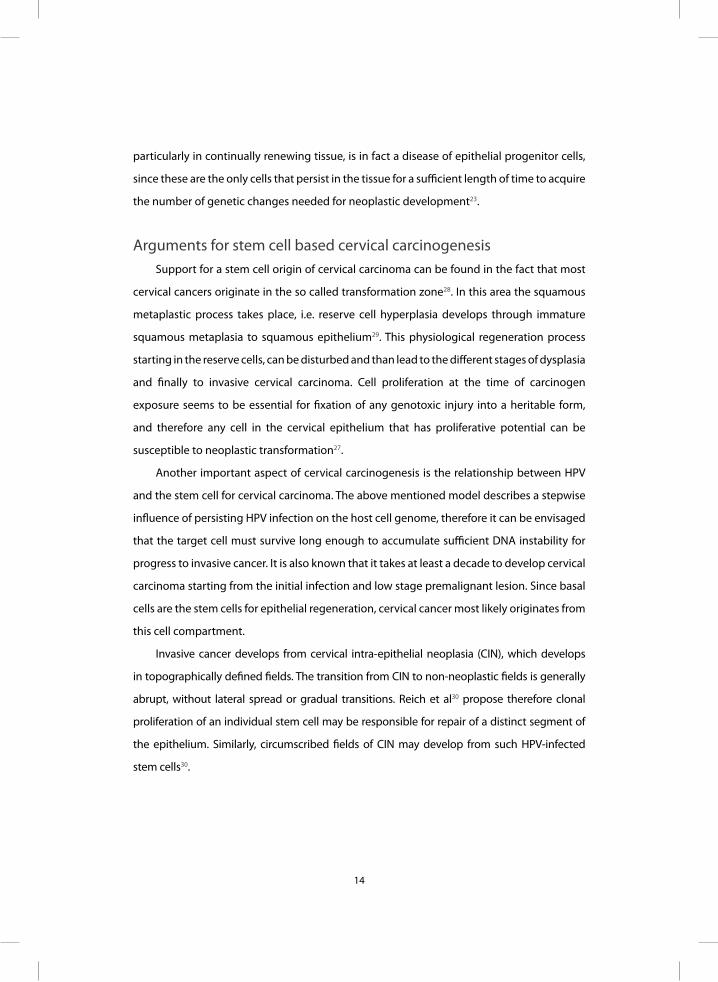
particularly in continually renewing tissue, is in fact a disease of epithelial progenitor cells,
since these are the only cells that persist in the tissue for a sufficient length of time to acquire
the number of genetic changes needed for neoplastic development23.
Arguments for stem cell based cervical carcinogenesis Support for a stem cell origin of cervical carcinoma can be found in the fact that most
cervical cancers originate in the so called transformation zone28. In this area the squamous
metaplastic process takes place, i.e. reserve cell hyperplasia develops through immature
squamous metaplasia to squamous epithelium29. This physiological regeneration process
starting in the reserve cells, can be disturbed and than lead to the different stages of dysplasia
and finally to invasive cervical carcinoma. Cell proliferation at the time of carcinogen
exposure seems to be essential for fixation of any genotoxic injury into a heritable form,
and therefore any cell in the cervical epithelium that has proliferative potential can be
susceptible to neoplastic transformation27.
Another important aspect of cervical carcinogenesis is the relationship between HPV
and the stem cell for cervical carcinoma. The above mentioned model describes a stepwise
influence of persisting HPV infection on the host cell genome, therefore it can be envisaged
that the target cell must survive long enough to accumulate sufficient DNA instability for
progress to invasive cancer. It is also known that it takes at least a decade to develop cervical
carcinoma starting from the initial infection and low stage premalignant lesion. Since basal
cells are the stem cells for epithelial regeneration, cervical cancer most likely originates from
this cell compartment.
Invasive cancer develops from cervical intra-epithelial neoplasia (CIN), which develops
in topographically defined fields. The transition from CIN to non-neoplastic fields is generally
abrupt, without lateral spread or gradual transitions. Reich et al30 propose therefore clonal
proliferation of an individual stem cell may be responsible for repair of a distinct segment of
the epithelium. Similarly, circumscribed fields of CIN may develop from such HPV-infected
stem cells30.

14 15
Stem cell candidates in the uterine cervix Stem cells can be defined as undifferentiated cells, often present in the basal layer
of an epithelium, with the capacity of self renewal and the ability to asymmetric division,
thus producing committed daughter cells that can migrate into the suprabasal layers and
differentiate.
The uterine cervix consists of an outer ectocervix and an inner endocervix.
The ectocervix is lined by a multilayered squamous epithelium and the endocervix by
glandular columnar epithelium, often with a subcolumnar layer of reserve cells. Three
potential stem cell candidates can be distinguished in the uterine cervical epithelium, i.e.
1. the basal cell of the squamous epithelium, 2. the columnar cell and 3. the subcolumnar
reserve cell. These three cell types are located at and interact with the basal membrane. As
described above, the cervical stem cell however, should be multipotent.
The reserve cell can not only renew the columnar cell population but
through the process of squamous metaplasia also the squamous cell population27.
This makes the reserve cells (or at least a subpopulation of the reserve cells) serious
candidates for a progenitor cell or even stem cell compartment.
The reserve cell The reserve cell has been subject of investigation for over a century and its function
and origin were intensely debated in the first half of the 20th century. In 1910 Meyer first,
describes the reserve cell and the process of reserve cell hyperplasia31. He suggests that
reserve cells originate at about six months of gestation when, because of the encroaching
growth of columnar epithelium in the cervix, the pre-existing squamous epithelium is
retracted, leaving basal-type squamous cells behind. Novak32 suggested an ingrowth of the
basal cells from the squamous epithelium at the squamo-columnar junction, undermining
the columnar epithelium. Fluhmann33 suggested that reserve cells originate from columnar
cells by means of unequal division. Song34 and Lawrence and Shingleton35 indicated that
reserve cells are derived from stromal cells. This was further supported by Reid et al36, who
suggested that stromal cells destined to be transformed into reserve cells are derived from

16 17
mononuclear cells of bone marrow origin. Recently Witkiewicz et al37 concluded that reserve
cells in microglandular hyperplasia can originate from specialized columnar cells.
The regenerative function of the reserve cell, as the cell from which the metaplastic
process originates, was first described by Fluhmann38. Carmichael 39 initiated the concept
of subcolumnar basal cells as reserve cell depots from which squamous and columnar cells
could originate. This was largely based on the presence of mucin in the subcolumnar basal
and metaplastic squamous cells. The multipotency of reserve cells was later confirmed
by immunohistochemical studies based on specific detection of individual cytokeratin
subtypes40;41.
Epithelial stem cell markers Tissue stem cells form the cellular base for organ homeostasis and repair.
Stem cells have the unusual ability to renew themselves over the lifetime of the organ
while producing daughter cells that differentiate into one or multiple lineages. Unlike
hematopoietic stem cells, whose pluripotency can be verified in vivo using cell-transfer
experiments, epithelial stem cells cannot readily be tested for functional competence.
They are difficult to isolate and their proliferative capacity is strongly influenced by their
environment, the so called stem cell niche42. Another problem is that epithelial stem cells
are difficult to characterize because of the absence of specific molecular markers for each
epithelium.
Candidate epithelial stem cell markers can be divided into several groups, including
transcription factors, signaling proteins, detoxifying proteins, markers of immortality, etc. In
the literature several stem cell markers have been used to detect these cells in the different
types of epithelia, but not all of these markers give unequivocally conclusive results. For
example, Oct 3/4 identifies pluripotent cells in human germ cell tumors43, but studies on
cervical carcinoma show no expression of this transcription factor44. Also stem cell abundant
proteins like nanog, nucleostemin and musashi-1 are not expressed in normal uterine
cervix45. Studies on Wnt-signaling pathways are limited to studies on mouse models where

16 17
several Wnt-signaling molecules have been shown to act together to establish the correct
development of the uterus46. Furthermore, studies on catenins indicate a role for this family
of proteins in the development and progression of neoplasm of the uterine cervix47;48.
Another class of candidate stem cell markers is the integrin family of transmembrane
receptors, whose members are responsible for the attachment of the basal cell layer to
the basement membrane49. It is possible that stem cells require strong adherence to the
basement membrane to maintain their stem cell characteristics or their position in the stem
cell niche. Despite this, most, if not all proliferating cells use integrins in adhesion. Therefore
the usefullness of integrins as stem cell markers is limited by the uncertainty of interpretation
of their levels of expression relative to the transit-amplifying cells.
Immunohistochemical markers
For our studies we used paraffin material which enabled us to correlate morphology
with immunohistochemical marker expression at single cell level. For the recognition of
the different cell populations this type of fixed material is essential to allow correlation
with aspects of development and differentiation of cell types. Only this type of material
enabled us to perform the study as described in chapters 3, 4 and 5. Particularly the study
on embryonic tissue, as described in chapter 2, would never have been possible without the
use of paraffin material. A consequence of this approach is however the limitation that we
could only apply antibodies that react in formalin fixed and paraffin embedded material. In
our investigation of cervical carcinogenesis we focussed on the reserve cell. Our aim was
to obtain more information on the characteristics, origin, phenotype, distribution pattern,
and possible progenitor or stem cell features of these cells. Our immunohistochemical
approach was based on newly proposed as well as previousy suggested stem cell markers
and differentiation markers found in the developing human uterine cervix, in adult normal
cervical epithelia and in its (pre)malign lesions.
We used the following immunohistochemical markers: bcl-2 plays a central role in
the inhibition of apoptosis, and localizes to basal cells of the squamous epithelium and
to subcolumnar reserve cells50. It was detected using an antibody to a synthetic peptid.

18 19
We hypothesize an epithelial stem cell must be protected against apoptosis to be able to
survive as long as possible.
Ki-67 is a well known marker for proliferation and can be used in cervical epithelium51.
Since progenitor cells are generally quiescent with a low proliferation capacity this marker
should in general be negative in such cells52.
P63, a homologue of the tumor suppressor p53, is a transcription factor
operating mainly in the embryonal stage of development and plays a role in
the regulation and maturation of the cervical epithelium in the adult phase53-57.
A critical role for p63 in the normal development of the cervical epithelium was found in
committings of early epithelial stem cells to a basal progenity54. In the absence of p63 basal
progenitor cells (which are normally p63-positive) are absent and are replaced by ciliated
columnar cells58. P63 is therefore important for the formation of progenitor cells at the basal
layer of stratified squamous epithelium. Mice deficient of p63, completely lack stratified
squamous epithelia as demonstrated by the lack of cytokeratin 14, a marker for commitment
to squamous epithelia. The mice die soon after birth and display a number of developmental
defects as well as several abnormalities in limb development53. P63 is particularly highly
expressed in progenitor or stem cell populations of a variety of epithelial tissues53;59-61.
Glutathione S-transferase p, a member of a multigene enzyme family that plays a role
in the detoxification of endogenous and exogenous compounds, mainly catalyzing their
conjugation with gluthatione62. It is found mainly in the cytoplasm and has been shown
to have a detoxificating capacity for carcinogens 63;64. Since stem cells by virtue of their role
in tissue regeneration should possess efficient defense mechanisms for protection against
DNA damaging agents, we expected this marker to occur in the stem cell of the uterine
cervix. Shiratori et al.64 have found positivity in koilocytotic cells and suggested that the
enzyme expression could be related to the presence of HPV.
Cytokeratins (CK) were shown in earlier experiments to represent strong differentiation
markers for different types of epithelial cells in the cervix 41. Secondly, reports from the
literature suggest that cytokeratins are suitable markers for progenitor or stem cells65-67.
18 19
The cytokeratins are a family of intermediate filament proteins that are characterized
by their molecular weight and in human epithelia numbered from 1 to 20. Cytokeratin
expression occurs in cell-type specific combinations and can be used for identification
and subclassification of epithelial tissues. In our study we used CK 5,14,17 and 19 for basal
cell differentiation, CK 7,8 and 18 for glandular differentiation and CK 13 for squamous
differentiation 41.
Aim of this thesis
The central theme of this investigation was a search for the progenitor/stem cell of the
human uterine cervical epithelium. It is hypothesized that the reserve cell plays a crucial role
in the regeneration of the different epithelial cell lineages of the uterine cervix, thus shedding
more light on the missing link in cervical carcinogenesis, a progenitor cell for cervical cancer,
the putative HPV-target cell.
Outline and scope
In chapter 2, fetal human tissue from different gestational ages was studied by using
antibodies to p63, bcl-2, Ki-67 and cytokeratins 5,7,8,13,17,18 and 19, to ascertain when
basal or stem cells first appear in cervical epithelium during fetal human development.
It was presumed that relatively high concentrations of progenitor cells would be present
during fetal human ontogenesis. This study also tried to solve the question of the origin of
the reserve cell.
In chapter 3, an immunohistochemical study is performed using monoclonal antibodies
against p63 and cytokeratin 17. A well defined subset of normal epithelium of the uterine
cervix and in preneoplastic samples ( CIN I, II and III) was used. By applying these markers for
basal cells we tried to identify the progenitor cell of the uterine cervical epithelium.
In chapter 4, the distribution pattern and immunoprofile of reserve cells along the entire
length of the adult cervix was examined by the use of p63, bcl-2 and cytokeratins 5,7,8 and
17 antibodies. Reserve cell subpopulations were studied with specific keratin phenotypes,
being the progenitor cell population of the different types of the cervical epithelium.

20 21
In chapter 5, the detoxification enzyme Gluthation S-tranferase p (GST p) is used
as a marker for progenitor cells to shed more light on the cervical carcinogenesis. In
the uterine cervix the presence of GST p has been associated with high grade cervical
intraepithelial neoplasia (CIN), but reports are conflicting. We investigated GST p expression
immunohistochemically in a well documented sequence from normal epithelium to cervical
cancer. In a search for the progenitor of the cervical epithelium, it was hypothesized that
a potential stem cell must be protected against toxifing agents. Focus was placed on the
expression of GST p in the basal epithelial cells and reserve cells.
Chapter 6 focusses on markers for progressive potential of premalignant lesions in
cytologic smears, with particular interest in the identification of dysplastic cells in smears, using
antibodies to cytokeratin 8 and cytokeratin 17. The use of these antibodies could be helpful
in detecting premalignant lesions in cytologic smears or be informative of their progressive
potential. Furthermore it was investigated whether reserve cells could be identified in cervical
smears using these antibodies.
The General discussion, in chapter 7, provides an overview of our main results in
relation to the literature on progenitor/stem cells in general and in carcinogenesis. We finally
conclude that the reserve cell serves as the progenitor cell in uterine cervical epithelium,
although functional studies still have to be performed.

20 21
Reference List
1 Parkin DM, Bray F, Ferlay J, and Pisani P. Global cancer statistics, 2002. CA Cancer J Clin 55: 74-108,
2005.
2 Stegner HE. Precursors of cervical cancer: ultrastructural morphology. Curr Top Pathol 70: 171-193,
1981.
3 Papanicolaou GN and Traut HF. The diagnostic value of vaginal smears in carcinoma of the uterus.
Am J Obstet Gynecol 42: 193-206, 1941.
4 The 1988 Bethesda System for reporting cervical/vaginal cytologic diagnoses. Developed and
approved at the National Cancer Institute workshop in Bethesda, MD, December 12-13, 1988.
Diagn Cytopathol 5: 331-334, 1989.
5 Schenck U, Herbert A, Solomon D, Amma NS, Collins RJ, Gupta SK, Jimenez-Ayala
M, Kobilkova J, Nielsen M, and Suprun HZ. Terminology. International Academy
of Cytology Task Force summary. Diagnostic Cytology Towards the 21st Century:
An International Expert Conference and Tutorial. Acta Cytol 42: 5-15, 1998.
6 Hopman EH, Kenemans P, and Helmerhorst TJ. Positive predictive rate of colposcopic
examination of the cervix uteri: an overview of literature. Obstet Gynecol Surv
53: 97-106, 1998.
7 Wright TC, Jr., Gagnon S, Richart RM, and Ferenczy A. Treatment of cervical intraepithelial neoplasia
using the loop electrosurgical excision procedure. Obstet Gynecol 79: 173-178, 1992.
8 Walboomers JM, Jacobs MV, Manos MM, Bosch FX, Kummer JA, Shah KV, Snijders PJ, Peto J, Meijer
CJ, and Munoz N. Human papillomavirus is a necessary cause of invasive cervical cancer worldwide.
J Pathol 189: 12-19, 1999.
9 Koss LG and Durfee G.R. Cytological changes preceding the appearance of in situ carcinoma of the
uterine cervix. Cancer 8: 295-301, 1955.
10 Koss LG, Stewart F, Foote FW, Jordan M.J., Bader GM, and Day E. Some histological aspects of
behavior of epidermoid carinoma in situ and related lesions of the uterine cervix. A long term
prospective study. Cancer 16: 1160-1211, 1963.

22 23
11 Durst M, Glitz D, Schneider A, and zur Hausen. Human papillomavirus type 16 (HPV 16) gene
expression and DNA replication in cervical neoplasia: analysis by in situ hybridization. Virology
189: 132-140, 1992.
12 zur Hausen H. Papillomaviruses causing cancer: evasion from host-cell control in early events in
carcinogenesis. J Natl Cancer Inst 92: 690-698, 2000.
13 Syrjanen KJ. Biology of human papillomavirus (HPV) infections and their role in squamous cell
carcinogenesis. Med Biol 65: 21-39, 1987.
14 Snijders PJ, van den Brule AJ, Schrijnemakers HF, Snow G, Meijer CJ, and Walboomers JM. The use
of general primers in the polymerase chain reaction permits the detection of a broad spectrum of
human papillomavirus genotypes. J Gen Virol 71 ( Pt 1): 173-181, 1990.
15 de Villiers EM, Fauquet C, Broker TR, Bernard HU, and zur Hausen. Classification of papillomaviruses.
Virology 324: 17-27, 2004.
16 Schiffman M, Castle PE, Jeronimo J, Rodriguez AC, and Wacholder S. Human papillomavirus and
cervical cancer. Lancet 370: 890-907, 2007.
17 Snijders PJ, Steenbergen RD, Heideman DA, and Meijer CJ. HPV-mediated cervical carcinogenesis:
concepts and clinical implications. J Pathol 208: 152-164, 2006.
18 Stoler MH. Human papillomaviruses and cervical neoplasia. a model for carcinogenesis. Int J
Gynecol Pathol 19: 16-28, 2000.
19 zur Hausen. Papillomaviruses and cancer: from basic studies to clinical application. Nat Rev Cancer
2: 342-350, 2002.
20 Dalerba P, Dylla SJ, Park IK, Liu R, Wang X, Cho RW, Hoey T, Gurney A, Huang EH, Simeone DM,
Shelton AA, Parmiani G, Castelli C, and Clarke MF. Phenotypic characterization of human colorectal
cancer stem cells. Proc Natl Acad Sci U S A 104: 10158-10163, 2007.
21 Daniely Y, Liao G, Dixon D, Linnoila RI, Lori A, Randell SH, Oren M, and Jetten AM. Critical role of p63
in the development of a normal esophageal and tracheobronchial epithelium. Am J Physiol Cell
Physiol 287: C171-C181, 2004.
22 Foster CS, Dodson A, Karavana V, Smith PH, and Ke Y. Prostatic stem cells. J Pathol 197: 551-565,
2002.
23 Alison MR, Poulsom R, Forbes S, and Wright NA. An introduction to stem cells. J Pathol 197: 419-
423, 2002.

22 23
24 Pierce GB. Neoplasms, differentiations and mutations. Am J Pathol 77: 103-118, 1974.
25 Potten CS and Loeffler M. Stem cells: attributes, cycles, spirals, pitfalls and uncertainties. Lessons for
and from the crypt. Development 110: 1001-1020, 1990.
26 Brittan M and Wright NA. Gastrointestinal stem cells. J Pathol 197: 492-509, 2002.
27 Vooijs GP. The problem of replacement and differentiation of the intestinal epithelium: its relation
to squamous metaplasia in the uterine cervix. Cancer 81: 317-322, 1997.
28 F.W Foote Jr and FW Stewart. The anatomical distribution of intraepithelial epidermoid carcinomas
of the cervix. Cancer 1: 431-440, 1948.
29 Vooijs GP. Benign proliferative reactions, intraepithelial neoplasia and invasive cancer of the uterine
cervix. Philadelphia/London/Toronto, W.B Saunders Company, 1991, pp 153-230.
30 Reich O, Pickel H, and Regauer S. Why do human papillomavirus infections induce sharply
demarcated lesions of the cervix? J Low Genit Tract Dis 12: 8-10, 2008.
31 Meyer R. Die epithelentwicklung der cervix und portio vaginalis uteri. Arch gynec Berl 91: 579-598,
1910.
32 Novak E. The pathologic diagnosis of early cervical and corporeal cancer with special reference to
the differentiation from pseudomalignant inflammatory lesions. Am J Obstet Gynecol 18: 440-471,
1929.
33 Fluhmann CF. Comparative studies of squamous metaplasia of the cervix uteri and endometrium.
Am J Obstet Gynecol 68: 1447-1463, 1954.
34 Song J. The human uterus. 1964, pp 91-105.
35 Lawrence WD and Shingleton HM. Early physiologic squamous metaplasia of the cervix: light and
electron microscopic observations. Am J Obstet Gynecol 137: 661-671, 1980.
36 Reid BL, Singer A, and Coppleson M. The process of cervical regeneration after electrocauterization.
Aust and NZ J Obstet Gynecol 7: 136-143, 1967.
37 Witkiewicz AK, Hecht JL, Cviko A, McKeon FD, Ince TA, and Crum CP. Microglandular hyperplasia:
a model for the de novo emergence and evolution of endocervical reserve cells. Hum Pathol 36:
154-161, 2005.
38 Fluhmann CF. The histogenesis of squamous cell metaplasia of the cervix and endometrium. Surg
Gynecol Obstet 97: 45-58, 1953.
39 Carmichael R JB. Basal cells in the epithelium of the human cervical canal. J Pathol Bacteriol 49: 63-
69, 1939.

24 25
40 Purkis PE, Steel JB, Mackenzie IC, Nathrath WB, Leigh IM, and Lane EB. Antibody markers of basal
cells in complex epithelia. J Cell Sci 97 ( Pt 1): 39-50, 1990.
41 Smedts F, Ramaekers F, Troyanovsky S, Pruszczynski M, Robben H, Lane B, Leigh I, Plantema F, and
Vooijs P. Basal-cell keratins in cervical reserve cells and a comparison to their expression in cervical
intraepithelial neoplasia. Am J Pathol 140: 601-612, 1992.
42 Tysnes BB and Bjerkvig R. Cancer initiation and progression: involvement of stem cells and the
microenvironment. Biochim Biophys Acta 1775: 283-297, 2007.
43 Looijenga LH, Stoop H, de Leeuw HP, Gouveia Brazao CA, Gillis AJ, van Roozendaal KE, van Zoelen
EJ, Weber RF, Wolffenbuttel KP, van Dekken H, Honecker F, Bokemeyer C, Perlman EJ, Schneider
DT, Kononen J, Sauter G, and Oosterhuis JW. POU5F1 (OCT3/4) identifies cells with pluripotent
potential in human germ cell tumors. Cancer Res 63: 2244-2250, 2003.
44 Carlson JW, Nucci MR, Brodsky J, Crum CP, and Hirsch MS. Biomarker-assisted diagnosis of
ovarian, cervical and pulmonary small cell carcinomas: the role of TTF-1, WT-1 and HPV analysis.
Histopathology 51: 305-312, 2007.
45 Ye F, Zhou C, Cheng Q, Shen J, and Chen H. Stem-cell-abundant proteins Nanog, Nucleostemin and
Musashi1 are highly expressed in malignant cervical epithelial cells. BMC Cancer 8: 108, 2008.
46 Miller C and Sassoon DA. Wnt-7a maintains appropriate uterine patterning during the development
of the mouse female reproductive tract. Development 125: 3201-3211, 1998.
47 de Boer CJ, van Dorst E, van Krieken H, Jansen-van Rhijn CM, Warnaar SO, Fleuren GJ, and Litvinov
SV. Changing roles of cadherins and catenins during progression of squamous intraepithelial
lesions in the uterine cervix. Am J Pathol 155: 505-515, 1999.
48 Shinohara A, Yokoyama Y, Wan X, Takahashi Y, Mori Y, Takami T, Shimokawa K, and Tamaya T.
Cytoplasmic/nuclear expression without mutation of exon 3 of the beta-catenin gene is frequent
in the development of the neoplasm of the uterine cervix. Gynecol Oncol 82: 450-455, 2001.
49 Watt FM. Role of integrins in regulating epidermal adhesion, growth and differentiation.
EMBO J 21: 3919-3926, 2002.
50 ter Harmsel B, Smedts F, Kuijpers J, Jeunink M, Trimbos B, and Ramaekers F. BCL-2 immunoreactivity
increases with severity of CIN: a study of normal cervical epithelia, CIN, and cervical carcinoma. J
Pathol 179: 26-30, 1996.

24 25
51 ter Harmsel B, Kuijpers J, Smedts F, Jeunink M, Trimbos B, and Ramaekers F. Progressing imbalance
between proliferation and apoptosis with increasing severity of cervical intraepithelial neoplasia.
Int J Gynecol Pathol 16: 205-211, 1997.
52 Khavari PA. Profiling epithelial stem cells. Nat Biotechnol 22: 393-394, 2004.
53 Yang A, Schweitzer R, Sun D, Kaghad M, Walker N, Bronson RT, Tabin C, Sharpe A, Caput D, Crum
C, and McKeon F. p63 is essential for regenerative proliferation in limb, craniofacial and epithelial
development. Nature 398: 714-718, 1999.
54 Ince TA, Cviko AP, Quade BJ, Yang A, McKeon FD, Mutter GL, and Crum CP. p63 Coordinates
Anogenital Modeling and Epithelial Cell Differentiation in the Developing Female Urogenital Tract.
Am J Pathol 161: 1111-1117, 2002.
55 Di Como CJ, Urist MJ, Babayan I, Drobnjak M, Hedvat CV, Teruya-Feldstein J, Pohar K, Hoos A, and
Cordon-Cardo C. p63 expression profiles in human normal and tumor tissues. Clin Cancer Res 8:
494-501, 2002.
56 van Bokhoven H and McKeon F. Mutations in the p53 homolog p63: allele-specific developmental
syndromes in humans. Trends Mol Med 8: 133-139, 2002.
57 Yang A, Kaghad M, Wang Y, Gillett E, Fleming MD, Dotsch V, Andrews NC, Caput D, and McKeon F.
p63, a p53 homolog at 3q27-29, encodes multiple products with transactivating, death-inducing,
and dominant-negative activities. Mol Cell 2: 305-316, 1998.
58 McKeon F. p63 and the epithelial stem cell: more than status quo? Genes Dev 18: 465-469, 2004.
59 Wang BY, Gil J, Kaufman D, Gan L, Kohtz DS, and Burstein DE. P63 in pulmonary epithelium,
pulmonary squamous neoplasms, and other pulmonary tumors. Hum Pathol 33: 921-926, 2002.
60 Pellegrini G, Dellambra E, Golisano O, Martinelli E, Fantozzi I, Bondanza S, Ponzin D, McKeon F, and
De Luca M. p63 identifies keratinocyte stem cells. Proc Natl Acad Sci U S A 98: 3156-3161, 2001.
61 Signoretti S, Waltregny D, Dilks J, Isaac B, Lin D, Garraway L, Yang A, Montironi R, McKeon F, and
Loda M. p63 is a prostate basal cell marker and is required for prostate development. Am J Pathol
157: 1769-1775, 2000.
62 Mannervik B. Identification of three classes of cytosolic glutathione transferase common to several
mammalian species: correlation between structural data and enzymatic properties. Proc Natl Acad
Sci U S A 82: 7202-7206, 1985.

26
63 Sato K, Satoh K, Tsuchida S, Hatayama I, Shen H, Yokoyama Y, Yamada Y, and Tamai K. Specific
expression of glutathione S-transferase Pi forms in (pre)neoplastic tissues: their properties and
functions. Tohoku J Exp Med 168: 97-103, 1992.
64 Shiratori Y, Soma Y, Maruyama H, Sato S, Takano A, and Sato K. Immunohistochemical detection of
the placental form of glutathione S-transferase in dysplastic and neoplastic human uterine cervix
lesions. Cancer Res 47: 6806-6809, 1987.
65 Zenzmaier C, Untergasser G, and Berger P. Aging of the prostate epithelial stem/progenitor cell.
Exp Gerontol 2008.
66 Cotsarelis G, Kaur P, Dhouailly D, Hengge U, and Bickenbach J. Epithelial stem cells in the skin:
definition, markers, localization and functions. Exp Dermatol 8: 80-88, 1999.
67 Lyngholm M, Hoyer PE, Vorum H, Nielsen K, Ehlers N, and Mollgard K. Immunohistochemical
markers for corneal stem cells in the early developing human eye. Exp Eye Res 87: 115-121, 2008.

26
2Reserve cells in human uterine cervical epithelium are
derived from Mullerian epithelium at midgestational age.
Jolise E. Martens M.D., Frank Smedts M.D. Ph.D.,
Ruud C.P.A. van Muyden M.D.,Coen Schoots M.D.,
Theo J.M. Helmerhorst M.D. Ph.D., Anton Hopman Ph.D.,
Frans C.S. Ramaekers Ph.D., Jan Willem Arends M.D. Ph.D.
International Journal of Gynecological Pathology 2007; 26(4) 463-468

29

29
Abstract
Introduction The role of endocervical reserve cells in squamous metaplasia and neoplasia is still
debated. Their origin in the cervix is open to speculation and it is unclear, how they are
targeted during carcinogenesis. In order to further understand the primary characteristics
of reserve cells we phenotyped them in the developing human cervix.
Material and Methods In 13 perinatal autopsies between 16 to 40 weeks of gestation, the human fetal cervix
was evaluated in serial sections. Immunostaining comprized a panel of antibodies for
cytokeratins, p63, bcl-2 and the sections were stained with alcian blue, as well as PAS before
and after diastase treatment.
Results Reserve cells are first identified at approximately 20 weeks of gestation. They are first
noted under Müllerian type columnar cells lining the developing uterine cavity. There is a
considerable overlap in expression profiles of Müllerian cells and reserve cells for p63, bcl-2
and cytokeratins 5, 8 and 18 at this stage of development, with increasing gestational age
expression localizes to respective cell compartments . Eventually the phenotype of these
cells correspond fully with that described for adult reserve cells and endocervical cells.
Conclusion Müllerian epithelial cells are the stemcells for endocervical reserve cells and endocervical
columnar cells. They have the capacity to transform into both endocervical columnar and
squamous type epithelium in the endocervix, during early cervical development.

30 31
Introduction
Cervical cancer generally originates in the uterine cervical squamous columnar
junction. This junction is lined by ectocervical squamous epithelium, endocervical columnar
epithelium and basally located reserve cells. The reserve cell layer is able to regenerate the
epithelium and is considered to harbour a stem cell population1. The process of reserve
cell hyperplasia often progresses to squamous metaplasia resulting in the formation
of squamous epithelium. Infection with high risk Human Papilloma Viruses (HPV) can
disrupt this physiologic process resulting in transformation into a premalignant epithelial
type2. Carcinogenetic theories speculate that epithelial stem cells are the target for HPV.
Reserve cells therefore probably play a central role in the development of cervical cancer.
Because little is known about these cells a comprehensive characterization could help in our
understanding of cervical carcinogenesis.
Endocervical reserve cells in the adult cervix contain the p53 homologue p63, and most
of these also cytokeratin 173.
At present there is no definite concept regarding the origin of these cells. A recent
paper by Witkiewicz et al4 on the evolution of reserve cells in microglandular hyperplasia
concludes these cells can originate in the adult from specialized columnar cells. In contrast,
other theories hypothesize that reserve cells are the progenitors from which columnar
epithelial cells originate1;5;6.
An early theory regarding the origin of these cells by Meyer in 1910, suggested that they
originate at about 6 months of gestation, when due to encroaching growth of columnar
epithelium in the cervix the pre-existing squamous epithelium is retracted, leaving basal
type squamous cells behind7.
Novak suggested an ingrowth of the basal cells from the squamous epithelium
at the squamous columnar junction undermining the columnar epithelial layer8.
Fluhmann was the first to conceive reserve cells to have a columnar cell origin,
stating that they originate from columnar cells by unequal division9. Song
and Lawrence and Singleton, believed that reserve cells derived from stromal

30 31
cells10-12. This was further supported by Reid who suggested that stromal cells
are destined to be transformed into reserve cells are derived from mononuclear
cells 13.
Using a totally different approach with molecular markers the group of Cunha suggests
that squamous differentiation in the cervix is related to the initiation of p63 expression in
the columnar type cells lining the Müllerian duct and part of the vagina14.
To shed more light on the development of the different epithelial cell lineage in the
cervix, we undertook a study in embryonic tissues attempting to identify the reserve cells as
early as possible during human fetal development, as it can be expected that relatively high
concentrations of reserve cells and their progenitors are present during early fetal human
ontogenesis.
We therefore profiled the various cell populations in the developing fetal
femaleurogenital tract using cytokeratin antibodies specific for luminal and basal cell
compartments, in combination with the stem cell markers p63 and bcl-2.

32 33
Material and methods
Specimens and processing From the files of the Department of Pathology of the Foundation of Collaborating
Hospitals in Eastern Groningen we selected 13 perinatal autopsies of gestational ages
between 16 to 40 weeks. Fetal age was determined using foot length tables and femur X rays.
Cases were selected, if 1. there was an evaluable junction between epithelial lining of the early
uterine corpus and the most cranial portion of the vagina, 2. autolysis was not too extensive,
3. there was enough tissue available for further evaluation in semiserial sections, and
4. there was no evidence of congenital malformations.
Table 1. Antibodies and antigens used in this study with retrieval procedures applied.
Antibody/antigen/ Cytokeratin Detected
Antibody cloneAntigen retrieval step/pH
Source
P63 4A4 +, Ab4 Y4A3 EDTA 8,0 Neomarkers, Fremont, California USA
Ki-67 7B11 EDTA 8,0 Zymed, San Francisco, USA
Bcl- 2 124 Tris/EDTA 9,0 Dako A/S, Glostrup, Denmark
CK5 XM-26 Tris/EDTA 9,0 Novocastra, Newcastle upon Tyne, UK
CK7 OV-TL 12/30 protease BioGenex, San Ramon,
California SA, USA
CK8 CAM 5.2 protease Becton Dickinson, USA
CK13 1C7 Citr 6,0 Neomarkers, Fremont, California, USA
CK17 E3 Citr 6,0 Neomarkers, Fremont, California USA
CK18 RCK106 Citr 6,0 MUbio Products BV, Maastricht, The Netherlands
CK19 RCK108 protease MUbio Products BV, Maastricht, The Netherlands

32 33
Results were catagorized into three gestational age groups i.e. from 16 to18 weeks
(n=2), from 19 to 24 weeks (n=6), and from 29 to 40 weeks (n=5). We also took paraffin
sections from adult cervix as controls (n= 2).
Serial sections were cut from the paraffin blocks and subjected to immunostaining
with a panel of antibodies (Table 1). We also stained sections for H&E, Alcian blue and PAS
before and after diastase treatment. The Medical Ethical Committee of the Foundation of
Collaborating Hospitals of Eastern Groningen approved this study.
Slide evaluation In each case we evaluated the columnar type epithelium lining the fused Müllerian
ducts. In those cases in which there was an evident junction between the squamous type
epithelium lining of the cranial part of the primitive vagina and columnar epithelium we
particularly investigated this area. The immunoreactivity of the various types of epithelium
was semiquantitatively scored. Immunostaining was scored as weak, moderate or strong in
comparison to expression in the adult cervix.
The staining results were independently evaluated by four of the authors (JM, FS, RvM
and JWA). In cases of discrepancy, slides were reviewed together and consensus was reached
in all cases.

34 35
Results
Histological features of cervix development In order to identify the reserve cell population in the fetal cervix, we focussed on the
Müllerian epithelium particularly in the proximal portion of the early uterine cavity.
Figure 1 shows an overview of the uterine cavity of three different gestational stages.
Figure 2 summarises the development of the immunohistochemical profile of the columnar
epithelium and reserve cells in the developing human uterine cervix.
a.) Overview of the lower genital tract in a fetus gestational age 18 weeks. The primitive uterine cavity is present (left) and there is a solid cord of epithelial cells in the distal portion of the uterine cavity (right, Mullerian ducts). The transition between the uterine cavity and solid epithelial cord has yet to be established.
b.) Overview of lower genital tract in a 19 week old fetus. The transition between uterine cavity and solid epithelial cord has been established (arrow) The observed lumen is an artefact due to retraction of the solid epithelial cord from the underlying stroma. In the proximal portion of the primitive uterine cavity there is no endocervical differentiation.
c.) Overview of the lower genital tract in a fetus gestational age 29 weeks. The arrow denotes the transition between the ectocervix, lined by non-keratinising squamous epithelium and the newly developed endocervix. This is very short (less than 1mm) and lined by endocervical and Mullerian type columnar cells.
Figure 1 (figure in color on page 141)

34 35
The development of reserve cells in the human fetal uterine cervix from 16-18 weeks, 19-24 weeks, 29-40 weeks of gestation to adult and the development of the expression pattern of p63, bcl-2, cytokeratins 8,18,17 and 5 in the reserve cells and the overlying columnar epithelium.
Figure 2 (figure in color on page 142)

36 37
In the fetuses of 16 and 18 weeks gestation there was no discernable junction between
squamous epithelium lining of the vagina and the proximal portion of the uterine cavity
which had not developed any cervical characteristics. The distal uterine cavity was funnel
shaped and was lined by columnar cells with large nuclei and little cytoplasm (figure 1 A).
In the fetuses between 19 and 24 weeks the funnel shaped uterine cavity first abutted a
solid epithelial cord, this could be considered an early squamocolumnar junction (figure 1 B).
Reserve cells were first noted at approximately 20 weeks of gestation. They were identified
under the Müllerian type epithelial cells, often also not directly adjacent to the early transitional
zone (figure 2 B). In a few cases, in which there was as yet no discernable contact between
Müllerian epithelium lining the primitive uterine cavity and the more distally located solid
squamous epithelial cord, reserve cells could also be identified under the Müllerian epithelial
cells.
At 29 weeks gestation a squamocolumnar junction showing adult features was first
recognized (figure 1 C). This consisted of the appearence of small numbers of columnar cells
with containing mucins. Furthermore as opposed to the Müllerian cells these cells showed
small nuclei basally located. The solid squamous like epithelial cord had split open and there
was an abrupt transition between the squamous epithelium and columnar epithelium.
(figure 2 C). Underlying basal cells were observed reminescent of the reserve cell population
in the adult squamocolumnar junction.
At 29 to 40 weeks there were still a considerable number of Müllerian type cells in the
transitional zone but many had transformed into slender columnar type cells with small
basally located nuclei and abundant cytoplasm containing acid mucopolysacharines. Under
both columnar cell types (Müllerian and endocervical), small numbers of reserve cells could be
distinguished in all cases.

36 37
Immunophenotyping studies In the 16 and 18 week gestation fetuses (n=2) there was no discernable junction
between the two different types of epithelium lining the lower genital tract. The uterine
cavity was lined by Müllerian type columnar cells. Upon close inspection basal cells under
the columnar cells were not identified. The columnar cells intensely expressed the luminal
type cytokeratins 8 and 18 (figure 2 I) and bcl-2 (figure 2 E) The basal cell marker cytokeratin
5 was present in a minority of cells (figure 2 Q). Cytokeratin 17 and p63 were not detected at
this stage (figure 2 A and figure 2 M).
The 19 to 24 week gestational age fetuses (n=6) showed a change in the immunostaining
compared to the younger fetuses. In these cases we were able to identify reserve cells in the
distal part of the uterine cavity. P63 and bcl-2 were intensely expressed in the reserve cells, the
overlying columnar cells also expressed these markers less intensely (figure 2 B and figure 2 F) .
The level of cytokeratin expression of the reserve cells was different from the overlying
columnar cells which helped in their identification. Of the simple cytokeratins, cytokeratin
7 was intensely expressed, while cytokeratins 8 and18 were less intensely expressed than in
the overlying columnar cells (figure 2 J). Basal type cytokeratin 5 (figure 2 R) was intensely
expressed along with cytokeratin 13. Cytokeratin 17 could not be detected in either
compartment (figure 2 N).
In the fetuses of 29-40 weeks gestation ( n=5) there was a recognizable squamocolumnar
junction with the appearence of small numbers of endocervical cells. P63 stained the reserve
cells, while overlying endocervical type columnar cells were negative (figure 2 C). However
Müllerian type columnar cells were still weakly positive. Furthermore there was staining
activity for bcl-2 in most reserve cells (figure 2 G). Cytokeratin 8 and 18 showed strong
expression in the Müllerian epithelium and moderate immunoreactivity in the reserve cells
as well as in the overlying columnar epithelium (figure 2 K).

38 39
Interestingly the reserve cells underlying the endocervical columnar cells from 29
weeks on showed a different cytokeratin expression pattern compared to the basal cells in
19 to 24 week gestational age fetuses. This was characterized by the additional expression
of cytokeratin 17 along with more intense expression of cytokeratin 5 (figure 2 O and figure
2 S), indicating maturation towards an adult reserve cell cytokeratin profile.
In the adult squamocolumnar junction the reserve cells are strongly positive for p63
while the overlying columnar epithelial cells are negative (figure 2 D). The reserve cell
population is also bcl-2 positive (figure 2 H), cytokeratin 5 and 17 positive (figure 2 T and
figure 2 P). Cytokeratin 8 and 18 show strong positivity for the overlying columnar cells and
lower activity in reserve cells (figure 2 L).

38 39
Discussion
The immunophenotype of reserve cells The human uterine cervix is lined by ectocervical squamous epithelium, endocervical
columnar epithelium and a subcolumnar compartment of reserve cells.
In the adult cervix reserve cells demonstrate a specific phenotype, expressing amongst
others bcl-2 and p63 as well as cytokeratin 17 3;15;16. The main issues addressed in this study
are 1. what is the origin of reserve cells and 2. in which way are reserve cells related to
columnar and squamous cells? To answer these questions we focussed on the development
of reserve cells in the fetal uterine tract.
This study shows that in the earliest stages of development of the uterine tract up to
19 weeks, reserve cells are not discernable. At about 20 weeks gestation a small number of
basal cells (reserve cells) are noted underlying the columnar Müllerian type cells. Initially,
these cells have an identical immuno phenotype as compared to the overlying cells, in terms
of expression of p63, cytokeratin 5 and bcl-2, but the intensity of the expression is higher in
the reserve cells.
We postulate that the Müllerian columnar cells are the progenitors of these reserve cells.
As gestational age increases, p63 and cytokeratin 5 expression in the overlying Müllerian
type columnar cells decreases in comparison to the reserve cells. At approximately 29 weeks
gestation a portion of the reserve cells initiate expression of cytokeratin 17, in distinction
to the overlying columnar cells. Also in our previous study on cytokeratin 17 in the adult
cervical epithelium cytokeratin 17 was identified as a specific marker for reserve cells 3.

40 41
Origin of reserve cells According to the literature there are several theories and questions regarding the origin
of reserve cells. In 1910, Meyer published data suggesting that reserve cells are remnants of
fetal squamous epithelium which lined the uterine tract at an earlier phase of development 7.
Our study provides no proof for this theory as the part of the Müllerian tubes we investigated
is lined by columnar epithelium during all phases of development and reserve cells appear
beneath these cells at approximately 20 weeks.
Song suggested that these reserve cells are derived from subepithelial stromal cells 12. Our
study does not support this theory, as stromal cells do not express p63, or cytokeratins which
has also been confirmed in adult studies 17.
Recently Witkiewicz et al published a study on microglandular hyperplasia suggesting
that reserve cells are created in adulthood through a transition from columnar noted
p63 expression in mature columnar cells (4) and the cytokeratin profile of these two cell
types are different. Of course we cannot rule out the possibility that during very explicite
conditions i.e. microglandular hyperplasia endocervical cells have the capacity to initiate
expression of p63, and switch their cytokeratin pattern. Alternatively, one could speculate
that the endocervical compartment is not homogeneous, consisting not only of mature
endocervical cells but also containing small numbers of pluripotent Müllerian type cells that
can dedifferentiate into reserve cells.
Our study indeed shows that Müllerian epithelium, transiently expresses p63 in early
developmental stages and during this period it is capable of generating reserve cells.
Transplantation experiments, in which urogenital sinus lined by Müllerian columnar cells of
embryonal mice is grafted onto adult mice, show that reserve cells develop from Müllerian
columnar type cells in the complete absence of squamous type cells, further supporting our
observation, that Müllerian type cells are progenitors for reserve cells 18.
In a mouse and human embryo study the group of Cunha 14 looked closely at the
development of squamous type epithelium in the female genital tract, amongst others
by investigating the expression of different isoforms of p63. They noted that p63 can be

40 41
expressed in Müllerian type epithelial cells, confirming our study. These authors suggest
that the expression of p63 imports a differentiation characteristic to this epithelium, which
enables it to transform into a squamous type epithelium. Our observations enabled us to
add to these observations by demonstrating that this transient expression enables the
development of a basal type cell, the reserve cell. This newly appeared cell-type, is most
likely the pool from which squamous metaplastic epithelium develops in later life. Although,
as Witkiewicz points out, this may not be the only mechanism for squamous metaplasia (4).
Model for epithelial development in the human cervix The Müllerian ducts are lined by columnar epithelial cells, up to the point where this
epithelium abuts the solid epithelial cord that develops from the urogenital sinus. Up to
about 20 weeks of gestation the Müllerian cells have limited differentation characteristics.
At approximately 20 weeks a population of cells under the overlying Müllerian columnar
cells arise. These cells contain the same markers as the Müllerian type cells, but p63 and
cytokeratin 5 expression are more extensive and more intense. We postulate these cells to be
the cell from which both reserve cells and columnar cells originate (figure 3). As gestational
age increases a subpopulation of reserve cells, expressing cytokeratin 17, can be identified.
We suggest this to be a reserve cell population, which has also the capacity to differentiate
into a squamous direction.
In summary, our study shows that developing Müllerian epithelium generates the
reserve cell population in the human cervix. Probably there are subpopulations of reserve
cells giving rise to columnar or squamous epithelium.

42 43
Müllerian type I
Müllerian type II
Endocervical columnar cellReserve cell
Squamous cell
P63 negative, CK 17 negative,CK 5 negative
P63 positive 10%, CK 17 negative CK 5 positive
P63 positiveCK 17 positiveCK 5 positive P63 negative, CK 17 negative,
CK 5 negative
(?)
P63 positiveCK 17 negative
Figure 3
Model for hierarchical cell lineage in the human uterine cervix. Early columnar type I (Müllerian) cells lining the Müllerian tubes express only simple keratins and are p63 negative. At approximately 20 weeks gestation expression of p63 and the basal cell marker CK5 is initiated in a small fraction of these cells, we call these cells columnar cell type II (Müllerian) cells. Simultanously we note the appearence of the first reserve cells under these type II columnar cells. Initially the p63 and keratin phenotype of both cells is identical. However, with increasing gestational age the reserve type cells aditionally express keratin 17. At approximately this time true endocervical cells appear. These cells arize either directly from columnar type II (Müllerian) cells during which process p63 and K5 are lost or from reserve cells. Whether endocervical cells give rise to reserve cells directly, has in our opinion not been proven satisfactorily to date. Reserve cells sustain their own numbers by low frequency mitoses and are of course the progenitor cells for squamous epithelium.

42 43
Reference List
1 Vooijs GP. Benign proliferative reactions, intraepithelial neoplasia and invasive cancer of the uterine
cervix. Philadelphia/London/Toronto: W.B Saunders Company, 1991: 153-230.
2 Walboomers JM, Jacobs MV, Manos MM, Bosch FX, Kummer JA, Shah KV et al. Human papillomavirus
is a necessary cause of invasive cervical cancer worldwide. J Pathol 1999; 189(1):12-19.
3 Martens JE, Arends J, Van der Linden PJ, De Boer BA, Helmerhorst TJ. Cytokeratin 17 and p63 are
markers of the HPV target cell, the cervical stem cell. Anticancer Res 2004; 24(2B):771-775.
4 Witkiewicz AK, Hecht JL, Cviko A, McKeon FD, Ince TA, Crum CP. Microglandular hyperplasia: a
model for the de novo emergence and evolution of endocervical reserve cells. Hum Pathol 2005;
36(2):154-161.
5 A tribute to Stanley F. Patten, Jr., M.D., Ph.D. Cancer 1997; 81(6):315-336.
6 Weikel W, Wagner R, Moll R. Characterization of subcolumnar reserve cells and other epithelia of
human uterine cervix. Demonstration of diverse cytokeratin polypeptides in reserve cells. Virchows
Arch B Cell Pathol Incl Mol Pathol 1987; 54(2):98-110.
7 Meyer R. Die epithelentwicklung der cervix und portio vaginalis uteri. Arch gynec Berl 1910;
91:579-598.
8 Novak E. The pathologic diagnosis of early cervical and corporeal cancer with special reference
to the differentiation from pseudomalignant inflammatory lesions. Am J Obstet Gynecol 1929;
XVIII:440-471.
9 Fluhmann CF. The histogenesis of squamous cell metaplasia of the cervix and endometrium. Surg
Gynecol Obstet 1953; 97(1):45-58.
10 Stegner HE. Precursors of cervical cancer: ultrastructural morphology. Curr Top Pathol 1981; 70:171-
193.
11 Lawrence WD, Shingleton HM. Early physiologic squamous metaplasia of the cervix: light and
electron microscopic observations. Am J Obstet Gynecol 1980; 137(6):661-671.
12 Song J. The human uterus: Morphogenesis and embryological basis for cancer. Springfield, Charles
C. Thomas publisher. 1964; 91-105.

44
13 Reid BL, Singer A, Coppleson M. The process of cervical regeneration after electrocauterization.
Aust and NZ J Obstet Gynecol 1967; 7:136-143.
14 Kurita T, Cunha GR, Robboy SJ, Mills AA, Medina RT. Differential expression of p63 isoforms in
female reproductive organs. Mech Dev 2005; 122(9):1043-1055.
15 ter Harmsel B, Smedts F, Kuijpers J, Jeunink M, Trimbos B, Ramaekers F. BCL-2 immunoreactivity
increases with severity of CIN: a study of normal cervical epithelia, CIN, and cervical carcinoma. J
Pathol 1996; 179(1):26-30.
16 Ince TA, Cviko AP, Quade BJ, Yang A, McKeon FD, Mutter GL et al. p63 Coordinates Anogenital
Modeling and Epithelial Cell Differentiation in the Developing Female Urogenital Tract. Am J Pathol
2002; 161(4):1111-1117.
17 Smedts F, Ramaekers F, Troyanovsky S, Pruszczynski M, Robben H, Lane B et al. Basal-cell keratins in
cervical reserve cells and a comparison to their expression in cervical intraepithelial neoplasia. Am
J Pathol 1992; 140(3):601-612.
18 Forsberg JG. Cervicovaginal epithelium: its origin and development. Am J Obstet Gynecol 1973;
115(7):1025-1043.

44
3Cytokeratin 17 and p63 are markers of the
progenitor cell of the human uterine cervical epithelium, a putative HPV target cell.
Jolise E. Martens M.D., JanWillem Arends M.D., PhD, Paul J.Q. van der Linden M.D.,PhD,
Bert A.G. de Boer C.T, Theo J.M. Helmerhorst MD,PhD.
Based on:
Anticancer Research 2004; (24) 771-776

47

47
Abstract
Background Basic research on HPV has focused on identifying the genetic changes that lead to cervical
carcinoma. However, while focusing on the molecular biology of the cancer, understanding
of its cellular biology has lagged: the target cell of the HPV infection is unknown.
Materials and Methods In this study we identified the stem cell population of the cervical epithelium by
monoclonal antibodies against p63, a homologue of the tumor suppressor gene p53 and
cytokeratin 17 (CK17).
Results We noted p63 expression consistently in the nuclei of reserve cells, hyperplasia of the
reserve cells and the basal layer of the ectocervical epithelium, while CK17 only stained
endocervical reserve cells and reserve cell hyperplasia.
Conclusion We conclude that both p63 and CK 17 are suitable markers for cervical progenitor/stem
cell identification. Both markers, therefore, qualify for the identification of the putative HPV
target cell.

48 49
Introduction
The relationship between the development of cervical cancer and infection with certain
types of Human Papilloma Viruses (high risk HPV) is well established1. Cell cycle is influenced
by molecular interactions of human papillomavirus gene products, particularly from the E6
and E7 open reading frames. These gene products have the ability to bind host regulatory
proteins and lead to degradation of the p53 tumor suppressor gene product E6 and functional
inactivation of the (tumor suppressor) retinoblastoma gene protein (pRb) E7 2;3.
However, the target cell of these transforming mutations, caused by high risk HPV
infection in the uterine cervical epithelium still remains unknown.
Pierce and Potten suggested that the target cell in the carcinogenic cascade is
the stem cell of the epithelium 4-6. Stem cells are defined as cells that have the ability to
perpetuate themselves by means of self renewal and to generate mature cells of a particular
tissue through differentiation. Signalling pathways that normally regulate stem cell self
renewal can lead to carcinogenesis when dysregulated, so stem cells may be the target of
transformation. Arguments supporting this hypothesis are the following: firstly, stem cells
have the machinery for self renewal already activated; secondly, stem cells often persist for
long periods of time, in contrast to dying after short periods of time like mature cells in highly
proliferative tissues. This means that there is a much greater opportunity for mutations to
accumulate in individual stem cells than in mature cell types 7.
Therefore, it may be presumed that the target cell for high risk HPV infection is the stem cell
of the uterine cervical epithelium. However, the exact nature of this stem cell is still under
debate.
The uterine cervical epithelium consists of ectocervical squamous epithelium,
endocervical columnar epithelium and subcolumnar reserve cells, so there are several
potential candidates to qualify as stem cells. From morphological studies we know that
reserve cells are undifferentiated, omnipotent cells which possess the capacity to undergo
squamous differentiation (metaplasia) 8. Basal cells of the ectocervical squamous epithelium

48 49
are more differentiated, dedicated to the formation of squamous cells, and therefore less
suitable as stem cells of the epithelium 9;10.
Another problem in the identification of the stem cell is that, so far, reliable markers are
not available to identify stem cells of certain epithelia. From earlier studies we noticed two
potential markers 10;11.
Recently, p63, a homologue of the tumor suppressor gene p53, has been described
as a transcription factor operating mainly in the embryonal stage of development 12.
In several tissues such as bronchial, prostate and cervical reserve cells, p63 has been
immunohistochemically demonstrated and hence is suggested to play a role in the
regulation and maturation of epithelium in the adult phase 11;13-15. In pulmonary epithelium,
p63 expression was seen in the bronchial reserve cells, which is consistent with the role of
p63 in maintaining a stem cell population 16. In prostate tissue, p63 specifically labelled basal
cells 17. P63 therefore could be a suitable marker for cervical stem cells 13.
Keratin polypeptide patterns can be used for identification or at least sub-classification
of epithelial tissues. The keratin expression pattern in cervical tissue has been well defined 18.
Previously, we observed that subcolumnar reserve cells, a potential stem cell population of
the uterine cervix, showed a typical keratin expression pattern of which cytokeratin 17 was
prominent and specific 19. To identify the stem cell of the uterine cervical epithelium, we
performed an immunohistochemical study using monoclonal antibodies against p63 and
CK 17 in a well defined subset of normal epithelium of the uterine cervix and in preneoplastic
samples (CIN I, II and III).
Materials and Methods
Tissue specimens All of the formalin-fixed and paraffin-embedded uterine cervix specimens were
retrieved from the files of the Department of Clinical Pathology, Deventer Hospital, The
Netherlands. Biopsies and diathermy loop excision specimens were taken from women with

50 51
cytologically verified dysplasia. Histopathological analyses were performed on H&E-stained
sections. The samples comprised CIN I (6 cases), CIN II (7 cases) and CIN III (7 cases). In these
samples normal ectocervical squamous epithelium was diagnosed in 14 cases: endocervical
columnar cells (20 cases), reserve cells (17 cases) and reserve cell hyperplasia (5 cases) were
also identified.
Immunostaining protocol The p63 mouse monoclonal antibody (Ab 4, clones 4A4+Y4A3, titre 1:100) was used
to study tissue samples. It was obtained from Neomarkers, Klinipath, NL. To perform the
immunological staining procedures we used the Ventana Medical Systems iViewTM DAB
Detection Kit, an indirect biotin streptavidin system of the Ventana BenchmarkTM (Ventana
Medical Systems, Inc., Tucson, Arizona, USA). The specificity of the staining with p63 antigen
was verified by an internal control system. In each case, stromal cells, that did not show p63
expression in animal models, were used as an internal negative control 11.
Mouse anti-cytokeratin 17 (clone E3) titre 1:200 was used to study the paraffin-
embedded tissue samples in the same way, also using the Ventana BenchmarkTM and the
Ventana iViewTM DAB Detection Kit. This antibody labels basal and myoepithelial cells of
complex human epithelia 20. The mouse anti-cytokeratin 17 antibody was obtained from
Dakocytomation (Glostrup, Denmark).
Immunoenzyme double-staining method We used an immunoenzyme double-staining method for the simultaneous detection
of cytokeratin 17 and p63 antigens. The first indirect method involved an unlabelled
monoclonal antibody cytokeratin 17 followed by the Ventana Medical Systems iViewTM DAB
Detection Kit. After a washing step with Tris-HCl buffer pH 7.6, the second indirect method
was applied using an unlabelled antibody p63 followed by the visualisation of this antibody
by the alkaline phosphatase conjugated EnvisionTM reagents, based on a unique enzyme
conjugated polymer backbone (Dakocytomation). The two antigens can be distinguished
clearly and selectively by the reaction products of the enzyme activities of horseradish
peroxidase (brown) and alkaline phosphatase (blue).

50 51
Evaluation of the staining reactions The staining results of p63 and CK17 were independently evaluated by three of the
authors (JM, BB and JWA). In cases of discrepancy, slides were reviewed together and
consensus was reached in all cases.
The number of cells positively stained for p63 and CK 17 was semi-quantitatively
evaluated. Four groups could be distinguished, i.e. cases with 1 to 25%, 26-50%, 51-75% and
76-100% of positive cells, respectively. For both p63 (nuclear staining reaction) and CK 17
(cytoplasmatic staining reaction), the expression within the different epithelial layers was
studied. CIN lesions were subdivided into three compartments, i.e. basal, intermediate and
superficial, each comprising, approximately one-third of the epithelial thickness. Staining in
ectocervical squamous epithelium, endocervical columnar epithelium and reserve cells as
well as reserve cell hyperplasia was separately identified.

52 53
Results
The immunohistochemical staining pattern of monoclonal antibody against p63 is
schematically represented in Figure 1 and illustrated in Figure 3 A-F. The expression of the
monoclonal antibody against CK 17 is represented in Figure 2 and further illustrated in
Figure 3G-H. The results of the combined immunohistochemical staining of p63 (nuclear)
and CK 17 (cytoplasmatic) staining illustrated in Figure 3 I.
Normal cervical epithelia P63 expression was restricted to the nucleus and was consistently expressed in all slides.
Staining intensity varied very little between individual cases. In ectocervical squamous
epithelium, nuclear p63 staining was restricted to the basal layer of the epithelium in 76% to
100% of the cells in all cases (Figure 3A). In sharp contrast, endocervical columnar epithelium
did not show any expression of p63. The subcolumnar reserve cells in the transitional zone
and endocervix, however, showed p63 expression in 76%-100% of cells in all cases (Figure 3B).
Clusters of reserve cell hyperplasia displayed intense nuclear immunoreactivity in all cases,
in 76%-100% of cells (Figure 3C). In regions with koilocytotic changes p63 expression was
completely absent.
CK 17 expression was restricted to the cytoplasm and was consistently expressed in all
slides. In ectocervical epithelium, CK 17 expression was noted in the basal compartment
in 26-50% of cells in all cases. In endocervical epithelium, CK 17 was not expressed ( Figure
2). The subcolumnar reserve cells in the transitional zone and in the endocervix, however,
showed CK 17 expression in 76%-100% of cells in all cases (Figure 3G–H). Since p63 expression
is nuclear and CK 17 expression is cytoplasmic, combined staining was possible and was
demonstrated in 76%- 100% of cells in all cases (Figure 3I).

52 53
0
20
40
60
80
100
reservecel
endocervix
ectocervixCIN I
CIN IICIN III
basalintermsuperf
0
20
40
60
80
100
reservecel
endocervix
ectocervixCIN I
CIN IICIN III
basalintermsuperf
P63 immunostaining pattern in normal ectocervical epithelium, endocervical epithelium, reserve cells and CIN I-II-III lesions.
The CK 17 staining pattern in normal ectocervical, endocervical epithelium, reserve cells and CIN I-II-III lesions.
Figure 2
Figure 1

54 55
P63 expression in normal ectocervical epithelium (A), endocervical epithelium and reserve cell (B), reserve cell hyperplasia (C), in CIN I (D), CIN II (E), CIN III (F). Cytokeratin 17 expression in endocervical epithelium and reserve cells (G), reserve cell hyperplasia (H) and double staining of p63 and CK 17 (I).
Figure 3 (figure in color on page 143)

54 55
Preneoplastic conditions In cervical intraepithelial neoplasia (CIN), p63 expression was linked to cell maturation.
In CIN I the basal compartment showed intense expression in 76%-100% cells, in all cases.
The cells with intense positive staining with monoclonal antibody against p63 of the
intermediate compartment showed expression in 26-50% cells in individual cases. The
superficial compartment did not show staining (Figure 3D). In CIN II lesions, the basal
compartment showed the same pattern as CIN I. In the intermediate compartment, a greater
percentage of cells, i.e. 76-100%, showed immunostaining. The superficial compartment did
not show staining (Figure 3E).
In CIN III lesions, the staining was noted throughout the full epithelial thickness,
extending into the upper layers in less well-differentiated lesions (Figure 3F).CK 17 expression
was noted in the basal compartment of all CIN lesions, irrespective of their grade, in 26-50%
of cells in all cases (Figure 2).

56 57
Discussion
In this report we described the identification of the stem cell population of the
uterine cervical epithelium using monoclonal antibodies against p63 and CK 17 in
normal ectocervical, endocervical and preneoplastic lesions. Reserve cells and reserve cell
hyperplasia showed strong staining expression of p63 (nuclear) and CK 17 (cytoplasmatic)
in all cases.
In all cases p63 immunostaining was strongly expressed in the basal layer of ectocervical
epithelia and in the basal layers of CIN (cervical intra epithelial neoplasia) lesions irrespective
of grade. Our findings are consistent with recent studies describing p63 as a marker for basal
squamous cells and subcolumnar reserve cells in cervical epithelium 13;21.
The cytoplasmatic staining reaction with the monoclonal antibody against CK 17 was
shown expression in the basal compartment of ectocervical epithelium and of CIN lesions
irrespective of their grade. Reserve cells and reserve cell hyperplasia showed strong
expression in all cases.
P63 is involved in the maintenance of basal, progenitor cell populations and guarantees
the capacity of tissues to develop and regenerate 22. The p63 protein appears to play a critical
role by regulating stem cell commitment and promotion of squamous differentiation in skin,
lung, cervix and other sites 16;23;24. Immunohistochemical studies detected p63 expression
in proliferating mouse and human tissues. Inactivation of murine p63 resulted in complex
deformities in the late mouse embryo. The abnormalities were accompanied by a non-
regenerative differentiation of the epidermis and squamous mucosa including that of
the genital tract, indicating that the p63 gene is critical to normal epithelial development
and function 11. In cervical carcinogenesis high-risk human papilloma viruses are able to
influence cell cycle control mechanisms leading to (pre) malignant transformation 1;25.
However, the exact target cell for the HPV infection is unknown. It is plausible that it is an
undifferentiated, multiplying cell, that makes it possible for a virus to incorporate into its
DNA during the cell cycle. A stem cell can be the origin of tumor growth by disturbance of

56 57
the “asymmetric” division process and can therefore be a candidate for the target cell of high
risk HPV infection 7;22;26.
The main feature of a stem cell population is the ability to undergo “asymmetric”
divisions, in such way that one daughter cell proceeds onto differentiation pathways, while
the other retains its stem cell identity and thus regenerative potential 26. Failure of such an
asymmetric cell division can lead to differentiation of all daughter cells, thereby causing
total depletion of stem cells and regenerative capacity 24. Changes that block the normal
maturation of cells toward a non dividing, terminally differentiated state or that prevent
normal programmed cell death play an essential role in the development of cancer. Such a
basal, stem cell population should be p63-positive 23. The combination of CK 17 expression
and p63 expression suggests that the reserve cell population is the stem cell population of
the uterine cervical epithelium.
Evidence has already been found for the stem cell role of the reserve cell 9;19. Furthermore,
a study on bcl-2, a marker for protection against apoptosis, showed the ubiquitous presence
of this marker in reserve cells 27. This protection of bcl-2-positive reserve cells against
apoptotic cell death is required to ensure survival of the epithelium and in this way points
out their stem cell function.
The well-known process of metaplasia in the transformation zone of the cervix also
suggests an important role for the reserve cell as the basal cell for columnar as well as
squamous epithelial regeneration 8. In our study we detected two important p63- and
CK 17- positive cell populations, i.e. the basal cells of the normal ectocervical epithelium and
the subcolumnar reserve cells. In conclusion, our study identified the reserve cell as the most
favourable candidate for progenitor cell of the epithelium of the uterine cervix and showed
the value of both CK 17 and p63 as markers for these stem cells.

58 59
References
1 Walboomers JM, Jacobs MV, Manos MM, Bosch FX, Kummer JA, Shah KV, Snijders PJ, Peto J, Meijer
CJ, and Munoz N. Human papillomavirus is a necessary cause of invasive cervical cancer worldwide.
J Pathol 189: 12-19, 1999.
2 zur Hausen. Papillomavirus infections--a major cause of human cancers. Biochim Biophys Acta
1288: F55-F78, 1996.
3 zur Hausen. Papillomaviruses causing cancer: evasion from host-cell control in early events in
carcinogenesis. J Natl Cancer Inst 92: 690-698, 2000.
4 Pierce GB. Neoplasms, differentiations and mutations. Am J Pathol 77: 103-118, 1974.
5 Potten CS and Loeffler M. Stem cells: attributes, cycles, spirals, pitfalls and uncertainties. Lessons for
and from the crypt. Development 110: 1001-1020, 1990.
6 Goldberg DM and Diamandis EP. Models of neoplasia and their diagnostic implications: a historical
perspective. Clin Chem 39: 2360-2374, 1993.
7 Reya T, Morrison SJ, Clarke MF, and Weissman IL. Stem cells, cancer, and cancer stem cells. Nature
414: 105-111, 2001.
8 Vooijs GP. Benign proliferative reactions, intraepithelial neoplasia and invasive cancer of the uterine
cervix. Philadelphia/London/Toronto, W.B Saunders Company, 1991, pp 153-230.
9 A tribute to Stanley F. Patten, Jr., M.D., Ph.D. Cancer 81: 315-336, 1997.
10 Smedts F, Ramaekers F, Leube RE, Keijser K, Link M, and Vooijs P. Expression of keratins 1, 6, 15, 16,
and 20 in normal cervical epithelium, squamous metaplasia, cervical intraepithelial neoplasia, and
cervical carcinoma. Am J Pathol 142: 403-412, 1993.
11 Yang A, Schweitzer R, Sun D, Kaghad M, Walker N, Bronson RT, Tabin C, Sharpe A, Caput D, Crum
C, and McKeon F. p63 is essential for regenerative proliferation in limb, craniofacial and epithelial
development. Nature 398: 714-718, 1999.
12 Yang A, Kaghad M, Wang Y, Gillett E, Fleming MD, Dotsch V, Andrews NC, Caput D, and McKeon F.
p63, a p53 homolog at 3q27-29, encodes multiple products with transactivating, death-inducing,
and dominant-negative activities. Mol Cell 2: 305-316, 1998.

58 59
13 Ince TA, Cviko AP, Quade BJ, Yang A, McKeon FD, Mutter GL, and Crum CP. p63 Coordinates
Anogenital Modeling and Epithelial Cell Differentiation in the Developing Female Urogenital Tract.
Am J Pathol 161: 1111-1117, 2002.
14 Di Como CJ, Urist MJ, Babayan I, Drobnjak M, Hedvat CV, Teruya-Feldstein J, Pohar K, Hoos A, and
Cordon-Cardo C. p63 expression profiles in human normal and tumor tissues. Clin Cancer Res 8:
494-501, 2002.
15 van Bokhoven H and McKeon F. Mutations in the p53 homolog p63: allele-specific developmental
syndromes in humans. Trends Mol Med 8: 133-139, 2002.
16 Wang BY, Gil J, Kaufman D, Gan L, Kohtz DS, and Burstein DE. P63 in pulmonary epithelium,
pulmonary squamous neoplasms, and other pulmonary tumors. Hum Pathol 33: 921-926, 2002.
17 Shah RB, Zhou M, LeBlanc M, Snyder M, and Rubin MA. Comparison of the basal cell-specific
markers, 34betaE12 and p63, in the diagnosis of prostate cancer. Am J Surg Pathol 26: 1161-1168,
2002.
18 Smedts F, Ramaekers FC, and Vooijs PG. The dynamics of keratin expression in malignant
transformation of cervical epithelium: a review. Obstet Gynecol 82: 465, 1993.
19 Smedts F, Ramaekers F, Troyanovsky S, Pruszczynski M, Robben H, Lane B, Leigh I, Plantema F, and
Vooijs P. Basal-cell keratins in cervical reserve cells and a comparison to their expression in cervical
intraepithelial neoplasia. Am J Pathol 140: 601-612, 1992.
20 Martens J, Baars J, Smedts F, Holterheus M, Kok MJ, Vooijs P, and Ramaekers F. Can keratin 8 and 17
immunohistochemistry be of diagnostic value in cervical cytology? A feasibility study. Cancer 87:
87-92, 1999.
21 Quade BJ, Yang A, Wang Y, Sun D, Park J, Sheets EE, Cviko A, Federschneider JM, Peters R, McKeon
FD, and Crum CP. Expression of the p53 homologue p63 in early cervical neoplasia. Gynecol Oncol
80: 24-29, 2001.
22 Watt FM. Epidermal stem cells: markers, patterning and the control of stem cell fate. Philos Trans R
Soc Lond B Biol Sci 353: 831-837, 1998.
23 Pellegrini G, Dellambra E, Golisano O, Martinelli E, Fantozzi I, Bondanza S, Ponzin D, McKeon F, and
De Luca M. p63 identifies keratinocyte stem cells. Proc Natl Acad Sci U S A 98: 3156-3161, 2001.

60
24 Yang A and McKeon F. P63 and P73: P53 mimics, menaces and more. Nat Rev Mol Cell Biol 1: 199-
207, 2000.
25 Steenbergen RD, Walboomers JM, Meijer CJ, van der Raaij-Helmer EM, Parker JN, Chow LT, Broker
TR, and Snijders PJ. Transition of human papillomavirus type 16 and 18 transfected human foreskin
keratinocytes towards immortality: activation of telomerase and allele losses at 3p, 10p, 11q and/
or 18q. Oncogene 13: 1249-1257, 1996.
26 Alison MR, Poulsom R, Forbes S, and Wright NA. An introduction to stem cells. J Pathol 197: 419-
423, 2002.
27 ter Harmsel B, Smedts F, Kuijpers J, Jeunink M, Trimbos B, and Ramaekers F. BCL-2 immunoreactivity
increases with severity of CIN: a study of normal cervical epithelia, CIN, and cervical carcinoma. J
Pathol 179: 26-30, 1996.

60
4Distribution pattern and markerprofile disclose
two subpopulations of reserve cells in the endocervical canal.
JE Martens MD, FMM Smedts MD, PhD, D.Ploeger Bsc, ThJM Helmerhorst MD,PhD, FCS
Ramaekers PhD, JW Arends MD, PhD, AHN Hopman PhD.
Submitted

63

63
Abstract
Introduction Our earlier study on the fetal uterine cervix provided evidence for the existence of two
subpopulations of reserve cells 1. Therefore reserve cells in the adult uterine cervix were
investigated for their distribution pattern and marker profile.
Materials and Methods Sagital sections from ten normal uteri, comprising the region from ectocervix to lower
uterine cavity, were histologically examined and immunostained for p63, bcl-2 and cytokeratins
(CKs) 5,7,8 and 17.
Results The endocervical canal consists of three regions, i.e a part lined by squamous epithelium,
a part lined by endocervical cells and a part lined by tubal type epithelial cells. Histologically
we found reserve cells in all 10 investigated cervices, with a higher abundance under the
endocervical columnar cells close to the squamous columnar junction, and high in the
endocervical canal where the invaginations exist of tubal type epithelium. In between an
area lined with endocervical columnar cells without reserve cells was identified. No reserve
cells were detected in the endometrial epithelium. We defined the end of the endocervix as
the point were the surface of the cervical canal and the invaginations are completely lined
by tubal type epithelium. From this point upwards reserve cells are no longer found.
Reserve cells show strong expression for p63 and CK 5, CK 7 and moderate expression
for bcl-2. CK 17 is strongly expressed in the area of the squamocolumnar junction and
decreases in proximal direction. Endocervical columnar cells usually express CKs 7 and 8,
sporadically also p63and CK 5. CK 17 was only found in these cells in the vicinity of CK 17
positive subcolumnar reserve cells. Tubal type epithelium was present in all samples and
contains bcl-2, along with CKs 5, 7 and 8. Bcl-2 and CK 5 expression distinguishes tubal
epithelium from endocervical columnar cells.

64 65
Conclusion We conclude that reserve cells are present in all investigated cervices along the entire
cervical canal. The concentration of reserve cells is highest proximal to the squamous
columnar junction and in the upper third of the cervix. The marker profile of reserve cells is
the same in all parts of the cervix exept for CK 17 which shows a decreasing gradient from
distal to proximal. This suggests a subpopulation of CK 17 negative reserve cells proximal in
the endocervical canal that can be the progenitor cells of columnar epithelium only.

64 65
Introduction
Premalignant lesions of the uterine cervix and subsequent cervical carcinoma generally
originate in the uterine cervical squamo-columnar junction 2;3, which is formed at the interface
between mature and/or immature squamous epithelium and endocervical columnar
epithelium. The original squamo-columnar junction i.e the point where native ectocervical
squamous columnar epithelium abuts endocervical columnar epithelium is often replaced
by metaplastic epithelium during reproductive years. The new or functional squamo-
columnar junction than becomes the region of active replacement of columnar endocervical
epithelium by squamous cells. The area between the old and new squamocolumnar junction
is the so-called transition zone. The endocervical canal is lined by a layer of columnar
epithelium consisting of mucin secreting cells, between which ciliated cells, goblet cells, and
usually dispersed subcolumnar reserve cells are found. It is suggested that from these reserve
cells squamous metaplastic epithelium can originate, which then replaces the columnar
epithelium. Reserve cell hyperplasia usually progresses to immature squamous metaplastic
epithelium, resulting in the formation of mature squamous epithelium 4.
The origin, function and fate of reserve cells however is still subject of discussion. In an
earlier study on adult uterine cervical epithelium we concluded that the reserve cell population
has a stem cell function, based on the presence of p63 and CK 17 in these cells 5. This stem
cell population plays a pivotal role in the process of carcinogenesis of. Since it is suggested
to be the target cell for Human Papilloma Virus (HPV) infection 5. Since cervical carcinoma
can be of squamous or of glandular type, the target cell for HPV must be able to differentiate
into both directions. In a recent study on human fetal uteri 1 we were able to show that the
marker profile of the reserve cells during embryogenesis shows two subpopulations, i.e. a
distal CK 17 positive reserve cell population that gives rise to both squamous epithelium
and columnar epithelium, and a proximal CK 17 negative subpopulation that gives rise to
columnar cells. In the underlying study we tried to identify subpopulations of reserve cells
in the adult uterine cervix.

66 67
Studies by Carmichael and Jeaffreson 6 and Weikel et al 7 describe the presence of
reserve cells in all investigated uterine cervices. The reserve cells were usually most abundant
in the upper half of the cervix. This in contrast to Burghardt’s 8 observations who described
a decreasing number of reserve cells with increasing distance from the squamocolumnar
junction.
In this study we focus on the distribution pattern and marker profile of reserve cells
along the entire length of the adult endocervical canal. Using a combination of cytokeratin
antibodies specific for simple and complex epithelia and the stem cell markers p63, bcl-2
and CK 17. 5;9-11, we aimed at identifying different reserve cell subpopulations in the adult
cervix to shed more light on the stem cell function of reserve cells.

66 67
Materials and Methods
Tissue specimens and processing Ten uterine cervices were selected from patients undergoing a hysterectomy for benign
indications, such as menstrual cycle disorders or uterine prolapse. Five specimens were
from premenopausal women and five from postmenopausal women. Informed consent was
obtained and the medical ethical committee of the Deventer Hospital gave its approval for this
study.
After measuring dimensions and external inspection, a central sagital section was taken
through the entire specimen. This allowed inspection of the endocervical canal, endometrial
cavity and the myometrium. More parallel sagital sections were then taken, and in cases
with no macroscopic abnormalities a second transverse section was taken approximately
3 mm lateral to the first central section. This section included the ectocervix, endocervix,
lower and upper uterine cavity, up to the fundus uteri. This section was formalin fixed and
paraffin embedded along with any other sections taken from points of interest. The sections
were routinely processed and hematoxylin-eosin (H&E) staining was used for selection of
continous epithelium from the ectocervix up to the fundus uteri and to inspect for epithelial
abnormalities.
Immunostaining procedures and antibodies. From the selected tissue blocks 4 mm thick paraffin sections were cut. The first and
the last sections were stained for H&E to check that epithelium was present along the
entire length of the uterine cavity. If this was the case the unstained slides were subjected
to immunohistochemical staining procedures. All slides were immunostained using an
automated Ventana BenchmarkTM , using the Ventana iView TM DAB detection kit. Four of the
cases were also examined after applying different retrieval procedures as described before 1.

68 69
Primary antibodies to the following antigens were applied: p63 (4A4+Y4A3, Neomarkers, Fremont, CA, USA): present in basal/stem cells in epithelial
compartments which can differentiate along a squamous pathway (12).
bcl-2 (124, Neomarkers, Fremont, CA, USA) specifically present in cells protected from
apoptotic cell death, and usually found in basal/stem cell compartments (10).
cytokeratin 5 (XM-26, Menarini, Athens, Hellas) found particularly in basal cell
compartments of squamous and complex epithelia (11).
cytokeratin 7 (OV-TL 12/30, Biogenex, San Ramon, CA, USA) present in glandular cells
of amongst others the urogenital tract, while mature squamous epithelia, generally, do not
contain cytokeratin 7 (11).
cytokeratin 8 (CAM 5.2, Becton and Dickinson, San Jose, CA, USA) present in glandular
cells of most epithelia (13).
cytokeratin 17 (E3, Dakopatts, Glostrup, Denmark) present in basal/stem cells in
complex epithelia and occasionally squamous epithelia (11).
Slide evaluation and scoring The H&E stained slides were used to inspect various morphological features of the
epithelial lining of the cervical canal. In each case we evaluated the epithelial lining of
the ectocervical squamous epithelium up to the endometrial cavity. In each section the
immunostaining was scored in the reserve cells, the endocervical columnar cells and
the tubal epithelium for intensity and frequency. Furthermore, we examined the various
antibodies for an immunostaining gradient along the entire length of the endocervical canal.
The staining results were indepently evaluated by 3 of the authors (J.M, F.S and JW.A). In
cases of discrepancy the slides were re-evaluated and consensus was reached in all cases.

68 69
Results
Histological features of the uterine cervical epithelium. On basis of the histological features encountered in the tissue sections the endocervical
canal was subdivided into three regions, each characterized as follows (figure 1):
Region I starts from the ectocervical epithelium and abruptly ends at the functional
squamo-columnar junction. From distal to proximal this section is subsequently lined by
ectocervical squamous epithelium, mature squamous epithelium, followed by immature
squamous epithelium, reserve cell hyperplasia, ending at the new or functional squamo-
columnar junction.
Overview of regions I-III of the epithelium of the uterine cervix with the distribution pattern of the reserve cells indicated. Also the original and new squamocolumnar junction (OsqJ and NSqJ), as well as the last reserve cell (rc) and first tubal invagination (arrowhead) are indicated.
ECTOCERV IX ENDOCERV IX ENDOM ETR IUM
reserve ce lls
O SqJ
Region I II III
NSqJ
Last RC
Figure 1

70 71
Region II starts from the point where the endocervical type of columnar epithelium lines
the cervical canal. The lining consist mostly of a single layer of mucin producing columnar
cells with basally located nuclei, and sporadic ciliated cells. This epithelium contains foci of
subcolumnar reserve cells. The endocervical invaginations in this region are also lined by
this endocervical columnar epithelium with sporadic reserve cells.
Region III comprises the area where the surface epithelium is still of the endocervical type,
but invaginations become lined by tubal epithelium (see arrowhead in figure 1). Eventually
all invaginations and the surface are covered by tubal epithelium. Tubal epithelium is defined
by the existence of three cell types i.e. ciliated cells, secretory (non-ciliated) cells and thin
intercalacted cells 14. We define the end of the endocervix as the point where the surface
epithelium is entirely of the tubal type and no more reserve cells are observed. This epithelium
continues up into the endometrial cavity with transitions into the endometroid epithelium.
Distribution pattern of reserve cells. Reserve cells were present in all ten cervical tissue samples, located beneath the
columnar cells lining the endocervical surface, as well as in the invaginations.
In general a discontinous distribution pattern of subcolumnar reserve cells was observed
in regions I-III (see figure 1 for schematic representation). As expected we found reserve cell
hyperplasia in region I in areas of squamous metaplasia. Larger streches of reserve cells were
usually situated at the proximal part of region II. Strinkingly this was followed by a part of the
endocervical columnar epithelium almost no reserve cells. Than, higher in the cervical canal
in region III, large stretches of reserve cells were detected. The last reserve cell was always
found before the beginning of the endometrial cavity. We never identified reserve cells in
the endometrial epithelium.

70 71
Immunostaining patterns for p63 (A-C), Bcl-2 (D-F), CK5 (G-I), CK8 (J-L) and CK 17 (M-O) of the ectocervical squamous epithelium (A,D,G,J,M), distally located columnar epithelium with underlying reserve cells (B,E,H,K,N) and more proximally located glandular epithelium without underlying reserve cells (C,F,I,L,O).
p63 A B C
region I region II
BCL-2 D E F
CK5 G H I
CK8 J K L
CK17 M N O
Figure 2 (figure in color on page 144)

72 73
Schematic overview of the immunomarker profile for CK5, CK7,CK8,CK17, bcl-2 and p63 in the different epithelial cell types from the distal to the proximal regions of the endocervical canal. Annotation in brackets indicates moderate expression and in double brackets indicates weak expression.
A: region I, ectocervical epitheliumB: region II, columnar epithelium with subcolumnar reserve cellsC: region II, columnar epithlium without subcolumnar reserve cellsD: region III, columnar epithelium with subcolumnar reserve cells E: region III, tubal type epithelium lining the invaginations
Figure 3

72 73
Marker study on reserve cells and related epithelia.
Region I In region I the expression pattern of several layers of the ectocervical epithelium
for the different cytokeratins, p63 and bcl-2 was consistent with earlier studies 5;10;15
(figure 2 A,D,G,J,M and figure 3A). The reserve cells in the transitional zone were all positive
for p63, bcl2, CK 5, CK 7 and CK 17. Only half of cases showed a 10% staining voor CK 8.
Region IIThe subcolumnar reserve cells in this area were all strongly positive for p63, CK 5 and CK 7 (figure
2B, H and figure 3B ). Bcl-2 was expressed in most reserve cells (figure 2E and figure 3B). The
reserve cells showed weak staining, in less than 5% of the cells, for CK 8 (figure 2K). CK 17 was
detected in approximately 80% of the reserve cells (figure 2N).
Endocervical columnar cells were all negative for bcl-2 but showed strong expression for CKs
7 and 8 (figure 2E, F, K, L and fgure 3B and C). Columnar cells in all cases showed some expression
for CK 5 (fgure 2H, I), from sporadic to moderate staining intensity. Staining for CK 17 was found
in 6 cases within endocervical columnar cells, sporadic staining in 2 cases and moderate staining
in 4 cases (figure 2N, O and figure 3B, C). CK 17 positivity was particularly evident in endocervical
columnar cells when subcolumnar reserve cells were observed in the same region (figure 2N
and figure 3B). Four cases showed no expression for CK 17 in the columnar cells. Furthermore
endocervical columnar cells showed sporadic immunoreactivity for p63 (figure 2B, C and figure
3B, C), although the frequency of cells staining was less than 1% and the intensity was variable.
Region III All reserve cells were strongly positive for p63 and CK 5 and positive for CK 7. CK 8 expression
was seen sporadically in only 2 cases (figure 3D). Bcl-2 and CK 17 were present in 80-100%
of these reserve cells, although less intense than in region I and II, showing a decreasing
gradient of expression from distal to proximal (compare figure 4A and 4B). The bcl-2
gradient was observed using both immunohistochemical detection procedure (see Material
and Methods), while the CK 17 gradient became particularly obvious using the automated
method (figure 4A and 4B compared with figure 4C and 4D).

74 75
The endocervical columnar cells showed a pattern that was slightly different from that
in region II. The basal type CK 5 was found in 50% of endocervical cells which overlay reserve
cells (figure 3D). The staining patterns for CKs 7,8, 17 and p63 and bcl- 2 were similar to those
in region II (figure 3D).
Tubal epithelium (figure 3E) in the invaginations was almost completely negative for
p63. Strong bcl-2 staining activity was detected in 60-90% of these tubal cells. CK 7 and CK
8 positive tubal epithelial cells were found with a discontinous patchy distribution pattern.
CK 5 was found in 50% of the tubal epithelial cells in 6 cases. The other cases showed a
discontinous pattern similar to observations for CK 7 and CK 8. CK 17 showed almost no
staining activity in five cases and very weak staining activity in the other five cases.
CK 17 expression patterns of distally (A,C) located subcolumnar reserve cells, close to the squamocolumnar junction, and (B,D) proximally located subcolumnar reserve cells in the upper third of the endocervix. Two different immunostaining protocols were applied, i.e. the automated staining procedure (A,B) and a staining protocol with optimized antigen retrieval (C,D).
Figure 4 (figure in color on page 145)

74 75
Discussion
This study focusses on the identification of subpopulations of reserve cells along the
uterine cervical canal up to the uterine cavity, based on their distribution pattern and
marker profile. We describe the epithelial lining along the entire length of the cervical canal
and define the end of the endocervix. We used a well defined set of immunohistochemical
markers that enabled us to support our hypothesis of subpopulations of cervical stem cells.
The distribution pattern of the reserve cells we describe confirms previous histochemical
studies by Carmichael and Jeaffreson6 and also Weikel 7. Like these authors we observed
reserve cells in all cervical tissue samples indepent of patient age. We can also confirm their
findings on the higher concentration of reserve cells in the upper part of the cervical canal.
They also describe a discontinuity in reserve cell distribution and reserve cell patches at a
certain distance from the squamocolumnar junction. These findings are in contradiction to
the distribution pattern of reserve cells described by Burghardt 16 and Richart 17 who suggest
a diminishing frequency of reserve cells as the distance from the squamo-columnar junction
increases.
We hypothesize that the concentration of reserve cells in the more distal part of
the cervical canal (close to the squamocolumnar junction) attributes to the formation of
squamous metaplasia. Reserve cells are used in this metaplastic process, via the pathway of
reserve cell hyperplasia which may explain the lower frequency of these cells in this region
as compared to the upper part of the endocervix. This theory has also been proposed by
Vooijs et al 4;18, who stated that: ” the predominant site of pure reserve cell hyperplasia is in
the proximal part (region close to the squamocolumnar junction) of the endocervical canal”.
The higher concentration of reserve cells proximal (in the upper part) in the endocervical
canal suggests an exclusive progenitor role for the endocervical columnar cells. From
distal to proximal at a certain point the surface is lined by tubal epithelium and almost no
reserve cells are found; we define this point as the end of the endocervix. After this point we
observed in only 3 cases some reserve cells.

76 77
Contrary to the report by O’Connell, we did not find any reserve cells or p63 expression
in the lower endometrial region 8. O’Connell also reports that in the fetus reserve cells are
found in the endometrial part of the uterine cavity. It has to be kept in mind however that
in the fetus it is not possible to accurately assess the parts of the uterine cavity that will
develop either into endometrium or into endocervix 1.
The marker profiles of reserve cells in the vicinity of the squamocolumnar junction
demonstrate strong expression for the stem cell markers p63, bcl-2 and squamous cell
markers CK 5 and CK 17 as well as the luminal cell marker CK 7. Expression of both the
luminal and basal markers could be considered proof for our hypothesis that reserve cells
are capable of dual differentiation. On the one hand they are capable of differentiating into
a squamous type epithelium, during which process they will loose the expression of CK
7, on the other they have the capacity to differentiate into columnar cells and than loose
expression of CK 5 and CK 17. The lower levels of CK 17 expression in the more proximal
region of the endocervix indicates that this subpopulation of reserve cells are predestined to
replenish the columnar cell compartment and have lost their ability to undergo squamous
differentiation.
The expression pattern of the columnar cells provides more proof for this hypothesis.
We observed CK 5 expression and CK 17 expression, albeit less frequently, in endocervical
columnar cells lying above reserve cells or adjacent to them. We interpret this remarkable
expression of CK 5 and CK 17 in these endocervical cells as proof that they have developed
from the underlying reserve cells.
In conclusion we found a discontinuous distribution pattern of reserve cells in all the
uterine cervix with concentrations close to the squamocolumnar junction and in the upper
third of the endocervical canal. In the middle part of the cervices only sporadic reserve cells
were observed. Based on their marker profile we define two subpopulations of reserve cells
in the adult human uterine cervical epithelium; i.e. a CK 17 positive subpopulation in the
lower part of the cervical canal having a stem cell function for the squamous and columnar
epithelium, and a subpopulation of CK 17 negative reserve cells having only a stem cell
function for only columnar cells.

76 77
Reference List
1 Martens J, Smedts F, van Muyden R, Helmerhorst TJ, Hopman T, Ramaekers F, and Arends J. Reserve
cells in human uterine cervical epithelium are derived from Mullerian epithelium at midgestational
age. Int J Gynecol Pathol 26: 463-468, 2007.
2 Schiffman M, Castle PE, Jeronimo J, Rodriguez AC, and Wacholder S. Human papillomavirus and
cervical cancer. Lancet 370: 890-907, 2007.
3 Ferenczy A and Wright Th. Anatomy and histology of the cervix. In: Blaustein’s pathology of the
female genital tract (Kurman RJ ed). New York, Springer-Verlag, 1995, pp 185-201.
4 Vooijs GP. Benign proliferative reactions, intraepithelial neoplasia and invasive cancer of the uterine
cervix. Philadelphia/London/Toronto, W.B Saunders Company, 1991, pp 153-230.
5 Martens JE, Arends J, Van der Linden PJ, De Boer BA, and Helmerhorst TJ. Cytokeratin 17 and p63
are markers of the HPV target cell, the cervical stem cell. Anticancer Res 24: 771-775, 2004.
6 Carmichael R JB. Basal cells in the epithelium of the human cervical canal. J Pathol Bacteriol 49: 63-
69, 1939.
7 Weikel W, Wagner R, and Moll R. Characterization of subcolumnar reserve cells and other epithelia
of human uterine cervix. Demonstration of diverse cytokeratin polypeptides in reserve cells.
Virchows Arch B Cell Pathol Incl Mol Pathol 54: 98-110, 1987.
8 O’Connell JT, Mutter GL, Cviko A, Nucci M, Quade BJ, Kozakewich HP, Neffen E, Sun D, Yang A,
McKeon FD, and Crum CP. Identification of a basal/reserve cell immunophenotype in benign and
neoplastic endometrium: a study with the p53 homologue p63. Gynecol Oncol 80: 30-36, 2001.
9 Ince TA, Cviko AP, Quade BJ, Yang A, McKeon FD, Mutter GL, and Crum CP. p63 Coordinates
Anogenital Modeling and Epithelial Cell Differentiation in the Developing Female Urogenital Tract.
Am J Pathol 161: 1111-1117, 2002.
10 ter Harmsel B, Smedts F, Kuijpers J, Jeunink M, Trimbos B, and Ramaekers F. BCL-2 immunoreactivity
increases with severity of CIN: a study of normal cervical epithelia, CIN, and cervical carcinoma. J
Pathol 179: 26-30, 1996.

78
11 Smedts F, Ramaekers F, Troyanovsky S, Pruszczynski M, Robben H, Lane B, Leigh I, Plantema F, and
Vooijs P. Basal-cell keratins in cervical reserve cells and a comparison to their expression in cervical
intraepithelial neoplasia. Am J Pathol 140: 601-612, 1992.
12 Yang A, Schweitzer R, Sun D, Kaghad M, Walker N, Bronson RT, Tabin C, Sharpe A, Caput D, Crum
C, and McKeon F. p63 is essential for regenerative proliferation in limb, craniofacial and epithelial
development. Nature 398. 714-718, 1999.
13 Smedts F, Ramaekers F, Link M, Lauerova L, Troyanovsky S, Schijf C, and Vooijs GP. Detection of
keratin subtypes in routinely processed cervical tissue: implications for tumour classification and
the study of cervix cancer aetiology. Virchows Arch 425: 145-155, 1994.
14 Ducatman BS, Wang HH, Jonasson JG, Hogan CL, and Antonioli DA. Tubal metaplasia: a cytologic
study with comparison to other neoplastic and non-neoplastic conditions of the endocervix. Diagn
Cytopathol 9: 98-103, 1993.
15 Martens J, Baars J, Smedts F, Holterheus M, Kok MJ, Vooijs P, and Ramaekers F. Can keratin 8 and 17
immunohistochemistry be of diagnostic value in cervical cytology? A feasibility study. Cancer 87:
87-92, 1999.
16 Burghardt E. Early histological diagnosis of cervical cancer. (George Thieme ed). Stuttgart, 1973, pp
61-65.
17 Richart RM. Cervical Intraepithelial Neoplasia. In: Pathol Annual (Sommers SC ed). New York,
Appleton, 1973, pp 301-328.
18 Vooijs GP. The problem of replacement and differentiation of the intestinal epithelium: its relation
to squamous metaplasia in the uterine cervix. Cancer 81: 317-322, 1997.

78
5Glutathione S-Transferase p is expressed in (pre) neoplastic lesions of the human uterine cervix irrespective of their degree of severity
Jolise E. Martens, Frank Smedts, Bram ter Harmsel , Theo J.M. Helmerhorst,
Frans C.S. Ramaekers.
Based on:
Anticancer Research 1997; (17) 4305-4310

81

81
Abstract Introduction Glutathione S-transferase p (GST p) is involved in a variety of cell detoxification processes.
In the uterine cervix its presence has been associated with high grade cervical intraepithelial
neoplasia (CIN), but the reports are conflicting. For this reason we immunohistochemically
investigated glutathione S-transferase p expression in a well documented sequence, leading
to cervical cancer.
Materials and methods The series of tissue samples that were examined comprised normal, metaplastic,
dysplastic (CIN I, II and III) and malignant cervix. GST p expression was examined in 15 cases
of uterine cervix lined with normal epithelia, in 11 cases of CIN I, 9 cases of CIN II, 10 cases of
CIN III, 6-cases of squamous cell cervical carcinomas and 5 cases of adenocarcinoma of the
cervix.
Results Both nuclear and cytoplasmic staining reactions were noted. In normal ectocervical
epithelia a moderately strong nuclear and cytoplasmic staining reaction was noted, while
in immature squamous metaplasia staining was more intense. Only 50% of the endocervical
cells were immunostained while almost 100% of the reserve cells stained moderately
positive, mainly restricted to the cytoplasm. Irrespective of severity, CIN lesions showed a
moderate staining intensity in both cytoplasm and nuclei. Cervical carcinoma, irrespective
of their type, showed significantly less staining activity.
Conclusion GST p occurs in normal cervical epithelium and in all stages of premalignant cervix,
suggesting an important role in the detoxification process in all these stages. All reserve

82 83
cells stained positive for GST p suggesting that these cells need special protection against
influences from outside. The ubiquitous presence of GST p indicates, in contrast to the earlier
reports, that the enzyme does not play a crucial role in the initiation of the carcinogenic
cascade. However, the absence of this detoxificating enzyme in the nucleus of the majority
of cervical carcinomas may indicate that xenobiotic compounds are not catabolized and
may therefore exert their mutagenic activity, resulting in tumor progression.

82 83
Introduction
In the uterine cervix well defined premalignant conditions may develop into cervical
carcinoma 1. Most cervical intraepithelial lesions (CIN) will, however, not progress to cervical
carcinoma 2. It is generally thought that low grade lesions, i.e. CIN I and CIN II, have a low
progressive potential and if left untreated probably less than 10% of the cases would progress.
In high grade lesions (CIN III) up to 50% are thought to progress to cervical carcinoma when
not treated 1. It is generally believed that non-progressive CIN lesions may remain stable for
years or even regress and eventually disappear.
On the basis of morphologic criteria it is relatively easy to distinguish the different grades
of CIN. It is, however, not possible to predict whether an individual CIN lesion is progressive,
stable or regressive in nature 2.
Application of molecular techniques in this field has provided new insight into the
cellular processes that are involved in determining the malignant nature of cervical tissue.
In the tumorigenesis of cervical carcinoma a number of toxic substances has been implicated,
and for this reason a number of studies have examined the expression of glutathione
S-transferase p (GST p) in CIN 3-7. GTS p is a member of a multigene enzyme family which
is found mainly in the cytoplasm, and has been shown to have a detoxificating capacity for
electrophilic compounds, including carcinogens 8;9. The first study by Shiratori et al 3 reported
increased immunohistochemical staining for GST p with increasing grade of CIN. This was in
accord with the results of Zhang and co workers 10 who demonstrated a close correlation
between the intensity of immunohistochemical staining with GST p and increasing grade of
preneoplasia in oral squamous epithelia. These results were not confirmed in later studies on
the cervix 5-8. Because results from studies investigating GST p expression in cervical epithelia
of various degrees of (pre)neoplasia are conflicting, we investigated the presence of GST p
in a well defined set of normal, metaplastic, dysplastic and malignant cervical lesions, in an
effort to improve our understanding of cervical carcinogenesis.

84 85
The role of the reserve cell in carcinogenesis is very interesting. We presume the reserve
cell to be the target cell for HPV infection 11. From the point of carcinogenic theories involving
the stem cell of the epithelium we hypothesized a subpopulation of reserve cells being the
progenitor cell of the cervical epithelium 11;12. This was the reason to focus on the expression
of GST p in reserve cells.

84 85
Materials and Methods
Tissue specimens Formalin fixed and paraffin embedded uterine cervix specimens were used in this study.
As controls, excision samples were taken from hysterectomy specimens removed, for benign
conditions in premenopausal women (n=15). Histologic diagnosis was performed on H&E
stained slides. In the 15 normal specimens ectocervical epithelium was diagnosed in 9 cases,
endocervical columnar epithelium in 14 cases, reserve cells in 7 cases, mature squamous
metaplasia in 8 cases, immature squamous metaplasia in 4 cases. Diathermy loop excision
specimens were taken from women with cytologically verified dysplasia. The samples showed
CIN I in 9 cases, CIN II in 11 cases and CIN III in 10 cases. In these slides normal epithelia
were also detected, with ectocervical epithelium found in 6 sample, endocervical columnar
epithelium in 22 samples, reserve cells in 9 samples, mature squamous metaplasia in 16
samples and immature squamous metaplasia in 3 samples. The tissue samples representing
cervical carcinoma were taken from hysterectomy specimens and included 7 cases of non-
keratinizing squamous cell carcinoma and 6 cases of adenocarcinoma.
Immunostaining protocol Two micron thick sections were cut from the representative paraffin blocks, mounted
on glutaraldehyde activated slides and dried overnight at 56ºC. After deparaffinazation
and blocking of endogenous peroxidase activity with 0.3% H2O2 the slides were incubated
with normal goat serum to block non-specific binding sites. This was followed by a 1 hour
incubation with the polyclonal GST p antibody (BioGenex, San Ramon, California, USA) at
room temperature. After three subsequent washing steps with phosphate buffered saline
(PBS) at pH 7.4, antibody binding was detected with the SuperSensitive Biotin-StreptAvidin
system (BioGenex) and detected with diaminobenzidine (DAB) as chromogenic substrate.
The sections were briefly counterstained with haematoxylin and mounted with Coverfilm.
In negative controls the primary antibody was omitted. Malignant and benign breast tissues
were used as positive controls for cytoplasmic as well as nuclear reaction of GST p.

86 87
Evaluation of staining reactions The staining results were evaluated by two of the authors (JM and FS). In cases of
discrepancy the slides were reviewed together and consensus was reached in all cases.
Immunostaining intensity was scored as negative, weak, moderate or strong. The number
of positively staining cells was semiquantitively evaluated, with four groups being
distinguished, i.e. cases with 1 to 25%, 25-50%, 50-75% and 75-100% of the cells positive.
Cytoplasmic and nuclear staining reactions were separately scored, while also the
localisation of the staining reaction within the epithelial layers were scored, Therefore CIN
lesions were subdivided into three compartments, i.e. basal, intermediate and superficial,
each consisting approximately one-third of the epithelial thickness. Also staining of non-
epithelial tissues was evaluated.
Results
The immunohistochemical staining results for GST p are schematically presented in Figure 1A
and 1B and illustrated Figure 2. For the sake of clarity we only describe the most salient features.
Normal cervical epithelia Cytoplasmic staining of moderate intensity was observed in all cases of ectocervical
non-keratinizing epithelium, through the full epithelial thickness (figure 1A). Nuclear staining
was located in the parabasal and intermediate layers (figure 1B and 2a). The cytoplasm of
endocervical columnar cells stained moderately strongly in 50% of cases while in almost all
cases a moderate nuclear staining reaction was observed (figure 2b). In all reserve cells the
cytoplasm stained moderately positive, with only 40% of the nuclei showing a moderate
immunostaining reaction (figure 2b). Full thickness cytoplasmic staining was observed in
mature squamous metaplasia with the most intense staining reaction in the intermediate
cell layer (figure 1A). Nuclear staining was only observed in 50% of cases in the parabasal
and intermediate layers and almost no nuclear staining reactivity was seen in the basal
and superficial cell compartments (figure 1B). Moderate to intense full thickness staining
was observed in the cytoplasm of cells in immature squamous metaplasia while an intense
nuclear staining reaction was observed in 50% of the cases (figure 2c).

86 87
Sche
mat
ic r
epre
sent
atio
n of
GST
p i
mm
unos
tain
ing
reac
tions
in
epith
elia
l tis
sues
of
the
uter
ine
cerv
ix c
ompr
ising
ect
ocer
vica
l ep
ithel
ium
(ec
toce
rv.),
end
ocer
vica
l ep
ithel
ium
(end
ocer
v.),
rese
rve
cells
(res
erve
), m
atur
e sq
uam
ous e
pith
eliu
m (m
atur
e), im
mat
ure
squa
mou
s met
apla
stic
epi
thel
ium
(im
mat
ure)
, CIN
I, CI
N II
, CIN
III, s
quam
ous
cell
carc
inom
a (s
q.ce
ll ca
.) an
d ad
enoc
arci
nom
a (a
deno
ca.
). Th
e ba
rs re
pres
ent
the
perc
enta
ges
of c
ases
with
imm
unor
eact
ivity
in c
ells
with
the
GST
p a
ntib
ody.
Whi
te
bars
indi
cate
a w
eak
stai
ning
reac
tion,
gra
y ba
rs in
dica
te a
mod
erat
e in
tens
ity a
nd th
e bl
ack
bars
indi
cate
str
ong
reac
tivity
. (A)
cyt
opla
smic
sta
inin
g (B
) nuc
lear
sta
inin
g Ab
brev
atio
ns: B
bas
al c
ell c
ompa
rtm
ent;
PB p
arab
asal
cel
l com
part
men
t; I i
nter
med
iate
cel
l co
mpa
rtm
ent;
S su
perfi
cial
cel
l com
part
men
t.
A (c
ytop
lasm
ic)
B (n
ucle
ar)
BB
PBPB
ecto
cerv
.I
ecto
cerv
.I
SS
endo
cerv
.en
doce
rv.
rese
rve
rese
rve
BB
PBPB
mat
ure
mat
ure
SS
BB
imm
atur
eI
imm
atur
eI
SS
BB
CIN
II
CIN
II
SS
BB
CIN
III
CIN
III
SS
BB
CIN
IIII
IS
CIN
IIIS
sq. c
ell c
a.sq
. cel
l ca.
aden
o ca
.ad
eno
ca.
5010
050
100
% p
ositi
ve c
ases
% p
ositi
ve c
ases
Figure 1

88 89
GST p immunostaining reactivity in ectocervical epithelium (a), endocervical epithelium and reserve cells (b), immature squamous metaplasia (c), CIN I (d), CIN II (e), CIN III (f ), squamous cell carcinoma (g), adenocarcinoma (h) and tubal metaplasia (i).
Figure 2 (figure in color on page 146)

88 89
Preneoplastic conditions Cytoplasmic staining in CIN I, II and III was moderate in almost all cells in the basal
and intermediate epithelial cell compartments and in most cells in the superficial cell
compartment (figure 1A). Nuclear staining reactions showed slight differences between the
various cell compartments, although the reaction pattern was almost the same in all grades
of CIN (figure 1B). Nuclear staining reactions in the basal compartment were observed in
30% of cases irrespective of CIN grade. In the intermediate cell layer moderate to strong
staining reactions were observed in 50% to 100% of nuclei while the superficial cells only
showed reactivity in a small proportion of the nuclei (figure 2d, 2e, 2f ).
Cervical carcinoma All squamous cell carcinomas and adenocarcinomas showed a moderate cytoplasmic
immunoreactivity. Nuclear staining of moderate intensity was observed in approximately
30% of the cases (figure 2g, 2h). In all cases weak immunoreactivity was focally found in the
stromal fibroblasts. Smooth muscle cells in blood vessels showed moderate immunostaining
and endothelial cells were usually negative. A striking cytoplasmic staining was observed in
the stroma of tubal metaplasia (figure 2i), which allowed easy recognition of this epithelial
tissue type.

90 91
Discussion
In the present study the role of the placental form of Glutathione S-transferase p
(GST p) in the initiation and progression of human cervical neoplasia was investigated, using
well defined tissue samples representing the subsequent steps in the process of cervix
carcinogenesis. Special attention was given to the expression of GST p in the reserve cell,
probably the stem cell of the cervical epithelium.
Previously, Shiratori et al 3 demonstrated increased expression of GST p, both nuclear
and cytoplasmic, in all grades of CIN as well as in invasive cervical lesions, as compared to the
absence of expression in normal ectocervical epithelium. These authors therefore suggested
that GST p could be a useful marker for (pre)neoplasia. Also Randall et al 7, found a more
intense nuclear staining activity in a greater proportion of the cells of all stages of CIN, and
staining of the nuclei in the upper epithelial layers of these lesions was noted, as compared
to normal ectocervical epithelium. However, these authors 7 stressed the presence of GST p
in the cytoplasm of normal cells and in cells of all stages of cervical tumorigenesis.
In a comprehensive study by de Camargo et al 6 cytoplasmic immunoreactivity was also
noted in the majority of normal epithelia as well as in dysplasia, CIN and cervical carcinoma.
An increase in nuclear staining intensity with increasing grade of CIN, like Shiratori et al 3
demonstrated, could also not be confirmed by de Camargo et al 6. The authors concluded
that GST p might be related to an increased cell turnover, but is not related to neoplastic
development.
In a study by Carder et al 5 the same pattern of staining was seen in normal squamous
epithelium as in CIN I. In CIN II and III, there was increased staining for GST p, with nuclear
reactivity being particularly more prominent. Furthermore, all squamous cell carcinomas
were strongly positive, although there was some heterogeneity within individual tumors.
The authors 5 showed, however, that this altered expression of GST p is not specific for
dysplasia, since identical changes were seen in non-dysplastic viral condylomata.

90 91
Maguire et al 4 found an accumulation of GST p in most CIN lesions and carcinomas,
but no differentiation between the various grades and types could be made. Furthermore,
immunoreactivity was noted in reserve cell hyperplasia and immature squamous metaplasia,
while practically all ectocervical squamous epithelia were negative.
From the above, it may be obvious that the literature is full of discrepancies concerning
the expression of GST p in the tumorigenic process. We could show that the cytoplasmic
activity of GST p is seen in all layers of normal ectocervical epithelium and also in all cell
layers of CIN irrespective of grade. The staining intensity varied slightly for the different cell
layers. Most striking was the strong cytoplasmic staining intensity in immature squamous
metaplasia. In general a lower percentage of cases showed nuclear staining activity than
cytoplasmic staining activity. The nuclear staining activity was concentrated in the parabasal
and intermediate cell layers in all tissue types. Nuclear staining was again most intense in
immature squamous metaplasia. For squamous cell carcinoma and adenocarcinoma of the
cervix we found nuclear immunoreactivity in a significantly lower percentage of the cases
as compared to the normal epithelia and preneoplastic lesions. These observations are in
striking contrast to those of Shiratori et al 3, and in line with the studies of Randall et al 7 who
showed variable, and in general less nuclear GST p immunostaining in cervical carcinoma as
compared to CIN.
In a recent study by Moskaluk et al 13, a drastic down-regulation of GST p are involved in
the initiation of cervical carcinogenesis.
In conclusion we would like to state that it is unlikely that changes in the level of
GST p are involved in the initiation of cervical carcinogenesis. However, the absence of this
detoxificating enzyme in the nucleus of the majority of cervical carcinomas may indicate
that xenobiotic compounds are not catabolized and may therefore exert their mutagenic
activity, resulting in tumor progression. The strong, cytoplasmatic, expression of GST p
in reserve cells supports the hypothesis that subpopulations of reserve cells can be the
progenitor cell of the cervical epithelium.

92 93
Reference List
1 Vooijs GP. Benign proliferative reactions, intraepithelial neoplasia and invasive cancer of the uterine
cervix. Philadelphia/London/Toronto: W.B Saunders Company, 1991: 153-230.
2 Ostor AG. Natural history of cervical intraepithelial neoplasia: a critical review. Int J Gynecol Pathol
1993; 12(2):186-192.
3 Shiratori Y, Soma Y, Maruyama H, Sato S, Takano A, Sato K. Immunohistochemical detection of
the placental form of glutathione S-transferase in dysplastic and neoplastic human uterine cervix
lesions. Cancer Res 1987; 47(24 Pt 1):6806-6809.
4 Maguire NC, Drozd K, Luff RD, Kantor RR. Immunohistochemical localization of glutathione
S-transferase in preneoplastic and neoplastic lesions of the human uterine cervix. Acta Cytol 1991;
35(1):94-99.
5 Carder PJ, al Nafussi A, Rahilly M, Lauder J, Harrison DJ. Glutathione S-transferase detoxication
enzymes in cervical neoplasia. J Pathol 1990; 162(4):303-308.
6 de Camargo JL, Tsuda H, Tatematsu M, Rodrigues MA, Yamada M, Tzuji K et al. Placental form of
glutathione S-transferase in normal and diseased human uterine cervical mucosa. Carcinogenesis
1989; 10(12):2317-2320.
7 Randall BJ, Angus B, Akiba R, Hall A, Cattan AR, Proctor SJ et al. Glutathione S-transferase (placental)
as a marker of transformation in the human cervix uteri: an immunohistochemical study. Br J
Cancer 1990; 62(4):614-618.
8 Mannervik B. Identification of three classes of cytosolic glutathione transferase common to several
mammalian species: correlation between structural data and enzymatic properties. Proc Natl Acad
Sci U S A 1985; 82:7202-7206.
9 Sato K, Satoh K, Tsuchida S, Hatayama I, Shen H, Yokoyama Y et al. Specific expression of glutathione
S-transferase Pi forms in (pre)neoplastic tissues: their properties and functions. Tohoku J Exp Med
1992; 168(2):97-103.

92 93
10 Zhang L, Xiao Y, Priddy R. Increase in placental glutathione S-transferase in human oral epithelial
dysplastic lesions and squamous cell carcinomas. J Oral Pathol Med 1994; 23(2):75-79.
11 Martens JE, Arends J, Van der Linden PJ, De Boer BA, Helmerhorst TJ. Cytokeratin 17 and p63 are
markers of the HPV target cell, the cervical stem cell. Anticancer Res 2004; 24(2B):771-775.
12 Pierce GB. Neoplasms, differentiations and mutations. Am J Pathol 1974; 77(1):103-118.
13 Moskaluk CA, Duray PH, Cowan KH, Linehan M, Merino MJ. Immunohistochemical expression of
pi-class glutathione S-transferase is down-regulated in adenocarcinoma of the prostate. Cancer
1997; 79(8):1595-1599.


6Can Cytokeratin 8 and 17 immunohistochemistry be
of diagnostic value in cervical cytology? A feasibility study.
Jolise Martens M.D. , Janny Baars C.T., Frank Smedts M.D., Ph.D., Marjon Holterheus C.T.,
Marie-Jose Kok C.T., Peter Vooijs M.D., Ph.D , Frans Ramaekers, Ph.D.
Cancer (Cancer Cytopathology) 1999;(87) 87-92

97

97
Abstract
Introduction Based on results from evaluation of tissue sections from premalignant lesions of the
uterine cervix, the authors examined the hypothesis that immunostaining of Papanicolaou-
stained cytologic smears with monoclonal antibodies to cytokeratins 8 and 17 allows
detection of cervical intraepithelial neoplasia (CIN) with progressive potential. They also
investigated whether detection of these two keratin subtypes could be of help in the analysis
of normal and/or poor quality cytology smears.
MethodsSixty-one Papanicolaou-stained smears, representing 25 normal smears, 8 CIN I, 7 CIN II, 18
CIN III, and 3 cervical carcinomas, were stained with CAM 5.2 and E3, which are capable of
detecting cytokeratin 8 and 17, respectively. The percentages of immunoreactive normal,
metaplastic, dysplastic, and malignant epithelial cells were determined.
Results In normal cervical smears, cytokeratin 8 was detected in endocervical columnar cells and
sporadically in immature squamous metaplastic cells. Cytokeratin 17 was identified in reserve
cells and frequently in immature squamous metaplastic cells. In CIN, the number of cases in
which cytokeratin 8 was present increased with the severity of the lesion. Cytokeratin 17 was
found in the majority of CIN lesions, irrespective of grade. Intensity of immunostaining and
number of cells stained per lesion varied and were also not related to the severity of CIN.
ConclusionThe use of the cytokeratin 8 antibody in normal cervical smears enabled the detection of
endocervical cells in cases where they were thought to be absent, particularly in cases with
severe inflammation. Staining with cytokeratin 17 enabled the identification of reserve
cells or immature metaplastic cells, which were often misinterpreted as parabasal cells. The
application of antibodies to these subtypes of keratins in cervical cytology can to a certain
extent help in the identification of CIN and may in future be tested in automated screening.

98 99
Introduction
Keratin phenotyping of normal, premalignant, and malignant epithelium of the uterine
cervix has revealed interesting changes in the expression patterns of cytokeratins 8 and 17
on progression of cervical intraepithelial neoplasia (CIN) 1-8. In tissue studies, cytokeratin 8
was found to occur in normal endocervical columnar cells but not in ectocervical squamous
epithelium 3-5;7-9. It was found to display a maturation-related expression pattern in immature
squamous metaplastic epithelium, being absent in more mature or fully matured squamous
metaplastic epithelium. In formalin fixed, paraffin embedded tissues, the number of lesions
expressing cytokeratin 8 was less than observed in fresh frozen tissue specimens 1;7. Based
on observations in fresh frozen tissue, cytokeratin 8 was detected in a minority of CIN I and
CIN II lesions and in approximately 80% of CIN III lesions. This observation prompted the
hypothesis that the persistent presence of cytokeratin 8 in a CIN lesion indicated that this
lesion was progressive in nature 2;4;5. This hypothesis was supported by the observation that
cytokeratin 8 was invariably present in cervical carcinomas 7;10. Cytokeratin 17 was found
in endocervical reserve cells and in immature squamous metaplastic epithelium, but it
was absent in endocervical columnar cells, ectocervical squamous epithelium, and mature
squamous metaplastic epithelium. It is noteworthy that with increasing severity of CIN,
cytokeratin 17 was more frequently found and the intensity of immunostaining was also
increased. In cervical carcinoma, cytokeratin 17 was always detected 7. These observations
prompted us to propose the theory that the combined presence of cytokeratin 8 and 17 in CIN
was reflection of its progressive potential. Based on these findings, we investigated whether
immunostaining of Papanicolaou stained cytology smears with monoclonal antibodies
to cytokeratin 8 and 17 could be used to detect CIN in these specimens. Furthermore, we
examined whether or not cytokeratin phenotyping of cervical smears could contribute to
the accuracy of cytodiagnosis and to our understanding of the pathogenesis of CIN and
cervical carcinoma.

98 99
Material and Methods
Cytologic Specimens The cytologic material used in this study had been stored for 11-13 years and was
retrieved from the archives of the Department of Pathology at the SSDZ/ Reinier De Graaf
Group, Delft, The Netherlands. It comprised 25 Papanicolaou-stained cervical smears with
no abnormalities, 8 smears with cytologic findings consistent with CIN I, 7 smears with CIN II,
18 with CIN III, and 3 with cervical squamous cell carcinoma. Cases with reasonable numbers
of diagnostic cells were selected.
Antibodies CAM 5.2 (IgG2a) was supplied by Becton Dickinson (San Jose, CA, USA). In immunoblotting
studies, CAM 5.2 was shown to be reactive with cytokeratin 8 and to a minor degree with
cytokeratin 7 5, but not with cytokeratins 18 and 19 2, as suggested previously. In tissue
studies it reacts with columnar cells of the endocervix. CAM 5.2 does not stain keratinizing
or nonkeratinizing squamous metaplastic epithelium. E3 (IgG 2b) was supplied by DAKO
A/S (Glostrup, Denmark). The E3 cytokeratin antibody is specific for cytokeratin 17. It reacts
with endocervical reserve cells and also with immature squamous metaplastic epithelium.
The primary monoclonal antibodies were tested on separate “normal” cervical smears
to determine the optimal antibody dilutions, which were 1:400 for CAM 5.2 and 1:4 for E3.
Staining Procedure All smears used in this study were Papanicolaou stained (hematoxylin, Orange G,
E50, Merck, Germany) for routine cervical screening at least 10 years prior to retrieval. In
the current study the smears were all rescreened. Areas of the smear containing reserve
cells, squamous metaplastic cells, and columnar cells were marked on the coverslip, as were
dysplastic cell groups in smears consistent with CIN. Photocopies were taken of the slide,
allowing relocation of the marked groups after immunohistochemical staining procedures
had been performed.

100 101
The coverslips were removed by immersing the slides in xylol for at least 24 hours. In
some cases this process was accelerated by a microwave step for 2 minutes at 700W. Each
slide was then divided in half by a paraffin bar across the middle of the slide perpendicular
to the long axis. In this way two separate parts of the original smear could be investigated.
Cells were rehydrated in a descending alcohol series (95%, 70% and 50%), after which
they were rinsed in distilled water for 10 minutes and phosphate-buffered saline (PBS, pH
7.4) at room temperature. Each slide was then incubated for 10 minutes in 5% bovine serum
albumin (Sigma, St. Louise, MO) in PBS. One half of the slide was incubated with CAM 5.2 and
the other half with E3.
The immunostaining procedure was as follows: Step1, incubation with the appropriately
diluted primary cytokeratin antibody for 1 hour at 37° C; Step 2, rinsing in PBS; Step 3,
incubation with biotinylated rabbit antimouse IgG, containing 2 mL Tris HCI buffer, 20 µL
streptavidin and biotinylated horseradish peroxidase dilution 1:600 for 30 minutes; Step 4,
rinsing in PBS; Step 5, rinsing in tap water; Step 6, incubation with 3-amino-9-ethylcarbozol
AEC (0.5 mg/mL Sigma); Step 7, rinsing in tap water for 30 minutes; Step 8, counterstaining
with haematoxylin, a repeat rinsing, and coverslip with glycerine/gelatine. Marks were
then replaced on the slides with use of the photocopies. The level of immunostaining was
evaluated by counting the number of immunostained cells and the total number of cells per
epithelial cell type. In this way percentages of metaplastic, dysplastic, and neoplastic cells
immunoreactive for the two antibodies were determined.

100 101
Results
Normal Cervical Epithelium Cells in Cytology Smears Results for normal cervical epithelial cells are given in Table 1. Cytokeratin 8 was not
detected in ectocervical squamous epithelial cells (Fig. 1a) In 2 cases a few superficial
ectocervical cells stained weakly with the cytokeratin 17 antibody. Strong immunoreactivity
was served with CAM 5.2 in both cell sheets and in dispersed endocervical columnar cells
(Fig. 1b). Groups of endocervical columnar cells present in inflammatory infiltrates, which
could not be classified previously, showed intense cytokeratin 8 staining that highlighted
their external contours and enabled identification (Fig. 1b).
Table 1. Expression of cytokeratins 8 and 17 in different cell types in normal cervical smears.
Keratin 8 Keratin 17 Keratin 8 and 17
Ectocervical squamous epithelial cells 0/25a 2/25a 0/25a
Endorcervical columnar cells 0/25 0/25 0/25
Reserve cells 4/4 4/4 4/4
Squamous metaplastic cells 3/21 10/21 2/21
a. number of positive cases over the total number of cases tested.
Immunoperoxidase of Papanicolaou-prestained slides with ectocervical squamous cells (A), endocervical columnar cells (B), reserve cells (C), and immature squamous metaplastic cells (D) are shown after staining for keratin 8.
Figure 1

102 103
Without exception, these cells were negative for the cytokeratin 17 antibody (Fig. 2a).
In four slides reserve cells were found. Both the cytokeratin 8 antibody (Fig. 1c) and the
cytokeratin 17 antibody (Fig. 2b) were strongly expressed in these cases. Naked nuclei,
classified by Boon et al 11 as reserve cells, demonstrated no immunoreactivity with either
antibody (Fig. 2c). Squamous metaplastic cells were found in 21 smears. Cytokeratin 8 was
detected in some dispersed cells in 2 cases (Fig. 1d), and cytokeratin 17 antibody stained
scattered cells in 10 cases with moderate intensity.
Immunoperoxidase staining patterns of Papanicolaou-prestained slides with ectocervical squamous cells (A), reserve cells (B) and naked nuclei (C) shown after staining for keratin 17.
Figure 2
Immunoperoxidase staining patterns of Papanicolaou-prestained slides with cervical intraepithelial neoplasia CIN I (A), CIN II (B), CIN III (C) are shown after staining for keratin 8.
Figure 3
Immunoperoxidase staining patterns of Papanicolaou-prestained slides with cervical intraepithelial neoplasia CIN I (A), CIN II (B), CIN III (C) are shown after staining for keratin 17.
Figure 4

102 103
Cervical Intraepithelial Neoplasia Results for CIN are given in Table 2. Of the 8 cases with CIN I, 4 displayed cytokeratin
8 positivity in approximately 10% of the cells (Fig. 3a) and 5 showed immunoreactivity for
cytokeratin 17 (fig. 4a), with approximately 60% of the cells positive. Intensity of staining for
both antibodies was mild to moderate.
In CIN II cytokeratin 8 was expressed in 4 of 7 cases (Fig. 3b), in which an average of 15%
of cells were positive with moderate intensity. The cytokeratin 17 antibody stained cells in 6
of 7 cases; 40% of cells stained intensely (Fig. 4b). Twelve of the 18 smears with CIN III were
immunoreactive for cytokeratin 8 (Fig. 3c); approximately 10% of cells stained with mild-to-
moderate intensity. Fourteen of 17 cases were immunoreactive for cytokeratin 17 (fig. 4c),
with an average of 25% of cells intensely immunoreactive.
Table 2. Expression of cytokeratins 8 and 17 in different cell types in normal cervical smears.
Keratin 8 Keratin 17 Keratin 8 and 17
CIN I 4/8 a 10% b+c 5/8 b+c 60% b+c 2/8 a
CIN II 4/7 15% ++ 6/7 40% +++ 3/7
CIN III 12/18 10% + 14/17 25% +++ 6/12
Cervical squamous cell carcinoma 2/3 25% +++ 2/3 25% +++ 1/3
a. number of positive cases over the total number of cases tested.
b. % of positive cells.
c. Intensity of immunoreaction: + weak, ++ moderate, +++ strong.

104 105
Squamous Cell Carcinoma These results are shown in Table 2 and Figure 5. Cytokeratin 8 was detected in 2 of the 3
cases of squamous cell carcinoma. Approximately 25% of the cells stained intensely (Fig. 5b).
Cytokeratin 17 stained a similar percentage of cells in the same cases (Fig. 3c).
Combined Results of Cytokeratin 8 and 17 Immunostaining In approximately 25% of the cases of CIN I, CIN II, and CIN III, both antibodies were
expressed.
Immunoperoxidase staining patterns of malignant cells from squamous cell carcinoma of the cervix are shown in the original Papanicolaou-stained slide (A) and after staining with keratin 8 (B) and keratin 17 (C).
Figure 5

104 105
Discussion
Comparison of Cytokeratin Expression Patterns in Cervical Cytology Smears and in Cervical Tissue Sections The monoclonal antibodies to cytokeratins 8 and 17 used in this study gave excellent
staining results in routinely Papanicolaou-stained cervical smears that had been stored for
longer than 10 years. Antigen retrieval procedures were not necessary, and we did not observe
any improvement in staining results when we compared the slides that had been subjected
to micro irradiation (in order to remove the coverslip) with smears in which this treatment was
not necessary. Background staining was completely absent, even in inflammatory exudates.
When we compared the cytokeratin expression patterns in cytologic specimens with those
previously observed in tissue slides, a number of differences were observed. Cytokeratin 8
expression in tissue specimens with CIN I was observed as very minor staining in about 10%
of cases, whereas cytologic smears harbouring CIN I stained in 50% of cases. In cytologic
smears harbouring CIN II, the number of cases that stained for cytokeratin 8 had slightly
increased compared with the smears form CIN I lesions; and again, compared with histologic
sections, the percentage of cases staining was very much higher. In cytologic smears with
CIN III 67% of cases expressed cytokeratin 8, with many cells displaying moderate staining
intensity, whereas in histologic sections 80% of cases staining. Again, cytokeratin 17
expression did not significantly increase with the severity of CIN in the smears.
In contrast to previous observations regarding histologic specimens, in this study the
number of CIN lesions showing simultaneous expression of cytokeratin 8 and 17 in smears
did not show the same dramatic increase with severity of CIN, as approximately 50% of the
positive cases were positive for both antibodies irrespective of CIN grade.
On the basis of histologic studies, we and other authors have suggested that expression
of cytokeratin 8 alone or in combination with cytokeratin 17 in CIN may signify that a group
of lesions could be more aggressive and develop into cervical carcinoma 1;2;5;10. This was
supported by the observation that low grade CIN expressed these cytokeratins in small

106 107
numbers of cases, whereas in high grade CIN large numbers of cases expressed these two
cytokeratins. Additional proof was based on the fact that cytokeratin expression is generally
conserved during neoplastic development. This also seems to be the case for cervical
carcinoma, which expresses cytokeratins 8 and 17 irrespective of type and grade.
The fact that the percentage of cytologic specimens combining cytokeratins 8 and 17
is higher than the positive fraction estimated from tissue section studies could indicate a
relatively high percentage of progression of low grade CIN lesions, as has been suggested
by Whittaker et al 12. However, it is generally thought that only a low percentage of low
grade CIN lesions are progressive in nature 13. The immunohistochemistry protocol we used
may have influenced our results. As we did not use an antigen retrieval technique for the
cytologic specimens, one could expect immunoreactivity to be even more intense if heat-
induced epitope retrieval was used; however, immunoreactivity in cytology specimens is
often excellent without a retrieval step 14.
We observed that cytokeratin immunostaining allowed better insight into the cellular
components of a cervical smear. For example, parts of the smear that showed numerous
inflammatory cells exhibited groups of cells that could not be categorized in Papanicolaou-
stained specimens. These cells showed an intense reactivity with cytokeratin 8 close to
their membranes. This made identifying endocervical cells easy. Small cells with relatively
large nuclei and a thin cytoplasmic rim that displayed beginning squamoid characteristics
could be identified as reserve cells or very immature squamous cells on the basis of their
cytokeratin 17 expression. Naked nuclei, identified by Boon as reserve cells 11, displayed no
immunoreactivity at all. The cytoplasm of these cells was undoubtedly stripped, and this
could of course explain their lack of immunoreactivity. On the other hand, it could also be
expected that at least some of these cells had some stainable cytoplasm. We consider these
nonimmunoreactive “cells” to be naked nuclei of different cell types from the cervix that may
have lost their cytoplasm during smear-taking or processing.

106 107
Future Prospects Cervical cytodiagnostics urgently need specific markers capable of distinguishing between
dysplastic and normal epithelial cells, as well as markers capable of distinguishing progressive
CIN lesions from those with more indolent behaviour. Studies of other types of (pre)malignancies
provide indications that antibodies to the so-called “simple cytokeratins” , such as cytokeratin 8,
could become such markers. Lane et al 15. Reported that when cytokeratin 8 positive cells were
detected in a smear from the oral cavity, this invariably indicated the presence of a carcinoma
of the oral cavity. To study the prognostic value of cytokeratins 8 and 17 in the uterine cervix,
cytology expression wil have to be examined in successive cervical smears from women with
progressive CIN lesions. Only this approach will relate the expression of keratins 8 and 17 to the
prognoses of patients with abnormalities of the cervix.
Cytokeratin 8 is found in a large number of dysplastic lesions, but it is also detected
in some reserve cells, some cases of immature squamous metaplastic epithelium, and all
endocervical cells. Usually these normal cells are easily identified, thus allowing slides with
“no abnormalities” that express cytokeratin 8 to be separated form slides showing CIN. Even
then, however, a large number of cases of CIN (approximately 40%) are not immunoreactive,
limiting the applicability of this method. Automated screening may benefit from case
selection on the basis of the method described above. The cytokeratin 17 antibody
recognizes in a high number of cases with CIN lesions, but approximately 20-30% of normal
cells will be recognized as false-positive and a number of CIN lesions will not stain. Obviously
this percentage is far too high, but when cytokeratin 17 positivity is used as an indicator
for manual rescreening of the specimens it may be acceptable, and it would therefore be
interesting to investigate the applicability of this method.

108 109
Reference List
1 Angus B, Kiberu S, Purvis J, Wilkinson L, Horne CH. Cytokeratins in cervical dysplasia and neoplasia:
a comparative study of immunohistochemical staining using monoclonal antibodies NCL-5D3,
CAM 5.2, and PKK1. J Pathol 1988; 155(1):71-75.
2 Bobrow LG, Makin CA, Law S, Bodmer WF. Expression of low molecular weight cytokeratin proteins
in cervical neoplasia. J Pathol 1986; 148(2):135-140.
3 Puts JJ, Moesker O, Kenemans P, Vooijs GP, Ramaekers FC. Expression of cytokeratins in early
neoplastic epithelial lesions of the uterine cervix. Int J Gynecol Pathol 1985; 4(4):300-313.
4 Smedts F, Ramaekers F, Robben H, Pruszczynski M, van Muijen G, Lane B et al. Changing patterns
of keratin expression during progression of cervical intraepithelial neoplasia. Am J Pathol 1990;
136(3):657-668.
5 Smedts F, Ramaekers F, Troyanovsky S, Pruszczynski M, Robben H, Lane B et al. Basal-cell keratins in
cervical reserve cells and a comparison to their expression in cervical intraepithelial neoplasia. Am
J Pathol 1992; 140(3):601-612.
6 Smedts F, Ramaekers F, Leube RE, Keijser K, Link M, Vooijs P. Expression of keratins 1, 6, 15, 16,
and 20 in normal cervical epithelium, squamous metaplasia, cervical intraepithelial neoplasia, and
cervical carcinoma. Am J Pathol 1993; 142(2):403-412.
7 Smedts F, Ramaekers F, Link M, Lauerova L, Troyanovsky S, Schijf C et al. Detection of keratin
subtypes in routinely processed cervical tissue: implications for tumour classification and the
study of cervix cancer aetiology. Virchows Arch 1994; 425(2):145-155.
8 Gigi-Leitner O, Geiger B, Levy R, Czernobilsky B. Cytokeratin expression in squamous metaplasia of
the human uterine cervix. Differentiation 1986; 31(3):191-205.
9 Levy R, Czernobilsky B, Geiger B. Subtyping of epithelial cells of normal and metaplastic human
uterine cervix, using polypeptide-specific cytokeratin antibodies. Differentiation 1988; 39(3):185-
196.
10 Smedts F, Ramaekers F, Troyanovsky S, Pruszczynski M, Link M, Lane B et al. Keratin expression in
cervical cancer. Am J Pathol 1992; 141(2):497-511.

108 109
11 Boon ME, Tabbers-Bouwmeester ML. Gynaecological cytology. London: Mac Millan Press Ltd,
1980.
12 Whittaker JR, Samy AM, Sunter JP, Sinha DP, Monaghan JM. Cytokeratin expression in cervical
epithelium: an immunohistological study of normal, wart virus-infected and neoplastic tissue.
Histopathology 1989; 14(2):151-160.
13 Ostor AG. Natural history of cervical intraepithelial neoplasia: a critical review. Int J Gynecol Pathol
1993; 12(2):186-192.
14 Mravunac M, Verbeek D, van Heusden C, Reuterink A, Hop G, Smedts F. Applications of
the microbiopsy technique in non-cervical cytology: where cytology and histology meet.
Histopathology 1998; 33(2):174-182.
15 Lane EB, Alexander CM. Use of keratin antibodies in tumor diagnosis. Semin Cancer Biol 1990;
1(3):165-179.


7General discussion

113

113
General discussion
Invasive cervical cancer develops from the different stages of intraepithelial neoplasia
(CIN) 1. The development of cervical neoplasia is the result of proliferation by high risk,
oncogenic human papillomavirus (HPV) transformed cells. Untill now it has not been
possible to identify and analyze these early infected cells or cell populations 2-4, which must
originate from an epithelial progenitor or stem cell like in other types of epithelial cancer 5-7.
This cancer stem cell hypothesis represents a relative new concept in cancer biology, and
research on cancer stem cells now runs in parallel with the research on normal embryonic
and adult stem cells. The current study focussed on trying to identify the stem/progenitor
cell population(s) in the normal and (pre)malignant human cervical epithelium by using
cervical tissue from fetal origin, as well as healthy and diseased adult tissue.
Stem cells It is known that all adult tissues possess tissue specific stem cells which serve to replace
worn out tissues 8. These cells ensure the maintenance and proper functioning of the
respective tissues throughout the lifetime of an organism. To this end they have adopted
self-renewal capacity by asymmetric division 9. An additional important aspect of stem
cells is their close interaction with their micro environment, the so called stem cell niche 10.
Although stem cells in general share common features, stem cells of individual tissues show
marked differences.
Characterization of stem cells in the context of tissue homeostasis is essential to our
understanding of tumorigenesis. Markers for stem cells or progenitor can be divided into
different subtypes, like markers of stem cell division and immortality (telomerase, Bcl-2,
DNA label retention), signaling protein markers, cell surface antigens, transcription factors,
detoxifying proteins as well as differentiation markers like nestin and cytokeratins 11.
Recently, progress has been made in understanding how epithelial stem cells develop into
tissues such as skin, the corneal epithelium and the intestine 12-14.

114 115
Progenitor cells in uterine cervical epithelium In human uterine cervical epithelium very few studies have so far been undertaken to
identify progenitor or stem cell populations. A study on Ep-Cam, an epithelial adhesion molecule,
showed that its expression in cervical squamous epithelium is associated with abnormal
proliferation of cell populations that are not committed to terminal differentiation 15. A study
on the Wnt-signaling proteins Wnt 4,5a,7a in the mouse reproductive tract, and catenin-
subtype immunostaining studies in cervical tissues, showed complex results, which are not
readily applicable for studies in paraffin material 16-18.
In our studies we selected immunohistochemical markers applicable in paraffin material
because of the importance to be able to properly judge the morphology of the tissue while
interpreting the immunohistochemical results. For example as summarized in chapter 1, p63
has been described to be a suitable marker for epithelial tissue progenitor cells in several
other epithelia including skin, esophagus and urothelia 19-21. Bcl-2 is used as a marker for
protection against apoptosis 22 while Ki-67 is a well defined marker for proliferation 23;24.
From the detoxifying enzyme group we choose to study gluthation S-transferase p 25.
Cytokeratins were used as differentiation markers to identify and characterize the different
epithelial cell populations in our tissue preparations. Studies on other epithelia suggested
the use of cytokeratin antibodies as progenitor cell markers in skin and eye 11;12. We used a
range of cytokeratins known as markers for basal cells including CK 5, 14, 17 and 19, as well
as markers for glandular differentiation i.e. CK 7,8,18, and cytokeratin markers for squamous
differentation, i.e. CK 13 26.
The reserve cell In our search for the progenitor or stem cells of cervical epithelium our special interest
was drawn to the subcolumnar reserve cell population. In the literature the origin of reserve
cells has long been a major subject of discussion in the first half of the 20th century 27-29.

114 115
At that time studies on uterine cervical carcinogenesis focussed on the regeneration
process in the uterine cervical epithelium. This process of prosoplasia 30, later called squamous
metaplasia, starts from reserve cell hyperplasia 31, and will normally result in normal mature
squamous epithelium. The prominent role of the reserve cell in this physiological repair
process suggests a progenitor role in cervical epithelium. We assumed that in fetal tissue a
relatively large fraction of progenitor cells should be present. Therefore we investigated fetal
human uteri at several stages of gestation and could in this way visualize the “birth” of the
endocervical reserve cell (chapter 2) 32. We conclude, on basis of their immunophenotype,
that reserve cells are derived from Mullerian epithelium at midgestational age and have
the same marker profile as adult reserve cells 32, hereby strongly supporting the progenitor
cell nature of reserve cells. As gestational age increases a subpopulation of reserve cells,
expressing cytokeratin 17, can be identified.
Progenitor cells should evidently be present in all samples of an adult epithelium, as
they guarantee the regeneration capacity of the tissue. We indeed observed reserve cells in
all adult cervices examined, along the entire cervical canal with concentrations of reserve
cells at the squamo-columnar junction and high in the endocervical canal (chapter 4).
Our phenotyping studies of reserve cells showed that these cells express gluthation
S-transeferase p, p63, bcl-2 and CK 5,7 and 8 33;34. Earlier studies revealed that these reserve
cells show no or only very limited immunoreactivity for Ki-67 23. This means that they exhibit
a basal cell type profile and are protected against apoptosis and toxifying influences,
suggesting a progenitor/stem cell funtion in the cervical epithelium.
Based on their CK 17 expression profile two distinct reserve cell subpopulations can
be defined. We propose, that a strongly CK 17 positive population close to the squamo-
columnar has a progenitor cell function for both the squamous epithelium and the columnar
cells in this region, while the subpopulation of reserve cells with a much weaker or absence
of CK 17 expression, in the higher endocervix, is suggested to have a progenitor cell funtion
for the endocervical columnar epithelium (chapter 4).

116 117
Cancer stem cells in cervical carcinoma There is evidence that cancer is maintained by a subpopulation of cells termed cancer
stem cells, which are responsible for tumour maintaince and for regrowth of cancer following
conventional treatment 35;36. At present little is known about the relationship between stem
cells in normal for example stratified epithelia and stem cells in squamous cell carcinoma
derived therefrom. A model proposed by Watt et al 37 suggests that during tumour
development the pathways that control tissue homeostasis e.g. proliferation and apoptosis,
are lost. There seems no evidence for expansion of the normal stem cell department.
For cervical (pre) malignancies only limited marker studies have so far been performed.
A recent study by Ye et al 38 describes the expression of, amongst others, nanog a protein
highly expressed in undifferentiated embryonic stem cells, in malignant cervical epithelial
cells. They conclude that stem cell proteins have a role in cervical carcinogenesis and that
they regulate the cell differentiation, proliferation and maintain cancer cell pluripotency.
Expression levels of these constituents were higher in carcinoma than in CIN lesions, and
higher in CIN than in normal epithelium. Expression of these markers in normal epithelium
of the cervix was not indicated by these authors. Oct 3/4, a transcription factor, highly
expressed in immature germ cell tumors was negative in all cases of normal cervix and the
lesions derived therefrom 39;40.
The immunohistochemical markers we used in order to identify the progenitor cell of
the human cervical epithelium showed also strong expression of reserve cell constituents
in CIN lesions and in squamous cell carcinoma. In Chapter 5 we conclude that all different
grades of CIN lesions and all squamous carcinomas show moderate cytoplasmic activity for
Glutathion S-transferase p. In Chapter 3 we showed p63 expression in all grades of CIN.
In low grade CIN lesions the staining was limited to the basal compartment, while in high
grade CIN lesions the staining was noted throughout the full thickness of the epithelium.
We also stated that p63 and CK 17 positive reserve cells could be the putative target cell for
HPV infection. Regauer et al 41 conclude, based on our findings, that well-delineated fields of
HPV-related intra epithelial neoplasia, may occur from these stem/reserve cell populations.

116 117
In Chapter 6 we describe that cytokeratins 8 and 17 can be used as a diagnostic tool
for cervical carcinoma detection in cytologic samples. We could identify the CK 17 positive
reserve cells 42, with the percentage of CK 17 positive cells being higher when the lesions
were of higher dysplastic degree, suggesting a proliferation of reserve cells as dysplasia
progresses. We therefore suggest that this marker can be regarded as a diagnostic and
prognostic indicator.
Conclusion
As a general conclusion from these studies it can be stated that the origin of the reserve
cell could be defined and that the compartment of the basal cytokeratin 17 positive cells
of the cervical epithelium exhibits stem cell features. These reserve cells are positive for
p63 and bcl-2 and negative for Ki-67. Based on immunohistochemical studies, a progenitor
role for these reserve cells is proposed in the normal and carcinogenic development of the
epithelial compartments in the uterine cervix.

118 119
Reference List
1. Ostor AG. Natural history of cervical intraepithelial neoplasia: a critical review. Int J Gynecol Pathol
12: 186-192, 1993.
2 Walboomers JM, Jacobs MV, Manos MM, Bosch FX, Kummer JA, Shah KV, Snijders PJ, Peto J, Meijer
CJ, and Munoz N. Human papillomavirus is a necessary cause of invasive cervical cancer worldwide.
J Pathol 189: 12-19, 1999.
3 Steenbergen RD, Hermsen MA, Walboomers JM, Joenje H, Arwert F, Meijer CJ, and Snijders PJ.
Integrated human papillomavirus type 16 and loss of heterozygosity at 11q22 and 18q21 in an
oral carcinoma and its derivative cell line. Cancer Res 55: 5465-5471, 1995.
4 Steenbergen RD, Walboomers JM, Meijer CJ, van der Raaij-Helmer EM, Parker JN, Chow LT, Broker
TR, and Snijders PJ. Transition of human papillomavirus type 16 and 18 transfected human foreskin
keratinocytes towards immortality: activation of telomerase and allele losses at 3p, 10p, 11q and/
or 18q. Oncogene 13: 1249-1257, 1996.
5 Marshman E, Booth C, and Potten CS. The intestinal epithelial stem cell. Bioessays 24: 91-98, 2002.
6 Pierce GB: Neoplasms, differentiations and mutations. Am J Pathol 77: 103-118, 1974.
7 Potten CS, Wilson JW, and Booth C. Regulation and significance of apoptosis in the stem cells of the
gastrointestinal epithelium. Stem Cells 15: 82-93, 1997.
8 Weissman IL, Anderson DJ, and Gage F. Stem and progenitor cells: origins, phenotypes, lineage
commitments, and transdifferentiations. Annu Rev Cell Dev Biol 17: 387-403, 2001.
9 Watt FM. Epidermal stem cells: markers, patterning and the control of stem cell fate. Philos Trans R
Soc Lond B Biol Sci 353: 831-837, 1998.
10 Tysnes BB and Bjerkvig R. Cancer initiation and progression: involvement of stem cells and the
microenvironment. Biochim Biophys Acta 1775: 283-297, 2007.
11 Natarajan TG and FitzGerald KT. Markers in normal and cancer stem cells. Cancer Biomark 3: 211-
231, 2007.
12 Lyngholm M, Hoyer PE, Vorum H, Nielsen K, Ehlers N, and Mollgard K. Immunohistochemical
markers for corneal stem cells in the early developing human eye. Exp Eye Res 87: 115-121, 2008.
13. Barker N, van de WM, and Clevers H. The intestinal stem cell. Genes Dev 22: 1856-1864, 2008.

118 119
14. Alonso L and Fuchs E: Stem cells of the skin epithelium. Proc Natl Acad Sci U S A 100 Suppl 1: 11830-
11835, 2003.
15. Litvinov SV, van Driel W, van Rhijn CM, Bakker HA, van Krieken H, Fleuren GJ, and Warnaar SO.
Expression of Ep-CAM in cervical squamous epithelia correlates with an increased proliferation and
the disappearance of markers for terminal differentiation. Am J Pathol 148: 865-875, 1996.
16. de Boer CJ, van Dorst E, van Krieken H, Jansen-van Rhijn CM, Warnaar SO, Fleuren GJ, and Litvinov
SV. Changing roles of cadherins and catenins during progression of squamous intraepithelial
lesions in the uterine cervix. Am J Pathol 155: 505-515, 1999.
17. Miller C and Sassoon DA. Wnt-7a maintains appropriate uterine patterning during the development
of the mouse female reproductive tract. Development 125: 3201-3211, 1998.
18. Shinohara A, Yokoyama Y, Wan X, Takahashi Y, Mori Y, Takami T, Shimokawa K, and Tamaya T.
Cytoplasmic/nuclear expression without mutation of exon 3 of the beta-catenin gene is frequent
in the development of the neoplasm of the uterine cervix. Gynecol Oncol 82: 450-455, 2001.
19. Ince TA, Cviko AP, Quade BJ, Yang A, McKeon FD, Mutter GL, and Crum CP. p63 Coordinates
Anogenital Modeling and Epithelial Cell Differentiation in the Developing Female Urogenital Tract.
Am J Pathol 161: 1111-1117, 2002.
20. McKeon F. p63 and the epithelial stem cell: more than status quo? Genes Dev 18: 465-469, 2004.
21. Blanpain C and Fuchs E. p63: revving up epithelial stem-cell potential. Nat Cell Biol 9: 731-733,
2007.
22. ter Harmsel B, Smedts F, Kuijpers J, Jeunink M, Trimbos B, and Ramaekers F. BCL-2 immunoreactivity
increases with severity of CIN: a study of normal cervical epithelia, CIN, and cervical carcinoma. J
Pathol 179: 26-30, 1996.
23. ter Harmsel B, Kuijpers J, Smedts F, Jeunink M, Trimbos B, and Ramaekers F. Progressing imbalance
between proliferation and apoptosis with increasing severity of cervical intraepithelial neoplasia.
Int J Gynecol Pathol 16: 205-211, 1997.
24. Khavari PA. Profiling epithelial stem cells. Nat Biotechnol 22: 393-394, 2004.
25. Shiratori Y, Soma Y, Maruyama H, Sato S, Takano A, and Sato K. Immunohistochemical detection of
the placental form of glutathione S-transferase in dysplastic and neoplastic human uterine cervix
lesions. Cancer Res 47: 6806-6809, 1987.

120 121
26. Smedts F, Ramaekers F, Link M, Lauerova L, Troyanovsky S, Schijf C, and Vooijs GP. Detection of
keratin subtypes in routinely processed cervical tissue: implications for tumour classification and
the study of cervix cancer aetiology. Virchows Arch 425: 145-155, 1994.
27. Meyer R. Die epithelentwicklung der cervix und portio vaginalis uteri. Arch gynec Berl 91: 579-598,
1910.
28. Novak E. The pathologic diagnosis of early cervical and corporeal cancer with special reference to
the differentiation from pseudomalignant inflammatory lesions. Am J Obstet Gynecol 18: 440-471,
1929.
29. Carmichael R JB. Basal cells in the epithelium of the human cervical canal. J Pathol Bacteriol 49: 63-
69, 1939.
30. Fluhmann C.F..The histogenesis of squamous cell metaplasia of the cervix and endometrium. Surg
Gynecol Obstet 97: 45-58, 1953.
31. Vooijs GP. The problem of replacement and differentiation of the intestinal epithelium: its relation
to squamous metaplasia in the uterine cervix. Cancer 81: 317-322, 1997.
32. Martens J, Smedts F, van Muyden R, Helmerhorst TJ, Hopman T, Ramaekers F, and Arends J. Reserve
cells in human uterine cervical epithelium are derived from Mullerian epithelium at midgestational
age. Int J Gynecol Pathol 26: 463-468, 2007.
33. Martens JE, Arends J, Van der Linden PJ, De Boer BA, and Helmerhorst TJ. Cytokeratin 17 and p63
are markers of the HPV target cell, the cervical stem cell. Anticancer Res 24: 771-775, 2004.
34. Martens JE, Smedts F, ter Harmsel B, Helmerhorst TJ, and Ramaekers FC. Glutathione S-transferase
pi is expressed in (pre) neoplastic lesions of the human uterine cervix irrespective of their degree
of severity. Anticancer Res 17: 4305-4309, 1997.
35. Locke M, Heywood M, Fawell S, and Mackenzie IC. Retention of intrinsic stem cell hierarchies in
carcinoma-derived cell lines. Cancer Res 65: 8944-8950, 2005.
36. Prince ME, Sivanandan R, Kaczorowski A, Wolf GT, Kaplan MJ, Dalerba P, Weissman IL, Clarke MF,
and Ailles LE. Identification of a subpopulation of cells with cancer stem cell properties in head and
neck squamous cell carcinoma. Proc Natl Acad Sci U S A 104: 973-978, 2007.
37. Jensen KB, Jones J, and Watt FM. A stem cell gene expression profile of human squamous cell
carcinomas. Cancer Lett 2008.

120 121
38. Ye F, Zhou C, Cheng Q, Shen J, and Chen H: Stem-cell-abundant proteins Nanog, Nucleostemin and
Musashi-1 are highly expressed in malignant cervical epithelial cells. BMC Cancer 8: 108, 2008.
39. Looijenga LH, Stoop H, de Leeuw HP, Gouveia Brazao CA, Gillis AJ, van Roozendaal KE, van Zoelen
EJ, Weber RF, Wolffenbuttel KP, van Dekken H, Honecker F, Bokemeyer C, Perlman EJ, Schneider
DT, Kononen J, Sauter G, and Oosterhuis JW. POU5F1 (OCT3/4) identifies cells with pluripotent
potential in human germ cell tumors. Cancer Res 63: 2244-2250, 2003.
40. Carlson JW, Nucci MR, Brodsky J, Crum CP, and Hirsch MS. Biomarker-assisted diagnosis of
ovarian, cervical and pulmonary small cell carcinomas: the role of TTF-1, WT-1 and HPV analysis.
Histopathology 51: 305-312, 2007.
41. Reich O, Pickel H, and Regauer S. Why do human papillomavirus infections induce sharply
demarcated lesions of the cervix? J Low Genit Tract Dis 12: 8-10, 2008.
42. Martens J, Baars J, Smedts F, Holterheus M, Kok MJ, Vooijs P, and Ramaekers F. Can keratin 8 and 17
immunohistochemistry be of diagnostic value in cervical cytology? A feasibility study. Cancer 87:
87-92, 1999.


8Summary

125

125
Summary
The current study was designed to give more insight in cervical carcinogenesis. It is well
known that a persisting infection with high risk human papillomavirus (HPV) is a necessary
condition in the carcinogenesis of cervical cancer. A lot of epithelial carcinoma originate from
a progenitor or stem cell population of the epithelium. We therefore assumed a progenitor
or stem cell model in the development of human uterine cervical cancer. By identifying the
progenitor or stem cell in the uterine cervical epithelium we tried to find the putative target
cell for high risk HPV infection. We used several immunohistochemical markers in paraffin
material which enabled us to correlate morphology with immunohistochemical marker
expression at single cell level. An important candidate for the progenitor cell of the cervical
epithelium is the reserve cell.
In chapter 2 we studied fetal human tissue from different gestational ages by using
p63, bcl-2, ki-67 and cytokeratins 5,7,8,13,17,18 and 19. We presumed high concentrations
of progenitor cells in fetal human ontogenesis. In this study we tried to identify the
progenitor/stem cell in human fetal uterine epithelium and studied the origin of the reserve
cell. We concluded that reserve cells are at first identified at approximately 20 weeks of
gestation. They are first noted under Müllerian type columnar cells lining the developing
uterine cavity. There is considerable overlap in expression profiles of Müllerian cells and
reserve cells for p63, bcl-2 and cytokeratins 5,8 and 18 at this stage of development, with
increasing gestational age expression localizes to respective cell compartments. Eventually
the phenotype of these cells correspond fully with that described for adult reserve cells and
endocervical cells. These observations support the idea that reserve cells are the progenitor
cells of the human cervical epithelium.
In chapter 3 we used p63 and cytokeratin 17 in normal cervical tissue and in precursor
lesions. By applying these markers for basal cells we tried to identify the progenitor cell of the

126 127
uterine cervical epithelium. We noted p63 expression consistently in the nuclei of reserve cells,
of hyperplastic reserve cells and of cells of the basal cell layer in the ectocervical epithelium.
Cytokeratin 17 only stained endocervical reserve cells and reserve cell hyperplasia. From
the combination of p63 positivity and CK 17 positivity in reserve cells we conclude that the
reserve cell could be the progenitor cell of the epithelium.
In chapter 4 we studied the distribution pattern and immunoprofile of reserve cells
along the entire length of the adult cervix by the use of p63, bcl-2, cytokeratins 5,7,8 and
17. We tried to identify reserve cell populations with specific keratin phenotypes being the
progenitor cell population of the cervical epithelium.
We observed reserve cells along the entire cervical canal in all cervical samples independent
of age of the patient. There were reserve cells in the vicinity of the transitional zone, followed
by a discontinous pattern under the endocervical columnar cells. High concentrations of
subcolumnar reserve cells were found proximal in the endocervical canal. The immunoprofile
of reserve cells for p63, bcl-2, cytokeratins 5,7 and 8 was the same in all parts of the cervix.
Cytokeratin 17 expression showed a diminishing gradient from distal to proximal in the
cervical canal. This suggests a subpopulation of cytokeratin 17 negative cells that can be
the progenitor cells of columnar epithelium. While the cytokeratin 17 positive reserve cell
population could be the progenitor cell population for the endocervical and ectocervical
epithelium.
In chapter 5 we used the detoxification enzyme Gluthation S-transferase p (GST p) in a
well defined subset of normal cervical tissue, precursor lesions (CIN I,II and III) and carcinoma
to try to predict progression or regression of the lesions. In literature there were conflicting
results. In our study we found no differences in GST p expression in the precursor lesions.
We also studied the expression profile in reserve cells. A progenitor cell should be protected
against toxifying agents to be able to survive as long as possible. We concluded reserve cells
and reserve cell hyperplasia to be strongly positive for GST p. In our opinion this finding
supports the possible progenitor role of the reserve cell.

126 127
In chapter 6 we focussed on markers for progressive potential of premalignant lesions in
cytologic smears. We used antibodies to cytokeratin 8 and 17 in order to identify dysplastic,
progenitor cells. We tried to predict the behaviour of the lesions. From this study we
concluded that the more cytokeratin 8 and 17 positivity, the more progressive potential.
Finally we conclude a central role for the reserve cell, as a the progenitor cell of the
uterine cervical epithelium.


9Samenvatting

131

131
Samenvatting
De studies die in dit proefschrift worden beschreven hebben als doel het inzicht in de
ontwikkeling van baarmoederhalskanker te vergroten. Het is algemeen bekend dat een
persisterende infectie met hoogrisico Humaan Papillomavirus (HPV) een noodzakelijke
voorwaarde is voor de ontwikkeling van baarmoederhalskanker. Veel typen epitheliale
kanker ontstaan vanuit een voorlopercel of stamcel populatie. Wij veronderstelden om die
reden dat een stam/voorlopercel een rol speelt in de ontwikkeling van de baarmoederhals
en baarmoederhalskanker. Door het identificeren van de voorlopercel van het epitheel van
de baarmoederhals hebben wij geprobeerd om de doelwitcel te vinden voor een hoogrisico
HPV infectie. Wij gebruikten diverse immunohistochemische markers in parafine materiaal.
Dit gaf de mogelijkheid om morfologie te correleren aan immunohistochemische marker
expressie op celniveau. Een belangrijke kandidaat voor de voorlopercel in de baarmoederhals
is de reservecel.
In hoofdstuk 2 werd foetaal humaan baarmoederhalsweefsel gebruikt van oplopende
amenorrhoe duur. Wij veronderstelden dat foetaal weefsel een ruime hoeveelheid
voorlopercellen bevat en probeerden de oorsprong van de reservecellen vast te stellen. De
weefsel coupes werden gekleurd met p63, bcl-2, ki67 en cytokeratines 5,7,8,13,17,18 en 19.
Reservecellen werden voor het eerst gezien bij een amenorrhoeduur van 20 weken.
Deze cellen werden gezien onder het Müllerse epitheel dat de zich ontwikkelende
baarmoederholte bekleedt. Er werd een duidelijke overlap gezien in het expressie profiel van
Müllers epitheel en foetale reservecellen voor p63, bcl-2 en cytokeratines 5,8 en 18. Met een
toename van de amenorrhoeduur ontwikkelde het phenotype van de foetale reservecellen
zich richting het expressiepatroon zoals dat in volwassen reservecellen wordt gezien. Uit
deze bevindingen concludeerden wij dat reservecellen ontstaan uit Müllers epitheel en dat
zij de voorlopercel voor het baarmoederhals epitheel zijn.

132 133
In hoofdstuk 3 richtten wij ons op de identificatie van de voorlopercel door het gebruik
van basaalcelmarkers. Wij gebruikten p63, een homoloog van het tumorsuppressorgen
p53 en cytokeratine 17 in normaal baarmoederhalsepitheel en premaligne afwijkingen.
Wij stelden een consistent expressiepatroon vast voor p63 in de kernen van reservecellen,
in reservecelhyperplasie en in de basale laag van het ectocervicale epitheel. Cytokeratine
17 kleurde reservecellen en reservecelhyperplasie. Wij concludeerden dat zowel p63
als cytokeratine 17 geschikte markers zijn voor het opsporen van de voorlopercel van
het baarmoederhalsepitheel. Aangezien zowel p63 als cytokeratine 17 de reservecellen
aankleurde stellen wij, dat deze cellen, de voorlopercelpopulatie vormen.
In hoofdstuk 4 bestudeerden wij het distributiepatroon en markerprofiel van
reservecellen langs het hele epitheeloppervlak van de baarmoederhals. Wij maakten
gebruik van p63, bcl-2, cytokeratine 5,7,8 en 17. Het doel van deze studie was om
subpopulaties van reservecellen met specifieke keratine expressiepatronen op te sporen
om voorlopercelpopulaties te kunnen vast stellen. In elke baarmoederhals zagen wij
reservecellen, onafhankelijk van de leeftijd van de patiënt. De reservecellen toonden een
specifiek distributiepatroon. Zij waren aanwezig in de buurt van de transformatiezone,
vervolgens discontinue onder het endocervicale cylinder epitheel en uiteindelijk hoog in het
cervicaal kanaal was weer een grote hoeveelheid reservecellen aanwezig. Het markerprofiel
van de reservecellen was identiek voor p63,bcl-2, cytokeratine 5,7 en 8. Voor cytokeratine
17 zagen wij een afnemende expressie van distaal naar proximaal. Dit suggereert een
subpopulatie van cytokeratine 17 negatieve reservecellen die de voorlopercelpopulatie
voor het cylinderepitheel zou kunnen zijn.
In hoofdstuk 5 gebruikten wij het detoxificatie enzym Gluthation S-transferase p (GST p)
in een reeks van normaal baarmoederhals epitheel, premaligne afwijkingen en carcinoom
om de aard van de laesie te voorspellen. De in de literatuur beschreven resultaten zijn
niet eenduidig. Wij stelden vast dat GST p alle premaligne afwijkingen op dezelfde manier
aankleurden. In deze studie richten wij ons op het expressieprofiel van de reservecellen.

132 133
Wij vonden een sterke aankleuring van alle reservecellen. Naar onze mening ondersteunt
dit de gedachte dat reservecellen de voorlopercellen van het baarmoederhals epitheel zijn.
Een voorlopercel dient immers beschermd te zijn tegen invloeden van buiten af.
In hoofdstuk 6 hebben wij onderzoek gedaan naar markers die informatie geven over de
kans op progressie van een premaligne laesie in cytologisch materiaal. In de veronderstelling
dat reservecellen betrokken zijn in het dysplastisch proces hebben we antilichamen tegen
cytokeratine 8 en cytokeratine 17 gebruikt om dysplastische cq reservecellen te kunnen
identificeren. Op deze manier probeerden wij het gedrag van de laesies in te schatten. Uit
deze studie concludeerden wij dat een toename van de immuunreactiviteit voor cytokeratine
8 en 17 gepaard gaat met een grotere kans op progressie van een afwijking.
In hoofdstuk 7 worden de belangrijkste resultaten bediscussieerd in relatie tot de
literatuur met betrekking tot stamcelonderzoek. De belangrijkste conclusie is dat de
reservecel een centrale rol heeft in de ontwikkeling baarmoederhalsepitheel.


Bibliography

137

137
Bibliography
1. Van der Aa MA, Schutter EM, Looijen-Salomon M, Martens JE, Siesling S.
Differences in screening history, tumour characteristics and survival between screen-
detected versus not screen-detected cervical cancer in the east of the Netherlands, 1992-
2001. Eur J Obstet Gynecol Reprod Biol 2008 aug;139(2): 204-209 (epub 2008 feb 21)
2. Martens JE, Smedts F, van Muyden RC, Schoots C, Helmerhorst TJ, Hopman A, Ramaekers
FC, Arends JW.
Reserve cells in human uterine cervical epithelium are derived from Mullerian epithelium at
midgestational age. Int J Gynecol Pathol 2007 oct;26(4):463-468
3. Venhuizen AC, Martens JE, van der Linden PJ.
Dermatomyositis as first presentation of ovarian cancer. Acta Obstet Gynecol Scand
2006;85(10);1271-1272
4. Martens JE, Arends JW, van der Linden PJ, de Boer BA, Helmerhorst TJ.
Cytokeratin 17 and p63 are markers of the HPV target cell, the cervical stem cell.
Anticancer Res. 2004 Mar-Apr;24(2B):771-775
5. Martens JE, Baars J, Smedts F, Holterheus M, Kok MJ, Vooijs P, Ramaekers F.
Can keratin 8 and 17 immunohistochemistry be of diagnostic value in cervical cytology?
A feasibility study. Cancer 1999 Apr 25;87(2):87-92
6. Martens JE, Verdel M, Wladimiroff JW
The seventh exchange meeting and exchange program of European Obstetricians and
Gynaecologists in training (13-15 november, 1997, in Athens). Eur J Obstet Gynecol Reprod
Biol. 1998 Sep;80(1):113-116

138
7. Martens JE, Smedts F, ter Harmsel B, Helmerhorst TJ, Ramaekers FC
Gluthation S- transferase p is expressed in (pre) neoplastic lesions of the human uterine cervix
irrespective of their degree of severity. Anticancer Res. 1997 nov-dec;17(6D):4305-4309
8. de Jonge N, Martens JE, Milani AL, Krijnen JL, van Krimpen C, Ponjee GA.
Haemolytic disease of the newborn due to anti-K antibodies. Eur J Obstet Gynecol Reprod
Biol. 1996 Jul;67(1):69-72

138
Color figures

141

141
a.) Overview of the lower genital tract in a fetus gestational age 18 weeks. The primitive uterine cavity is present (left) and there is a solid cord of epithelial cells in the distal portion of the uterine cavity (right, Mullerian ducts). The transition between the uterine cavity and solid epithelial cord has yet to be established.
b.) Overview of lower genital tract in a 19 week old fetus. The transition between uterine cavity and solid epithelial cord has been established (arrow) The observed lumen is an artefact due to retraction of the solid epithelial cord from the underlying stroma. In the proximal portion of the primitive uterine cavity there is no endocervical differentiation.
c.) Overview of the lower genital tract in a fetus gestational age 29 weeks. The arrow denotes the transition between the ectocervix, lined by non-keratinising squamous epithelium and the newly developed endocervix. This is very short (less than 1mm) and lined by endocervical and Mullerian type columnar cells.
Chapter 2 - Figure 1

142 143
The development of reserve cells in the human fetal uterine cervix from 16-18 weeks, 19-24 weeks, 29-40 weeks of gestation to adult and the development of the expression pattern of p63, bcl-2, cytokeratins 8,18,17 and 5 in the reserve cells and the overlying columnar epithelium.
Chapter 2 - Figure 2

142 143
P63 expression in normal ectocervical epithelium (A), endocervical epithelium and reserve cell (B), reserve cell hyperplasia (C), in CIN I (D), CIN II (E), CIN III (F). Cytokeratin 17 expression in endocervical epithelium and reserve cells (G), reserve cell hyperplasia (H) and double staining of p63 and CK 17 (I).
Chapter 3 - Figure 3

144 145
Immunostaining patterns for p63 (A-C), Bcl-2 (D-F), CK5 (G-I), CK8 (J-L) and CK 17 (M-O) of the ectocervical squamous epithelium (A,D,G,J,M), distally located columnar epithelium with underlying reserve cells (B,E,H,K,N) and more proximally located glandular epithelium without underlying reserve cells (C,F,I,L,O).
p63 A B C
region I region II
BCL-2 D E F
CK5 G H I
CK8 J K L
CK17 M N O
Chapter 4 - Figure 2

144 145
CK 17 expression patterns of distally (A,C) located subcolumnar reserve cells, close to the squamocolumnar junction, and (B,D) proximally located subcolumnar reserve cells in the upper third of the endocervix. Two different immunostaining protocols were applied, i.e. the automated staining procedure (A,B) and a staining protocol with optimized antigen retrieval (C,D).
Chapter 4 - Figure 4

146
GST p immunostaining reactivity in ectocervical epithelium (a), endocervical epithelium and reserve cells (b), immature squamous metaplasia (c), CIN I (d), CIN II (e), CIN III (f ), squamous cell carcinoma (g), adenocarcinoma (h) and tubal metaplasia (i).
Chapter 5 - Figure 2

146
Dankwoord

149

149
Dankwoord
Promoveren kun je niet alleen. Graag wil ik gebruik maken van de gelegenheid om allen
te bedanken door wier medewerking dit proefschrift tot stand kon komen. In de periode dat
de studies van dit proefschrift zijn gedaan hebben velen vanuit allerlei windstreken hun
bijdrage aan dit proefschrift geleverd. De steun en bereidheid van jullie allen om mij te
helpen dit onderzoek tot een goed einde te brengen waardeer ik zeer.
Byzondere dank gaat uit naar de promotoren en copromotoren voor hun jarenlange
steun en vertrouwen in de goede afloop: Prof dr ThJM Helmerhorst, Prof dr FCS Ramaekers,
dr AHN Hopman, dr FMM Smedts.
Veel dank ben ik verschuldigd aan de leden van de Kleine Commissie: Prof dr CW
Burger, Prof dr CJLM Meijer, Prof dr JW Oosterhuis. Ik dank hen voor het kritisch lezen van
het manuscript en de opbouwende opmerkingen naar aanleiding daarvan.
De leden van de Grote Commissie wil ik bedanken voor hun bereidheid te oponeren:
Prof dr JW Arends, Prof dr JWW Coebergh, Prof dr E Dzierzak, Prof dr EJ Kuijpers, Prof dr GP
Vooijs.
Alle co-auteurs wil ik hartelijk bedanken voor hun bijdrage aan de studies en daarmee
aan dit proefschrift: Jan Willem Arends, Janny Baars, Bert de Boer, Bram ter Harmsel, Theo
Helmerhorst, Marjon Holterheus, Ton Hopman, Marie Jose Kok, Paul van der Linden, Ruud
van Muyden, Diane Ploeger, Frans Ramaekers, Coen Schoots, Frank Smedts en Peter Vooijs.
De medewerkers van de laboratoria in Delft, Deventer, Maastricht en Winschoten. Hun
werk was onmisbaar!

150 151
Secretaresses Bea Bral, Yolanda Verhaegen, Diane van den Bos-Monteny, Ingrid van den
Bos, Chrisa van Dijk, Els Kruit en Marja van der Meer-Scheijgrond, dank voor de logistieke en
administratieve ondersteuning.
Bibliothecaris van het Maasstad ziekenhuis Jacco van Meel en de bibliothecaresse van
het Deventer ziekenhuis Marianne Boerstoel, veel dank voor jullie uitstekende literatuur-
service.
Mattie Simons wil ik hartelijk bedanken voor de digitale ondersteuning en het maken
van de figuren.
Paranimfen: Astrid Baalbergen, in de Daniel den Hoed kliniek begon onze samenwerking.
Onze gemeenschappelijk interesse in de oncologie, snelle auto’s en het moederschap op
latere leeftijd vormen de basis voor onze vriendschap. Nu jij nog?!
Lieve Ruud, dank voor je liefdevolle ondersteuning in alle facetten van ons leven!

150 151
Curriculum Vitae
Jolise Martens werd geboren op 23 juli 1965 te Haarlem. Zij behaalde in 1983 haar
eindexamen VWO-B aan het Peter Stuyvesant college te Curaçao. In dat zelfde jaar startte zij haar
studie informatica aan de Rijksuniversiteit Leiden. Na het halen van het propedeutisch examen
besloot zij in 1985 de overstap naar de studie geneeskunde te maken. In 1989 slaagde zij voor haar
doctoraal examen en in 1992 voor het artsexamen. Na een periode van anio-schap gynaecologie
in het IJsselland ziekenhuis te Capelle aan de IJssel en het Reinier de Graaf gasthuis te Delft begon
zij in 1995 aan de opleiding tot gynaecoloog in het Rotterdams opleidingscluster.
Het eerste jaar opleidingsjaar werd in het Reinier de Graaf gasthuis (opleider dr J.C.
Kuijpers), gevolgd, haar academische periode 1996-1999 in het Erasmus MC te Rotterdam
(opleiders: Prof dr H. Wallenburg en Prof dr Th.J.M. Helmerhorst). Zij vervolgde haar opleiding
in het Zuiderziekenhuis (opleider dr M. van Lent) en heeft zij haar opleiding afgesloten met
een differentiatie jaar oncologie in de Daniël den Hoed kliniek te Rotterdam. Haar opleiding tot
gynaecoloog werd op 1 maart 2001 afgerond.
Vervolgens begon zij haar werkzame leven als gynaecoloog, met taakstelling oncologie,
in het Deventer Ziekenhuis. Om de oncologische vaardigheden verder te ontwikkelen werd
een nulaanstelling in het AMC verkregen. Dit gaf de mogelijkheid om deel te nemen aan
multidisciplinaire oncologische besprekingen. Tevens kreeg zij de gelegenheid haar operatieve
vaardigheden te vergroten. Vanaf 1 november 2007 werd haar carrière als gynaecoloog
voortgezet in het Maasstadziekenhuis. De overgang van Deventer naar Rotterdam geeft haar
de mogelijkheid het oncologisch deelgebied verder te ontwikkelen en uit te diepen.
De basis voor het onderhavig onderzoek werd als anio in Delft gelegd. Geïnspireerd door
de zeer enthousiaste patholoog en huidige copromotor, dr F. Smedts. Gedurende de opleiding
bleef de wetenschap trekken en werd de in Delft ingezette lijn voortgezet. De samenwerking
met de afdeling moleculaire celbiologie te Maastricht in de persoon van Prof dr F.C.S. Ramaekers
en dr A. Hopman werd een feit. Het fulltime werk in de perifere praktijk, in combinatie met het
moederschap, heeft er toe geleid dat dit promotie onderzoek pas na 10 jaar wordt afgerond.
Naast haar klinische werkzaamheden was zij steeds bestuurlijk actief.
Jolise is gehuwd, met mij, Ruud van Muyden en samen hebben wij twee zonen, Sweder
(2004) en Witte (2006).
