doppler and compression british dermatology conference london 7th july 2011
TRANSCRIPT
Latest Technology in practical ABPI Assessments understanding compression
Elaine Gibson BSc(Hons) DipN, RGN
Medical Affairs UKI ConvaTec
Tissue Viability Nurse Specialist
East Kent University Hospitals Foundation TrustThanks to Dr Jon Evans
Vascular Business Unit Manager Huntleigh Healthcare
Ellie Lindsay Leg Club Foundation
Aims of this session
• Practical hints and tips when performing Doppler assessment
• Calculating ABPI
• Alternatives to Doppler• Understanding compression therapy

what about “leg ulcers”?

Examination of the arterial patient
Past Medical History• Cardiac: angina; arrhythmias; MI
• Diabetes
• Hypertension• Renal• Neurological: cerebrovascular; peripheral
• Injuries
• Arthritis / collagen disease• Clotting abnormalities

Clinical features of the ischaemic foot
• Cold
• Pale colour
• Glass like skin
• Little callous
• Pulse less
• Dependent rubor
• Claudication
• Rest pain
• Ulcers on edges© copyright Cardiff and Vale Trust

Doppler Assessment
•Doppler probes come in several Frequencies 2-10 MHz
•It is important to use contact gel, use at 45 degree angle
•8MHz probe is ideal for measuring ABPI

• Position patient supine and rest for 15-20 minutes
• Measure both Brachial pressures
• Measure two pedal pressures per foot
• Calculate ABPI using highest ankle/highest brachial pressure
Doppler ABPI Measurements
ABPI > 1.0 - 1.3
ABPI = 0.8 - 1.0
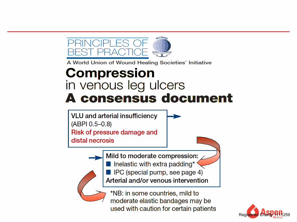
ABPI = 0.5 - 0.8
ABPI < 0.5
ABPI > 1.3 �
Unlikely to be arterial in origin
Mild peripheral
disease
Moderate arterial
disease
Severe arterial disease
Measure toe pressures or refer to specialist
Apply compression therapy
Apply compression therapy with caution
Do not compressrefer to specialist
Do not compress - refer urgently to vascular
specialist.

Formula to Calculate ABPI
Highest Ankle pressureAT/PT/DP for that leg=___________Highest Brachial pressure whether it is left or r ight.
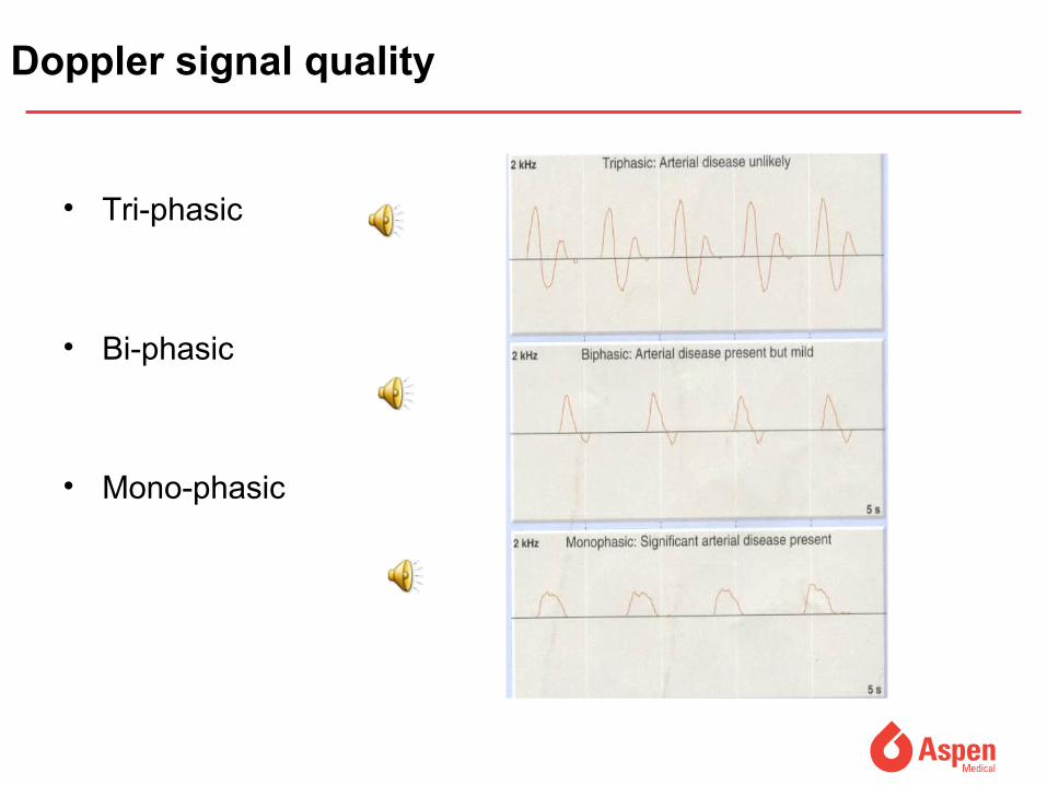
Doppler signal quality
• Tri-phasic
• Bi-phasic
• Mono-phasic

Other useful tests
• Wave form assessment• Exercise Doppler• Segmental pressures• Buergers test• Slow capillary return after blanching• Pole test
Pole Test
Waveform analysis
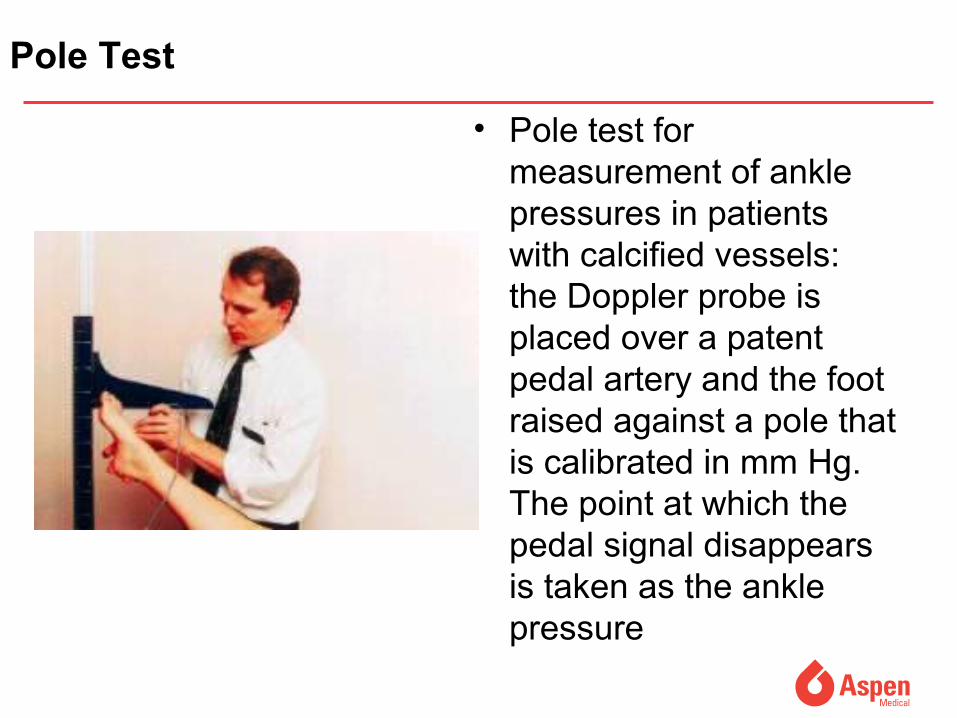
Pole Test
• Pole test for measurement of ankle pressures in patients with calcified vessels: the Doppler probe is placed over a patent pedal artery and the foot raised against a pole that is calibrated in mm Hg. The point at which the pedal signal disappears is taken as the ankle pressure

Other useful tests
Toe pressures: Doppler or photoplethysmography
• Toe/brachial pressure>0.6 = normal• Rest pain usually present in patients with index < 0.15• Absolute pressure in the toes of 20-30mmHg is
usually associated with rest pain

Inflate cuff to 60mmHg
Hold for 10 secs
Inflate by 10mmHg
Up to 100mmHg Then inflate by 20 mmHg
When the signal disappears take the reading below
If present at 180mmHg record this as the reading
How to use Pulse Oximetry
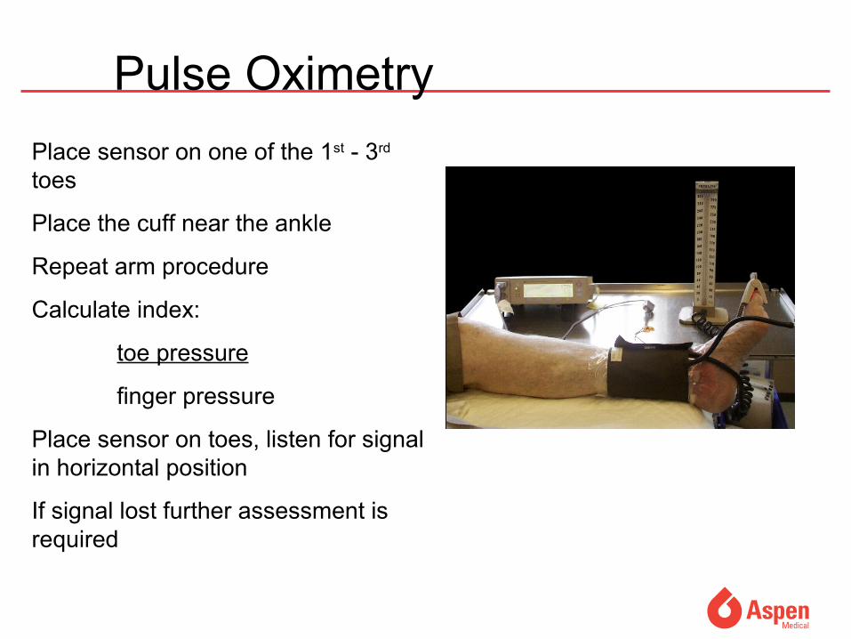
Place sensor on one of the 1st - 3rd toes
Place the cuff near the ankle
Repeat arm procedure
Calculate index:
toe pressure
finger pressure
Place sensor on toes, listen for signal in horizontal position
If signal lost further assessment is required
Pulse Oximetry

Pulse Oximetry Limitations
• Light reflection can be affected by hyper calcified nails
• Patients wearing nail varnish
• Patients suffering from chronic obstructive airways disease
• Can be affected with macro-vessel disease in diabetic patients

Problems with measuring ABPI using Doppler
• Difficult to maintain vessel contact during inflation and deflation
• A reasonable knowledge of anatomy is required
• Difficult to locate vessels
• Typical average time for ABPI is 11mins + 15-20 mins rest
(Ipsilon and Get ABI Study 2006)
• Clinicians must be trained and monitored (RCN Guidelines 2006)
• Doppler ABPIs taken by junior doctors disagreed with vascular technicians by 30%. This improved to 15% after formal training (Ray et al 1994)

The New Dopplex Ability
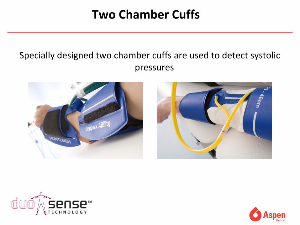
Specially designed two chamber cuffs are used to detect systolic pressures
Two Chamber Cuffs
• Extremely easy to use and fully automatic
• Rapid bi-lateral ABI measurement in < 5mins
(Doppler based ABI typically takes 30mins)• No need to rest patient for 15mins• ABI can now be undertaken by less skilled staff
• Only have to apply 4 cuffs
• Physiologically more accurate• No need to remove socks and tights• Integral printer for documentation of results and
waveforms• Automatic interpretation
• Clinically validated (Lewis et al, 2010)
Advantages of Auto ABI
• Improve venous return • Promote a healthy wound environment
Improve condition of skin/patient comfort • Reduce oedema • Control exudate and odour Reduce pain
Registered Charity 1111259

researchers at Charing Cross hospital in the late 80s
demonstrated that venous leg ulcers could be encouraged to
heal by the use of four-component pressure
bandaging;even chronic ulcers of many
years duration would healfor the first time
Registered Charity 1111259
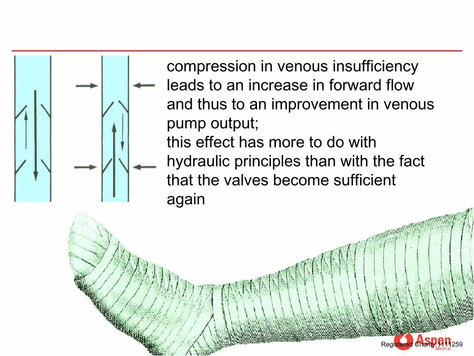
compression in venous insufficiency leads to an increase in forward flow and thus to an improvement in venous pump output;this effect has more to do with hydraulic principles than with the fact that the valves become sufficient again
Registered Charity 1111259
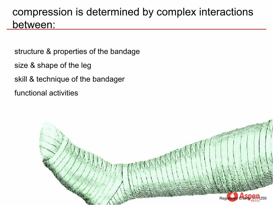
structure & properties of the bandage
size & shape of the leg
skill & technique of the bandager
functional activities
Registered Charity 1111259
compression is determined by complex interactions between:
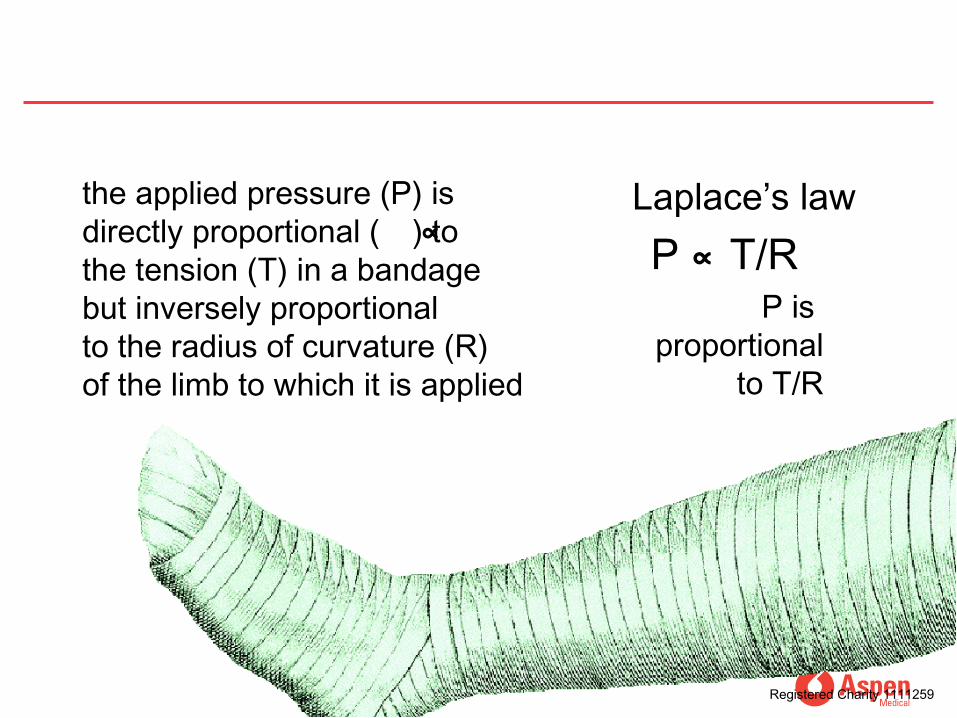
the applied pressure (P) is directly proportional ( ) to the tension (T) in a bandage but inversely proportional to the radius of curvature (R) of the limb to which it is applied
∝
P is proportional
to T/R
Laplace’s lawP ∝ T/R
Registered Charity 1111259
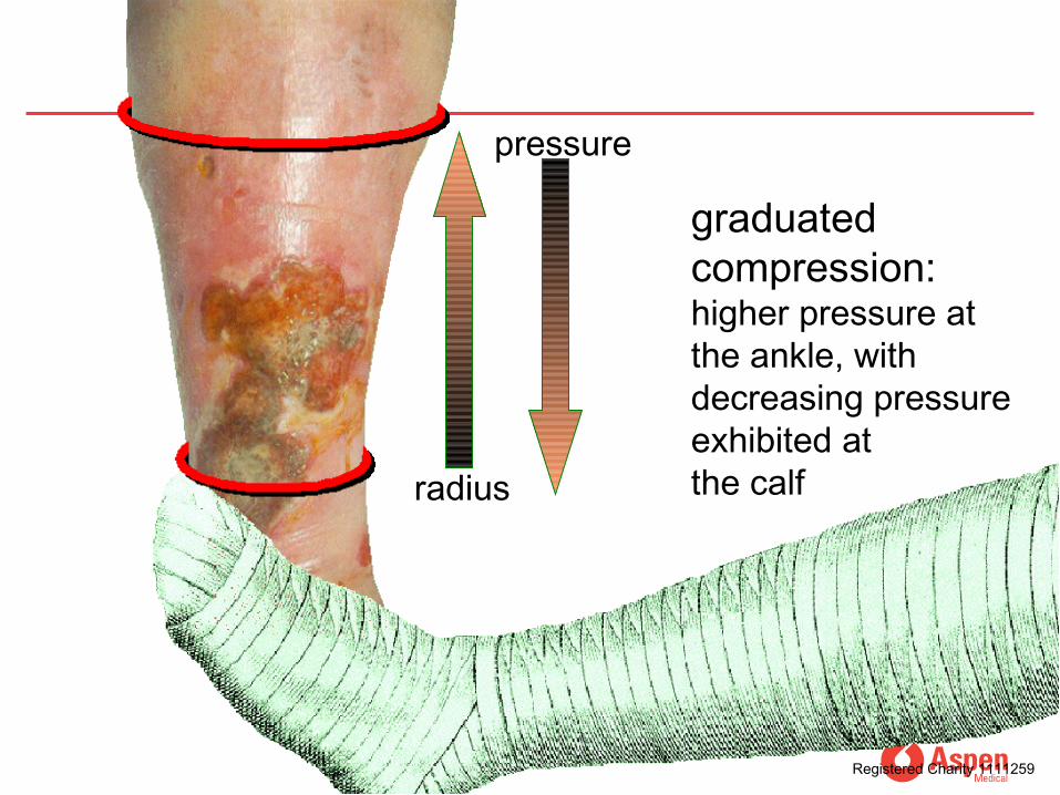
radius
pressure
graduated compression:higher pressure at the ankle, with decreasing pressure exhibited atthe calf
Registered Charity 1111259

BS 7505: 1995
Type 1 : Conforming and Retention
Type 2: Light Support
Type 3: Compression 3a: light compression - up to 20mmHg 3b: medium compression – up to 30mmHg3c: high compression – up to 40mmHg3d: very high compression – up to 50mmHg
Registered Charity 1111259
Bandage Classif ication
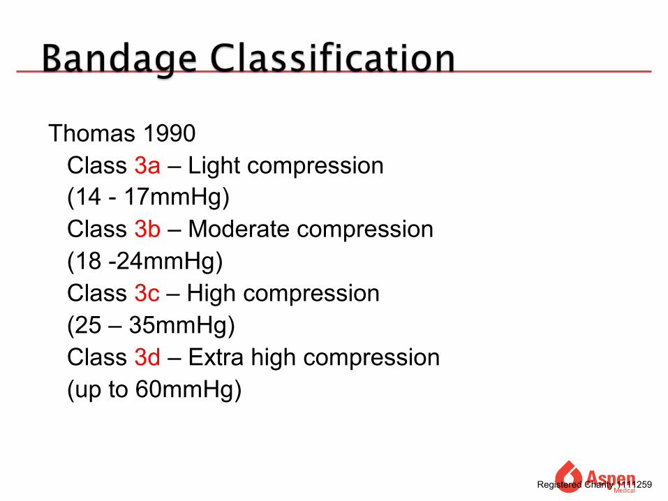
Thomas 1990Class 3a – Light compression (14 - 17mmHg) Class 3b – Moderate compression (18 -24mmHg)Class 3c – High compression (25 – 35mmHg)Class 3d – Extra high compression (up to 60mmHg)
Registered Charity 1111259

Registered Charity 1111259

Registered Charity 1111259
Registered Charity 1111259

Key points
• The majority of leg ulcers are venous in origin• Compression therapy is the treatment of choice• Choice of compression systems enhances
concordance• No ulcer will heal without a good blood supply• Other conditions are rare but need to be
considered.• Ensure you have time to get an good history with
relevant investigations to support your diagnosis• If in doubt document and refer to
multidisciplinary team

Finally
• Before diving in make sure you have assessed the risks.
• Thank you for your attention