dopamine
TRANSCRIPT

DopaminePET Imaging and Parkinson Disease
Shichun Peng, PhDa, Doris J. Doudet, PhDb,Vijay Dhawan, PhDa, Yilong Ma, PhDa,*
KEYWORDS
� Neurodegenerative disorders � Parkinsonism � Dopamine � PET � Imaging biomarkers � Diagnosis� Disease progression � Therapeutic intervention
KEY POINTS
� Dopamine (DA)-specific PET radioligands provide viable imaging biomarkers to describe dysfunc-tional molecular substrates underlying Parkinson disease (PD) and parkinsonian disorders.
� Presynaptic DA markers allow accurate discrimination of idiopathic PD from atypical PD and corre-late with the severity of clinical symptoms in individual patients.
� Postsynaptic DA markers enable quantitative measurement of endogenous DA release in chal-lenging conditions.
� Longitudinal changes in these DA markers have revealed critical insights on the neurobiologicalmechanisms associated with compensatory processes in the brain and therapeutic efficacy in clin-ical trials.
INTRODUCTION
Parkinson disease (PD) is a major movement dis-order resulting from the progressive degenerationof dopaminergic (DA) neurons in the nigrostriatalsystem, as well as non-DA neurons of serotonergicand noradrenergic origins in the brainstem andtheir related neural pathways. The loss of nigralneurons leads to the deficiency of dopamine(DA), a monoamine neurotransmitter, and is thebasis of DA replacement therapy with medicationsor cellular regenerative interventions. The loss ofserotoninergic and noradrenergic innervationsmay play a role in several nonmotor symptomssuch as mood and sleep disorders. Although theseneuronal deficits are often confined in a few iso-lated neurochemical pathways, they have signifi-cant consequences on the integrity of the brain
Disclosure: The authors have nothing to disclose.Support: This work was supported in part by NIH R01 NSClinical Research Center (M01 RR 018535) and the Morris71675) at The Feinstein Institute for Medical Research.a Center for Neurosciences, The Feinstein Institute for Me11030, USA; b Department of Neurology, University ofBritish Columbia V2Z 2J9, Canada* Corresponding author.E-mail address: [email protected]
PET Clin 8 (2013) 469–485http://dx.doi.org/10.1016/j.cpet.2013.08.0031556-8598/13/$ – see front matter � 2013 Elsevier Inc. All
by way of the widespread interconnectivity amongvarious brain circuits. It is this systemic impairmentin functional and anatomic substrates that under-lies motor and nonmotor symptoms in PD.1,2
Despite progressive striatal depletion, there is anextended presymptomatic phase in individual pa-tients, implying a sustained compensation pro-cess in the brain. Recent research suggests thatonset of motor symptoms may be associatedwith the failure of such preclinical compensatorymechanisms in PD.3
The diagnosis of idiopathic PD is based chieflyon the clinical manifestations of motor symptomsof resting tremor, rigidity, bradykinesia, and po-stural instability. The diagnostic decision is oftencomplicated by the similarity of symptoms in earlypatients with atypical parkinsonian disorders suchas multiple system atrophy (MSA), progressive
35069, R01 NS 32368, P50 NS 38370 and the GeneralK. Udall Center of Excellence for PD Research (P50 NS
dical Research, 350 Community Drive, Manhasset, NYBritish Columbia, 2221 Wesbrook Mall, Vancouver,
rights reserved. pet.theclinics.com

Peng et al470
supranuclear palsy (PSP), and corticobasal gangli-onic degeneration (CBGD). A misdiagnosis cansignificantly affect clinical management of patientsover the early course of the disease. A definitiveclinical diagnosis can only be established in pa-tients by long-term follow-up of 2 to 4 years byexperienced movement disorder specialists.Although quantitative rating scales have beenused to quantify the severity of clinical symptomsin patients, they are subjective and unable todetect any covert changes during preclinicalstages, and they are often insensitive to early signsof disease onset. It is therefore difficult to accu-rately distinguish idiopathic PD from atypical PDon clinical grounds alone.A wide variety of novel radiotracers have been
developed to characterize DA and non-DAdysfunction in PD using both positron emission to-mography (PET) and single-photon emissioncomputed tomography (SPECT). Specific neuro-chemical indicators of the disease process maybe measured more directly using radioligands thattarget particular neurotransmitters, neurotrans-porters, and neuroreceptors. By contrast, comple-mentary information on the disease-mediatedfunctional changes may be extracted indirectly interms of regional differences in cerebral perfusionand metabolism over the brain. In particular, thiseffort is directed toward the identification and vali-dation of disease-specific descriptors that canserve as imaging biomarkers for clinical andbiomedical translational studies. A viable imagingbiomarker should ideally meet the conditions sum-marized in Box 1.The applications of non–DA-specific imaging
methods in PD have previously been reviewed
Box 1Criteria for a viable imaging biomarker forneurodegenerative disorders
� High sensitivity and specificity for detectingdisease onset and for early differentialdiagnosis
� Clinical correlation with independent mea-sures of disease severity or behavioralabnormality
� High sensitivity to track longitudinal changeover the time course of disease progression
� High accuracy to assess therapeutic responsesand correlations with clinical outcomemeasures
� High test-retest reliability in patients withinan imaging center and excellent reproduc-ibility in independent patients acrossdifferent imaging centers
from the perspectives of neurochemistry4 andcerebral blood flow and metabolism.5 It has beenreported that brain network analysis of 2-deoxy-2-[18F]fluoro-D-glucose (FDG) PET images canprovide characteristic metabolic patterns to distin-guish patients with idiopathic PD or atypicalPD from healthy volunteers. Monkeys with spe-cific 1-methyl-4-phenyl-1,2,3,6-tetrahydropyridine(MPTP)–induced DA nigral lesions display thesame metabolic pattern as patients with PD.6
The expression of these disease-specific brainnetworks can be measured prospectively on asingle-case basis in individual subjects. Manystudies with FDG-PET imaging have been pub-lished in recent years and suggest that networkscores seem to fulfill the key criteria to serve asviable imaging biomarkers (see Ref.7).This article describes the latest development in
major DA-related radioligands and imaging tech-niques that are used clinically in human subjectsand parkinsonian patients. The emphasis of thearticle is on the resting-state studies in PD popula-tions without dementia. The article begins with abrief introduction to the normal neuroanatomyinvolved in DA projection systems. It also summa-rizes practical imaging protocols and the optimalanalytical methods designed to simplify the imple-mentation of these imaging techniques for clinicaland research applications. The article focuses ontheir use in providing imaging biomarkers for diag-nosis, progression, and treatment response.
NORMAL ANATOMY/IMAGING TECHNIQUE
There are 4 major pathways involved in the neuro-transmission of DA in the brain: the mesolimbic,mesocortical, nigrostriatal, and tuberoinfundibularpathways. Each of these pathways has a distinc-tive connection with the subdivisions of the stria-tum. The putamen is mainly involved in motorfunction, whereas the ventral striatum and thecaudate nucleus are primarily involved in limbicand cognitive processes. In addition, there is agradient of DAmotor innervation in the striatal sub-regions. This gradient is evident in the degenera-tion of the nigral neurons in the substantia nigrapars compacta in PD leading to a loss of DA termi-nals, beginning in the posterior putamen and, asthe disease progresses, spreading gradually tothe anterior putamen and caudate nucleus, butmostly sparing the ventral striatum. Imaging ofDA radiotracers reveals vital information on theintegrity of the nigrostriatal projection systemsand related cortical pathways from the perspec-tive of presynaptic and postsynaptic DA function.DA-specific radioligands are designed to havefast, high, and selective accumulation in the target

Evaluation of Dopamine Dysfunction in PD 471
brain region with rapid washout in the nonspecificreference region. Many optimal imaging protocolsand analytical methods have also been devised foraccurately measuring parameters of neurobiolog-ical interest in the brain.
Presynaptic Dopaminergic Function
Dopamine synthesisAssay of 6-[18F]fluoro-L-dopa (FDOPA) can pro-vide a sensitive measure of synthesis, storage,and turnover of DA in presynaptic DA nerve termi-nals.8–12 FDOPA PET imaging reflects the activityof aromatic amino acid decarboxylase (AADC),an enzyme largely responsible for catalyzing theconversion of L-dopa into DA.13,14 Other radio-fluorinated L-m-tyrosines, including 6-[18F]fluoro-L-m-tyrosine ([18F]FMT), are also substrates ofAADC and provide an index of DA synthesis andstorage,15,16 but do not undergo the same meta-bolic fate and do not provide information on DAturnover.17
Dopamine transporterDopamine transporter (DAT) is responsible forthe regulation of synaptic reuptake of DA in thenigrostriatal projection systems. Cocaine ana-logues basedon tropanes like [11C]-labeled 2b-car-bomethoxy-3b-(4-fluorophenyl)tropane ([11C]CFT)or methylphenidate ([11C]MP) and [18F]-labeledN-3-fluoropropyl-2-b-carboxymethoxy-3-b-(4-io-dophenyl)nortropane ([18F]FPCIT) are among theradioligandsmost commonly used for PET imagingof presynaptic DA nerve terminals in humans.18–20
Imaging of DAT has become more widely availablewith the use of commercial SPECT radioligands,also from the tropane family, such as [123I]-labeled(1R)-2-b-carbomethoxy-3-b-(4-iodophenyl)-tropane([123I]b-CIT)21 and its fluoroalkyl esters [123I]FPCIT.22 DAT binding seems to be unaffected byL-dopa treatment in monkey studies with SPECTand postmortem examination.23
Vesicular monoamine transporter type 2Vesicular monoamine transporter type 2 (VMAT2)is the transporter responsible for packing mono-amine neurotransmitters (DA, serotonin, andnoradrenaline) from the cytoplasm into vesiclesfor storage and subsequent synaptic release.[11C]dihydrotetrabenazine (DTBZ) can be usedas a reliable measure of monoaminergic nerve ter-minal density.24 More recently, [18F]-labeled 9-flu-oropropyl-(1)-DTBZ ([18F]AV-133) has beensuccessfully synthesized as a higher affinity fluo-ropropyl derivative of DTBZ.25 VMAT2 bindingseems to be less sensitive to drug-mediated orlesion-mediated regulation than other presynapticligands26 but it is not completely insensitive.27
However, this method has a lower signal/noise ra-tio than FDOPA uptake and DAT binding, but doesnot have specificity for DA terminals.2
Postsynaptic Dopaminergic Function
Radioligands such as [11C]raclopride and [11C]N-methylspiperone can provide sensitive mea-sures of local DA D2 receptor density in thestriatum. Newer tracers such as [11C]-labeled(S)-N-((1-Ethyl-2-pyrrolidinyl)methyl)-5-bromo-2-[11C]methoxy-3-methoxybenzamide ([11C]FLB 457)and [18F]fallypride are more sensitive in detectingchanges in D2 receptor binding in extrastriatalregions such as prefrontal and anterior cingulatecortices during the performance of cognitive func-tional tasks in normal subjects.28,29 Becauseligands of the benzamide family have limited affin-ity for the D2 receptors and compete with theendogenous ligand for the binding sites, changesin their binding potential reflect variations inendogenous DA levels in challenge situations.PET studies of these tracers have shown striataland prefrontal DA release in response to behav-ioral activations30,31 and pharmacologic stimula-tions32,33 in healthy volunteers.
In developing and validating the neurobiologicalusefulness of a novel DA-specific radioligand it isoften necessary to define its brain distribution insubcortical and cortical structures in healthy vol-unteers. An age-related decline of 5% to 8% perdecade is observed in striatal regions of normalcontrols and monkeys in most of the markers ofDA function described earlier.24,34,35 Age-relatedchanges in FDOPA uptake are absent as a resultof AADC upregulation in the aged DA terminals.The normative database can provide the magni-tude and range in regional uptake/binding of theradioligand as a function of healthy aging. This in-formation is necessary for accurately detectingfunctional abnormality on a single-case basis in in-dividual patients. Table 1 gives a summary of themost commonly used radiotracers and referencescited throughout the text.
Imaging Protocols and Analytical Methods
The salient features of imaging protocols for DA-related PET radioligands are included in Box 2.PET images are typically acquired in dynamicmode up to a period of 60/120 minutes for [11C]-labeled or [18F]-labeled radioligands immediatelyafter intravenous radiotracer injection. Imagingwith [11C]-based radioligands usually affords ahigher injected dose and shorter scanning dura-tion to compensate for the rapid decay of radioac-tivity caused by their shorter half-lives. In contrast,imaging with [18F]-based radioligands generally

Table 1PET radioligands for imaging dopaminergicfunction in PD
Radiolabel Radiotracers References
AADC
F18 FDOPA 8–14,17,22,35–37,
43,44,48,49,52,
56–60,63–65,
70,73–75,77,
78,81,85
F18 FMT 15–17,83,84
DAT
C11 CFT 18,40,65,86
C11 MP 19,34–37,41,56,
70,74,77,88
C11 FECIT 46
C11 PE2I 57
F18 FPCIT 20,50,62,71,72
VMAT2
C11 DTBZ 24,26,35–37,41,
56,64,65,70,
74,76,77,88
F18 AV-133 25,45,47
D2 receptor
C11 Raclopride 11,30–32,34,41,
57,66–68,76,
79,85–87
C11 FLB 457 28,69
F18 Fallypride 29,33
F18 Desmethoxyfally-pride
51
Abbreviations: FECIT, 2-b-carbomethoxy-3-b-(4-fluoro-phenyl)-tropane; PE2I, N-(3-iodoprop-2 E-enyl)-2b-carbo-methoxy-3b-(4-methylphenyl)nortropane.
Box 2Imaging protocols for DA-specific PETradioligands
� The participants are scanned following atleast 12 hours off medications of L-dopa andother central nervous system (CNS) drugs
� Radiotracers (5–15 mCi/185–555 MBq) areadministrated intravenously either as a singlebolus or by continuous infusion over a periodof time
� Dynamic imaging usually starts immediatelyafter radiotracer administration and consistsof a set of contiguous frames beginningwith finer sampling over early times andending with longer sampling over late times
� As an alternative, static image(s) may be ac-quired over a predetermined time windowwhen the radiotracer reaches a steady-stateequilibrium in uptake/binding
� Photon attenuation correction data are ob-tained using transmission images acquiredeither before or after the administration ofradiotracers, which is performed with PETwith point sources or with the computed to-mography (CT) part of a PET/CT dependingon the configuration of imaging systems
Peng et al472
requires a lower injected dose to compensate forhigher dosimetry, longer scanning duration, buthigher signal/noise ratio because of their longerhalf-lives. [18F]-based radioligands offer theunique ability for easy distribution of radiotracersfrom a cyclotron facility to satellite sites nearby.However, from a research perspective, with[11C]-labeled ligands, more than one radiotracermay be administered to a single subject in a day,leading to combined rapid evaluation of multipleaspects of the DA system.36,37
The time sampling is shorter early but is gradu-ally increased at late times in accordance withthe unique uptake characteristics of each partic-ular radiotracer. Projection data from dynamicacquisition are corrected online for physical ef-fects of photo attenuation, scatter, randoms, de-tector efficiency variation, and electronic dead
time. Attenuation is usually corrected using anattenuation map measured from a transmissionscan acquired either before or after the administra-tion of the radiotracer for a dedicated PET camera.For a PET/computed tomography (CT) scanner,this is achieved with a low-energy CT scan,adjusted for energy differences relative topositron-emitting radiopharmaceutical agents.The projection data are then reconstructed intothree-dimensional image volumes with analyticalmethods based on filtered back projection or iter-ative methods based on ordered subsets expecta-tion maximization algorithms.In addition to the clinical use for visualizing
functional abnormality, PET images are mostlyanalyzed to extract neurobiological parametersrepresenting uptake or binding of radioligands inthe brain. Over the last 2 decades, analyticaltechniques have evolved from absolute quantifi-cation with input functions derived from arterialblood sampling or dynamic imaging proceduresto semiquantitative measures with the use ofreference tissue models.38–42 Brain regions cho-sen as a reference tissue tend to be occipitallobe or cerebellum, known to have low to negli-gible binding sites for specific DA radioligands,which is best confirmed by postmortem analysisof particular imaging agents such as VMAT2
Box 3What the referring physician needs to know
� At the time of imaging, the patients have hadto withdraw from L-dopa or DA agonists forat least 12 hours and are not taking other
Evaluation of Dopamine Dysfunction in PD 473
protein in human brains.27 Both multiple-timegraphical analyses and target/background ratiomethods are simple, but valuable in estimatingparameters of interest for disease discriminationand clinical correlation.43–45
Two general approaches have been in use byclinicians and biomedical investigators. The firstmethod is based on the analysis of images overa set of predefined volumes of interest (VOIs).This hypothesis-driven approach is most suitablefor quantifying regional neurobiological parame-ters in the native space of each brain. The secondmethod is based on the brain mapping analysis ofimages in a standard anatomic space to providecomplementary information on disease-mediatedchanges in these measures over the brain. Thisdata-driven approach is most useful for localizingbrain regions showing functional abnormality orclinical correlates without an a priori hypothesis.
In recent years, it has become feasible tosimplify imaging protocols from dynamic to staticacquisition at later stages following radiotraceradministration. A simple method of standardizeduptake value ratio can be used for image analysiswhen the uptake of a tracer establishes a steadystate.43–45 As a result, imaging acquisition for thistype of radioligands can be conducted like anFDG-PET study. Scanning can begin after the sub-jects complete the necessary period of radiotraceruptake off the scanner. This simplification in-creases compliance, especially in patients with se-vere disability, decreases the patient discomfortassociated with a longer scanning session, and in-creases the throughput in the imaging facility.
CNS medications, such as neuroleptics
� There may be large intraindividual and inter-individual variability in DA-specific imagingoutcomes because of differences in subjectage and possibly gender
� DA-specific imaging markers decrease withsubject age, and comparison with age-matched database values is necessary foradequate interpretation
� Severity of clinical symptoms may be associ-ated with lower image quality in elderly sub-jects or patients with advanced disease
� Differential diagnosis of idiopathic or atyp-ical PD may be confirmed clinically over afollow-up period of at least 2 years or/andby the striatal distribution pattern of the pre-synaptic DA tracer
� Some individuals with normal presynaptic ni-grostriatal DA function may show signs ofmanifesting parkinsonism, such as essentialtremor and dystonic tremor, over the earlycourse of the disease
IMAGING FINDINGS/PATHOLOGY
Clinicians identify patients with idiopathic PD bythe appearance of at least 2 of the 4 cardinal motorfeatures described earlier. The patients are evalu-ated clinically in the practically defined Off statewith Unified Parkinson Disease Rating Scale(UPDRS), Hoehn and Yahr Scale (HY), andSchwab and England Activities of Daily Living(ADL) Scale. In general, a higher score on one ormore of these scales is associated with a longerdisease duration in an individual patient. Such rat-ings are less useful in patients showing the earliestsigns of motor dysfunction but more useful in pa-tients confirmed to have idiopathic PD. In partic-ular, these measures may not be sensitiveenough to reflect subtle incremental changes re-sulting from disease progression and followingneuroprotective or symptomatic therapies. Earlypatients with atypical parkinsonism similarly maynot be separated adequately by clinical ratingscales.
The molecular imaging modality with DA-specific PET radioligands has played importantroles in the assessment of PD pathophysiologyand differential diagnosis between idiopathic andatypical patients with PD, and for the evaluationof the efficacy of novel experimental therapeutics.Among presynaptic DA markers, FDOPA uptakeand DAT binding are used more frequently, fol-lowed by VMAT2 binding. In contrast, D2 receptorbinding is the most common postsynaptic DAmarker. This marker alone is not particularly use-ful for diagnosis purposes, but is valuable in com-bination with FDOPA/DAT or for measuringendogenous DA release. Both VOI-based andvoxel-based analytical methods are often usedeither separately or concurrently in practice.31,46,47
Basic information for physicians considering theuse of DA-specific imaging is presented in Box 3.
Disease Diagnosis
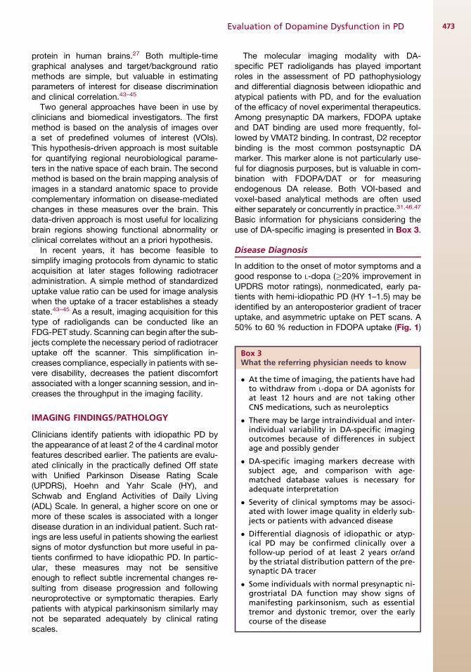
In addition to the onset of motor symptoms and agood response to L-dopa (�20% improvement inUPDRS motor ratings), nonmedicated, early pa-tients with hemi-idiopathic PD (HY 1–1.5) may beidentified by an anteroposterior gradient of traceruptake, and asymmetric uptake on PET scans. A50% to 60 % reduction in FDOPA uptake (Fig. 1)
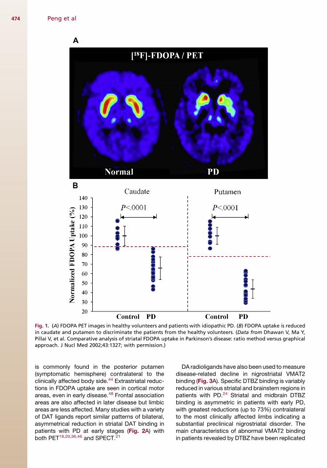
Fig. 1. (A) FDOPA PET images in healthy volunteers and patients with idiopathic PD. (B) FDOPA uptake is reducedin caudate and putamen to discriminate the patients from the healthy volunteers. (Data from Dhawan V, Ma Y,Pillai V, et al. Comparative analysis of striatal FDOPA uptake in Parkinson’s disease: ratio method versus graphicalapproach. J Nucl Med 2002;43:1327; with permission.)
Peng et al474
is commonly found in the posterior putamen(symptomatic hemisphere) contralateral to theclinically affected body side.44 Extrastriatal reduc-tions in FDOPA uptake are seen in cortical motorareas, even in early disease.48 Frontal associationareas are also affected in later disease but limbicareas are less affected. Many studies with a varietyof DAT ligands report similar patterns of bilateral,asymmetrical reduction in striatal DAT binding inpatients with PD at early stages (Fig. 2A) withboth PET18,20,36,46 and SPECT.21
DA radioligands have also been used tomeasuredisease-related decline in nigrostriatal VMAT2binding (Fig. 3A). Specific DTBZ binding is variablyreduced in various striatal and brainstem regions inpatients with PD.24 Striatal and midbrain DTBZbinding is asymmetric in patients with early PD,with greatest reductions (up to 73%) contralateralto the most clinically affected limbs indicating asubstantial preclinical nigrostriatal disorder. Themain characteristics of abnormal VMAT2 bindingin patients revealed by DTBZ have been replicated

Fig. 2. (A) FPCIT PET images in healthy volunteers and patients with idiopathic PD. DAT binding is reduced incaudate and putamen to discriminate the patients from the healthy volunteers. (B) Increased motor symptomsand progressive reduction in presynaptic DA biomarker in patients with idiopathic PD measured with FPCITPET. (Data from Huang C, Tang C, Feigin A, et al. Changes in network activity with the progression of Parkinson’sdisease. Brain 2007;130:1838–9; with permission.)
Evaluation of Dopamine Dysfunction in PD 475
more recently with [18F]AV-13345,47 as well as inpostmortem analysis.27 The diagnostic criteria forPET imaging in PD are given inBox 4. Different pre-synaptic markers show an excellent diagnostic ac-curacy between early-stage idiopathic PD andhealthy controls, with a sensitivity of 95% to100% and specificity of 90% to 95%.
PET imaging with presynaptic markers has beenreplicated in nonhuman primate models.14 One
study analyzed FDOPA, MP, and DTBZ images inthe same monkeys with mild to severe systemicMPTP lesions.35 The mean reduction of the bind-ing of the three ligands was variable in the caudateand putamen and depended on the symptomseverity. Binding of all three ligands was also var-iably reduced in the anterior cingulate cortex,brainstem, and thalamus, reflecting toxicity ofMPTP for extrastriatal catecholamine innervations.
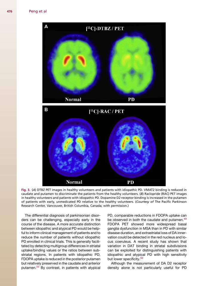
Fig. 3. (A) DTBZ PET images in healthy volunteers and patients with idiopathic PD. VMAT2 binding is reduced incaudate and putamen to discriminate the patients from the healthy volunteers. (B) Raclopride (RAC) PET imagesin healthy volunteers and patients with idiopathic PD. Dopamine D2 receptor binding is increased in the putamenof patients with early, unmedicated PD relative to the healthy volunteers. (Courtesy of The Pacific ParkinsonResearch Center, Vancouver, British Columbia, Canada; with permission.)
Peng et al476
The differential diagnosis of parkinsonian disor-ders can be challenging, especially early in thecourse of the disease. A more accurate distinctionbetween idiopathic and atypical PDwould be help-ful to inform clinical management of patients and toreduce the number of patients without idiopathicPD enrolled in clinical trials. This is generally facili-tated by detectingmultigroup differences in striataluptake/binding values or the ratios between sub-striatal regions. In patients with idiopathic PD,FDOPA uptake is reduced in the posterior putamenbut relatively preserved in the caudate and anteriorputamen.22 By contrast, in patients with atypical
PD, comparable reductions in FDOPA uptake canbe observed in both the caudate and putamen.49
FDOPA PET showed more widespread basalganglia dysfunction in MSA than in PD with similardisease duration, and extrastriatal loss of DA inner-vation could be detected in the red nucleus and lo-cus coeruleus. A recent study has shown thatvariation in DAT binding in striatal subdivisionscan be exploited for distinguishing patients withidiopathic and atypical PD with high sensitivitybut lower specificity.50
Although the measurement of DA D2 receptordensity alone is not particularly useful for PD

Box 4Diagnostic criteria of idiopathic PD
Clinical criteria
PD may be diagnosed clinically by identifyingthe earliest signs of motor dysfunction and agood response to an acute dose of L-dopa(�20% improvement in objective disease mea-sures of motor symptoms).
Motor symptoms usually appear unilaterally atdisease onset and include resting tremor, rigid-ity, bradykinesia, and postural instability.
Imaging criteria
PD may be diagnosed with PET imaging by thenonuniform distribution of presynaptic DAmarkers.
The early-stage PD is characterized by an asym-metry and gradient in presynaptic DA functionwith the largest reduction in the contralateraldorsal-posterior putamen compared with thecaudate nucleus and the ventral-anteriorputamen.
A quantitative diagnostic confirmation may bemade when striatal DA loss exceeds a certainthreshold. The threshold is determined in poste-rior putamen in reference to a normative data-base.
Box 5Differential diagnosis of idiopathic andatypical PD
The differential diagnosis between idiopathicand atypical PD is important for adequate clin-ical care of patients in early phases of the dis-ease course and for phenotypical patientscreening before enrollment in clinical trials.
Key diagnostic features
Presynaptic DA markers may be useful in earlydetection of idiopathic PD by taking into ac-count the asymmetry of clinical symptoms andthe uneven distribution of striatal DAhypofunction.
Patients with atypical PD tend to show symmet-ric clinical symptoms of parkinsonism with com-parable reductions in presynaptic markers inboth the caudate and putamen. Patients withMSA develop autonomic symptoms such assevere orthostatic hypotension and cerebellarsymptoms like ataxia. By contrast, patientswith PSP develop parkinsonism with earlypostural instability, falls, and oculomotorabnormalities.
Enhancement of diagnostic accuracy
The diagnostic performance can be improvedwith the use of rigorous statistical metrics suchas receiver operating characteristic analysis.The accuracy of differential diagnosis may beenhanced by including other information likeintrastrial ratios of imaging markers as addi-tional outcome measures.
Evaluation of Dopamine Dysfunction in PD 477
diagnosis, DA D2 receptor binding allows for thedifferentiation of patients with idiopathic and atyp-ical PD with high accuracy.51 This study confirmsthe relative sparing of D2 receptors in theDA-denervated putamen of idiopathic patients, incontrast with a more substantial loss of striatalDA receptors in atypical patients. However, thisdifferential DA topography is often insufficient todiscriminate idiopathic PD from atypical PD atearly clinical stages. Furthermore, some (10%–15%) patients have parkinsonian-type motorsymptoms but normal DA substrates based onFDOPA PET and DAT SPECT scans,52,53 a phe-nomenon nicknamed scans without evidence ofDA deficiency [SWEDD] and thought to be relatedto dystonic tremor.54 The factors influencing thedifferential diagnosis are provided in Box 5.Although DA imaging with multitracers canimprove the accuracy of differential diagnosis,55
this is usually not cost-effective or feasible outsideof research-oriented specialty centers.
PET imaging has revealed both similarities anddifferences between idiopathic PD and familialPD. An early study found that carriers of PD-specific mutations such as leucine-rich repeatkinase 2 (LRRK2) are nearly indistinguishable
from idiopathic cases on PET markers of DAdysfunction.56 Both presynaptic and postsynapticstriatal DA markers are reduced to similar levels inpatients with early-onset PD with or without muta-tions of the parkin gene,57 with FDOPA uptakereduced by 32% and 60% in caudate and puta-men, and DAT binding decreased by 44% and59% compared with normal control values.Further analysis revealed extrastriatal decreasesof FDOPA uptake or DAT binding in substantial ni-gra. The reduction in presynaptic markers wasalso observed in mutation carriers of PTEN-induced putative kinase 1 (PINK1) causing reces-sively inherited early-onset PD.58 Striatal FDOPAuptake declined not only in homozygous mutationcarriers but also, to a lesser extent, in the putamenof heterozygous mutation carriers.
Clinical Correlation
A robust imaging biomarker has to show clinicalcorrelations with objective measures of disease

Peng et al478
severity and cognitive impairment in PD. UPDRSmotor ratings correlated consistently with striatalvalues in presynaptic DA markers such as FDOPAuptake,43,59 DAT binding,19,20 and VMAT2 bind-ing.47 By contrast, striatal reductions in VMAT2binding correlated significantly with PD durationand SE scores, but not with HY stage or with motorUPDRS subscale scores.24 The side and severityof DAT/VMAT2 binding reduction significantlycorrelated with the severity and asymmetry of clin-ical motor scores.24,46 In addition, extrastriatalDAT dysfunction could predict the conversionfrom HY 1 to HY 2.18
PET imaging with DA radioligands has also beenuseful in identifying nonmotor functional abnor-mality such as cognitive correlates in caudateand putamen. Mental and motor components ofexecutive dysfunction were reported to correlatewith FDOPA uptake in the caudate and putamenin patients with advanced PD.60 DAT binding los-ses in the putamen correlated with motor andcognitive dysfunction involved in the recognitionof emotional gestures in PD.19 This loss was posi-tively related to the reduction of ventrolateral pre-frontal activation. DAT dysfunction in the caudatebut not the putamen was associated with restingbrain metabolic changes underlying cognitiveimpairment.61,62 These studies support the notionthat a loss of DA neurotransmission in the puta-men/caudate results in impairment in motor andcognition domains.Several studies have investigated behavioral
correlations between PET imaging of presynap-tic DA markers and postmortem measurementsin MPTP-lesioned nonhuman primate modelsof experimental parkinsonism. The degree ofmotor dysfunction correlated negatively withFDOPA uptake and DTBZ binding potential,tyrosine hydroxylase (TH)–immunoreactive cellcounts in substantia nigra, striatal DA markers(TH, DAT, and VMAT2), and striatal DA concen-tration.14,63,64 There was a critical threshold ofnigral cell loss and DA innervation distinguishingbetween the asymptomatic and symptomaticparkinsonian monkeys. This intoxication proto-col established a close correlation between cellloss in the substantia nigra, striatal DA depletion,and the severity of motor symptoms. In monkeyswith hemiparkinsonism imaged with FDOPAand tracers of DAT and VMAT2,65 striatal uptakefor each radiotracer correlated strongly with ste-reological nigral cell counts for nigral loss lessthan 50% but very strongly with striatal DAover the full range of depletion. This findingmay explain differences between neuroimagingand clinical end points reported in some humantrials.
Imaging of Endogenous DA
Some D2-specific radiotracers (eg, raclopride) areeasily displaced by endogenous DA in challengeconditions, making them suitable for imaging DArelease in intact or unaffected DA terminals. Forinstance, this ability allowed demonstration ofreduced DA release in patients with PD in variousbehavioral and pharmacologic challenges,32,66
and observation of a placebo effect in patientswith PD.67 DA release in patients with early-stagePD was reduced in the dorsal caudate but pre-served in the medial prefrontal cortex during theperformance of cognitive tasks related to execu-tive dysfunction.31 This work shows that executivedeficits in patients with early PD are associatedwith impaired nigrostriatal DA function, resultingin impairment in the corticobasal ganglia circuit,but mesocortical DA transmission seems well pre-served in these patients. DA release was also de-tected in the ventral striatum and in the midbrainin patients with PD with impulse control disorderas a result of treatment with DA agonists,68,69
showing the abnormal DA processing underlyingthe medication-induced impulse control disorderin PD.
Disease Progression
Natural history studies of disease progression arecritical for assessing the efficacy of neuroprotec-tive and therapeutic trials to prevent continueddeterioration in the functional integrity of DA nerveterminals. The former are designed to delay thesymptom onset in at-risk populations, whereasthe latter are designed to slow the rate of progres-sion in patients. Over the last 10 years, multitracerPET imaging has increasingly been used to followthe degeneration of DA innervation in PD and ani-mal models. This approach has not only offeredgreater insights on the neurobiological mechanismof neurodegeneration but has also allowed theassessment of the relative performance of PET li-gands as potential biomarkers of PD. The prosand cons of DA-specific imaging are listed inBox 6.Many longitudinal studies have been published
on neurochemical progression in presynaptic DAintegrity in subjects at risk of developing PD andin patients with this disorder. PET studies withFDOPA and tracers of DAT and VMAT2 showthat striatal indices in 3 presynaptic DA markerswere variably decreased in patients with PD36
and over the course of progression in asymptom-atic members of parkinsonian LKKR2 kindred.70
Reduced DAT binding was the earliest indicationof subclinical DA dysfunction and progression toclinical disease was generally associated withthe emergence of abnormal FDOPA uptake.
Box 6Pearls, pitfalls, and variants
Pearls for DA PET imaging
A single scan acquired some time after radiotracer injection may be sufficient for separating patientswith idiopathic PD from healthy controls and patients with atypical PD in a clinical researchenvironment.
Presynaptic DA markers are reduced relative to healthy controls and decreased with the increasedseverity of clinical symptoms in patients with advanced disease. There is significant correlation betweenasymmetry of striatal values and clinical asymmetry measured with the motor UPDRS in patients withidiopathic PD.
Atypical patients with PD have more uniform reductions in striatal DA markers compared with healthycontrols.
Pitfalls for DA PET imaging
A. Binding to non-DA nerve terminals
FDOPA and VMAT2 are nonspecific to DA andmay label noradrenaline. DAT is more specific for DAeven though some radiotracers may have very low binding to serotonin transporters.
B. Upregulation: FDOPA, D2 receptor; downregulation: DAT, VMAT2
Most VMAT2 ligands are localized on DA nerve terminals and are less susceptible to compensatorychanges during the course of the disease.
C. Influence of medications
DAT and VMAT2 are less affected by antiparkinsonian dopaminergic medications such as L-dopa.
D. Variability of methodological factors
DA-specific markers are influenced by differences in imaging protocols and characteristics oftomographic systems.
Variants for DA PET imaging
Clinical diagnosis and rating scales of the disease may be subjective and depend on medications and theeffect of placebo.
Most presynaptic and postsynaptic DA markers decrease with age, leading to lower image quality inolder subjects.
Presynaptic DA markers show a flooring effect with disease progression resulting in lower statisticalpower. DAT is sensitive to the floor effect early on.
Be aware of parkinsonian patients with other forms of secondary parkinsonism or having SWEDD.
Evaluation of Dopamine Dysfunction in PD 479
In one study, 15 patients initially with hemi-PDwith disease duration less than or equal to 2 yearswere scanned with a DAT tracer and FDG-PET toevaluate longitudinal changes in striatal DAT den-sity and PD-related metabolic brain network(PDRP) expression at baseline and at 2 and4 years.71 Increased rates in UPDRS motor ratingscorrelated with loss rates of striatal DAT bindingmeasured in both caudate and putamen over thecourse of the follow-up (see Fig. 2B). The deterio-ration of motor symptoms correlated similarlywith changes in striatal DAT binding and PDRPnetwork activity. This finding had been replicatedwhen conducting the same analysis separately in
hemispheres ipsilateral or contralateral to themore affected limbs based on clinical diagnosisat baseline.72 Putamen DAT binding in the clinicalhemisphere was lower but progressedmore slowlycompared with the preclinical hemisphere. Bycontrast, decreased caudate DAT binding orincreased PDRP activity was also evident in pre-clinical hemispheres with rates of change similarto those measured concurrently in clinical hemi-spheres. Although most work used a linear regres-sion to describe the trajectory of progression over ashort interval, a nonlinear decline in FDOPA uptakehas been reported in the contralateral putamen,being faster at the beginning of the disease.73
Peng et al480
In another study, 78 patients and 35 controlswere scanned using PET with FDOPA and tracersof DAT and VMAT2 at baseline and 4 and 8 yearsof follow-up.74 Greater reductions in the three pre-synaptic DA markers were related exponentially tolonger symptom duration in individual patients.Further, the degree of denervation at diseaseonset was different between striatal subregionsbut the relative rate of disease progression wassimilar among these subregions. Although the an-teroposterior gradient of severity was unchangedfor DA synthesis, storage, and reuptake, the asym-metry between themore and less affected striatumbecame less prominent over the disease course.These findings agree with those from DAT bind-ing71,72 and FDOPA uptake.59,75
In a multitracer longitudinal study in MPTP mon-keys, an early (2 months) decrease (46%) of striatalVMAT2 in asymptomatic animals precededchanges in DAT and D2 receptors, despite the pro-gressive loss of all presynaptic DA markers in thestriatum with expression of parkinsonism.76 Thisfinding concurs with the finding in long-termMPTP monkeys with mild unilateral lesions.35
These results suggest that decreased VMAT2 isa key pathogenic event that precedes nigrostriatalDA neuron degeneration. The loss of VMAT2 mayresult from an association with alpha-synucleinaggregation induced by oxidative stress.
Compensatory Mechanisms in PD
The longitudinal paradigm with DA-specific radio-ligands has also been used for PET imaging ofcompensatory mechanisms in PD.3 A study ofDA synthesis capacity in normal aging showedgreater striatal uptake in older adults than inyounger adults,16 indicating possible compensa-tion for deficits elsewhere in the DA system. A lon-gitudinal study with 3 presynaptic DA markers ofDA synthesis, DAT, and VMAT2 reported thatyounger patients with PD may have more efficientcompensatory mechanisms.77 At symptom onset,the loss of putamen DTBZ binding was substan-tially greater in younger compared with older pa-tients with PD. However, the rate of DTBZbinding loss was significantly slower in youngerpatients. The estimated presymptomatic phaseof the disease spanned more than 20 years inyounger patients, compared with 10 years in olderpatients. These observations suggest that youngerpatients with PD progress more slowly and areable to endure more damage to the DA systembefore the appearance of first motor symptoms.The age effects on disease progression revealed
by DTBZ binding were absent in measures fromFDOPA and DAT binding. The analysis of this
dataset also yielded evidence for possible upregu-lation of DA synthesis and downregulation of DATin the more severely affected putamen in the earlystage of PD.37 However, the normalized FDOPAand DAT binding values tended to approachVMAT2 binding values in the putamen in laterstages of disease, when the rates of decline inthe PET measures were similar for all the markers,suggesting that compensatory mechanismsdecrease with the progression of PD.Compensatory responses in intact monoamine
neuron perikarya have also been examined bymeasuring extrastriatal FDOPA uptake.75 Progres-sive loss in FDOPA uptake occurred in putamen(8.1%), locus coeruleus (7.8%), internal globuspallidus (7.7%), caudate (6.3%), and hypothala-mus (6.1%). At baseline, levels of FDOPA uptakein internal pallidum and locus coeruleus increasedin PD compared with controls, indicating possiblecompensatory upregulation. These increasedlevels normalized (internal pallidum) or becamesubnormal (locus coeruleus) at follow-up, suggest-ing exhaustion of compensation within the firstyears of disease. This finding agrees with an earlycross-sectional study showing normal orincreased FDOPA uptake in the substantia nigra,midbrain raphe, and locus coeruleus in early butnot advanced PD.48 The red nucleus, subthala-mus, ventral thalamus, and pineal gland werealso eventually involved. However, FDOPA uptakemay also measure serotoninergic function in themedian raphe nuclei complex, which has low orno DA innervation.78 These studies show thatloss of monoaminergic function in extrastriatal re-gions is delayed and occurs independently fromnigrostriatal degeneration.An increase of striatal DA D2 receptor binding
has been reported with raclopride in patientswith early untreated PD particularly in the putamencontralateral to the more affected body side (seeFig. 3B).79 D2 receptor upregulation is evident inthe striatum of human patients or animals withMPTP-induced parkinsonism. However, the initialupregulation in putamen may reverse with DAtreatment, and increasing disease severity andbinding values in the range of control subjects orlower may be encountered in patients withadvanced PD. Because changes in raclopridebinding and FDOPA uptake are associatedthroughout the disease course, it is likely that DAD2 receptor changes result from the decline in pre-synaptic DA drive.In the study with FDOPA, MP, and DTBZ in long-
term MPTP monkeys,35 the decline in MP bindingin the ventral striatum (�75%) exceeded the de-clines of DTBZ binding and FDOPA uptake inthat region (�65%), suggesting that compensatory

Evaluation of Dopamine Dysfunction in PD 481
downmodulation of uptake sites may occur in thestriatal regions with the least DA depletion. In thesubacute unilateral MPTP model,65 indices ofstriatal uptake of FDOPA, DAT, and VMAT2 corre-lated most strongly with each other, supporting alack of differential regulation within 2 months afternigrostriatal injury. Another study reported thatcompensatory changes in nigrostriatal DA activityoccurred in the recovered and parkinsonian mon-keys when DA depletion was at least 88% ofcontrol,64 which may be too late to explaincompensatory mechanisms in the early asymp-tomatic period.
Evaluation of Antiparkinsonian Therapeutics
PET imaging with DA-specific radioligands hasbeen successfully used to assess the therapeuticefficacy and possible complications of noveldisease-modifying therapies, which is neededbecause clinical measures of disease severitymay be subjected to placebo effects often presentin neuropsychiatric patients participating inresearch trials. As an imaging marker, FDOPA up-take in the striatum is considered the gold stan-dard (Fig. 4) in several open-label as well as
Fig. 4. Changes in motor symptoms and presynaptic DA bibased experimental therapies. (A) FDOPA PET images in onand 4 (POST-4Y) years after fetal DA cell transplantation. (Buptake in the putamen are observed at 1, 2, and 4 years intransplantation. *P<.05, **P<.01, 1P<.005, 11P<.0005. (Datransplantation in Parkinson’s disease: challenge and persp
double-blind, placebo-controlled clinical trials ofstriatal DA cell transplantation in patients withadvanced PD (see Ref.80). It has been shownthat clinical improvement after fetal DA tissuegrafting is associated with increased FDOPA up-take in the transplanted putamen over a periodof 4 to 15 years.81,82 The efficacy of gene therapyhas been evaluated in phase 1 trials using PET im-aging with the AADC-specific radiotracer FMT.Short-term clinical improvement in the patientswas related to increased FMT uptake up to 2 yearsafter adeno-associated virus (AAV) vector-mediated gene delivery of AADC into the putamenof patients with PD.83,84 The multitracer approachhas proved to be useful in long-term follow-upstudies after the implantation of fetal cells in pa-tients with PD,82 as well as retinal pigment epithe-lial cells and neural stem cells in nonhumanprimate models of PD.85,86 These studies showthat increased FDOPA uptake is correlated withgraft-induced DA release in the putamenmeasured by changes in raclopride binding.
It has been suggested that loss of DA nerve ter-minals in association with changes in postsynapticDA receptors may underlie motor complicationsoccurring in the course of treatment of PD. In
omarkers in patients with idiopathic PD following cell-e PD patient before (PRE) and 1 (POST-1Y), 2 (POST-2Y)) Decreased UPDRS motor ratings and increased FDOPA15 patients with advanced PD following fetal DA cell
ta from Ma Y, Peng S, Dhawan V, et al. Dopamine cellective. Br Med Bull 2011;100:177–9; with permission.)
Peng et al482
patients with PD with and without dyskinesia, 1hour after oral administration of levodopa/carbi-dopa,87 levodopa-induced increases in synapticDA levels in the striatum correlated positivelywith duration of PD symptoms. Patients withpeak-dose dyskinesias had larger 1-hour in-creases in synaptic DA levels than stable re-sponders, with no differences 4 hours afterlevodopa. Intermittent, large, levodopa-inducedincreases in synaptic DA concentration may leadto dramatic changes in receptor pharmacologyand have been associated with the emergence ofpeak-dose dyskinesias in PD. In another study,88
the putaminal DAT/VMAT2 binding ratio wasdecreased in patients with dyskinesia comparedwith those without dyskinesia. DAT downregula-tion may minimize symptoms by contributing toincreased synaptic DA levels in early PD.
SUMMARY
PET studies have proved to be indispensable inproviding information about PD using radioligandstargeting nigrostriatal DA terminals. The develop-ment of presynaptic radiotracers for DA synthesis,DAT, and VMAT2 has led to viable means for imag-ing the nigrostriatal DA system. Some radiotracersbinding to postsynaptic D2 receptors offer a quan-titative approach to the measurement of endoge-nous DA release in relation to behavioral orpharmacologic stimulations. Many optimized im-aging protocols and analytical methods are usedto generate imaging biomarkers for clinical andresearch applications. The bulk of the work isfocused on investigating the interaction betweenDA-specific lesions and impaired brain circuitsthat subserve motor and cognitive symptoms inPD. The results have provided new insights intothe relationship between localized DA dysfunctionand downstream effects within widely distributedfunctional brain networks over the course of dis-ease onset and progression. This endeavor hasled to advances in early differential diagnosis, inthe understanding of molecular compensatoryprocesses, and in the design of therapeutic trialsin PD.
REFERENCES
1. Barrio JR, Huang SC, Phelps ME. Biological imag-
ing and the molecular basis of dopaminergic dis-
eases. Biochem Pharmacol 1997;54:341–8.
2. Stoessl AJ. Functional imaging studies of non-
motoric manifestations of Parkinson’s disease.
ParkinsonismRelat Disord 2009;15(Suppl 3):S13–6.
3. Appel-Cresswell S, de la Fuente-Fernandez R,
Galley S, et al. Imaging of compensatory
mechanisms in Parkinson’s disease. Curr Opin
Neurol 2010;23:407–12.
4. Brooks DJ, Pavese N. Imaging biomarkers
in Parkinson’s disease. Prog Neurobiol 2011;95:
614–28.
5. Ma Y, Peng S, Dhawan V, et al. Cerebral glucose
metabolism and blood flow in Parkinson’s disease.
In: Eidelberg D, editor. Imaging in Parkinson’s
disease. New York: Oxford University Press;
2011. p. 21–31.
6. Ma Y, Peng S, Spetsieris PG, et al. Abnormal meta-
bolic brain networks in a nonhuman primate model
of parkinsonism. J Cereb Blood Flow Metab 2012;
32:633–42.
7. Poston KL, Eidelberg D. FDG PET in the evaluation
of Parkinson’s disease. PET Clin 2010;5:55–64.
8. Huang SC, Yu DC, Barrio JR, et al. Kinetics and
modeling of L-6-[18F]fluoro-dopa in human posi-
tron emission tomographic studies. J Cereb Blood
Flow Metab 1991;11:898–913.
9. Cumming P, Leger GC, Kuwabara H, et al. Pharma-
cokinetics of plasma 6-[18F]fluoro-L-3,4-dihydroxy-
phenylalanine ([18F]Fdopa) in humans. J Cereb
Blood Flow Metab 1993;13:668–75.
10. Takikawa S, Dhawan V, Chaly T, et al. Input func-
tions for 6-[fluorine-18]fluorodopa quantitation in
parkinsonism: comparative studies and clinical
correlations. J Nucl Med 1994;35:955–63.
11. Antonini A, Vontobel P, Psylla M, et al. Complemen-
tary positron emission tomographic studies of the
striatal dopaminergic system in Parkinson’s dis-
ease. Arch Neurol 1995;52:1183–90.
12. Sossi V, Doudet DJ, Holden JE. A reversible tracer
analysis approach to the study of effective dopa-
mine turnover. J Cereb Blood Flow Metab 2001;
21:469–76.
13. Melega WP, Grafton ST, Huang SC, et al. L-6-[18F]
fluoro-dopa metabolism in monkeys and humans:
biochemical parameters for the formulation of
tracer kinetic models with positron emission tomog-
raphy. J Cereb Blood Flow Metab 1991;11:890–7.
14. Yee RE, Huang SC, Stout DB, et al. Nigrostriatal
reduction of aromatic L-amino acid decarboxylase
activity in MPTP-treated squirrel monkeys: in vivo
and in vitro investigations. J Neurochem 2000;74:
1147–57.
15. Barrio JR, Huang SC, Yu DC, et al. Radiofluorinated
L-m-tyrosines: new in-vivo probes for central dopa-
mine biochemistry. J Cereb Blood Flow Metab
1996;16:667–78.
16. Braskie MN, Wilcox CE, Landau SM, et al. Relation-
ship of striatal dopamine synthesis capacity to age
and cognition. J Neurosci 2008;28:14320–8.
17. Doudet DJ, Chan GL, Jivan S, et al. Evaluation
of dopaminergic presynaptic integrity: 6-[18F]
fluoro-L-dopa versus 6-[18F]fluoro-L-m-tyrosine.
J Cereb Blood Flow Metab 1999;19:278–87.

Evaluation of Dopamine Dysfunction in PD 483
18. Yagi S, Yoshikawa E, Futatsubashi M, et al. Pro-
gression from unilateral to bilateral parkinsonism
in early Parkinson disease: implication of mesocort-
ical dopamine dysfunction by PET. J Nucl Med
2010;51:1250–7.
19. Lotze M, Reimold M, Heymans U, et al. Reduced
ventrolateral fMRI response during observation of
emotional gestures related to the degree of dopa-
minergic impairment in Parkinson disease.
J Cogn Neurosci 2009;21:1321–31.
20. Wang J, Zuo CT, Jiang YP, et al. 18F-FP-CIT PET
imaging and SPM analysis of dopamine trans-
porters in Parkinson’s disease in various Hoehn &
Yahr stages. J Neurol 2007;254:185–90.
21. Ravina B, Marek K, Eberly S, et al. Dopamine trans-
porter imaging is associated with long-term out-
comes in Parkinson’s disease. Mov Disord 2012;
27:1392–7.
22. Eshuis SA, Jager PL, Maguire RP, et al. Direct com-
parison of FP-CIT SPECT and F-DOPA PET in pa-
tients with Parkinson’s disease and healthy
controls. Eur J Nucl Med Mol Imaging 2009;36:
454–62.
23. Fernagut PO, Li Q, Dovero S, et al. Dopamine
transporter binding is unaffected by L-DOPA
administration in normal and MPTP-treated mon-
keys. PLoS One 2010;5:e14053.
24. Bohnen NI, Albin RL, Koeppe RA, et al. Positron
emission tomography of monoaminergic vesicular
binding in aging and Parkinson disease. J Cereb
Blood Flow Metab 2006;26:1198–212.
25. Zhu L, Liu Y, Plossl K, et al. An improved radiosyn-
thesis of [18F]AV-133: a PET imaging agent for ve-
sicular monoamine transporter 2. Nucl Med Biol
2010;37:133–41.
26. Frey KA, Koeppe RA, Kilbourn MR, et al. Presyn-
aptic monoaminergic vesicles in Parkinson’s dis-
ease and normal aging. Ann Neurol 1996;40:
873–84.
27. Tong J, Boileau I, Furukawa Y, et al. Distribution of
vesicular monoamine transporter 2 protein in hu-
man brain: implications for brain imaging studies.
J Cereb Blood Flow Metab 2011;31:2065–75.
28. Ko JH, Ptito A, Monchi O, et al. Increased dopa-
mine release in the right anterior cingulate cortex
during the performance of a sorting task: a
[11C]FLB 457 PET study. Neuroimage 2009;46:
516–21.
29. Ceccarini J, Vrieze E, Koole M, et al. Optimized
in vivo detection of dopamine release using 18F-
fallypride PET. J Nucl Med 2012;53:1565–72.
30. Koepp MJ, Gunn RN, Lawrence AD, et al. Evi-
dence for striatal dopamine release during a video
game. Nature 1998;393:266–8.
31. Sawamoto N, Piccini P, Hotton G, et al. Cognitive
deficits and striato-frontal dopamine release in Par-
kinson’s disease. Brain 2008;131:1294–302.
32. Piccini P, Pavese N, Brooks DJ. Endogenous dopa-
mine release after pharmacological challenges in
Parkinson’s disease. Ann Neurol 2003;53:647–53.
33. Slifstein M, Kegeles LS, Xu X, et al. Striatal and ex-
trastriatal dopamine release measured with PET
and [(18)F] fallypride. Synapse 2010;64:350–62.
34. Volkow ND, Wang GJ, Fowler JS, et al. Parallel
loss of presynaptic and postsynaptic dopamine
markers in normal aging. Ann Neurol 1998;44:
143–7.
35. Doudet DJ, Rosa-Neto P, Munk OL, et al. Effect of
age on markers for monoaminergic neurons of
normal and MPTP-lesioned rhesus monkeys: a
multi-tracer PETstudy. Neuroimage 2006;30:26–35.
36. Lee CS, Samii A, Sossi V, et al. In vivo positron
emission tomographic evidence for compensatory
changes in presynaptic dopaminergic nerve termi-
nals in Parkinson’s disease. Ann Neurol 2000;47:
493–503.
37. Nandhagopal R, Kuramoto L, Schulzer M, et al.
Longitudinal evolution of compensatory changes
in striatal dopamine processing in Parkinson’s dis-
ease. Brain 2011;134:3290–8.
38. Patlak CS, Blasberg RG, Fenstermacher JD.
Graphical evaluation of blood-to-brain transfer con-
stants from multiple-time uptake data. J Cereb
Blood Flow Metab 1983;3:1–7.
39. Logan J. Graphical analysis of PET data applied
to reversible and irreversible tracers. Nucl Med
Biol 2000;27:661–70 [Record as supplied by
publisher].
40. Gunn RN, Lammertsma AA, Hume SP, et al. Para-
metric imaging of ligand-receptor binding in PET
using a simplified reference region model. Neuro-
image 1997;6:279–87.
41. Sossi V, Holden JE, Chan G, et al. Analysis of four
dopaminergic tracers kinetics using two different
tissue input function methods. J Cereb Blood
Flow Metab 2000;20:653–60.
42. Ichise M, Liow JS, Lu JQ, et al. Linearized refer-
ence tissue parametric imaging methods: applica-
tion to [11C]DASB positron emission tomography
studies of the serotonin transporter in human brain.
J Cereb Blood Flow Metab 2003;23:1096–112.
43. Dhawan V, Ma Y, Pillai V, et al. Comparative anal-
ysis of striatal FDOPA uptake in Parkinson’s dis-
ease: ratio method versus graphical approach.
J Nucl Med 2002;43:1324–30.
44. Jokinen P, Helenius H, Rauhala E, et al. Simple ratio
analysis of 18F-fluorodopa uptake in striatal subre-
gions separates patients with early Parkinson dis-
ease from healthy controls. J Nucl Med 2009;50:
893–9.
45. Lin KJ, Lin WY, Hsieh CJ, et al. Optimal scanning
time window for 18F-FP-(1)-DTBZ (18F-AV-133)
summed uptake measurements. Nucl Med Biol
2011;38:1149–55.
Peng et al484
46. Panzacchi A, Moresco RM, Garibotto V, et al.
A voxel-based PET study of dopamine transporters
in Parkinson’s disease: relevance of age at onset.
Neurobiol Dis 2008;31:102–9.
47. Okamura N, Villemagne VL, Drago J, et al. In vivo
measurement of vesicular monoamine transporter
type 2 density in Parkinson disease with (18)F-AV-
133. J Nucl Med 2010;51:223–8.
48. MooreRY,WhoneAL, BrooksDJ. Extrastriatalmono-
amine neuron function in Parkinson’s disease: an
18F-dopa PETstudy. Neurobiol Dis 2008;29:381–90.
49. Lewis SJ, Pavese N, Rivero-Bosch M, et al. Brain
monoamine systems in multiple system atrophy: a
positron emission tomography study. Neurobiol
Dis 2012;46:130–6.
50. Oh M, Kim JS, Kim JY, et al. Subregional patterns
of preferential striatal dopamine transporter loss
differ in Parkinson disease, progressive supranu-
clear palsy, and multiple-system atrophy. J Nucl
Med 2012;53:399–406.
51. la Fougere C, Popperl G, Levin J, et al. The value of
the dopamine D2/3 receptor ligand 18F-desme-
thoxyfallypride for the differentiation of idiopathic
and nonidiopathic parkinsonian syndromes.
J Nucl Med 2010;51:581–7.
52. Eckert T, FeiginA, LewisDE, et al. Regionalmetabolic
changes in parkinsonian patients with normal dopa-
minergic imaging. Mov Disord 2007;22:167–73.
53. Silveira-Moriyama L, Schwingenschuh P,
O’Donnell A, et al. Olfaction in patients with sus-
pected parkinsonism and scans without evidence
of dopaminergic deficit (SWEDDs). J Neurol Neuro-
surg Psychiatry 2009;80:744–8.
54. Schneider SA, Edwards MJ, Mir P, et al. Patients
with adult-onset dystonic tremor resembling
parkinsonian tremor have scans without evidence
of dopaminergic deficit (SWEDDs). Mov Disord
2007;22:2210–5.
55. Sudmeyer M, Antke C, Zizek T, et al. Diagnostic ac-
curacy of combined FP-CIT, IBZM, and MIBG
scintigraphy in the differential diagnosis of degen-
erative parkinsonism: a multidimensional statistical
approach. J Nucl Med 2011;52:733–40.
56. Adams JR, van Netten H, Schulzer M, et al. PET in
LRRK2 mutations: comparison to sporadic Parkin-
son’s disease and evidence for presymptomatic
compensation. Brain 2005;128:2777–85.
57. Ribeiro MJ, Thobois S, Lohmann E, et al.
A multitracer dopaminergic PET study of young-
onset parkinsonian patients with and without parkin
gene mutations. J Nucl Med 2009;50:1244–50.
58. Eggers C, Schmidt A, Hagenah J, et al. Progres-
sion of subtle motor signs in PINK1 mutation car-
riers with mild dopaminergic deficit. Neurology
2010;74:1798–805.
59. Gallagher CL, Oakes TR, Johnson SC, et al.
Rate of 6-[18F]fluorodopa uptake decline in striatal
subregions in Parkinson’s disease. Mov Disord
2011;26:614–20.
60. van Beilen M, Portman AT, Kiers HA, et al. Striatal
FDOPA uptake and cognition in advanced non-
demented Parkinson’s disease: a clinical and
FDOPA-PET study. Parkinsonism Relat Disord
2008;14:224–8.
61. Polito C, Berti V, Ramat S, et al. Interaction of
caudate dopamine depletion and brain metabolic
changes with cognitive dysfunction in early Par-
kinson’s disease. Neurobiol Aging 2012;33:206.
e229–39.
62. Niethammer M, Tang CC, Ma Y, et al. Parkinson’s
disease cognitive network correlates with caudate
dopamine. Neuroimage 2013;78:204–9.
63. Yee RE, Irwin I, Milonas C, et al. Novel observations
with FDOPA-PET imaging after early nigrostriatal
damage. Mov Disord 2001;16:838–48.
64. Blesa J, Pifl C, Sanchez-Gonzalez MA, et al. The ni-
grostriatal system in the presymptomatic and
symptomatic stages in the MPTP monkey model:
a PET, histological and biochemical study. Neuro-
biol Dis 2012;48:79–91.
65. Karimi M, Tian L, Brown CA, et al. Validation of ni-
grostriatal positron emission tomography mea-
sures: critical limits. Ann Neurol 2013;73:390–6.
66. de la Fuente-Fernandez R, Lu JQ, Sossi V, et al.
Biochemical variations in the synaptic level of
dopamine precede motor fluctuations in Parkin-
son’s disease: PET evidence of increased dopa-
mine turnover. Ann Neurol 2001;49:298–303.
67. de la Fuente-Fernandez R, Ruth TJ, Sossi V, et al.
Expectation and dopamine release: mechanism
of the placebo effect in Parkinson’s disease. Sci-
ence 2001;293:1164–6.
68. Steeves TD, Miyasaki J, Zurowski M, et al.
Increased striatal dopamine release in Parkinso-
nian patients with pathological gambling: a [11C]
raclopride PET study. Brain 2009;132:1376–85.
69. Ray NJ, Miyasaki JM, Zurowski M, et al. Extrastria-
tal dopaminergic abnormalities of DA homeostasis
in Parkinson’s patients with medication-induced
pathological gambling: a [11C] FLB-457 and PET
study. Neurobiol Dis 2012;48:519–25.
70. Nandhagopal R, Mak E, Schulzer M, et al. Progres-
sion of dopaminergic dysfunction in a LRRK2
kindred: a multitracer PET study. Neurology 2008;
71:1790–5.
71. Huang C, Tang C, Feigin A, et al. Changes in
network activity with the progression of Parkinson’s
disease. Brain 2007;130:1834–46.
72. Tang CC, Poston KL, Dhawan V, et al. Abnormal-
ities in metabolic network activity precede the
onset of motor symptoms in Parkinson’s disease.
J Neurosci 2010;30:1049–56.
73. Bruck A, Aalto S, Rauhala E, et al. A follow-up
study on 6-[18F]fluoro-L-dopa uptake in early

Evaluation of Dopamine Dysfunction in PD 485
Parkinson’s disease shows nonlinear progression
in the putamen. Mov Disord 2009;24:1009–15.
74. Nandhagopal R, Kuramoto L, Schulzer M, et al.
Longitudinal progression of sporadic Parkinson’s
disease: a multi-tracer positron emission tomogra-
phy study. Brain 2009;132:2970–9.
75. Pavese N, Rivero-Bosch M, Lewis SJ, et al. Pro-
gression of monoaminergic dysfunction in Parkin-
son’s disease: a longitudinal 18F-dopa PET study.
Neuroimage 2011;56:1463–8.
76. Chen MK, Kuwabara H, Zhou Y, et al. VMAT2 and
dopamine neuron loss in a primate model of Parkin-
son’s disease. J Neurochem 2008;105:78–90.
77. de la Fuente-Fernandez R, Schulzer M, Kuramoto L,
et al. Age-specific progression of nigrostriatal dys-
function in Parkinson’s disease. Ann Neurol 2011;
69:803–10.
78. Pavese N, Simpson BS, Metta V, et al. [18F]FDOPA
uptake in the raphe nuclei complex reflects seroto-
nin transporter availability. A combined [18F]
FDOPA and [11C]DASB PET study in Parkinson’s
disease. Neuroimage 2012;59:1080–4.
79. Rinne JO, Laihinen A, Ruottinen H, et al. Increased
density of dopamine D2 receptors in the putamen,
but not in the caudate nucleus in early Parkinson’s
disease: a PETstudy with [11C]raclopride. J Neurol
Sci 1995;132:156–61.
80. Ma Y, Peng S, Dhawan V, et al. Dopamine cell
transplantation in Parkinson’s disease: challenge
and perspective. Br Med Bull 2011;100:173–89.
81. Ma Y, Tang C, Chaly T, et al. Dopamine cell implan-
tation in Parkinson’s disease: long-term clinical and
(18)F-FDOPA PET outcomes. J Nucl Med 2010;51:
7–15.
82. Politis M, Wu K, Loane C, et al. Serotonin neuron
loss and nonmotor symptoms continue in Parkin-
son’s patients treated with dopamine grafts. Sci
Transl Med 2012;4:128ra41.
83. Christine CW, Starr PA, Larson PS, et al. Safety and
tolerability of putaminal AADC gene therapy for
Parkinson disease. Neurology 2009;73:1662–9.
84. Muramatsu S, Fujimoto K, Kato S, et al. A phase I
study of aromatic L-amino acid decarboxylase
gene therapy for Parkinson’s disease. Mol Ther
2010;18:1731–5.
85. Doudet DJ, Cornfeldt ML, Honey CR, et al. PET
imaging of implanted human retinal pigment
epithelial cells in the MPTP-induced primate
model of Parkinson’s disease. Exp Neurol 2004;
189:361–8.
86. Muramatsu S, Okuno T, Suzuki Y, et al. Multitracer
assessment of dopamine function after transplan-
tation of embryonic stem cell-derived neural stem
cells in a primate model of Parkinson’s disease.
Synapse 2009;63:541–8.
87. de la Fuente-Fernandez R, Sossi V, Huang Z, et al.
Levodopa-induced changes in synaptic dopamine
levels increase with progression of Parkinson’s dis-
ease: implications for dyskinesias. Brain 2004;127:
2747–54.
88. Troiano AR, de la Fuente-Fernandez R, Sossi V, et al.
PET demonstrates reduced dopamine transporter
expression in PD with dyskinesias. Neurology
2009;72:1211–6.