donor specific response of estrogen and progesterone on ... · to investigate the effects of...
TRANSCRIPT
INTRODUCTION
The cellular and subcellular targets and the mechanismsof action of estrogen and progesterone in human skin pig-mentation are largely unknown. Only a few studies haveexamined the actions of estrogen and progesterone on theepidermal microenvironment. Most of the actions of femalesex hormones appear to be exerted via their specific receptorson target cells and intracellular receptors that function as li-gand-activated transcription factors, regulating the synthe-sis of specific RNAs and proteins. Human progesteronereceptors (PR) have been identified in normal keratinocytes(1).
High estrogen levels in the serum have been reported tobe associated with an increase in skin pigmentation (2). How-ever, it is unclear whether the sex hormones play a significantrole in the proliferatiom of cultured human melanocytes andtheir tyrosinase activity.
The issue of melanocytic expression via estrogen receptors(ER) has been quite controversial (3, 4). Although manyresearchers have shown the ability of melanocyte to bind tothe hormones, such as estrogen and progesterone, using ahormone-binding assay, true hormone receptors have not
been molecularly identified in human melanocytes (5, 6).The present study used immunocytochemistry and reverse
transcriptase polymerase-chain reaction (RT-PCR) to deter-mine the ER and PR status in cultured melanocytes and theeffects of estrogen and progesterone in human pigmentationin vitro in an attempt to elucidate the mechanism of thepregnancy-induced pigmentation, melasma.
MATERIALS AND METHODS
Melanocyte culture
Normal human melanocytes were derived from the adultforeskin as previously described (7) and maintained in agrowth medium consisting of MCDB-153, 4% heat-inacti-vated fetal bovine serum (FBS) (Gibco BRL, Grand Island,NY, U.S.A.), 10-7 M -melanocyte stimulating hormone( -MSH) (Sigma Chemical Co., St. Louis, MO, U.S.A.), 8nM 12-O-tetradecanoyl phorbol 13-acetate (TPA) (SigmaChemical Co., St. Louis, MO, U.S.A.), 0.6 ng/mL humanrecombinant basic fibroblast growth factor (FGF) (Cal-biochem, San Diego, CA, U.S.A.), 5 g/mL insulin (Sigma
Sungbin Im, Eun-So Lee, Wankee Kim*,Whayoung On, Jaehyun Kim, Miok Lee,Won Hyoung Kang
Departments of Dermatology and Institute* for Medical Science, Ajou University School ofMedicine, Suwon, Korea
Received : 12 July 2001Accepted : 25 September 2001
Address for correspondenceWon Hyoung Kang, M.D.Department of Dermatology, Ajou University Schoolof Medicine, 5 Wonchon-dong, Paldal-gu, Suwon442-721, KoreaTel : +82.31-219-5190, Fax : +82.31-219-5189E-mail : [email protected]
*S.I., E-S.L., and W.H.K. were supported by grantof the Graduate School of Medicine, 1997, AjouUniversity, and were partially supported by grant(HMP-98-M-2-0021) of the 1998 Good Health R&DProject, Ministry of Health and Welfare, Republic ofKorea.
58
J Korean Med Sci 2002; 17: 58-64ISSN 1011-8934
Copyright � The Korean Academyof Medical Sciences
Donor Specific Response of Estrogen and Progesterone on CulturedHuman Melanocytes
The mechanisms of estrogen and progesterone in human cutaneous pigmenta-tion are largely unknown. The molecular identification of estrogen receptor (ER)and progesterone receptor (PR) in the human melanocytes is of great importanceto understand the mechanisms. We performed immunocytochemistry analysisand demonstrated that ER and PR were expressed in the cytoplasms and nucleiof human melanocytes. Reverse transcriptase-polymerase chain reaction (RT-PCR) and sequence analysis confirmed the expression of ER and PR at thetranscriptional level. Despite of the presence of ER and PR, the physiologicaland pregnant levels of estrogen and progesterone showed inconsistent effectson the proliferation and tyrosinase activity of cultured human melanocytes. Theseresults suggest that human melanocytes express ER and PR, which have a donor-specific action in human pigmentation. Further studies are needed to elucidatethe induction mechanism and functions of these receptors, and the role of estro-gen and progesterone in melanocytes.
Key Words : Progesterone; Estrogens; Receptors, Progesterone; Melanocytes
Chemical Co., St. Louis, MO, U.S.A.), 1 g/mL -toco-pherol (Sigma Chemical Co., St. Louis, MO, U.S.A.), and1% penicillin-streptomycin (10,000 units/mL and 10,000 g/mL, respectively) (Sigma Chemical Co., St. Louis, MO,U.S.A.).
All the subjects were Korean with Fitzpatrick skin typebetween III-VI. In all experiments for the expression of ERand PR, the melanocytes were maintained in culture MCDB153 with chelexed FBS (Hyclone, Logan, UT, U.S.A.) andlacking -MSH for 3 days prior to beginning. ConventionalFBS were reported to have estrogen.
Immunocytochemistry of ER and PR
Melanocytes grown on Lab-Tek chambers were fixed in 4%paraformaldehyde for 30 min at room temperature, and per-meated in methanol followed by 0.1% Triton X-100 to achievea nuclear permeance. Slides were placed in methanol contain-ing 0.3% hydrogen peroxidase for 10 min and the tissue non-specific activity was blocked by normal goat serum for 10min. Then they were incubated with mouse monoclonalprimary antibody against recombinant protein correspond-ing to the full-length estrogen receptors (Novocastra, Clare-mont Palace, U.K.) overnight at 0℃ at 1:40 dilution. Bio-tinylated antibody both against mouse and rabbit (DAKO,Carpinteria, CA, U.S.A.), was applied for 20 min. Subse-quently, the slides were incubated with streptavidin peroxi-dase (DAKO, Carpinteria, CA, U.S.A.), for 20 min at roomtemperature. The substrate chromogen 3-amino-9-ethyl-carbazole (AEC) (Biomeda Corp., Foster city, CA, U.S.A.),was applied for approximately 20 min. The sections werecounterstained with Mayer hematoxylin (Merck, Darmstadt,Germany), and mounted with universal mount. Negativecontrols were made by applying phosphate-buffered saline(PBS) instead of the primary antibody. They consistentlyyielded the negative results. MCF-7 cell line grown on theLabTek chamber were used as a positive control (8).
Reverse Transcriptase-Polymerase Chain Reaction
Total RNA was extracted from cells by using a silica gel-based membrane (RNeasy total RNA kits; QIAGEN, Chat-sworth, CA, U.S.A.) and incubated for 15 min at room tem-perature with 1 U RNase-free DNase I (Gibco BRL, GrandIsland, NY, U.S.A.), to remove contaminating genomic DNA.Following inactivation of DNase I with ethylenediamineteraacetic acid (EDTA), the samples were heated and 3 g oftotal RNA was reverse-transcribed using Oligo-dT andSuperscript II RNase H reverse-transcriptase (Gibco BRL,Grand Island, NY, U.S.A.). One tenth of RT product wasmixed with 22 mM Tris-HCl (pH 8.4), 55 mM KCl, 165mM MgCl2, 0.2 mM each of dATP, dCTP, dGTP, and dTTP,22 U recombinant Taq DNA polymerase (PCR SuperMixsystem; Gibco BRL, Grand Island, NY, U.S.A.), and 0.5 M
each of the sense and anti-sense primer pairs. The followingdeoxyoligonucleotides were used as specific primers for thePCR. Human PR (9), sense 5′-TGGCTAAAGTGGTGCATGAT-3′, anti-sense 5′-GTTTCACCATCCCTGCCAAT-3;human ER (10), sense 5′-GCACCCTGAAGTCTCTGGAA-3′, anti-sense 5′-TGGCTAAAGTGGTGCATGAT-3′;GAPDH, sense 5′-GGTCGGAGTCAACGCATTTG-3′,anti-sense 5′-ATGAGCCCCAGCCTTCTCCAT-3′. Afterheating the mixtures at 94℃ for 2 min, the PCR reactionwas performed for 35 cycles [30 sec at 94℃, 30 sec at 65℃(PR and GAPDH) or 55℃ (ER), and 2 min at 72℃]. PCRproducts were visualized with ethidium bromide staining(Promega, Madison, WI, U.S.A.).
DNA Sequencing
Amplified PCR products were electrophoresed on a 2%agarose gel. The cDNA fragments were eluted from theagarose gel slices by a QIAquick gel extraction kit (QIA-GEN, Chatsworth, CA, U.S.A.). Single strand DNA tem-plate was purified by a Big Dye Terminator cycle sequenc-ing kit (Perkin-Elmer, Norwalk, CT, U.S.A.). Sequencingreactions were performed using the dideoxy chain-termina-tion method and analyzed, using an ABI 733 sequencer(Perkin-Elmer, Norwalk, CT, U.S.A.).
Skin organ culture
Normal skin of the breast cancer patients during the mas-tectomy were used for organ culture. All the skin sampleswere used immediately after the excision. Subcutaneous fatwas removed and discs were taken from the skin sampleswith a 4-mm punch biopsy. Each skin sample was placedon a millicelles (1.2 m pore size, Millipore, Bedford, MA,U.S.A.) and then inserted on a 24-well culture plate in sucha way that the medium was only in contact with the dermalside of the sample so that the epidermis remained constantlyexposed to the air. Each piece was maintained in a DMEMmedium lacking phenol-red (Gibco BRL, Grand Island,NY, U.S.A.) and containing 10% Chelexed FBS (Hyclone,Logan, UT, U.S.A.). Phenol-red and conventional FBS werereported to have an estrogenic stimulatory effect. Treatmentwith estrogen and progesterone (1 and 100 nM each) andmedium change were performed every other day for 6 days.At 6th day, the cultures were fixed in 4% buffered formalde-hyde and embedded in paraffin. Routine hematoxylin andeosin stain and Fontana-Masson stain were done on eachsample.
Determination of melanocyte proliferation and tyrosi-nase activity after treatment with estrogen and pro-gesterone
To investigate the effects of estrogen and progesterone on
Response of Estrogen and Progesterone on Cultured Human Melanocytes 59
the melanocyte proliferation and tyrosinase activity, cellswere plated at a density of 1.5×10⁶ cells/60-mm dish. Theculture medium was replenished, and a fresh treatment (1,100 nM estrogen and progesterone) was done every other dayfor a total of 6 days. At the end of each experiment, melano-cytes from each individual dish were harvested and countedusing a Coulter counter (Coulter Electronics, Hialcah, FL,U.S.A.). During the final 24 hr of the experiment, (3, 5-3H)L-tyrosine (specific activity, 52 mCi/mmol; Dupont NEN,Boston, MA, U.S.A.), at a concentration of 0.7 Ci/ mL (totalof 2.1 Ci/3 mL/flask) was added to the culture medium.The conditioned medium from each well was then saved todetermine tyrosinase activity. The tyrosine hydroxylase activi-ty was determined in situ by a modification of the Pomer-
antz charcoal absorption method as previously described (11).This assay is based on the measurement of the amount of3H2O released into the culture medium as [3H]tyrosine isconverted to L-dihydroxyphenylalanine (L-DOPA) by tyro-sinase.
RESULTS
Expression of ER and PR in the cultured melanocytes
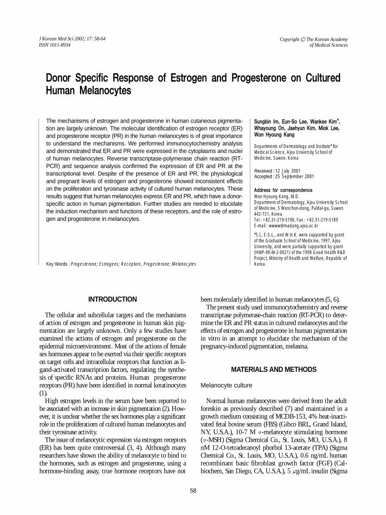
Melanocytes grown on slides were analyzed by immunos-taining using the same human ER and PR antibodies. Thehuman adult melanocytes expressed ER and PR in the cyto-
60 S. Im, E.-S. Lee, W. Kim, et al.
C
A B
Fig. 1. Immunocytochemistry of ER and PR in cultured melanocytes(original magnification, ×400). A: Melanocyte-negative control.B: Melanocyte-ER positive (arrow). Cultured melanocytes showcytoplasmic and nuclear immunoreactivity. C: Melanocyte-PRpositive (arrowhead). Cultured melanocytes show cytoplasmicand nuclear immunoreactivity.

plasm and nucleus (Fig. 1). The human breast cancer cellline MCF-7 shows positive ER and PR immunoreactivity
in the nucleus (data not shown) (12, 13).To confirm that the ER and PR immunoreactivity was
originated from the encoded proteins, we subsequently exam-ined the ER and PR transcripts by PCR. The predicted 195bp and 470 bp PCR products of PR and ER, respectively,were found in MCF-7 and human adult melanocytes (Fig.2). Neonatal melanocytes expressed the same immunoreactiv-ity and mRNA transcripts of ER and PR (data not shown).Sequencing analysis of the RT-PCR products of ER and PRshowed 99.9% homology with the reported human ER andPR sequences (data not shown). These results confirmed thatmRNA coding for ER and PR is transcribed and translatedin human melanocytes, providing a strong evidence thathuman melanocytes are the target for the estrogen and pro-gesterone action.
Effect of estrogen and progesterone on pigmentationin skin samples grown in organ culture
To investigate the influence of estrogen and progesteroneon pigmentation, skin organ culture was used as a modelsystem more closely resembling in vivo circumstances thanthose of the melanocyte monolayer culture. Because the steroidhormones are capable of penetrating the skin easily as com-pared with the peptide hormones, the skin explants werefirst immersed in the media and estrogen and progesteronewere treated for 6 days. During the culture, the overall in
Response of Estrogen and Progesterone on Cultured Human Melanocytes 61
Fig. 2. RT-PCR profiles of cultured human melanocyte. RT-PCR ofMCF-7 and human melanocytes yielded the predicted 195 bpPCR product for PR and 470 bp for ER at mRNA level. Lane 1,molecular size marker, Lane 2, GAPDH (MCF-7), Lane 3, GAPDH(melanocyte), Lane 4, ER ( MCF-7), Lane 5, ER (melanocyte), Lane6, PR (MCF-7), Lane 7, PR (melanocyte), Lane 8, molecular sizemarker.
1 2 3 4 5 6 7 8
320 bp→
←470 bp
←195 bp
A B
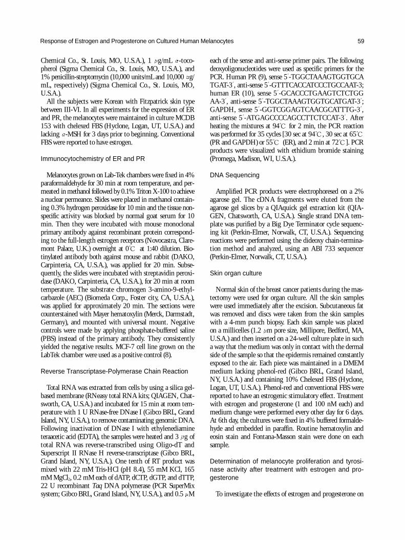
Fig. 3. Effect of estrogen and progesterone on pigmentation in skin samples grown in organ culture. Estrogen and progesterone at aphysiologic concentration (1 nM) and concentration in pregnancy (100 nM) did not show any significant epidermal changes including thethickness and rete ridge pattern. The concentrations also did not affect the epibolic outgrowth. In 2 samples out of 5, estrogen at 1 nMand 100 nM concentration increased the basal pigmentation as compared with the control. However, progesterone either at 1 nM or at100 nM concentration did not increase the melanin content in the epidermis in all samples. A: control, B: estrogen at 100 nM concentra-tion increased the basal pigmentation (Fontana-Masson stain, original magnification, ×400).
vivo cell morphology of keratinocytes was maintained for aperiod of 6 days. Although a slight dermoepidermal separa-tion was noted in some experiment, the explants were quitesatisfactory for testing. Epidermal cells of the explant prolif-erated and migrated to form a new epidermis (epibolus) thatcompletely surround the explant within 6 days. Estrogen andprogesterone either at a physiologic concentration (1 nM) orat a concentration in pregnancy (100 nM) did not show anysignificant epidermal changes including the thickness andrete ridge pattern. The concentrations did not affect the epi-bolic outgrowth, either. In 2 samples out of 5, estrogen at 1nM and 100 nM concentration both increased the basal pig-mentation as compared with the control (Fig. 3). However,progesterone either at 1 nM or at 100 nM concentration didnot increase the melanin content in the epidermis in all sam-ples.
Melanogenic and mitogenic effects of estrogen andprogesterone on human melanocytes
Because conventional FBS and phenol red in MCDB mediahave a mild estrogenic effect, we first maintained the culturedmelanocytes in phenol red-free DMEM with chelexed serumthat was deprived of estrogen by charcoal. In this condition,melanocytes started to float on day 4 and we could not main-tain the melanocytes more than 6 days. We next comparedthe effect of estrogen and progesterone on melanocytes inMCDB with phenol red and chelexed serum vs regular serum,and observed no difference. Thus the subsequent experimentswere done using MCDB with chelaxed serum.
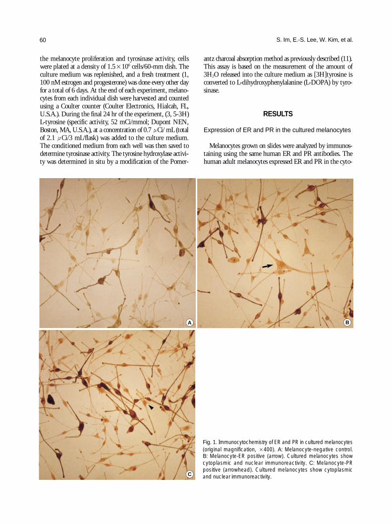
Melanocytes from 8 donors treated with 1 and 100 nMestrogen and progesterone for 6 days showed a donor-specif-ic response (Table 1). The number of melanocytes and tyro-sine hydroxylase activity increased in melanocytes from 3
62 S. Im, E.-S. Lee, W. Kim, et al.%
con
trol
control E 1 nM E 100 nM P 1 nM P100 nM
180
160
140
120
100
80
60
40
20
0A
AM-2
Porliferation
THA
% c
ontr
ol
control E 1 nM E 100 nM P 1 nM P 100 nM
140
120
100
80
60
40
20
0 B
AM-6
Porliferation
THA
Fig. 4. Effect of estrogen and progesterone on the proliferation and tyrosinase activity of human melanocytes. A: The number ofmelanocytes and tyrosine hydroxylase activity both increased in melanocytes from AM-2 donor in response to 1 and 100 nanomolarconcentrations of estrogen and progesterone. B: Neither proliferation nor tyrosinase activity showed any changes due to estrogenand progsterone treatment in AM-6 donor.
AM-1 AM-2 AM-3 AM-4 AM-5 AM-6 AM-7 AM-8
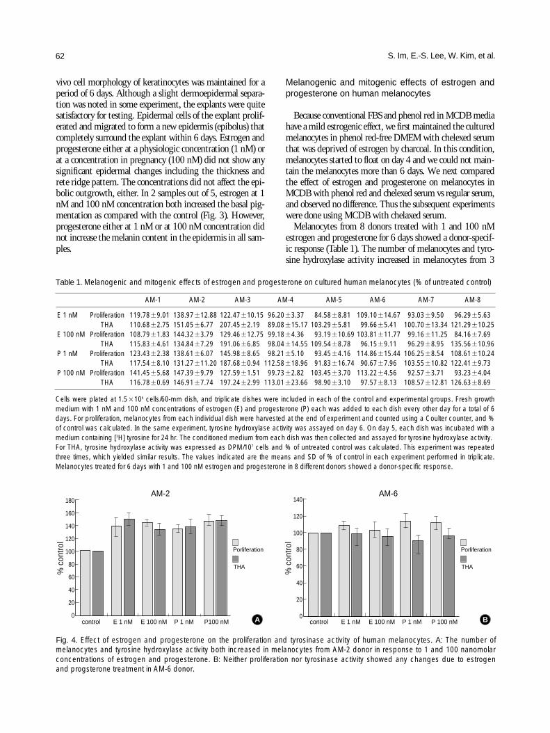
E 1 nM Proliferation 119.78±9.01 138.97±12.88 122.47±10.15 96.20±3.37 84.58±8.81 109.10±14.67 93.03±9.50 96.29±5.63THA 110.68±2.75 151.05±6.77 207.45±2.19 89.08±15.17 103.29±5.81 99.66±5.41 100.70±13.34 121.29±10.25
E 100 nM Proliferation 108.79±1.83 144.32±3.79 129.46±12.75 99.18±4.36 93.19±10.69 103.81±11.77 99.16±11.25 84.16±7.69THA 115.83±4.61 134.84±7.29 191.06±6.85 98.04±14.55 109.54±8.78 96.15±9.11 96.29±8.95 135.56±10.96
P 1 nM Proliferation 123.43±2.38 138.61±6.07 145.98±8.65 98.21±5.10 93.45±4.16 114.86±15.44 106.25±8.54 108.61±10.24THA 117.54±8.10 131.27±11.20 187.68±0.94 112.58±18.96 91.83±16.74 90.67±7.96 103.55±10.82 122.41±9.73
P 100 nM Proliferation 141.45±5.68 147.39±9.79 127.59±1.51 99.73±2.82 103.45±3.70 113.22±4.56 92.57±3.71 93.23±4.04THA 116.78±0.69 146.91±7.74 197.24±2.99 113.01±23.66 98.90±3.10 97.57±8.13 108.57±12.81 126.63±8.69
Table 1. Melanogenic and mitogenic effects of estrogen and progesterone on cultured human melanocytes (% of untreated control)
Cells were plated at 1.5×10⁶ cells/60-mm dish, and triplicate dishes were included in each of the control and experimental groups. Fresh growthmedium with 1 nM and 100 nM concentrations of estrogen (E) and progesterone (P) each was added to each dish every other day for a total of 6days. For proliferation, melanocytes from each individual dish were harvested at the end of experiment and counted using a Coulter counter, and %of control was calculated. In the same experiment, tyrosine hydroxylase activity was assayed on day 6. On day 5, each dish was incubated with amedium containing [3H] tyrosine for 24 hr. The conditioned medium from each dish was then collected and assayed for tyrosine hydroxylase activity.For THA, tyrosine hydroxylase activity was expressed as DPM/107 cells and % of untreated control was calculated. This experiment was repeatedthree times, which yielded similar results. The values indicated are the means and SD of % of control in each experiment performed in triplicate.Melanocytes treated for 6 days with 1 and 100 nM estrogen and progesterone in 8 different donors showed a donor-specific response.
adult foreskin donors in response to 1 and 100 nM estrogenand progesterone, respectively (Fig. 4A). In one donor, onlythe tyrosinase activity increased. In the remaining 4 donors,neither proliferation nor tyrosinase activity of melanocytesshowed any significant changes due to estrogen and proges-terone (Fig. 4B). Interestingly, the dose response to 1 and100 nanomolar concentrations of either hormone did notshow any significant differences. -MSH treatment, usedfor a positive control, was proved to be both mitogenic andmelanogenic in melanocytes. However when -MSH wastreated concomitantly with estrogen and progesterone, noadditive response was observed, and no difference was notedbetween neonatal and adult melanocytes (data not shown).
DISCUSSION
Since many dermatoses are related to a female predisposi-tion and pregnancy, it is feasible that the female sex hormonemay influence the skin directly. Estrogen appears to increasethe vascularization of the skin and suppress the sebaceousgland activity. Also estrogen increases the pigment cellactivity (14).
Skin pigmentation in vivo is determined by genetic, envi-ronmental, local, and endocrine factors, which influence bothmelanin synthesis within each melanocyte and the distribu-tion of melanin throughout the epidermis. Melasma, a com-mon acquired brownish pigmentation occurring exclusivelyin the sun-exposed areas on the face, is exacerbated by preg-nancy and oral contraceptives (15-20). Sato reported a signifi-cantly high level of progesterone in the serum of Japanesepatients with melasma (2). On the other hand, Perez et al.reported the increased serum levels of LH alone, and lowerlevels of serum estradiol in patients with melasma than innormal controls (21). We, therefore, assumed that the ovarianhormones such as estrogen and progesterone may be involvedin the pathogenesis of melasma, i.e., indirect influence ofovarian hormones on the melanocytes in vivo remains to beclarified.
The issue of melanocytic expression via ER has been quitecontroversial. Investigators found that tyrosinase can mimicthe tight binding of 3H-estradiol, giving 3H-labeled prod-ucts. In this way tyrosinase oxidation of 3H-estradiol led tospurious positive results in early biochemical assays of estro-gen binding. Although the true estradiol binding could notbe distinguished from tyrosinase activity with the dextran-coated charcoal assay, the use of immunocytochemistry andRT-PCR obviated this problem.
In this study we provide the first direct evidence thathuman melanoytes show ER and PR immunoreactivity. ERand PR belong to a superfamily of ligand-induced transacti-vators that exerts their regulatory activity on discrete genesthrough DNA binding at individual hormone-responsiveelements. Understanding the molecular characterization of
ER and PR is of particular importance in assessing the hor-monal dependence of target cells. The significance of ER andPR in the melanocytes is an area of considerable interest. Toclarify the existence of ER and PR, we analyzed the expressionof ER and PR in cultured melanocytes and designed an exper-iment to evaluate the role of estrogen and progesterone onhuman pigmentation using an in vitro culture system. Ourdata provides the evidence for the presence of ER and PR incultured melanocytes by immunocytochemistry, RT-PCR,and sequencing.
Recent studies have demonstrated the effects of estrogenand progesterone on human melanocytes. However, it isunclear whether the effects were directly exerted by the sexhormones or by tyrosinase activity.
Ranson et al., for the first time, reported that the incuba-tion of neonatal melanocytes with beta-estradiol (10-10-10-9
M) for 24 hr resulted in a dose-dependent stimulation ofthe tyrosinase activity (22). The increased melanogenesiswas accompanied by reduced cell number and enhancedmelanin extrusion into the medium. They are also aware that,most studies on the melanocytes have extrapolating any ofthe results to the adult situation.
On the contrary, Jee et al. reported that the treatment of17 -estradiol (10-12 to 10-9 M) for 10 days significantly showedthe increased neonatal melanocyte number, and for 1 dayexhibited the decreased tyrosinase activity and melanin con-tent (6). However, they suggested that the tyrosinase activi-ty and melanin content were expressed on per cell bases. It issuggested that the proliferating activity stimulated by estra-diol was greater than that by the tyrosinase activity and themelanin producing activity.
Recent data from the adult melanocytes of Japanese maleforeskin showed that estradiol in the concentration range of0.01 to 1.0 g/mL and 1 g/mL progesterone significantlyincreased the amount of TRP-1, and no significant effect onDOPA oxidase activity was detected after estradiol and pro-gesterone treatment (23). Estradiol and progesterone alsoincreased the area, the dendrites and the perimeter per cell.
Kippenberger et al. applied the reverse transcriptase-com-petitive multiplex PCR to normal human melanocytes, andreported that 20 M treatment of diethylstilbestrol and estra-diol for 48 hr lead to an increase of about 1.5 to 2.5 fold oftyrosinase and TRP-2 transcripts (24). The authors reportedfor the first time that the sex steroids cause an increase ofmelanogenic enzyme transcripts in normal human mela-nocytes. An increase in the tyrosinase activity could provokethe switch from pheomelanogenesis to eumelanogenesis.Activation of tyrosinase by estradiol might be an alternativeto a direct receptor-mediated mechanism for the growthinhibitory effect observed in vivo and in vitro.
In this study, in spite of the presence of receptor for estro-gen and progesterone, they had a donor-specific effect onproliferation of melanocytes. There are several plausibleexplanations for this lack of effect on melanocyte growth in
Response of Estrogen and Progesterone on Cultured Human Melanocytes 63
vitro. The first is that certain prone melanocytes only canrespond to estrogen and progesterone with a stimulation ofgrowth. The genetic predisposition and ultraviolet exposuremay be prerequisite or costimulating factors. Alternativeexplanations for the lack of either a stimulatory response toestrogen and progesterone in vitro include the limited amountof receptor present, a requirement for an estrogen- or pro-gesterone-induced autocrine or paracrine growth factor-mediated growth. The dilution of such factors in culture hasbeen proposed to account for the observed absence of estro-gen- or progesterone-stimulated cell growth in the breasttumor cells that otherwise respond to estrogen and proges-terone in vivo by increased tumor growth.
We provide the first direct evidence that human culturedmelanoytes contain ER and PR immunoreactivity. Humanmelanocytes uniformly expressed ER and PR in their cyto-plasm and nucleus. ER and PR mRNA were present in cul-tured melanocytes. In spite of the presence of receptor forestrogen and progesterone, estrogen and progesterone had adonor-specific effect on proliferation of melanocytes andtyrosine hydroxylase activity.
REFERENCES
1. Im S, Lee E-S, Kim W, Song J, Kim J, Lee M, Kang WH. Expressionof progesterone receptor in human keratinocytes. J Korean Med Sci2000; 15: 647-54.
2. Sato N. Endocrine environment in adult females with chloasma. JpnJ Dermatol 1987; 97: 937-43.
3. Hodgins MB, Spike RC, Mackie RM, McLean AB. An immunohisto-chemical study of androgen, oestrogen and progesterone receptorsin the vulva and vagina. Br J Obstet Gynaecol 1998; 105: 216-22.
4. Jee SH, Lee SY, Chiu HC, Chang CC, Chen TJ. Effect of estrogenand estrogen receptor in normal human melanocytes. Biochem Bio-phys Res Commun 1994; 199: 1407-12.
5. McCarty KS Jr, Wortman J, Stowers S, Lubahn DB, McCarty KSSr, Seigler HF. Sex steroid receptor analysis in human melanoma.Cancer 1980; 46: 1463-70.
6. Ellis DL, Wheeland RG, Solomon H. Estrogen and progesteronereceptors in melanocytic lesions. Arch Dermatol 1985; 121: 1282-5.
7. Kim NS, Cho JH, Kang WH. Behavioral differences between donorsite-matched adult and neonatal melanocytes in culture. Arch Der-matol Res 2000; 292: 233-9.
8. Cho H, Aronica SM, Katzenellenbogen BS. Regulation of proges-terone receptor gene expression in MCF-7 breast cancer cells: acomparison of the effects of cyclic adenosine 3′, 5′-monophosphate,
estradiol, insulin-like growth factor-I, and serum factor. Endocrinol-ogy 1994; 134: 658-64.
9. Ingamells S, Campbell IG, Anthony FW, Thomas EJ. Endometrialprogesterone receptor expression during the human menstrual cycle.J Reprod Fertil 1996; 106: 33-8.
10. Sabbah M, Kang KI, Tora L, Redeuilh G. Oestrogen receptor facili-tates the formation of preinitiation complex assembly: involvement ofthe general transcription factor TFIIB. Biochem J 1998; 336: 639-46.
11. Pomerantz SH. L-tyrosine-3, 5-3H assay for tyrosinase developmentin skin of newborn hamsters. Science 1969 16; 164: 838-9.
12. Greene GL, Gilna P, Waterfield M, Baker A, Hort Y, Shine J.Sequence and expression of human estrogen receptor complementaryDNA. Science 1986; 231: 1150-4.
13. Misrahi M, Atger M, d’Auriol L, Loosfelt H, Meriel C, FridlanskyF, Guiochon-Mantel A, Galibert F, Milgrom E. Complete aminoacid sequence of the human progesterone receptor deduced fromcloned cDNA. Biochem Biophys Res Commun 1987; 143: 740-8.
14. Shahrad P, Marks R. A pharmacological effect of oestrone on humanepidermis. Br J Dermatol 1977; 97: 383-6.
15. Newcomer VD, Lindbert MC, Stenbert TH. A melanosis of the face(“chloasma”). Arch Dermatol 1961; 83: 284-97.
16. Sanchez NP, Pathak MA, Sato S, Fitzpatrick TB, Sanchez JL, MihmMC Jr. Melasma: A clinical, light microscopic, ultrastructural, andimmunofluorescence study. J Am Acad Dermatol 1981; 4: 698-710.
17. Grimes PE. Melasma: etiologic and therapeutic considerations. ArchDermatol 1995; 131: 1453-7.
18. Resnick S. Melasma induced by oral contraceptive drugs. JAMA1967; 199: 95-9.
19. Nordlund JJ, Boissy RE, Hearing VJ, King RA, Ortonne JP, editors.Pigmentary system: physiology and pathophysiology. New York:Oxford University Press; 1997.
20. Pathak MA, Riley FC, Fitzpatrick TB. Melanogenesis in human skinfollowing exposure to long-wave ultraviolet and visible light. J InvestDermatol 1962; 39: 435-43.
21. Perez M, Sanchez JL, Aguilo F. Endocrinologic profile of patientswith idiopathic melasma. J Invest Dermatol 1983; 81: 543-5.
22. Ranson M, Posen S, Mason RS. Human melanocytes as a target tis-sue for hormones: in vitro studies with 1 alpha-25, dihydroxyvitaminD3, alpha-melanocyte stimulating hormone, and beta-estradiol. JInvest Dermatol 1988; 91: 593-8.
23. Maeda K, Naganuma M, Fukuda M, Matsunaga J, Tomita Y. Effectof pituitary and ovarian hormones on human melanocyte in vitro.Pigment Cell Res 1996; 9: 204-12.
24. Kippenberger S, Loitsch S, Solano F, Bernd A, Kaufmann R. Quan-tification of tyrosinase, TRP-1, and TRP-2 transcripts in humanmelanocytes by reverse transcriptase-competitive multiplex PCR-regulation by steroid hormones. J Invest Dermatol 1998; 110: 364-7.
64 S. Im, E.-S. Lee, W. Kim, et al.