donglu zhang, kan he, john j herbst, janet kolb ... -...
TRANSCRIPT
1
Characterization of Efflux Transporters Involved in Distribution and Disposition of
Apixaban
Donglu Zhang, Kan He, John J Herbst, Janet Kolb, Wilson Shou, Lifei Wang, Praveen
Balimane, Yong-Hae Han, Jinping Gan, Charles E Frost, W Griffith Humphreys
Pharmaceutical Candidate Optimization (DZ, KH, LW, PB, YH, JG, WGH), Discovery
Medicine and Clinical Pharmacology (CEF), Bristol-Myers Squibb, Princeton, NJ 08543;
Lead Profiling (JJH, JK, WS), Bristol-Myers Squibb, Wallingford, CT 06492
DMD #50260 DMD Fast Forward. Published on February 4, 2013 as doi:10.1124/dmd.112.050260
Copyright 2013 by the American Society for Pharmacology and Experimental Therapeutics.
This article has not been copyedited and formatted. The final version may differ from this version.DMD Fast Forward. Published on February 4, 2013 as DOI: 10.1124/dmd.112.050260
at ASPE
T Journals on M
ay 29, 2018dm
d.aspetjournals.orgD
ownloaded from
2
Running title: Efflux transporters in apixaban disposition
Correspondence To:
Donglu Zhang, Ph.D.
Pharmaceutical Candidate Optimization
Bristol-Myers Squibb Research and Development,
Princeton, NJ 08543
Text pages (not including references): 25
# of Table: 4
# of Figures: 4
# of references: 45
# of words in abstract: 250
# of words in introduction: 450
# of words in discussion: 1500
1Abbreviations used: BCS, Biopharmaceutical Classification Systems; BCRP, breast
cancer resistance protein; CsA, cyclosporin A; DMSO, dimethyl sulfoxide; DMEM,
Dulbecco's Modified Eagle's Medium; ER, efflux ratio - permeability of basolateral to
apical versus that of apical to basolateral; HBSS, Hank’s balance salt solution; HEPES,
N-(2-Hydroxyethyl) piperazine-N’-(2-ethanesulfonic acid); keto, ketoconazole; LC/MS,
liquid chromatography and mass spectrometry; MDR1, multiple drug resistance protein
(P-gp); NSAID, non-steroidal anti-inflammatory drugs; PcA-B, permeability of apical to
basolateral direction; PcB-A, permeability of basolateral to apical direction; TEER, trans-
epithelial electrical resistance
DMD #50260This article has not been copyedited and formatted. The final version may differ from this version.
DMD Fast Forward. Published on February 4, 2013 as DOI: 10.1124/dmd.112.050260 at A
SPET
Journals on May 29, 2018
dmd.aspetjournals.org
Dow
nloaded from
3
Abstract
The studies reported here were conducted to investigate the transport characteristics of
apixaban and to understand the impact of transporters on apixaban distribution and
disposition. In human P-gp- and BCRP-cDNA transfected cell monolayers as w ell as
Caco-2 cell monolayers, the apparent efflux ratio of basolateral to apical (PcB-A) versus
apical to basolateral permeability (PcA-B) of apixaban was >10. The P-gp- and BCRP-
facilitated transport of apixaban was concentration- and time-dependent and did not show
saturation over a w ide range of concentrations (1-100 μM). The efflux transport of
apixaban was also demonstrated by the lower mucosal to serosal permeability than that of
the serosal to mucosal direction in the isolated rat jejunum segments. Apixaban did not
inhibit digoxin transport in Ca co-2 cells. Ketoconazole decreased the P-gp-mediated
apixaban efflux in Caco-2 and the P-gp-cDNA transfected cell monolayers, but did not
affect the apixaban efflux to a m eaningful extent in the BCRP-cDNA transfected cell
monolayers. Co-incubation of a P-gp inhibitor (ketoconazole or c yclosporin A) and a
BCRP inhibitor (Ko134) provided more complete inhibition of apixaban efflux in Caco-2
cells than separate inhibition by individual inhibitors. Naproxen inhibited apixaban efflux
in Caco-2 cells, but showed only a minimal effect on apixaban transport in the BCRP-
transfected cells. Naproxen was the first NSAID that was demonstrated as a weak P-gp
inhibitor. These results demonstrate that apixaban is a substrate for efflux transporters P-
gp and BCRP, which can help explain its low brain penetration, low fetus exposures, and
milk excretion in rats.
DMD #50260This article has not been copyedited and formatted. The final version may differ from this version.
DMD Fast Forward. Published on February 4, 2013 as DOI: 10.1124/dmd.112.050260 at A
SPET
Journals on May 29, 2018
dmd.aspetjournals.org
Dow
nloaded from
4
Introduction
Efflux transporters are ATP Binding Cassette (ABC) proteins containing multi-trans-
membrane spanning domains with homologous ATP-binding sites. Several members of
this family are primary drug transporters which pump substrates out of cells by using
ATP as the energy source, thus significantly modulating the absorption, distribution,
metabolism, and elimination of endogen ous compounds, drugs and other xenobiotics
(Leslie et al., 2005; Shitara et al., 2006; Zhou, 2008; Koshiba et al., 2008; Xia et al.,
2005a; Giacomini et al., 2010). P-Glycoprotein (P-gp, encoded by MDR1, ABCB1), a
member of the ABC transporter superfamily, is expressed in th e human intestine, liver,
brains, and other tissues, and plays an important role in oral bioavailability and tissue
distribution of drug molecules that are substrates for this transporter (Zhou 2008). The
breast cancer resistance protein (BCRP, ABCG2), another ATP-binding cassette efflux
drug transporter (Doyle and Ross, 2003; Mao and Unadkat, 2005; Krishnamurthy and
Schuetz, 2006), is highly expressed in various normal tissues such as placenta, small
intestine, liver, and mammary glands (Maliepaard et al., 2001). BCRP can transport a
broad spectrum of substrates including chemotherapeutic agents, organic anions, and
xenobiotics (Doyle and Ross, 2003; Mao and Unadkat, 2005) and plays an important role
in drug disposition (Koshiba et al., 2008).
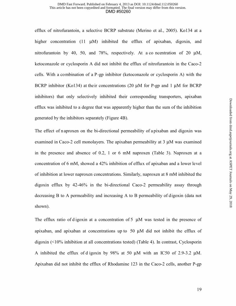
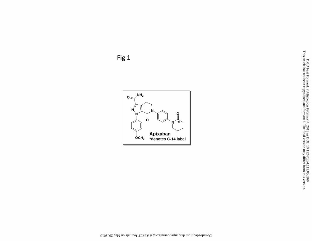
Apixaban, 1-(4-methoxyphenyl)-7-oxo-6-(4-(2-oxopiperidin-1-yl)phenyl)-4,5,6,7-
tetrahydro-1H-pyrazolo[3,4-c]pyridine-3-carboxamide, is a highly selective, oral, direct
inhibitor of Factor Xa, a protease enzyme that plays a piv otal role in t he coagulation
cascade. Direct and selective inhibition of Factor Xa represents a su perior approach to
anticoagulation therapy compared to the current treatments including use of warfarin.
DMD #50260This article has not been copyedited and formatted. The final version may differ from this version.
DMD Fast Forward. Published on February 4, 2013 as DOI: 10.1124/dmd.112.050260 at A
SPET
Journals on May 29, 2018
dmd.aspetjournals.org
Dow
nloaded from

5
Apixaban is currently approved for the prevention of venous thromboembolism (VTEp)
and the prevention of stroke in patients with atrial fibrillation (AF) (Lassen et al., 2007;
Connolly et al., 2011; Granger et al., 2011). Apixaban has balanced elimination pathways
including renal excretion, metabolism, intestinal/biliary excretion in humans (Raghavan
et al., 2009; Zhang et al., 2010; Wong et al., 2011). Metabolism was responsible for 25%
of apixaban clearance in humans and urinary excretion was an important elimination
pathway (~27%). Tissue distribution studies in rats showed that there were low exposures
in rat brain and fetal tissues; in addition, apixaban was highly excreted into milk (Wang
et al., 2011). These characteristics of apixaban seem to indicate a role of active transport
in disposition of this compound.
Interaction with transporters such as P-gp and BCRP has been widely studied using direct
cell-based assays in Caco-2 or drug transporter cDNA-transfected cell lines derived from
porcine or canine kidney cells (Taipalensuu et al., 2001; Elsby et al., 2008; Englund et
al., 2006). The Caco-2 cell line is derived from a human colon adenocarcinoma, and the
cell monolayers differentiate in culture to resemble the epithelial lining of t he human
small intestine and express a number of transporters including P-gp, BCRP, and MRP2
(Elsby et al., 2008). In comparison, cDNA-transfected cell lines are characterized by
selective expression of P-gp or BCR P, and are recommended in vitro systems to test
transporter properties of a c ompound by the International Transporter Consortium
(Giacomini et al., 2010). This study was conducted to evaluate apixaban as a p otential
substrate of common efflux transporters.
DMD #50260This article has not been copyedited and formatted. The final version may differ from this version.
DMD Fast Forward. Published on February 4, 2013 as DOI: 10.1124/dmd.112.050260 at A
SPET
Journals on May 29, 2018
dmd.aspetjournals.org
Dow
nloaded from
6
Materials and Methods
Materials
Apixaban, 1-(4-methoxyphenyl)-7-oxo-6-(4-(2-oxopiperidin-1-yl)phenyl)-4,5,6,7-
tetrahydro-1H-pyrazolo[3,4-c]pyridine-3-carboxamide, and radiolabeled [14C]apixaban
(102 μCi/mg) were synthesized at Bristol-Myers Squibb. The structure of apixaban is
shown in Figure 1. Other chemicals were purchased from Sigma-Aldrich (St Louis, MO).
All chemicals were of analytical grade. Stock solutions of apixaban (10 mM), Ko134 (4.4
mM), digoxin (10 mM), prazosin (5 m M), mannitol (20 mM), nitrofurantoin (1 mM),
ketoconazole (5 mM), and cyclosporin A (5 mM) were prepared in DMSO. A stock
solution of nap roxen (10 mM) was prepared in 10 mM HEPES assay buffer (pH 7.4).
[14C]Apixaban (102 μCi/mg) was prepared in DMSO (2.2 mM and 100 μCi/mL).
[3H]Digoxin (0.3 mCi/mg) was prepared in the transport assay buffer (5 μM and 1.3
μCi/mL). [3H]Prazosin (0.2 mCi/mg) was diluted in ethanol containing 0.01 M HCl (13
mM and 1 mCi/mL). [14C]Mannitol (0.1 mCi/mg) was diluted in ethanol:water (9:1) (20
mM, 0.4 mCi/ml). None of these chemicals used affected the pH of the transport buffer at
the applied concentrations. The final concentration of organic solvent in the assay buffer
was 1% (V/V).
Transporter cell lines
Human P-gp cDNA transfected cells were prepared using porcine kidney-derived LLC-
PK1 cells and control LLC-PK1 cells (BD GentestTM, MA) contained the vector without
human P-gp cDNA (Mock). The cells were seeded onto a collagen-coated polycarbonate
filter membrane (port size 1 μ, diameter 6.5 mm) on th e transwell inserts (0.7 cm2)
DMD #50260This article has not been copyedited and formatted. The final version may differ from this version.
DMD Fast Forward. Published on February 4, 2013 as DOI: 10.1124/dmd.112.050260 at A
SPET
Journals on May 29, 2018
dmd.aspetjournals.org
Dow
nloaded from
7
(Millipore, Billerica, MA) at a density of 50,000 cells/well and cultured in Medium 199
supplemented with 0.05 mg/mL gentamycin and 7% fetal bovine serum in BD Falcon 24-
transwell plates, at 37°C, 5% CO2 and 95% relative humidity for 7 days, with medium
change once every 3-4 days.
Human BCRP cDNA-transfected cells were prepared using canine kidney-derived
MDCKII (Madin Darby Canine Kidney) cells and control MDCKII cells (SOLVO
Biotechnology, Hungary) contained the vector without human BCRP cDNA (Mock).
Cells were cultured in DMEM Medium (Carbrex, NJ) at 37°C in an atmosphere of 5%
CO2 in cell culture flasks. Cells (50,000 cells/well) were seeded onto 24-well transwell
inserts (0.7 cm2). Transfected and control MDCKII cells were cultured on the inserts for
4 days prior to assay. Medium was changed daily and supplemented with 10 mM sodium
butyrate 24 hours before the experiment.
Caco-2 cells were obtained from American Type Culture Collection (Rockville, MD).
Caco-2 cells were maintained in flasks and seeded onto 24-well transwell Corning or BD
Falcon plates for culture and assays. Caco-2 cells were seeded onto the filter membrane
of transwell inserts (0.33 or 0.7 cm2) at a density of 45,000 or 70,000 cells/well and
grown in culture medium consisting of DMEM supplemented with 10% heat-inactivated
fetal bovine serum (Hyclone, Logan, UT), 0.5 mM HEPES, 1% nonessential amino acids,
1% L-glutamine, 100 U/ml penicillin-G, and 100 μg/ml streptomycin (all from Gibco
BRL, Gaithersburg, MD). The culture medium was replaced every two days and the cells
maintained at 37°C, 95% relative humidity, and 5% CO2. Caco-2 cell passage numbers of
17-40 were used for these studies and were monitored for P-gp expression with positive
DMD #50260This article has not been copyedited and formatted. The final version may differ from this version.
DMD Fast Forward. Published on February 4, 2013 as DOI: 10.1124/dmd.112.050260 at A
SPET
Journals on May 29, 2018
dmd.aspetjournals.org
Dow
nloaded from
8
substrates within five days of projected use in the assay, usually 13 - 22 days post-
seeding.
Monolayer integrity was evaluated by pre-experimental trans-epithelial electrical
resistance (TEER) measurements performed using the EVOM resistance meter from
World Precision Instruments or post-experimental Lucifer Yellow apical to basolateral
flux determinations for e ach cell monolayer. After all samples were collected, Lucifer
Yellow was added to each monolayer at a final concentration of 100 µM. The inserts
were placed in a new receiver plate containing the transport buffer. After 30-min
incubation on an orbital shaker (50 rpm) at 37°C, with ambient humidity and CO 2,
receiver samples were removed to measure percent Lucifer Yellow flux. Monolayer
integrity was also evaluated by determining permeability of mannitol and comparing with
the permeability values of m annitol without co-assay compound. For P-gp or BCRP
cDNA-expressed cells and Caco-2 cells, [3H]digoxin (at 5 μM) or [3H]prazosin (at 5 μM)
was measured in a bi-directional assay and the efflux ratios were determined.
Permeability determination procedures
Permeability studies were performed in triplicate at pH 7.4 in Hank’s balanced salt
solution (Gibco BRL, Gaithersburg, MD) containing 10 mM HEPES, and pH adjusted
with NaOH. For apical-to-basolateral (A to B) permeability (absorptive direction), the
donor solution was placed in the apical compartment, and for basolateral-to-apical (B to
A) permeability (secretive direction), the donor solution was placed in the basolateral
compartment. Donor solutions of test compounds were prepared at t he desired
concentrations for each step of the study by diluting aliquots of the test compound stock
solution into transport buffer with the receiver buffer prepared by adding the same
DMD #50260This article has not been copyedited and formatted. The final version may differ from this version.
DMD Fast Forward. Published on February 4, 2013 as DOI: 10.1124/dmd.112.050260 at A
SPET
Journals on May 29, 2018
dmd.aspetjournals.org
Dow
nloaded from
9
concentration of an organic solvent (1%). After incubations, the media from the cell
culture was aspirated, and both apical and basolateral portions of the transwell plate were
washed three times with the transport buffer. The transport buffer solution with the test
compound was then transferred to appropriate wells. After incubation at 37°C, aliquots
(35-100 μL) were taken from the receiver chambers to determine the translocated amount
of a co mpound. Samples were taken from the donor compartments before and af ter
incubation to determine the initial concentration (C0) and recovery of apixaban and other
test compounds. Radioactive samples were removed to scintillation vials.
Permeability determination in P-gp-cDNA expressed cells
Monolayers of P-gp-cDNA transfected LLC-PK1 cells were incubated on an orbital
shaker (50 rpm) at 37°C, with ambient humidity and CO2 for the duration of the transport
assay. The donor and receiver solutions were added to the apical (400 μL) or basolateral
(600 μL) chambers of the monolayers. The time-dependent transport of apixaban at 10
μM was evaluated, and samples from the receiver chambers were taken at three time
points (60, 90, 120 min) and replaced by an equal volume of receiver solution. Samples
from the donor chambers were taken at one time point of 120 min. To determine the
extent of non-specific binding of apixaban to the assay plate, apixaban donor solution
was incubated under the conditions described above in a 24-well assay plate with no cells
present.
[14C]Mannitol (50 μM) and [3H]digoxin (5 μM) were used as the low permeability
control and P-gp substrate control, respectively. [ 14C]Apixaban was assayed at six
concentrations (2.5, 5.0, 10, 25, 50, 100 µM) bi-directionally in both the P-gp transfected
and the vector carrying LLC-PK1 cell monolayers. Samples from the donor and receiver
DMD #50260This article has not been copyedited and formatted. The final version may differ from this version.
DMD Fast Forward. Published on February 4, 2013 as DOI: 10.1124/dmd.112.050260 at A
SPET
Journals on May 29, 2018
dmd.aspetjournals.org
Dow
nloaded from
10
chambers were taken at one time point of 90 min. Apixaban was also assayed bi-
directionally at tw o concentrations (5.0 and 50 µM) in the P-gp transfected cell
monolayers in the presence of i ncreasing concentrations (0, 1, 3, 10, 30 µM) of
ketoconazole and cyclosporin A in both the donor and receiver chamber.
Permeability determination in BCRP-cDNA expressed cells
Incubations with BCRP-cDNA tranfected MDCKII cell monolayers were carried out in
modified Krebs-Henseleit buffer plus 5 mM glucose at 37°C. Cells were pre-incubated
in buffer for 10 min to allow cells to adjust to the medium, then the buffer was added to
the apical (400 μL) or basolateral chamber (800 μL). The transport of [3H]prazosin was
also evaluated in the presence of 1 μM of Ko134, a known inhibitor for BCRP transporter
(Allen et al., 2002). Nitrofurantoin at 5 μM was used as a positive substrate for BCRP
mediated transport. The time-dependent transport of apixaban was evaluated at 15, 30, 60
and 120 min. The concentration-dependent transport of apixaban was also evaluated at 1,
5, 25, and 100 μM in the A to B and B to A directions in the BCRP-transfected and the
vector-carrying MDCKII cells. The efflux inhibition of prazosin (5 μM) was evaluated in
the presence of naproxen (8 mM), a potential inhibitor of efflux transporters based on
results of a clinical drug-drug interaction study with apixaban. The high concentration of
naproxen was selected because gastrointestinal concentration of naproxen could reach 5-
10 mM following oral administration of 500 mg naproxen based on 250 mL of gastro-
fluid volume in humans. The bi-directional permeability of apixaban (5 μM) was also
determined in the presence of ketoconazole (20 and 50 μM) and naproxen (3 and 8 mM).
Permeability determination in Caco-2 cell monolayers
DMD #50260This article has not been copyedited and formatted. The final version may differ from this version.
DMD Fast Forward. Published on February 4, 2013 as DOI: 10.1124/dmd.112.050260 at A
SPET
Journals on May 29, 2018
dmd.aspetjournals.org
Dow
nloaded from
11
For Caco-2 cell monolayers, apical compartments received 200 μL, and basolateral
compartments received 600 μL of permeability solutions. The cell transwells were then
placed on an orb ital shaker (50 rpm ) and incubated for 2 hours at 37°C. Following
incubation, samples were removed for a nalysis. Donor solutions were prepared for
permeability comparators of a l ow permeability control [14C]mannitol (50 µM, 1
µCi/mL) and a control P-gp substrate [3H]digoxin (5 µM). The bi-directional
permeability values of [14C]apixaban at 3.0 or 30 μM were also determined in the
presence of the two transporter inhibitors, ketoconazole and cyclosporin A (at 50 μM) as
well as naproxen (at 0.2, 1 or 6 mM) present in both donor and receiver chambers. The
bi-directional permeability of apixaban (3.0 μM), digoxin (5.0 μM), nitrofurantoin (5.0
μM) was also determined in the presence of ketoconazole (20 μM), cyclosporin A (20
μM), and Ko134 (1 and 11 μM) separately or in a combination. The efflux inhibition of
digoxin (5 μM) was also evaluated in the presence of naproxen (8 mM). Probenecid (200
μM and 1 mM) was co-incubated with apixaban at 20 μM in Caco-2 monolayers to
measure apixaban permeability (Loe et al., 1996; Horikawa et al., 2002).
The potential inhibition of P-gp by apixaban was explored by co-incubation of apixaban
(up to 50 μM) with digoxin (5 μM), or apixaban (200 μM) with Rhodamine 123 (20 μM)
in Caco-2 monolayers to determine the permeability of digoxin or Rhodamine 123, a P-
gp substrate (van der Sandt et a l., 2000). Rhodamine 123 was quantified using a
fluorescence plate reader (HTS 7000, PerkinElmer, Norwalk, CT) with excitation of 492
nm and emission of 530 nm. Additional experiments using a range of api xaban
concentrations in Caco-2 cells were conducted to assess the potential inhibition of
apixaban on P-gp-mediated transport of digoxin. In this study, apixaban at 0.1 to 50 μM
DMD #50260This article has not been copyedited and formatted. The final version may differ from this version.
DMD Fast Forward. Published on February 4, 2013 as DOI: 10.1124/dmd.112.050260 at A
SPET
Journals on May 29, 2018
dmd.aspetjournals.org
Dow
nloaded from
12
was tested and Cyclosporin A was also tested at the same range of concentrations (0.1 to
50 μM) and served as a positive inhibitor. To investigate the involvement of paracellular
transport, palmitoyl-L-carnitine, a paracellular absorption enhancer, was co-incubated
with apixaban in the Caco-2 monolayers (Hochman et al., 1994).
Permeability in rat intestinal segments
Rat intestinal permeability was examined using excised segments from various intestinal
sites of 8 week-old male rats as described previously (Kilic et al., 2004; Sinko et al.,
1995). The intestinal segments were mounted on side-by-side diffusion chambers
(Corning Incorporated, Acton, MA). Donor solution consisted of 200 μM apixaban and
1% DMSO in Tyrode’s buffer (pH 7.4). Permeability studies were conducted at 37°C
either in the mucosal (m) → serosal (s) or s → m direction using 4 mL of donor solution
and 4 mL of receiver solution (pH 7.4, Tyrode’s buffer containing 4% BSA). Samples
were taken at 30, 60, 90, and 120 min from receiver compartments. Donor compartments
were sampled at 120 min. Apixaban was quantified by LC/MS/MS.
LC/MS analysis of apixaban and nitrofurantoin
The LC-MS/MS system consists of binary Shimadzu 10ADvp pumps controlled by a
SCL-10Avp controller, a Leap HTS autosampler, and a Sciex 4000 QTrap hybrid triple
quadrupole – l inear ion trap mass spectrometer (Applied Biosystems, Forest City, CA).
MS/MS detection was in positive mode for apixaban (m/z 460.1 to 443.2) and in a
negative mode for nitrofurantoin (m/z 237.1 to 152.1). The internal standard was the
stable labeled [13CD3]apixaban (m/z 464.1 to 447.2) for apixaban and tobutamide (m/z
269.2 to 169.9) for nitrofurantoin. Standard curves contained a four-point calibration
curve (at 5, 50, 500 and 5000 nM) for apixaban and a eight-point curve (at 5, 10, 50,
DMD #50260This article has not been copyedited and formatted. The final version may differ from this version.
DMD Fast Forward. Published on February 4, 2013 as DOI: 10.1124/dmd.112.050260 at A
SPET
Journals on May 29, 2018
dmd.aspetjournals.org
Dow
nloaded from
13
100, 500, 1000, 2500, 5000 nM) for nitrofurantoin prepared in 1:1 Hank’s
buffer:acetonitrile. Study samples or c alibration standards (50 μL) were first extracted
with 50 μL of internal standard in methanol, and then 5 μL of the mixture was injected
onto a Mac-Mod Halo C18, 2.7 µm, 2.1 × 30 mm at room temperature for analysis. For
apixaban, the mobile phases were (A) 0.2% formic acid in water and (B) 0.2% formic
acid in acetonitrile with a flow rate of 0.5 mL/min; the gradient started with 2% B,
increased to 100% B in 0.5 min, then held at 100% B for 0.25 min before decreased to
2% B in 0.1 min. F or nitrofurantoin, the mobile phases were (A) 2 m M ammonium
acetate containing 0.2% formic acid and 2% actonitrile and (B) acetonitrile containing
0.2% formic acid and 2% water; the gradient was 5% to 100% B in 0. 5 min with a flow
rate of 0.65 mL/min. Peak area ratios of apixaban or nitrofurantoin and internal standards
were used for quantitation with a linear regression using a 1/x2 weighting. The
concentrations of study samples were then calculated from the calibration curve.
Sample analysis for radioactive samples
Samples of 100 μL were removed to scintillation vials, and 8 mL of EcoLite™
scintillation fluid (Irvine, CA) added to each vial. V ials were counted on either a 3H
DPM program or a 14C DPM program (with background subtraction) on a PerkinElmer
Tri-Carb Scintillation Counter Tri-Carb 3100TR (PerkinElmer Life Sciences, Boston,
MA).
Data analysis
The apparent permeability coefficient (Pc, nm/sec) was calculated according to the
following equation: Pc = dA/ (dt·S·C0), where dA/dt is the flux of compound across the
DMD #50260This article has not been copyedited and formatted. The final version may differ from this version.
DMD Fast Forward. Published on February 4, 2013 as DOI: 10.1124/dmd.112.050260 at A
SPET
Journals on May 29, 2018
dmd.aspetjournals.org
Dow
nloaded from

14
monolayer (nmole/sec), S i s the surface area of the cell monolayer, and C0 is the initial
concentration (μM) in the donor compartment. PcB-A is the Pc value measured in the B to
A direction and PcA-B is the Pc value measured in the A to B direction. The efflux ratio
was calculated as: Efflux Ratio = PcB-A/ PcA-B.
The intrinsic activity (IA) of P-gp was calculated as the sum of P-gp or BCRP facilitated
B to A transport in P-gp or BCRP cells relative to control cells and the negative effect of
P-gp or BCRP on A to B transport in P-gp cells relative to control cells:
IA = (d - c) – (b - a), where IA is the intrinsic activity (pmol of test compound
transported during the assay); a is the A to B transport of test compound in control cells
(pmol); b is the B to A transport of test article in control cells (pmol); c is the A to B
transport of test article in P-gp or BCRP expressing cells (pmol); d is the B to A transport
of test compound in P-gp or BCRP expressing cells (pmol). The rate of P-gp or BCRP
facilitated transport is then calculated as: v = IA / (A x t), where v is the rate of P-gp or
BCRP facilitated drug transport (pmol/cm2/hr), IA is the intrinsic activity (pmol
transported during the assay), and A is the surface area of the filter membrane.
Percent inhibition of ap ixaban efflux was determined by comparing the difference
between the Pc values of apixaban incubated in the presence or absence of inhibitor
(Balimane et al., 2006). The percent inhibition of apixaban efflux is calculated as:
Percent Inhibition = 1-[(PcB-A of test compound in presence of inhibitor) - (PcA-B of test
compound in presence of inhibitor)] / [(PcB-A of test compound in absence of inhibitor) -
(PcA -B of test compound in absence of inhibitor)]. Data are expressed as mean ± standard
deviation. To estimate the IC50 value of transport inhibition, the data were fitted by means
DMD #50260This article has not been copyedited and formatted. The final version may differ from this version.
DMD Fast Forward. Published on February 4, 2013 as DOI: 10.1124/dmd.112.050260 at A
SPET
Journals on May 29, 2018
dmd.aspetjournals.org
Dow
nloaded from
15
of nonlinear least-squares regression analysis using XL fit or WinNonlin (Scientific
Consulting Inc., Cary, NC).
Results
Cell Monolayer integrity and transporter characteristics
Pre-experimental TEER measurements and post-experimental Lucifer Yellow A to B flux
determinations were used to confirm monolayer integrity. The functionality of the cell
monolayers was confirmed with the positive control substrates in the absence and
presence of p ositive control inhibitors. TEER values were in agreement with the
historical range of > 500 Ω•cm2. The Lucifer Yellow flux was <2% with individual
values < 3-fold higher than the mean. In addition, the permeability comparator mannitol
showed permeability values of 6-30 nm/s that were within historical ranges. All of these
data indicated that the cell monolayers were appropriate for use in experiments
examining cellular permeability.
For Caco-2 and LLC-PK1-P-gp cells, the efflux ratios of digoxin (5 μM), a known P-gp
substrate, were > 7, which were inhibited >80% by ketoconazole (at 20 or 50 μM), a
known P-gp inhibitors. For MDCKII-BCRP cells, prazosin (5 μM), a known BCRP
substrate, showed an efflux ratio of approximately 10, and the efflux was inhibited by
Ko134 (1 μM), an analogue of fumitremorgin C, and a potent and selective inhibitor of
BCRP (Allen et al., 2002), yielding an efflux ratio of approximately 1. The results with
the positive control substrates and inhibitors demonstrated that the efflux transporters in
these cell models were functioning properly.
Compound recovery
DMD #50260This article has not been copyedited and formatted. The final version may differ from this version.
DMD Fast Forward. Published on February 4, 2013 as DOI: 10.1124/dmd.112.050260 at A
SPET
Journals on May 29, 2018
dmd.aspetjournals.org
Dow
nloaded from
16
Recovery of apixaban and other compounds used in this study from the apical and
basolateral chambers at the end of the assay (mass balance) as well as from the assay
plate without cells was high (82-100%) indicating that the degree of non-specific binding
to the cells or a bsorption to the transwell apparatus did not affect the assay under the
conditions used.
Permeability in cell models
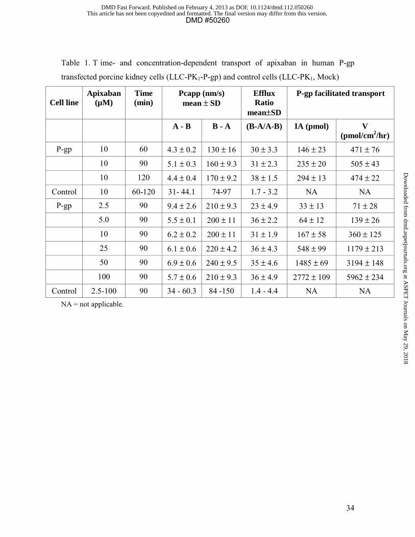
The bi-directional permeability of apixaban was studied in LLC-PK1-P-gp cell
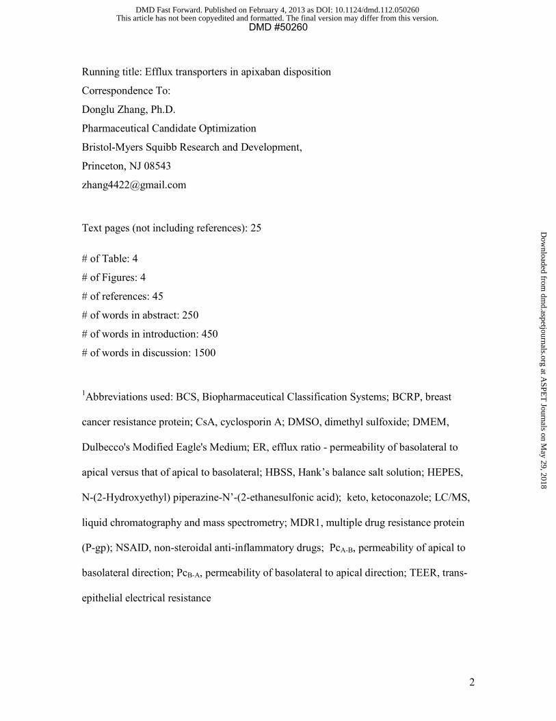
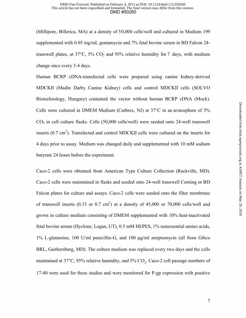
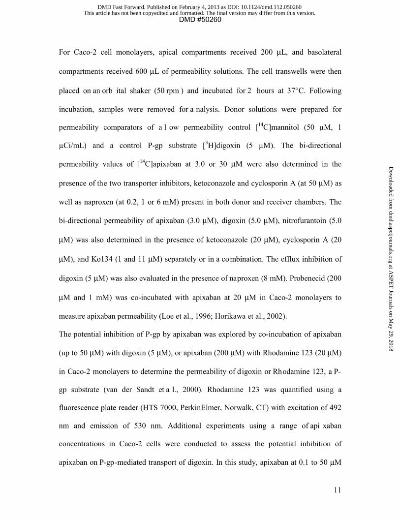
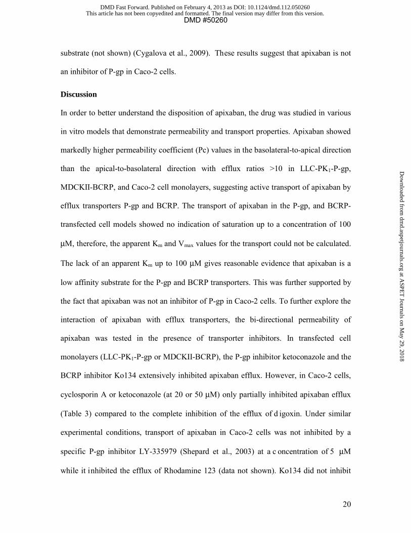
monolayers. Digoxin at 5 μM showed an efflux ratio of 7.4, reduced >90% by
ketoconazole (30 μM) (Figure 2A) or cyclosporin A (10 μM) (Data not shown). Apixaban
was subject to active basolateral to apical transport with efflux ratios in LLC-PK1-P-gp
cells ranging from 23 to 38 compared to ratios of 1.4 to 4.4 in control cells (Table 1).
Apixaban transport was linear over th e time course (60, 90, 120 min) (Table 1). P-gp
facilitated transport of apixaban was concentration-dependent and did not show saturation
over the concentration range (2.5-100 µM) tested. Therefore, the apparent Km and Vmax
values could not be calculated. Ketoconazole (at 0, 1, 3, 10, and 30 µM) caused a
concentration- dependent inhibition of apixaban transport in P-gp expressing cells; the
efflux ratios were 27, 24, 17, 8.3, and 3.2 at 5 µM apixaban and 29, 23, 15, 7.3, and 3.2 at
50 µM apixaban, respectively. Efflux ratios were reduced by approximately 10-fold at the
two apixaban concentrations with ketoconazole IC50 values of 2.9 - 5.4 µM (Figure 2B).
This finding indicates P-gp facilitated transport of apixaban across LLC-PK1-P-gp cell
monolayers.
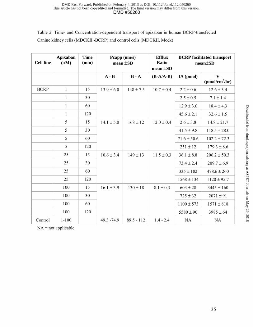
The bi-directional permeability of [3H]prazosin (5 μM) and [14C]apixaban (1 to 100 μM)
was studied in vector-containing MDCKII cell and BCRP cDNA-transfected MDCKII
DMD #50260This article has not been copyedited and formatted. The final version may differ from this version.
DMD Fast Forward. Published on February 4, 2013 as DOI: 10.1124/dmd.112.050260 at A
SPET
Journals on May 29, 2018
dmd.aspetjournals.org
Dow
nloaded from
17
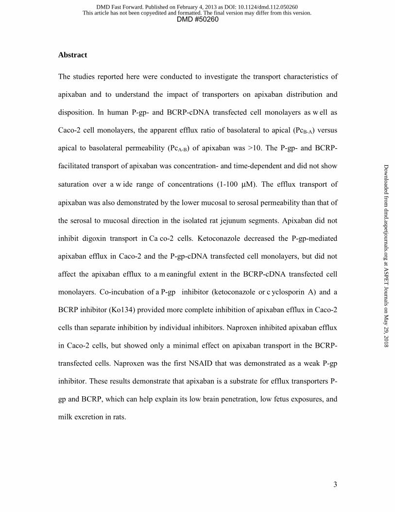
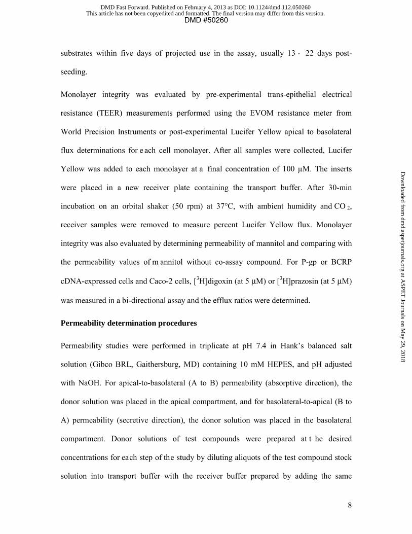
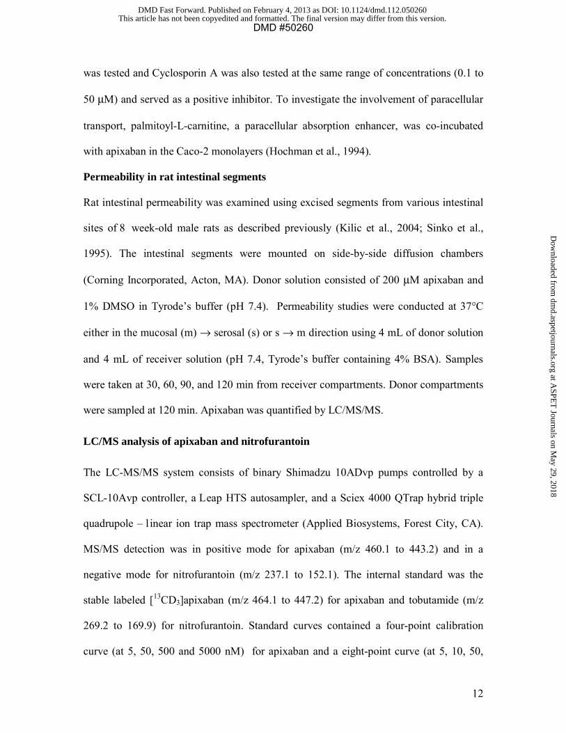
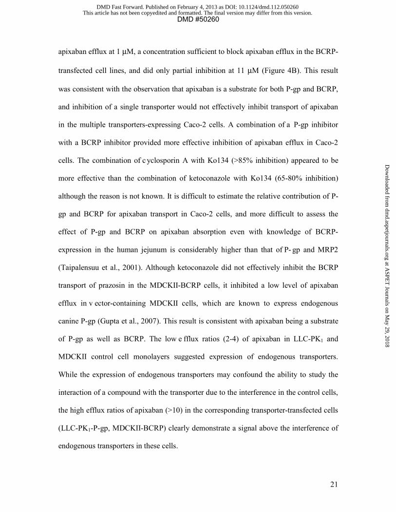
cells (Table 2, Figure 3A and B). Prazosin efflux was completely inhibited by 1 μM of
Ko134. Ketoconazole showed marginal inhibition on prazosin efflux at 20 μM but at 50
μM ketoconazole showed more robust inhibition. In the same experiment, naproxen
showed a very low level of inhibition of prazosin efflux with a minor increase in PcA-B
and decrease in PcB-A at the highest concentration (8 mM) tested, which was also
accompanied by a sl ightly higher mannitol permeability of 30 nm/sec. The apixaban
efflux ratio was 1.4 to 2.4 in the control cells (Table 2), whereas the values in BCRP
cDNA-transfected cells were between 8 and 12. Ko134 strongly inhibited apixaban
transport in th e BCRP-transfected cell lines (Figure 3B). These results indicate that
apixaban is a substrate for the BCRP transporter. Ketoconazole had a minimal effect at
20 μM and a modest effect (33% inhibition) at 50 μM on apixaban transport (Figure 3B);
however, ketoconazole completely inhibited the low level of apixaban efflux in the
vector-containing MDCKII cells (Data not shown). Inhibition of a pixaban efflux in
BCRP-transfected cells by naproxen at 3 and 8 mM was not firmly established.
The bi-directional permeability of apixaban was studied in Caco-2 cell monolayers,
which express a number of efflu x transporters including P-gp and BCRP (Xia et al.,
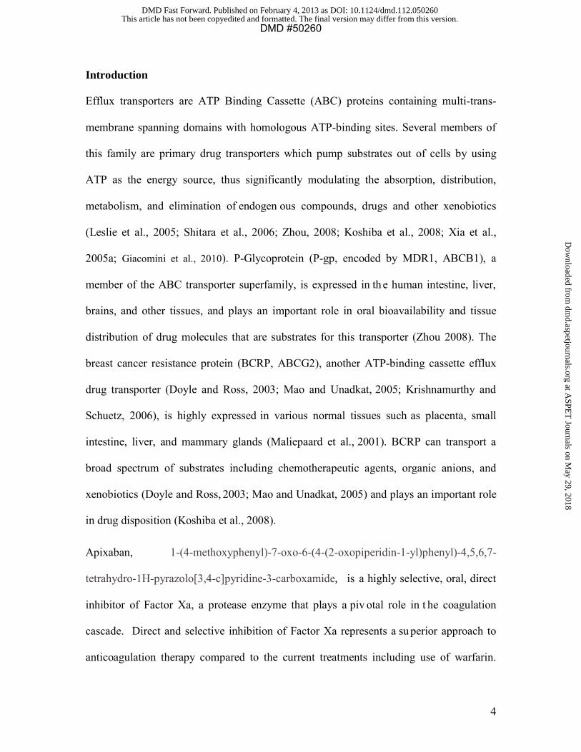
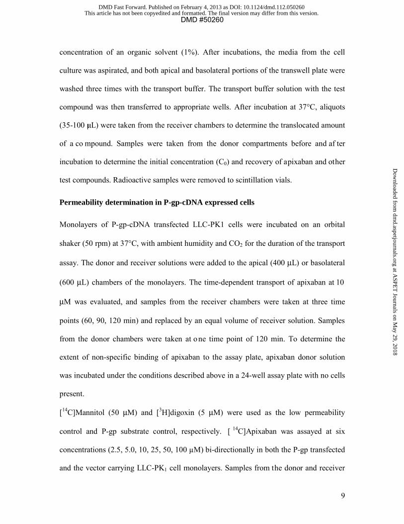
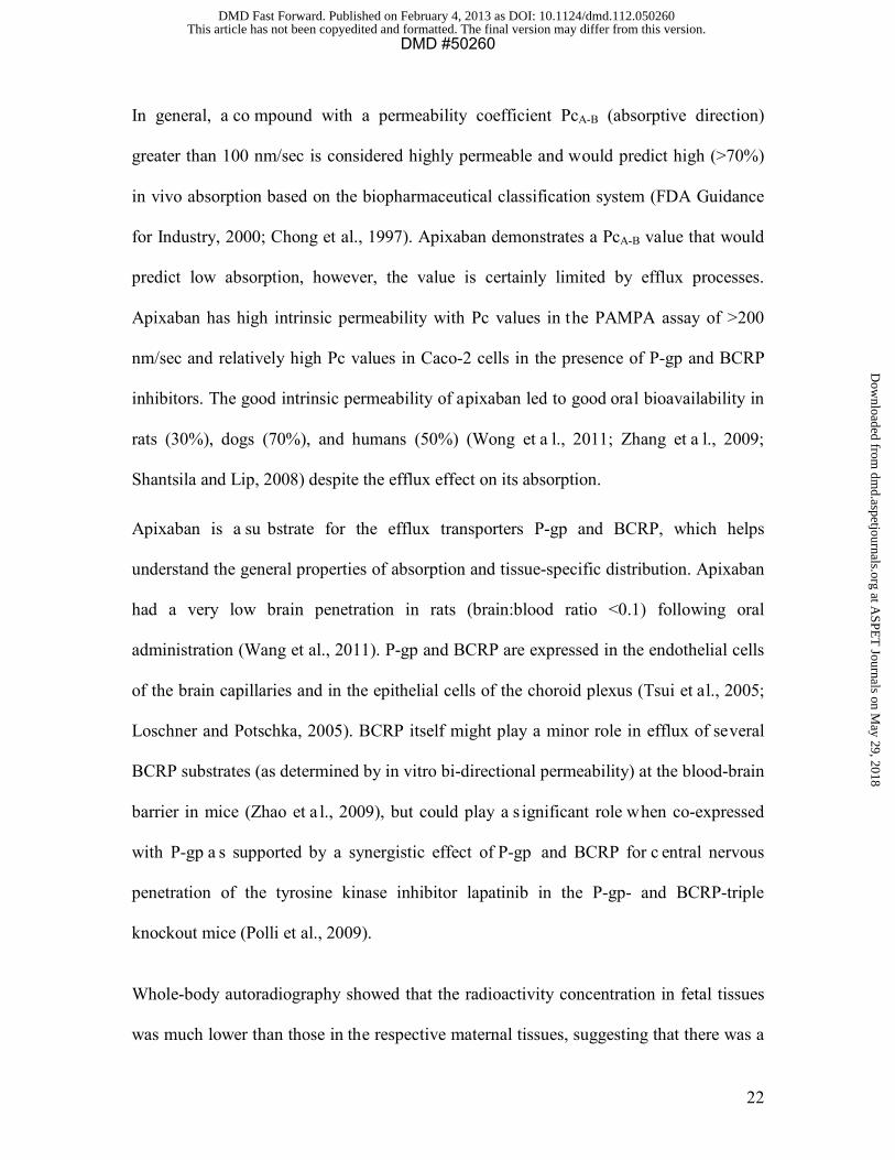
2005b). In this model, the efflux ratio of apixaban ranged between 12 and 37 with the
apparent permeability coefficient (PcA-B) values of approximately 6 to 16 nm/sec in the
apical-to-basal direction and 140 to 387 nm/sec in the basal-to-apical direction (Table 3,
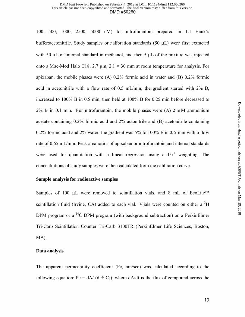
Figure 4A). Both cyclosporin A (50 μM) and ketoconazole (50 μM), known inhibitors of
P-gp, partially reduced the efflux of apixaban at 3 μM by about 43 and 71% (Table 3),
respectively, supporting that apixaban was a P-gp substrate, and potentially a substrate
for other transporters. In the same experiment, Cyclosporin A and ketoconazole
DMD #50260This article has not been copyedited and formatted. The final version may differ from this version.
DMD Fast Forward. Published on February 4, 2013 as DOI: 10.1124/dmd.112.050260 at A
SPET
Journals on May 29, 2018
dmd.aspetjournals.org
Dow
nloaded from
18
completely inhibited 98% of efflux of digoxin at 5 μM (a well-characterized substrate of
P-gp) (Table 3).
The apparent permeability (Pc) of apixaban in Caco-2 cells in e ither direction was not
affected by probenecid, an inhibitor for MRP2 (Table 3), suggesting that MRP2 was not
involved in the efflux of a pixaban in the Caco-2 monolayer. Palmitoyl-L-carnitine, a
known para-cellular absorption enhancer, significantly increased the Pc of apixaban from
10 ± 0.5, 28 ± 2.3, 37 ± 5.1, 47 ± 5.2 nm/sec, approximately 1, 3. 5, 5, and 6 folds at
concentrations of 0 , 0.2, 0.3, 0.5 mM, respectively, suggesting that apixaban showed
potential paracellular as well as intra-cellular transport in Caco-2 cells.
Intestinal permeability
The intestinal absorption of apixaban was studied using isolated rat duodenum, jejunum,
ileum, and colon segments (Kilic et al., 2004; Sinko et al., 1995). The mucosal-to-serosal
Pc values of apixaban were lower at 33± 12, 73± 28, 51± 38, and 31± 22 nm/sec in the
duodenum, jejunum, ileum, and colon segments, respectively, than the Pc values of
apixaban in the serosal-to-mucosal direction of 170 ±23 nm/sec in jejunum. These results
are consistent with intestinal efflux transport in the jejunum segment. The efflux potential
of apixaban in other segments of rat intestines is not known. .
Inhibition studies in Caco-2 cell monolayers
Both ketoconazole and cy cloporin A at co ncentrations of 20 μM partially inhibited
apixaban efflux compared to nearly complete inhibition of digoxin efflux in Caco-2 cells
(Figure 4B). Although Ko134 at 1 μM completely inhibited apixaban efflux in MCDKII-
BCRP cells, it did not inhibit apixaban efflux in Caco-2 cells, minimally inhibited the
DMD #50260This article has not been copyedited and formatted. The final version may differ from this version.
DMD Fast Forward. Published on February 4, 2013 as DOI: 10.1124/dmd.112.050260 at A
SPET
Journals on May 29, 2018
dmd.aspetjournals.org
Dow
nloaded from
19
efflux of nitrofurantoin, a selective BCRP substrate (Merino et al., 2005). Ko134 at a
higher concentration (11 μM) inhibited the efflux of apixaban, digoxin, and
nitrofurantoin by 40, 50, and 78%, respectively. At a co ncentration of 20 μM,
ketoconazole or cyclosporin A did not inhibit the efflux of nitrofurantoin in the Caco-2
cells. With a combination of a P-gp inhibitor (ketoconazole or cyclosporin A) with the
BCRP inhibitor (Ko134) at their concentrations (20 μM for P-gp and 1 μM for BCRP
inhibitors) that only selectively inhibited their corresponding transporters, apixaban
efflux was inhibited to a degree that was apparently higher than the sum of the inhibition
generated by the inhibitors separately (Figure 4B).
The effect of n aproxen on the bi-directional permeability of a pixaban and digoxin was
examined in Caco-2 cell monolayers. The apixaban permeability at 3 μM was examined
in the presence and absence of 0.2, 1 or 6 mM naproxen (Table 3). Naproxen at a
concentration of 6 mM, showed a 42% inhibition of efflux of apixaban and a lower level
of inhibition at lower naproxen concentrations. Similarly, naproxen at 8 mM inhibited the
digoxin efflux by 42-46% in the bi-directional Caco-2 permeability assay through
decreasing B to A permeability and increasing A to B permeability of digoxin (data not
shown).
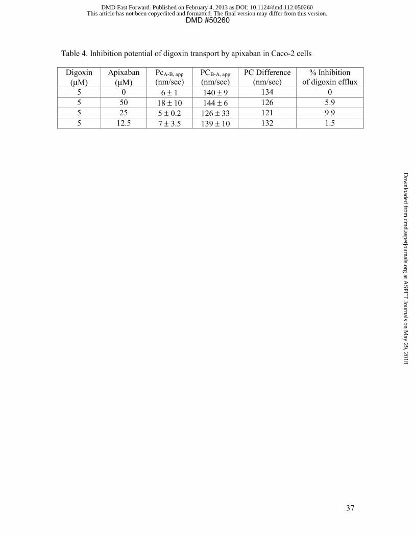
The efflux ratio of d igoxin at a concentration of 5 μM was tested in the presence of
apixaban, and apixaban at concentrations up to 50 μM did not inhibit the efflux of
digoxin (<10% inhibition at all concentrations tested) (Table 4). In contrast, Cyclosporin
A inhibited the efflux of d igoxin by 98% at 50 μM with an IC50 of 2.9-3.2 μM.
Apixaban did not inhibit the efflux of Rhodamine 123 in the Caco-2 cells, another P-gp
DMD #50260This article has not been copyedited and formatted. The final version may differ from this version.
DMD Fast Forward. Published on February 4, 2013 as DOI: 10.1124/dmd.112.050260 at A
SPET
Journals on May 29, 2018
dmd.aspetjournals.org
Dow
nloaded from
20
substrate (not shown) (Cygalova et al., 2009). These results suggest that apixaban is not
an inhibitor of P-gp in Caco-2 cells.
Discussion
In order to better understand the disposition of apixaban, the drug was studied in various
in vitro models that demonstrate permeability and transport properties. Apixaban showed
markedly higher permeability coefficient (Pc) values in the basolateral-to-apical direction
than the apical-to-basolateral direction with efflux ratios >10 in LLC-PK1-P-gp,
MDCKII-BCRP, and Caco-2 cell monolayers, suggesting active transport of apixaban by
efflux transporters P-gp and BCRP. The transport of apixaban in the P-gp, and BCRP-
transfected cell models showed no indication of saturation up to a concentration of 100
μM, therefore, the apparent Km and Vmax values for the transport could not be calculated.
The lack of an apparent Km up to 100 μM gives reasonable evidence that apixaban is a
low affinity substrate for the P-gp and BCRP transporters. This was further supported by
the fact that apixaban was not an inhibitor of P-gp in Caco-2 cells. To further explore the
interaction of apixaban with efflux transporters, the bi-directional permeability of
apixaban was tested in the presence of transporter inhibitors. In transfected cell
monolayers (LLC-PK1-P-gp or MDCKII-BCRP), the P-gp inhibitor ketoconazole and the
BCRP inhibitor Ko134 extensively inhibited apixaban efflux. However, in Caco-2 cells,
cyclosporin A or ketoconazole (at 20 or 50 μM) only partially inhibited apixaban efflux
(Table 3) compared to the complete inhibition of the efflux of d igoxin. Under similar
experimental conditions, transport of apixaban in Caco-2 cells was not inhibited by a
specific P-gp inhibitor LY-335979 (Shepard et al., 2003) at a c oncentration of 5 μM
while it inhibited the efflux of Rhodamine 123 (data not shown). Ko134 did not inhibit
DMD #50260This article has not been copyedited and formatted. The final version may differ from this version.
DMD Fast Forward. Published on February 4, 2013 as DOI: 10.1124/dmd.112.050260 at A
SPET
Journals on May 29, 2018
dmd.aspetjournals.org
Dow
nloaded from
21
apixaban efflux at 1 μM, a concentration sufficient to block apixaban efflux in the BCRP-
transfected cell lines, and did only partial inhibition at 11 μM (Figure 4B). This result
was consistent with the observation that apixaban is a substrate for both P-gp and BCRP,
and inhibition of a single transporter would not effectively inhibit transport of apixaban
in the multiple transporters-expressing Caco-2 cells. A combination of a P-gp inhibitor
with a BCRP inhibitor provided more effective inhibition of apixaban efflux in Caco-2
cells. The combination of c yclosporin A with Ko134 (>85% inhibition) appeared to be
more effective than the combination of ketoconazole with Ko134 (65-80% inhibition)
although the reason is not known. It is difficult to estimate the relative contribution of P-
gp and BCRP for apixaban transport in Caco-2 cells, and more difficult to assess the
effect of P-gp and BCRP on apixaban absorption even with knowledge of BCRP-
expression in the human jejunum is considerably higher than that of P- gp and MRP2
(Taipalensuu et al., 2001). Although ketoconazole did not effectively inhibit the BCRP
transport of prazosin in the MDCKII-BCRP cells, it inhibited a low level of apixaban
efflux in v ector-containing MDCKII cells, which are known to express endogenous
canine P-gp (Gupta et al., 2007). This result is consistent with apixaban being a substrate
of P-gp as well as BCRP. The low e fflux ratios (2-4) of apixaban in LLC-PK1 and
MDCKII control cell monolayers suggested expression of endogenous transporters.
While the expression of endogenous transporters may confound the ability to study the
interaction of a compound with the transporter due to the interference in the control cells,
the high efflux ratios of apixaban (>10) in the corresponding transporter-transfected cells
(LLC-PK1-P-gp, MDCKII-BCRP) clearly demonstrate a signal above the interference of
endogenous transporters in these cells.
DMD #50260This article has not been copyedited and formatted. The final version may differ from this version.
DMD Fast Forward. Published on February 4, 2013 as DOI: 10.1124/dmd.112.050260 at A
SPET
Journals on May 29, 2018
dmd.aspetjournals.org
Dow
nloaded from
22
In general, a co mpound with a permeability coefficient PcA-B (absorptive direction)
greater than 100 nm/sec is considered highly permeable and would predict high (>70%)
in vivo absorption based on the biopharmaceutical classification system (FDA Guidance
for Industry, 2000; Chong et al., 1997). Apixaban demonstrates a PcA-B value that would
predict low absorption, however, the value is certainly limited by efflux processes.
Apixaban has high intrinsic permeability with Pc values in the PAMPA assay of >200
nm/sec and relatively high Pc values in Caco-2 cells in the presence of P-gp and BCRP
inhibitors. The good intrinsic permeability of apixaban led to good oral bioavailability in
rats (30%), dogs (70%), and humans (50%) (Wong et a l., 2011; Zhang et a l., 2009;
Shantsila and Lip, 2008) despite the efflux effect on its absorption.
Apixaban is a su bstrate for the efflux transporters P-gp and BCRP, which helps
understand the general properties of absorption and tissue-specific distribution. Apixaban
had a very low brain penetration in rats (brain:blood ratio <0.1) following oral
administration (Wang et al., 2011). P-gp and BCRP are expressed in the endothelial cells
of the brain capillaries and in the epithelial cells of the choroid plexus (Tsui et al., 2005;
Loschner and Potschka, 2005). BCRP itself might play a minor role in efflux of several
BCRP substrates (as determined by in vitro bi-directional permeability) at the blood-brain
barrier in mice (Zhao et a l., 2009), but could play a s ignificant role when co-expressed
with P-gp a s supported by a synergistic effect of P-gp and BCRP for c entral nervous
penetration of the tyrosine kinase inhibitor lapatinib in the P-gp- and BCRP-triple
knockout mice (Polli et al., 2009).
Whole-body autoradiography showed that the radioactivity concentration in fetal tissues
was much lower than those in the respective maternal tissues, suggesting that there was a
DMD #50260This article has not been copyedited and formatted. The final version may differ from this version.
DMD Fast Forward. Published on February 4, 2013 as DOI: 10.1124/dmd.112.050260 at A
SPET
Journals on May 29, 2018
dmd.aspetjournals.org
Dow
nloaded from

23
placental membrane restriction to the transfer of apixaban into the fetus (Wang et al.,
2011). The placental barrier contains efflux transporters such as BCRP, P-gp, and Mrp2
in the apical membrane of placental syncytiotrophoblasts (Prouillac and Lecoeur, 2010;
Unadkat et al., 2004; Evseenko et al., 2006) that can pump compounds from the fetal
compartment to the maternal circulation (Lankas et al., 1998; Jonker et al., 2005;
Evseenko et al., 2006). BCRP is the most abundant transporter expressed in the placenta,
and the mRNA level of BCRP was found to be close to 10 times greater than that of P-gp
in human placenta (Maliepaard et a l., 2001). The abundant expression in the placenta
supports that BCRP plays an important role in l imiting the transfer of apixaban to the
fetus in rats.
Apixaban was extensively secreted into milk in lactating rats following oral
administration (Wang et al., 2011). The high milk/plasma ratio (about 8 for Cmax and 30
for AUC), which is well above that predicted based on physiochemical properties of
apixaban, strongly suggests that the active transport was involved in the lacteal secretion
of apixaban. BCRP is strongly induced in the mammary gland of mice, cows and humans
during lactation and is responsible for the active secretion of clinically important
substrates (Jonker et al., 2005). In comparison, other efflux transporters such as P-gp,
Mrp1 and Mrp2 were found to be absent from breast tissue in lactating mouse, suggesting
these efflux transporters may not be as important as BCRP.
Naproxen is a NSAID used for the reduction of moderate to severe pain, fever,
inflammation and stiffness. This study investigated the effect of naproxen on the efflux of
apixaban in MDCKII-BCRP cell monolayers. Naproxen did not affect prazosin efflux at
DMD #50260This article has not been copyedited and formatted. The final version may differ from this version.
DMD Fast Forward. Published on February 4, 2013 as DOI: 10.1124/dmd.112.050260 at A
SPET
Journals on May 29, 2018
dmd.aspetjournals.org
Dow
nloaded from
24
the concentrations that did not disrupt cell monolayers of MDCKII-BCRP cells and,
therefore, naproxen would at best be a very weak inhibitor of BCRP. The bi-directional
permeability of digoxin (5 μM) and apixaban (3 μM) in Caco-2 cell monolayers was also
examined in th e presence of naproxen, apixaban efflux was inhibited up to 42% at a
concentration of 6 mM. An oral dose of 500 mg of naproxen would provide an intestinal
concentration of 6 -10 mM given an u pper gastrointestinal volume of 0 .25 liter, and
naproxen would potentially enhance absorption of a P -gp substrate through intestinal
transporter inhibition. Naproxen has been identified as an inhibitor of organic cation
transporters OAT1 and OAT3 (Khamdang et al., 2002) and it is now identified as the first
NSAID as a weak P-gp inhibitor relative to ketoconazole and cyclosporin A. Clinical
results suggest that the naproxen co-administration increased the Cmax and AUC of
apixaban by approximately 50% without affecting its elimination phase, suggesting the
pharmacokinetic interaction of apixaban was at absorption (data not shown). In addition,
naproxen and its major metabolite (naproxen glucuronide) did not inhibit CYP3A4
(direct or time-dependent) at co ncentrations up to 300 μM, a c linically relevant
concentration of naproxen. Naproxen glucuronide did not inhibit P-gp at a 1 m M
concentration. Therefore, the clinical observation of increased absorption of apixaban
could be explained by a mechanism of inhibition of intestinal efflux of apixaban.
Ketoconazole at 20 μM only slightly affected the efflux ratio of prazosin measured in
experiments with MDCKII-BCRP cell monolayers and did not appear to greatly inhibit
BCRP-mediated transport of nitrofurantoin in Caco-2 cells. These data suggest that
ketoconazole is not a potent inhibitor of BCRP. However, ketoconazole is a s trong
inhibitor for both the efflux transporter P-gp and CYP3A, the main enzyme responsible
DMD #50260This article has not been copyedited and formatted. The final version may differ from this version.
DMD Fast Forward. Published on February 4, 2013 as DOI: 10.1124/dmd.112.050260 at A
SPET
Journals on May 29, 2018
dmd.aspetjournals.org
Dow
nloaded from
25
for metabolism of apixaban (Wang et al., 2010). Although available data would not allow
quantitative assessment of the relative contribution of P-gp and BCRP or quantitative
prediction of clinical drug-drug interaction, the results would predict that co-
administration of k etoconazole might result in an interaction at absorption and drug
metabolism of apixaban given that a c linical Cmax value of >10 µM of ketoconazole
(Kaeser et al., 2009) and IC50 values of <5 µM for ketoconazole inhibition of both P-gp
and CYP3A4. Indeed, a clinical drug-drug interaction study has shown that ketoconazole
increased apixaban AUC and Cmax by 100% and 54%, respectively. Apixaban is not an
inhibitor of P-gp or BCRP at clinical relevant concentrations, which would predict an
unlikely drug-drug interaction with substrates of P-gp and BCRP.
In summary, permeability studies in P-gp and BCRP expressed cell lines and Caco-2 cells
demonstrated that apixaban is a substrate for efflux transporters P-gp and BCRP. These
efflux transporters may play a r ole in the disposition of apixaban such as low brain
penetration, low fetal exposure, and milk excretion in rats. Inhibition of these transporters
provides an explanation for the observed low level of drug-drug interactions with
ketoconazole and naproxen. This study also demonstrates the application of multiple
approaches including in vitro models, probe substrates, and inhibitors of transporters that
are needed to study a co mpound, especially when it is a substrate for multiple
transporters.
Acknowledgments. Authors thank Brad Maxwell for p reparation of radiolabeled
apixaban, Berend Oosterhuis and G reg Loewen from Solvo Biotechnology and
DMD #50260This article has not been copyedited and formatted. The final version may differ from this version.
DMD Fast Forward. Published on February 4, 2013 as DOI: 10.1124/dmd.112.050260 at A
SPET
Journals on May 29, 2018
dmd.aspetjournals.org
Dow
nloaded from
26
XenoTech, and Dr. Elke S. Perloff from BD Gentest for conducting some of these
transporter experiments. Authors also thank Dr Bruce D. Car, and John H. Lawrence for
helpful discussions.
Authorship Contributions:
Participated in Research design: Zhang, He, Humphreys
Conducted experiments: Herbst, Kolb, Wang, Shou, Zhang
Contributed new reagents or analytic tools: Shou, Balimane, Han
Performed data analysis: Zhang, Herbst, Wang, Kolb, He, Han, Balimane, Gan,
Humphreys
Wrote or contributed to the writing of the manuscript: Zhang, Frost Gan, Humphreys
DMD #50260This article has not been copyedited and formatted. The final version may differ from this version.
DMD Fast Forward. Published on February 4, 2013 as DOI: 10.1124/dmd.112.050260 at A
SPET
Journals on May 29, 2018
dmd.aspetjournals.org
Dow
nloaded from

27
References
Allen JD, van Loevezijn A, Lakhai JM, van der Valk M, van Telingen O, Reid G, Schellens JHM, Koomen GJ, and Schinkel AH (2002) Potent and specific inhibition of the breast cancer resistance protein multidrug transporter in vitro and in mouse intestine by a novel analogue of fumitremorgin C. Mol Cancer Ther 1(6): 417-425. Balimane PV, Ma rino A, and Chong S (20 06) P-gp I nhibition Potential in Cell-Based Models: Which “Calculation” Method is the Most Accurate?: The AAPS J 10(4): 577-586.
Chong S, Dan do SA, and Morrison RA (1997) Evaluation of Biocoat intestinal epithelium differentiation environment (3-day cultured Caco-2 cells) as an absorption screening model with improved productivity. Pharm Res 14: 1835-1837. Connolly SJ, Eikelboom J, Joyner C, Diener HC, Hart R, Golitsyn S, Flaker G, Avezum A, Hohnloser SH, Diaz R, Talajic M, Zhu J, Pais P, Budaj A, Parkhomenko A, Jansky P, Commerford P, Tan RS, Sim KH, Lewis BS, Mieghem WV, Lip GYH, Kim JH, Lanas-Zanetti F, Gonzalez-Hermosillo A, Dans AL, Munawar M, O’Donnell M, Lawrence J, Lewis G, Afzal R, and Yusuf S (2011) Apixaban in patients with atrial fibrillation. N Engl J Med 364: 806-817. Cygalova LH, Hofman J, Ceckova M, and Staud F (2009) Transplacental pharmacokinetics of glyburide, rhodamine 123, and BODIPY FL prazosin: effects of drug efflux transporters and lipid solubility. J Pharmacol Exp Ther 331: 1118-1125. Doyle LA and Ross DD (2003) Multidrug resistance mediated by the breast cancer resistance protein BCRP (ABCG2). Oncogene 22: 7340–7358. Elsby R, Surry DD, Smith VN, and Gray AJ (2008) Validation and application of Caco-2 assays for the in vitro evaluation of development candidate drugs as substrates or inhibitors of P-gl ycoprotein to support regulatory submissions. Xenobiotica 38: 1140-1164. Englund G, Rorsman F, Ronnblom A, Karlbom U, Lazorova L, Grasjo J, Kindmark A, and Artursson P (2006) Regional levels of drug transporters along the human intestinal tract: co-expression of ABC and SLC transporters and comparison with Caco-2 cells. Eur J Pharm Sci 29:269-277. Evseenko D, Paxton JW, and Keelan J (2006) Active transport across the human placenta impact on drug efficacy and toxicity. Expert opinion Drug Metab Toxicol 2(1): 51-69.
FDA guidance for industry (2000) Waiver of in vi vo bioavailability and bioequivalence studies for im mediate-release solid oral dosage forms based on a biopharmaceutics classification system. Center for Drug Evaluation and Research (CDER).
Giacomini KM, Huang SM, Tweedie DJ, Benet LZ, Brouwer KL, Chu X, Dahlin A, Evers R, Fischer V, Hillgren KM, Hoffmaster KA, Ishikawa T, Keppler D, Kim RB, Lee
DMD #50260This article has not been copyedited and formatted. The final version may differ from this version.
DMD Fast Forward. Published on February 4, 2013 as DOI: 10.1124/dmd.112.050260 at A
SPET
Journals on May 29, 2018
dmd.aspetjournals.org
Dow
nloaded from
28
CA, Niemi M, Polli JW, Sugiyama Y, Swaan PW, Ware JA, Wright SH, Yee SW, Zamek-Gliszczynski MJ, and Zhang L (2010) Membrane transporters in drug development. Nat Rev Drug Discov 9:215-236.
Granger CB, Alexander JH, McMurray JJ, Lopes RD, Hylek EM, Hanna M, Al-Khalidi HR, Ansell J, Atar D, Avezum A, Bahit MC, Diaz R, Easton JD, Ezekowitz JA, Flaker G, Garcia D, Geraldes M, Gersh BJ, Golitsyn S, Goto S, Hermosillo AG, Hohnloser SH, Horowitz J, Mohan P, Jansky P, Lewis BS, Lopez-Sendon JL, Pais P, Parkhomenko A, Verheugt FW, Zhu J, and Wallentin L (2011) Apixaban versus warfarin in patients with atrial fibrillation. N Engl J Med 365:981-992.
Gupta A, Unadkat JD, and Mao Q (2007) Interactions of azole antifungal agents with the human breast cancer resistance protein (BCRP). J Pharm Sci 96(12):3226-3235.
Hochman, JH, Fix JA, and LeCluyse EL (1994) In vitro and in vivo analysis of the mechanism of absorption enhancement by palmitoylcarnitine. J Pharmacol Exp Ther 269(2): 813-822. Horikawa M, Kato Y, Tyson CA, Sugiyama Y (2002) The potential for an interaction between Mrp2 (ABCC2) and various therapeutic agents. Probenecid as a candidate inhibitor of biliary excretion of irinotecan metabolites. Drug Metab Pharmacokinet 17: 23-33. Jonker JW, Merino G, Musters S, van Herwaarden AE, Bolscher E, Wagenaar E, Mesman E, Dale TC, and Schinkel AH (2005) The breast cancer resistance protein BCRP (ABCG2) concentrates drugs and carcinogenic xenotoxins into milk. Nat Med 11: 127-129. Kaeser B, Zandt H, Bour F, Zwanziger E, Schmitt C, and Zhang X (2009) Drug-drug interaction study of ketoconazole and ritonavir-boosted saquinavir. Antimicrob Agents Chemother 53: 609-614. Kilic FS, Batu O, Sirmagul B, Yildirim E, and Erol K (2004) Intestinal absorption of digoxin and interaction with nimodipine in rats. Pol J Pharmacol 56:137-141. Khamdang S, Takeda M, Noshiro R, Narikawa S, Enomoto A, Ansai N, Piyachaturawat P, and Endou H (2002) Interactions of orga nic anion transporters and human organic cation transporters with non-steroidal anti-inflammatory drugs. J Pharmacol Exp Ther 303: 534-539. Koshiba S, An R, Saito H, Wakabayashi K, Tamura A, and Ishikawa T (2008) Human ABC transporters ABCG2 (BCRP) and ABCG4. Xenobiotica 38: 863-888. Krishnamurthy P and Schuetz JD (2006) Role of ABCG2/BCRP in biology and medicine. Annu Rev Pharmacol Toxicol 46: 381–410.
DMD #50260This article has not been copyedited and formatted. The final version may differ from this version.
DMD Fast Forward. Published on February 4, 2013 as DOI: 10.1124/dmd.112.050260 at A
SPET
Journals on May 29, 2018
dmd.aspetjournals.org
Dow
nloaded from

29
Lankas GR, Wise LD, Cartwright ME, Pippert T, and Umbenhauer DR (1998) Placental P-glycoprotein deficiency enhances susceptibility to chemically induced birth defects in mice. Reprod Toxicol 12: 457–463. Lassen MR, Davidson BL, Gallus A, Pineo G, Ansell J, and Deitchman D (2007) The efficacy and safety of apixaban, an oral, direct factor Xa inhibitor, as thromboprophylaxis in patients following total knee replacement. J Thromb Haemost 5: 2368-2375.
Leslie EM, Deeley RG, and Cole SP (2005) Multidrug resistance proteins: role of P-glycoprotein, MRP1, MRP2, and BCRP (ABCG2) in tissue defense. Toxicol Appl Pharmacol. 204(3):216-37
Löschner W and Potschka (2005) Blood-Brain Barrier Active Efflux Transporters: ATP-Binding Cassette Gene Family. J Amer Soc Exper NeuroTher 2: 86-89.
Loe DW, Deeley RG, and Cole SP (1996) Biology of the multidrug resistance-associated protein, MRP. Eur J Cancer 32a(6): 945-57. Maliepaard M, Scheffer GL, Faneyte IF, van Gastelen MA, Pijnenborg AC, Schinkel AH, van De Vijver MJ, Scheper RJ, and Schellens JH (2001) Subcellular localization and distribution of the breast cancer resistance protein transporter in normal human tissues. Cancer Res 61: 3458–3464. Mao Q and Unadkat JD (2005) Role of the breast cancer resistance protein (ABCG2) in drug transport. AAPS J 7: E118–E133. Merino G, Jonker JW, Wagenaar E, van Herwaarden AE, and Schinkel AH (2005) The breast cancer resistance protein (BCRP/ABCG2) affects pharmacokinetics, hepatobiliary excretion, and milk secretion of the antibiotic nitrofurantoin. Mol Pharmacol 67: 1758–1764.
Polli JM, Olson KL, Chism JP, John-Williams LS, Yeager RL, Woodard SM, Offo V, Castellianos S, and Demby VE (2009) An unexpected synergist role of p-glycoprotein and breast cancer resistance protein on central nervous system penetration of tyrosine kinase inhibitor lapatinib. Drug Metab Dispos 37: 439-442.
Prouillac C and lecoeur S (2010) The role of the placenta in fetus exposure to xenobiotics: Importance of membrane transporters, human models for transfer studies. Drug Metab Dispos #33571.
Raghavan N, Frost CE, Yu Z, He K, Zhang H, Humphreys WG, Pinto D, Chen S, Bonacors S, Wong PC, and Zhan D (2009) Apixaban metabolism and pharmacokinetics after oral administration to humans. Drug Metab Dispos 37: 74-81.
DMD #50260This article has not been copyedited and formatted. The final version may differ from this version.
DMD Fast Forward. Published on February 4, 2013 as DOI: 10.1124/dmd.112.050260 at A
SPET
Journals on May 29, 2018
dmd.aspetjournals.org
Dow
nloaded from

30
Shepard RL, Cao J, Starling JJ, and Dantzig AH (2003) Modulation of P-glycoprotein but not MRP1- or BCRP-mediated drug resistance by LY335979. Int J Cancer 103: 121-125. Shitara Y, Horie T, and Sugiyama Y (2006) Transporters as a de terminant of drug clearance and tissue distribution. Eur J Pharm Sci 27(5):425-46.
Sinko PJ, Hu P, Waclawski AP, and Patel NR (1995) Oral absorption of anti-AIDS nucleoside analogues. Intestinal transport of didanosine in rat and rabbit preparations. J Pharm Sci 84: 959-965.
Taipalensuu J, Tornblom H, Lindberg G, Einarsson C, Sjoqvist F, Melhus H, Garberg P, Sjostrom B, Lundgren B, and Artursson P (2001) Correlation of gene expression of ten drug efflux proteins of the ATP-binding cassette transporter family in normal human jejunum and in human intestinal epithelial Caco-2 cell monolayers. J Pharmacol Exp Ther 299: 164-170.
Tsui A (2005) Blood Small Molecular Drug Transfer across the Blood-Brain Barrier via Carrier-Mediated Transport Systems. J Amer Soc Exper NeuroTher 12:54-62.
Unadkat JD, Dahlin A, and Vijay S (2004) Placental drug transporters. Curr Drug Metab 5: 125–131. van der Sandt IC, Blom-Roosemalen MC, de Boer AG, and Br eimer DD (2000) Specificity of d oxorubicin versus rhodamine-123 in assessing P-glycoprotein functionality in the LLC-PK1, LLC-PK1:MDR1 and Caco-2 cell lines. Eur J Pharm Sci 11(3): 207-14.
Wang L, Zhang D, Raghavan N, Yao M, Ma L, Frost CE, Maxwell BD, Chen SY, He K, Goosen TC, Humphreys WG, and Grossman SJ (2010) In vitro assessment of metabolic drug-drug interaction potential of apixaban through cytochrome P450 phenotyping, inhibition, and induction studies. Drug Metab Dispos 38:448-458. Wang L, He K, Maxwell B, Grossman SJ, Tremaine LM, Humphreys WG, and Zhang D (2011) Tissue distribution and elimination of [14C]apixaban in rats. Drug Metab Dispos 39:256-264. Wong PC, Pinto DJ, and Zhang D (2011) Preclinical discovery of apixaban, a direct and orally bioavailable factor Xa inhibitor. J Throm Thrombol 31:478-492. Xia CQ, Yang JJ, and Gan LS (2005a) Breast cancer resistence protein in pharmacokinetic and drug-drug interactions. Expert Opin Drug Metab Toxicol 1: 595-611. Xia CQ, Liu, N, Yang JJ, Miwa G, and Gan LS (2005b) Expression, localization, and functional characteristics of breast cancer resistence protein in Caco-2 cells. Drug Metab Dispo 33: 637-643. Zhang D, He K , Raghavan N, Wang L, Mitroka J, Maxwell BD, Knabb R, Frost R, Schuster A, Hao F, Gu Z, Humphreys WG, and Grossman S (2009) Species comparison of in vitro and in vi vo metabolism of [14C]apixaban in m ice, rats, rabbits, dogs, and humans. Drug Metab Dispos 37: 1738-1748.
DMD #50260This article has not been copyedited and formatted. The final version may differ from this version.
DMD Fast Forward. Published on February 4, 2013 as DOI: 10.1124/dmd.112.050260 at A
SPET
Journals on May 29, 2018
dmd.aspetjournals.org
Dow
nloaded from
31
Zhao R, Raub TJ, Sawada GA, Steven C, Kasper SC, Bacon KJ, Bridges AS, and Pollack GM (2009) Breast cancer resistance protein interactions with various compounds in vitro, but plays a minor role in substrate efflux at the blood brain barrier. Drug Metab Dispos 37: 1251-1258. Zhou SF (2008) Structure, function and regulation of p-glycoprotein and its clinical relevance in drug disposition. Xenobiotica 38: 802-832.
DMD #50260This article has not been copyedited and formatted. The final version may differ from this version.
DMD Fast Forward. Published on February 4, 2013 as DOI: 10.1124/dmd.112.050260 at A
SPET
Journals on May 29, 2018
dmd.aspetjournals.org
Dow
nloaded from

32
Footnotes:
All authors are current or former employees of Bristol-Myers Squibb.
DMD #50260This article has not been copyedited and formatted. The final version may differ from this version.
DMD Fast Forward. Published on February 4, 2013 as DOI: 10.1124/dmd.112.050260 at A
SPET
Journals on May 29, 2018
dmd.aspetjournals.org
Dow
nloaded from

33
Legends of Figures
Figure 1. Structure of apixaban
Figure 2. (A) Permeability coefficients of digoxin (5 µM) and apixaban (5 µM) in LLC-
PK1-P-gp cell monolayers in the absence and presence of ketoconazole. (B) Inhibition of
apixaban efflux by ketoconazole at several concentrations in LLC-PK1-P-gp cell lines
(the numbers after compounds are concentrations in µM for keto = ketoconazole).
Figure 3. Permeability coefficients of prazosin (5 µM) (A) and apixaban (5 µM) (B) in
MDCKII-BCRP cell monolayers in the presence or absence of Ko134 (1 µM),
ketoconazole (20 and 50 µM), or naproxen (3 and 8 mM) (the numbers after compounds
are concentrations in µM for keto = ketoconazole and Ko134 and mM for naprox =
naproxen).
Figure 4. (A) Permeability coefficients of digoxin (5 µM) and apixaban (3 µM) in Caco-2
cell monolayers in the absence and presence of ketoconazole. (B) Inhibition of
permeability efflux of d igoxin (5 µM), nitrofurantoin (5 µM), and apixaban (3 µM) by
individual and combined transporter inhibitors in Caco-2 cell monlayers (the numbers
after compounds are concentrations in µM for keto = ketoconazole and CsA =
cyclosporin A and mM for naprox = naproxen).
DMD #50260This article has not been copyedited and formatted. The final version may differ from this version.
DMD Fast Forward. Published on February 4, 2013 as DOI: 10.1124/dmd.112.050260 at A
SPET
Journals on May 29, 2018
dmd.aspetjournals.org
Dow
nloaded from
34
Table 1. T ime- and concentration-dependent transport of apixaban in human P-gp
transfected porcine kidney cells (LLC-PK1-P-gp) and control cells (LLC-PK1, Mock)
Cell line
Apixaban (µM)
Time (min)
Pcapp (nm/s) mean ± SD
Efflux Ratio
mean±SD
P-gp facilitated transport
A - B B - A (B-A/A-B) IA (pmol) V (pmol/cm2/hr)
P-gp 10 60 4.3 ± 0.2 130 ± 16 30 ± 3.3 146 ± 23 471 ± 76
10 90 5.1 ± 0.3 160 ± 9.3 31 ± 2.3 235 ± 20 505 ± 43
10 120 4.4 ± 0.4 170 ± 9.2 38 ± 1.5 294 ± 13 474 ± 22
Control 10 60-120 31- 44.1 74-97 1.7 - 3.2 NA NA
P-gp 2.5 90 9.4 ± 2.6 210 ± 9.3 23 ± 4.9 33 ± 13 71 ± 28
5.0 90 5.5 ± 0.1 200 ± 11 36 ± 2.2 64 ± 12 139 ± 26
10 90 6.2 ± 0.2 200 ± 11 31 ± 1.9 167 ± 58 360 ± 125
25 90 6.1 ± 0.6 220 ± 4.2 36 ± 4.3 548 ± 99 1179 ± 213
50 90 6.9 ± 0.6 240 ± 9.5 35 ± 4.6 1485 ± 69 3194 ± 148
100 90 5.7 ± 0.6 210 ± 9.3 36 ± 4.9 2772 ± 109 5962 ± 234
Control 2.5-100 90 34 - 60.3 84 -150 1.4 - 4.4 NA NA NA = not applicable.
DMD #50260This article has not been copyedited and formatted. The final version may differ from this version.
DMD Fast Forward. Published on February 4, 2013 as DOI: 10.1124/dmd.112.050260 at A
SPET
Journals on May 29, 2018
dmd.aspetjournals.org
Dow
nloaded from
35
Table 2. Time- and Concentration-dependent transport of apixaban in human BCRP-transfected
Canine kidney cells (MDCKII -BCRP) and control cells (MDCKII, Mock)
Cell line
Apixaban (μM)
Time (min)
Pcapp (nm/s) mean ±SD
Efflux Ratio
mean ±SD
BCRP facilitated transport mean±SD
A - B B - A (B-A/A-B) IA (pmol) V (pmol/cm2/hr)
BCRP 1 15 13.9 ± 6.0 148 ± 7.5 10.7 ± 0.4 2.2 ± 0.6 12.6 ± 3.4
1 30 2.5 ± 0.5 7.1 ± 1.4
1 60 12.9 ± 3.0 18.4 ± 4.3
1 120 45.6 ± 2.1 32.6 ± 1.5
5 15 14.1 ± 5.0 168 ± 12 12.0 ± 0.4 2.6 ± 3.8 14.8 ± 21.7
5 30 41.5 ± 9.8 118.5 ± 28.0
5 60 71.6 ± 50.6 102.2 ± 72.3
5 120 251 ± 12 179.3 ± 8.6
25 15 10.6 ± 3.4 149 ± 13 11.5 ± 0.3 36.1 ± 8.8 206.2 ± 50.3
25 30 73.4 ± 2.4 209.7 ± 6.9
25 60 335 ± 182 478.6 ± 260
25 120 1568 ± 134 1120 ± 95.7
100 15 16.1 ± 3.9 130 ± 18 8.1 ± 0.3 603 ± 28 3445 ± 160
100 30 725 ± 32 2071 ± 91
100 60 1100 ± 573 1571 ± 818
100 120 5580 ± 90 3985 ± 64
Control 1-100 49.3 -74.9 89.5 - 112 1.4 - 2.4 NA NA
NA = not applicable.
DMD #50260This article has not been copyedited and formatted. The final version may differ from this version.
DMD Fast Forward. Published on February 4, 2013 as DOI: 10.1124/dmd.112.050260 at A
SPET
Journals on May 29, 2018
dmd.aspetjournals.org
Dow
nloaded from

36
Table 3. Inhibition of digoxin and apixaban efflux in Caco-2 cells
Test Conditions
PcA-B, app (nm/sec)
PCB-A, app (nm/sec)
% Inhibition of Efflux ± SD
Efflux Ratio
Digoxin (5 μM)a 23 ± 3 205 ± 7 NA 8.9
Digoxin (5 μM)+ Cyclosporin A (50 μM) 86 ± 15 89 ± 17 98 ± 1 1.0
Digoxin (5 μM)+ Ketoconazole (50 μM) 96 ± 3 95 ± 6 100 ± 0 1.0
Apixaban (3 μM) 16 ± 1 387 ± 54 NA 24.2
Apixaban (3 μM) + Cyclosporin A (50 μM) 67 ± 9 278 ± 25 43 ± 5 4.1
Apixaban (3 μM) + Ketoconazole (50 μM) 70 ± 8 177 ± 21 71 ± 5 2.5
Apixaban (3 μM) + Naproxen (0.2 mM) 12 ± 1 307 ± 13 21 ± 3 25.6
Apixaban (3 μM) + Naproxen (1 mM) 16 ± 3 279 ± 29 29 ± 7 17.4
Apixaban (3 μM) + Naproxen (6 mM) 35 ± 4 251 ± 25 42 ± 6 7.2
Apixaban (30 μM) 10 ± 2 292 ± 13 NA 29.2
Apixaban (30 μM) + Cyclosporin A (50 μM) 39 ± 4 182 ± 18 50 ± 6 4.7
Apixaban (30 μM) + Ketoconazole (50 μM) 69 ± 9 147 ± 21 79 ± 14 2.2
Apixaban (20 μM)b 6 ± 0 140 ± 10 NA 23.3
Apixaban (20 μM)+ Probenecid (200 μM)b 7 ± 0.2 123 ± 6 14 ± 5 17.6
Apixaban (20 μM) + Probenecid (1 mM)b 9 ± 0.5 137 ± 6 5 ± 2 15.2 a as a positive substrate. bthese experiments were done at a different time from rest of experiments in the table. NA, not applicable. SD = Standard Deviation.
DMD #50260This article has not been copyedited and formatted. The final version may differ from this version.
DMD Fast Forward. Published on February 4, 2013 as DOI: 10.1124/dmd.112.050260 at A
SPET
Journals on May 29, 2018
dmd.aspetjournals.org
Dow
nloaded from
37
Table 4. Inhibition potential of digoxin transport by apixaban in Caco-2 cells
Digoxin (μM)
Apixaban (μM)
PcA-B, app (nm/sec)
PCB-A, app (nm/sec)
PC Difference (nm/sec)
% Inhibition of digoxin efflux
5 0 6 ± 1 140 ± 9 134 0 5 50 18 ± 10 144 ± 6 126 5.9 5 25 5 ± 0.2 126 ± 33 121 9.9 5 12.5 7 ± 3.5 139 ± 10 132 1.5
DMD #50260This article has not been copyedited and formatted. The final version may differ from this version.
DMD Fast Forward. Published on February 4, 2013 as DOI: 10.1124/dmd.112.050260 at A
SPET
Journals on May 29, 2018
dmd.aspetjournals.org
Dow
nloaded from
NN
N
N
ONH2
O
OCH3
O
Apixaban*denotes C-14 label
*
Fig 1
This article has not been copyedited and form
atted. The final version m
ay differ from this version.
DM
D Fast Forw
ard. Published on February 4, 2013 as DO
I: 10.1124/dmd.112.050260
at ASPET Journals on May 29, 2018 dmd.aspetjournals.org Downloaded from

Fig 2
0
20
40
60
80
100
120
0.1 1 10 100
Ketoconazole (microM )
% of control 5 microM apixaban50 microM apixaban
0
20
40
60
80
100
120
0.1 1 10 100
Ketoconazole (µM )
5 µM apixaban50 µM apixaban
% of
control
B
0
50
100
150
200
250
Digoxin 5 Digoxin keto 30 Apixaban 5 Apixaban keto 30
Pc
(nm
/sec
)
Apical to Basolateral Basolateral to Apical
A This article has not been copyedited and form
atted. The final version m
ay differ from this version.
DM
D Fast Forw
ard. Published on February 4, 2013 as DO
I: 10.1124/dmd.112.050260
at ASPET Journals on May 29, 2018 dmd.aspetjournals.org Downloaded from
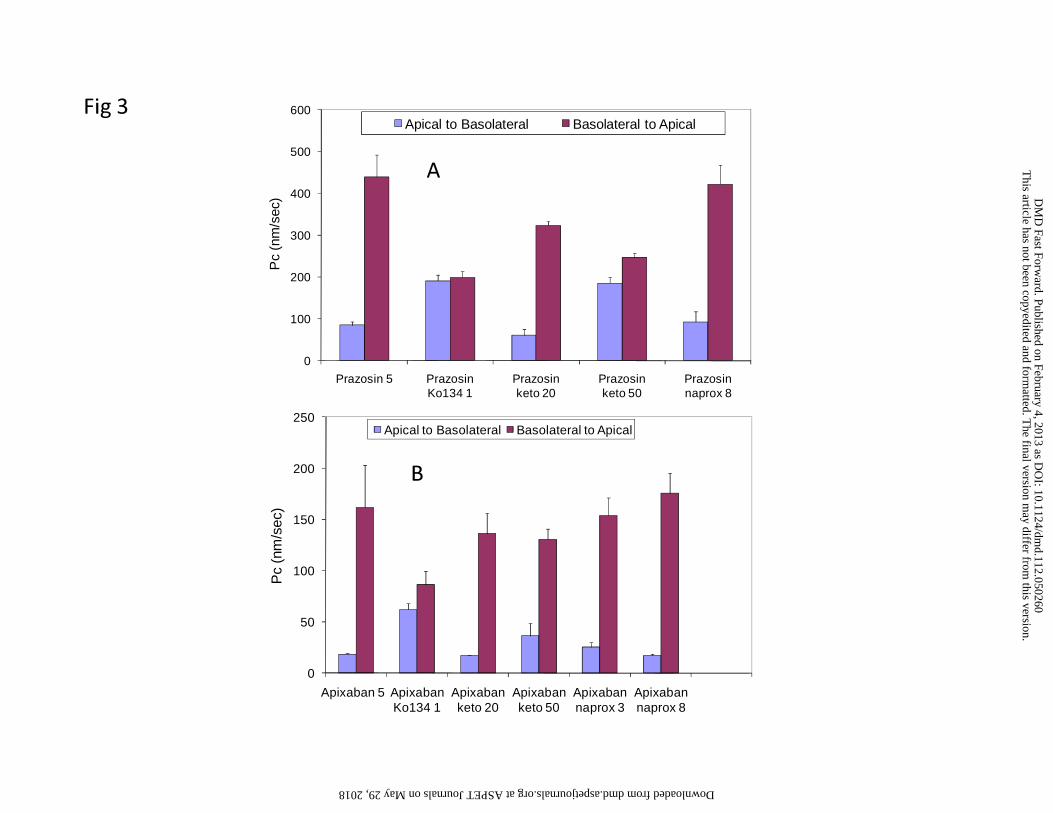
Fig 3
0
100
200
300
400
500
600
Prazosin 5 Prazosin Ko134 1
Prazosin keto 20
Prazosin keto 50
Prazosin naprox 8
Pc
(nm
/sec
)
Apical to Basolateral Basolateral to Apical
0
50
100
150
200
250
Apixaban 5 Apixaban Ko134 1
Apixaban keto 20
Apixaban keto 50
Apixaban naprox 3
Apixaban naprox 8
Pc
(nm
/sec
)
Apical to Basolateral Basolateral to Apical
A
B
This article has not been copyedited and form
atted. The final version m
ay differ from this version.
DM
D Fast Forw
ard. Published on February 4, 2013 as DO
I: 10.1124/dmd.112.050260
at ASPET Journals on May 29, 2018 dmd.aspetjournals.org Downloaded from
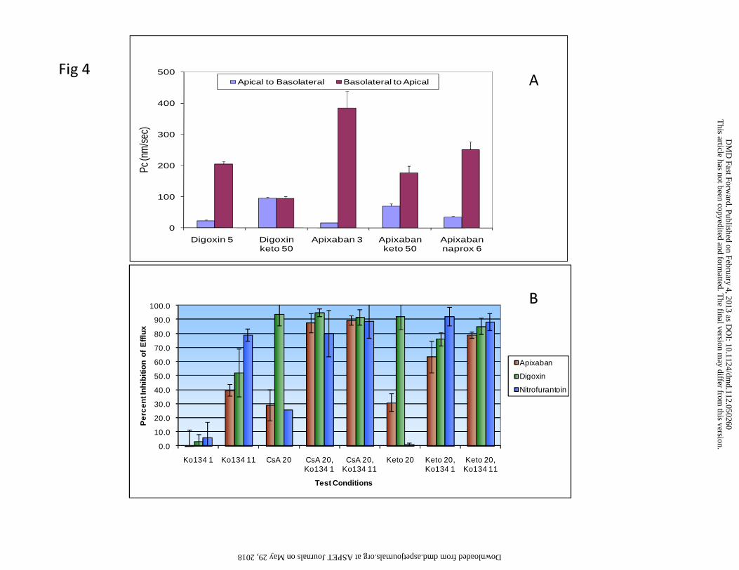
Fig 4
0.0
10.0
20.0
30.0
40.0
50.0
60.0
70.0
80.0
90.0
100.0
Ko134 1 Ko134 11 CsA 20 CsA 20, Ko134 1
CsA 20, Ko134 11
Keto 20 Keto 20, Ko134 1
Keto 20, Ko134 11
Pe
rcen
t In
hib
itio
n of
Eff
lux
Test Conditions
Apixaban
Digoxin
Nitrofurantoin
B
0
100
200
300
400
500
Digoxin 5 Digoxin keto 50
Apixaban 3 Apixaban keto 50
Apixaban naprox 6
Pc (n
m/s
ec)
Apical to Basolateral Basolateral to Apical A
This article has not been copyedited and form
atted. The final version m
ay differ from this version.
DM
D Fast Forw
ard. Published on February 4, 2013 as DO
I: 10.1124/dmd.112.050260
at ASPET Journals on May 29, 2018 dmd.aspetjournals.org Downloaded from