doi doc url //eprints.lib.hokudai.ac.jp/dspace/bitstream/2115/...complex, a number of smooth- and a...
TRANSCRIPT

Instructions for use
Title EOSINOPHILS OF OVINE PERIPHERAL BLOOD IN ELECTRON MICROSCOPY
Author(s) YAMADA, Yutaka; SONODA, Mitsuo
Citation Japanese Journal of Veterinary Research, 18(3): 117-123
Issue Date 1970-09
DOI 10.14943/jjvr.18.3.117
Doc URL http://hdl.handle.net/2115/1961
Type bulletin
File Information KJ00002369881.pdf
Hokkaido University Collection of Scholarly and Academic Papers : HUSCAP

Jaj>. J. '['Ct. Res., 18, 117-123 (1970)
EOSINOPHILS OF OVINE PERIPHERAL BLOOD IN ELECTRON MICROSCOPY
Yutaka YAMADA and Mitsuo SONODA
Department of Veterinary Internal Aledicine Faculty of Veterinary Medicine
Hokkaido Unh'ersity, Sapporo, Japan
(Received for publication, June 8, 1970)
The fine structures of the eosinophils of the peripheral blood obtained from 5
clinically normal sheep were examined under an electron microscope. The results
thus obtained were summarized as follows: 1) The cells with nuclei of two to four
lobes were most commonly observed on the cut planes. 2) The nuclear lobes of the
cells showed a clear maculous appearance with two distinct densities, one light and the
other dark, according to the amount of the chromatin condensation. 3) On the basis
of the internal structures of the specific granules, they were classified into six types
and the basic type of the granules seemed to be those containing middle plates.
4) A small number of mitochondria measuring 0.3 by 0.7 p on the average, Golgi
complex, a number of smooth- and a few rough-surfaced endoplasmic reticulum, and
some other micro-organelles were observed in the cytoplasm.
INTRODUCTION
There have been a lot of publications on the fine structures of the eosinophils In the peripheral blood of the human1 ,2,5,lO,1l,16,18,20,35,37,38) and other animals6 - 8 ,14,17,
21,23,29-3],34), and it is well known that the internal structures of their specific
granules are very different in accordance with the difference of the animal species. However, up to the present tiIlle, there are no reports on ovine eosinophils in
electron microscopy.
In this paper, the fine structures of the eosinophils III the peripheral blood
obtained from clinically normal sheep will be described.
MA TERIALS AND METHODS
Blocks for electron microscopy and the methods of observation were just the
same as those of the previous paper41).
OBSERV ATIONS
The general shapes of the eosinophils were round or oval. The contours of the cells
were usually smooth; however, some of them were irregular because of having many small
or large pseudopodic projections.

118 YAMADA, Y. & SONODA, M.
1 Nucleus
The nuclei of the eosinophils showed one to several separated nuclear lobes on the cut planes. In the present observations, however, the nuclei with 2 to 4 lobes were common
in number, but the cells with more than 5 nuclear lobes were rarely observed.
In the nuclei of the eosinophils, there were two parts of distinct densities, one light
and the other dark, depending upon the amount of the chromatin condensation. The dark
part of the two was located peripherally in the nuclear lobes and constituted a thick band
subjacent to the nuclear membrane. On the other hand, the light part occupied chiefly the central area of the nuclear lobes, however, it occasionally extended toward the nuclear
membrane.
2 Cytoplasm
The cytoplasm of the cells was delineated by thin cell membranes. In the cytoplasm,
there were a lot of the specific granules, a small number of mitochondria, Golgi complex and some other micro-organelles.
The specific granules The specific granules of the eosinophils were distributed
randomly in the cytoplasm except for parts of the pseudopodic projections, the Golgi and
narrow peri-nuclear areas. These granules had extraordinary high electron density and were
opaque. They were surrounded by a distinct unit membrane. On the cut planes, they
varied considerably in size and shape depending upon the cut directions in which the granules were sectioned.
Almost all of the granules had variable internal structures; however, some of them
were homogeneous without any internal structures. On the basis of the internal structures,
they were divided into 6 types of granules as fol1ows.
1) The 1st type
These were the homogeneously compact high or moderate dense granules without any internal structures. In general, the granules of this type were round in shape, and their
size was on the average 0.60 (0.4-0.8) p in diameter.
2) The 2nd type These were the granules containing so-called middle plates in their ground substances.
The middle plates were more dense than that of the ground substances of the granules,
and they consisted of substances with stratified structures or without any structures. Each granule usually contained one to several middle plates. In the longitudinal sections of the
granules, the middle plates appeared like bandages running parallel to the long axis of the
granule profiles. The middle plates were situated usually in the central portions of the
granules, but sometimes they were seen eccentrically in the side areas of the granules. The
granules of this type were usually oval or spindle-like and a few were round in form. In
size, they were 1.1 (0.8-1.5) J1 long by 0.3 (0.2 -0.6) J1 wide on the average.
3) The 3rd type These were the granules contammg substances with myelin-like structures, viz., they
had lamellar structures arranged concentrically, so that they looked just like hair balls.
In some of the granules of this type, the substances consisted of 2-3 myelin-like structures,
and a dense substance in the central area or periphery were observed. The granules of

Ovine eosinophils in electron microscopy
this type were usually round. They were 0.7 (0.4-1.0) f1 in diameter on the average.
4) The 4th type
119
These were the granules with fine reticulated structures in some parts of the ground
substances. The parts of the ground substances without reticulated structures were homo
geneous. By observing enlarged pictures, it was clear that the reticulated structures were
composed of fine granular materials arranged checkedly. The granules of this type were
round or oval in shape and they were much the same as those of the 2nd and the 3rd type
granules in size.
5) The 5th type
These were the granules contammg dense homogeneous round structures. They were
usually round in shape and much the same size as those of the 1st type granules.
6) The 6th type
These were the granules containing simultaneously some substances of the internal
structures described in the 2nd to the 5th type granules. Namely, for example, the granules
containing the reticulated structure or myelin-like structure together with the middle plate
were observed, frequently. Of course, in others, the ones with various complicated combi
nations of the internal structures were observed. The granules of this type were round,
oval or slightly irregularly round in shape, and approximately similar m size to those of
the 2nd and the 3rd type granules.
Among these specific granules, the granules with round or oval vacant defects were
observed rarely.
In the appearance rates of the specific granules of each type in the cytoplasm of the
cells, the granules of the 2nd type were observed most predominantly and the granules of
the 3rd and the 6th types were observed next most predominantly. The granules of other
types were only observed rarely.
Mitochondria A few mitochondria distributed randomly among the specific granules
throughout the cytoplasm. They were round or oval in shape. Their size was 0.3 (0.2-0.4) f1
by 0.7 (0.4-1.0) p on the average.
Golgi complex Golgi complex consisted of lamellar membranes and vesicles were frequently observed in the central areas of the cells.
Endoplasmic reticulum A large number of smooth-surfaced endoplasmic reticulum
were seen randomly throughout the cytoplasm. They were smaller than the specific
granules in size and were filled with more or less dense substances or had clear contents.
A few rough-surfaced endoplasmic reticulum were observed in the cytoplasm. They were
short or long thin canalicular in shape.
Others A number of free ribosomes and polysomes were present in the cytoplasm.
The multivesicular bodies enclosed by the unit membrane and containing a few small
granular or vesicular substances were rarely observed. The bodies with the structures
similar to those of thrombocytes were present in the cytoplasms of a few cells. They were
supposed to be phagocyted thrombocytes.

120 YAMADA, Y. & SONODA. M.
CONSIDERA TIONS
It has been well known that the fine structures of the specific granules of
the eosinophils are very variable according to the difference of animal species.
On the basis of the observadons reported already, the fine structures of their
specific granules will be fundamentally grouped into the following four types,
viz., they are the type of granules containing middle plates as seen in those of the human1,2,5,9-11,16,18,20,21,35,37-39,42l, guinea pigs12 ,13,20,21 ,36-38), rabbits21 ,40,43), mice20),
rats20,22-2~), orangutans14) and chimpanzees14), the ones with the middle trunks21 )
which are composed of a very characteristic lamellar structure as seen in those
of cats2- 4,21,37-39), the ones with two or three thick stratified concentric structures
as seen in those of cattle34\ dogs31 ,32) and minks19), and homogeneous dense
granules without any internal structures as seen in those of horses6 ,7,15,21,30,39),
pigs27), gorillas14) and chickens8,39).
In the present observations of the ovine eosinophils, the specific granules of
the cells have showed various internal structures. Namely, the homogeneous
granules without any internal structures, the ones with middle plates, the ones
with myelin-like structures, the ones with small fine reticulated structures, the
ones with homogeneous dense round substances, and the granules containing simultaneously some of the internal structures described above were observed.
It was very interesting to discover that in the individual eosinophils, the
internal structures of the granules were very variable from one granule to another.
Furthermore, considering the cubic structure of the granules, the 1st, the
2nd and the 5th type granules are supposed to be granules with similar internal
structures. The morphological differences among them on the cut planes are
thought to be derived from the different directions of cutting.
On the basis of the apperance rate of the granules of each type, the basic
type of the specific granules of the ovine eosinophils seems to be the granules
with the middle plates.
Recently, attention has been paid to the fine structures of the substances contained in the granules. SHELDON & ZETTERQUIST and othersZO ,35) reported that
the middle plates or crystals of the specific granules had a repeat period of about
30 to 40 A in the human, rats, mice and guinea pigs. MILLER et a1. suggested
that the crystals of the granules had cubic lattices. RUDOLPH observed that the
specific granules of the eosinophils in the mast cell tumor of dogs had lamellar
structures in the peripheral or central areas of their homogeneous matrix.
In the present observations of the ovine eosinophils, it was seen that the
middle plates or crystalloids of the specific granules had a lamellar structure
similar to that of the observations described above. In addition, the authors

Ovinc eosinophils in electron microscopy 121
observed the fine reticulated and lattice-like structures in the granules as shown
in the micrographs of the 4th type granules. This is the first report on the
presence of such structures in the granules of the eosinophils. Furthermore, the
5th and the 6th type granules classified by the authors have not been reported yet.
The granules with round or oval vacant defects in the ovine eosinophils
were rarely observed by the authors. SONODA and KOBAYASHI observed round
or oval vacant defects in the granules of the canine eosinophils, and supposed
that they were morphological findings related to the function of the eosinophils.
The presence of a unit membrane of the specific granules of the eosinophils has been disputed by many workers1 ,8,1l,16,20,21,'25,26,29,31,32,35,37,39). In the present
observations, it was clear that the specific granules of the ovine eosinophils had
a unit membrane consisting of three layers.
In the cytoplasm of the human eosinophils, moderately dense, homogeneous
and irregularly shaped bodies with much the same size as the specific granules
have been observed by some workers1,9,18,39,42). Especially Low & FREEMAN called
them basophilic bodies and WATANABE39 ) supposed that these bodies were the
lipid bodies. However, in the present study on the ovine eosinophils, any of
such bodies in the cytoplasm had not been observed at all.

122 YAMADA, Y. & SONODA. M.
REFERENCES
1) ANDERSON, D. R. (1966): Ultrastructure of normal and leukemic leukocytes m
human peripheral blood, J. Ultrastruct. Res., Suppl. 9, 1
2) BARGMAN, W. & KNOOP, A. (1956): Z. Zellforsch. mikrosk. Anat., 44, 282
3) BARGMAN, W. & KNOOP, A. (1956): Ibid., 44, 692
4) BARGMAN, W. & KNOOP, A. (1958): Ibid., 48, 130
5) BESSIS, M. & THIERY, 1. P. (1961): Int. Rev. Cytol., 12, 199 6) BOCCIARELLI, D. S., TENTORI, L. & VIVALDI, G. (1969): Rc. 1st. sup. San ita, 22,
1059
7) BRAUNSTEINER, H. & PAKESCH, F. (1962): Acta haemat., 28, 163
8) DHINGRA, L. D., PARRISH, W. B. & VENZKE, W. G. (1969): Am. J. vet. Res., 30,
637
9) FREEMAN, 1. A. (1964): Cellular fine structure, New York, Toronto, London:
McGraw-Hill Book Company
10) GOODMAN, J. R., REILLY, E. B. & MOORE, R. E. (1957): Blood, 12, 428
11) HIRSCH, J. G. & FEDORKO, M. E. (1968): J. Cell Biol., 38, 616
12) HUDSON, G. (1966): Expl. Cell Res., 41, 265
13) HUDSON, G. (1967): Ibid., 46, 121
14) HUSER, H. 1. & WEBB, C. M. (1967): Experientia, 23, 669 15) ITO, Y. (1964); Norinsho Kachiku-eisei-shikenjyo Nenpo, 4, 386 (in Japanese)
16) KAUTZ, J. & DEMARSH, Q. B. (1954): Blood, 9, 24
17) KNOCKE, K.-W. (1963): Folia haemat. Neue Folge, 7, 129
18) Low, F. N. & FREEMAN, J. A. (1958): Electron microscopic atlas of normal and
leukemic human blood, New York, Toronto, London: McGraw-Hill Book Com
pany, Inc.
19) LUTNER, M. A., TIERNEY, J. H. & BENDITT, E. P. (1965): Lab. Invest., 14, 2063
20) MILLER, F., DEHARVEN, E. & PALADE, G. E. (1966): J. Cell Biol., 31, 349
21) OSAKO, R. (1959): Acta haemat. jap., 22, 134 (in Japanese with English summary)
22) PEASE, D. C. (1956): Blood, 11, 501
23) RINEHART, J. F. (1955): Am. J. din. Path., 25, 605
24) Ross, R. & KLEBANOFF, S. 1. (1966): J. expo Med., 124, 653 25) RUDOLPH, R. (1969); Dt. tie ra rztl. Wschr., 76, 176
26) SCHULZE, P. (1966): Arch. expo VetMed., 20, 767
27 ) SCHULZE, P. (1967): Ibid., 21, 1305
28 ) SHELDON, H. & ZETTERQUIST, H. (1955): Bull. Johns Hopkins Hasp., 96, 135
29) SHIVELY, J. N., FELDT, C- & DAVIS, D. (1969): Am. J. vet. Res., 30, 893
30) SONODA, M. (1963): Proceedings of the 55th Meeting of the Japanese Society of
Veterinary Science, Jap. J. vet. Sci., 25, 394 (summary in Japanese)
31) SONODA, M. (1969): Proceedings of the 67th Meeting of the Japanese Society of
Veterinary Science, Ibid., 31, SuppL, 98 (summary in Japanese)
32) SONODA, M. & KOBAYASHI, K. (1970): Jap. J. vet. Res., 18, 43
33) SONODA, M. & MARSHAK, R. R. (1970): Ibid., 18, 9
34) SONODA, M., MIFUNE, Y. & OHY A, S. (1964): Proceedings of the 57th Meeting

Ovine eosinophils in electron microscopy 123
of the Japanese Society of Veterinary Science, Jap. J. vet. Sci., 26, 440 (summary
in Japanese)
35) WATANABE, 1., DONAHUE, S. & HOGGATT, N. (1967): J. Ultrastruct. Res., 20, 366
36) WATANABE, Y. (1954): J. Electron Microsc., Chiba Cy, 2, 34
37) W AT ANABE, Y. (1956): Acta haemat. jap., 19, 327 (in Japanese with English
summary)
38) W AT ANABE, Y. (1957): J. Electron Microsc., Chiba Cy, 5, 46
39) WATANABE, Y. (1963): (translated title) "The fine structures of leukocyte", Com
pendium of haematology, 1, Ed. AMANO, S. & HIBINO, S., 1 ed., 327, Tokyo, Maru
zen (in Japanese)
40) WETZEL, B. K., HORN, R. G. & SPICER, S. S. (1967): Lab. Invest., 16, 249
41 ) YAMADA, Y. & SONODA, M. (1970): Jap. J. vet. Res. 18, 83
42) ZUCKER-FRANKLIN, D. (1963): J. Ultrastruct. Res., 9, 325
43) ZUCKER-FRANKLIN, D. & HIRSCH, J. G. (1964): J. expo Med., 120, 569
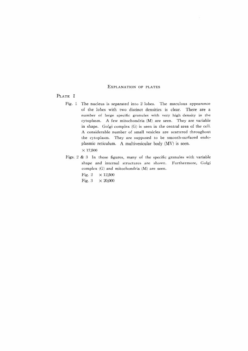
EXPLANATION OF PLATES
PLATE I
Fig. 1 The nucleus is separated into 2 lobes. The maculous appearance
of the lobes with two distinct densities is clear. There are a
number of large specific granules with very high density in the
cytoplasm. A few mitochondria (M) are seen. They are variable
in shape. Golgi complex (G) is seen in the central area of the cell.
A considerable number of small vesicles are scattered throughout
the cytoplasm. They are supposed to be smooth-surfaced endo
plasmic reticulum. A multivesicular body (MV) is seen.
x 17,500
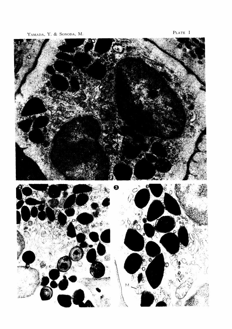
Figs. 2 & 3 In these figures, many of the specific granules with variable
shape and internal structures are shown. Furthermore, Golgi
complex (G) and mitochondria (M) are seen.
Fig. 2
Fig. 3 x 12,500
X 20,000
YAMADA, Y. & SONODA, M. PLATE I

PLATE II
Fig. 4 A small round homogeneous granule (the 1st type, I), a spindleform· granule with wide middle plate (the 2nd type, II), two short
spindle-form granules containing simultaneously narrow middle
plates and round myelin-like structures (the 6th type, VI) and one
other granule are seen.
x 75,000
Fig. 5 A small round granule with fine reticulated structures (the 4th type,
IV) and a large almost round granule with a highly dense annular
structure (the 3rd type, III) are seen.
X 75,000
Fig. 6 An oval granule with a wide middle plate (the 2nd type) is seen.
The middle plate has lamellar structures parallel to each other. The unit membrane consisting of three layers is clear.
x 150,000
Fig. 7 A granule with myelin-like structures (the 3rd type) is shown. Its
core consists of three small hair ball-like substances.
X 150,000

YAMADA, Y. & SONODA, M. PLATE II

PLATE III
Fig. 8 This is a round granule with almost homogeneous matrix (the 1st
type).
X 90,000
Fig. 9 A round granule with round dense homogeneous structures (the
5th type) is shown.
X 90,000
Fig. 10 An elliptical granule containing a wide middle plate and fine
reticulated structures (the 6th type) is shown.
X 90,000
Fig. 11 A round granule containing a bent needle-like middle plate and
a concentric myelin-like structure in its matrix (the 6th type) is
shown.
X 90,000
Fig. 12 An elliptical granule containing two narrow belt-like middle plates
and a myelin-like structure (the 6th type) is shown.
X 90,000
Fig. 13 A slightly irregularly round granule containing myelin-like struc
ture in the peripheral area and a fine reticulated structure in the
central area (the 6th type) is shown.
X 90,000

YAMADA, Y. & SONODA, r..1. PLATE III
f i