does high tibial osteotomy change the tibia length? · incision over the pes anserinus. this...
TRANSCRIPT
Abstract— High Tibial Osteotomy (HTO) is an accepted
means of improving the symptoms of a knee deformity such as
genu varum. Patient’s symptoms may improve by off-loading
the diseased medial compartment of the knee by changing the
axis of alignment. This means that loading of the knee joint
changes to the less diseased lateral compartment by an
osteotomy and opening wedge thereby reducing pain, swelling,
and stiffness associated with arthritis. By implication this is
major surgery and requires careful preoperative assessment
taking into account, patient factors, joint geometry and degree
of disease in affected and less affected compartments. This
study analyzes the effect of surgery on the ultimate length of the
tibia compared to its preoperative state. Both Opening and
Closing Wedge osteotomies were investigated in this study. The
results showed that the length of tibia would increase following
the Opening Wedge HTO and it would decrease by a Closing
Wedge HTO. Although the change in the length of the tibia was
small in comparison to its overall length the possibility of bone
lengthening must be considered carefully when determining an
Opening or Closing Wedge HTO, especially when a large
correction angle is required.
Index Terms— High Tibial Osteotomy, Genu Varum, Tibia
length, Opening and Closing Wedge HTO
I. INTRODUCTION
High Tibial Osteotomy (HTO) surgery was first described
by Jackson in 1958 [1] to prevent the symptoms of
osteoarthritis in the knee, and to limit its progression, thereby
prolonging the function of the diseased knee. It is a method to
treat unicondylar osteoarthritis at least temporarily, as
satisfaction begins to fall after 5 years. Ultimately patients
Manuscript received January 15, 2013; revised April 05, 2013. All of the
authors have no financial relationship to any private companies and
organizations.
Pierre-Louis RICCI is with the National Engineering School of Metz (ENIM), Metz, France and the School of Design and Engineering, Brunel
University West London, UK
Adrien Durandet is with the National Engineering School of Metz (ENIM), Metz, France and the School of Design and Engineering, Brunel
University West London, UK
Amir Hossein Saveh is with the Shahid Beheshti University of Medical Sciences, Akhtar Orthopaedic Research Centre, Tehran, Iran
Qureish Vanat is with the Queen Elizabeth Hospital, Woolwich, UK
Bin Wang is with the School of Design and Engineering, Brunel University West London, UK
Mahmoud Chizari is with Center of Orthopaedic Research at the School
of Design and Engineering, Brunel University West London, UK, (corresponding author: Mahmoud Chizari. Phone: +447886454320; e-mail:
who are very symptomatic go on to have total knee
athroplasty.
There are two methods of achieving the goal of the
osteotomy: An Opening Wedge HTO (OWHTO) or a
Closing Wedge HTO (CWHTO) [2] [3] [4] [5] [6] [7].
It was observed that an OWHTO tended to lengthen the
tibia whereas a CWHTO had the opposite effect, to shorten it.
[8] [9] [10]. In the first case, the opening of the bone cut
(osteotomy site) was responsible for lengthening [9] [10]
whereas shortening was seen in the second method as this
required removal of a wedge of bone from the tibia and
closing its ends [11] [12]. Although these observations were
made, just a few studies focus on the limb lengths after HTO
[13].
Currently, long length plain 2D radiographs are used to
calculate the mechanical axis of the lower limb. This static
image however cannot take in to account dynamic axial
loading. This is why groups like Andriacchi et al. [14], claim
that the static axial loading axis cannot correlate to a
reproducible clinical outcome. Unfortunately inaccuracy of
dynamic axis acquisition during gait is due to change in the
lower limb positions such as flexion and rotation at the knee.
This makes formation of a reliable focus from which to
reference and template difficult, and hence calculation of
axes inaccurate. [2] [15] [16].
In this study we focused on pre and post OWHTO
operative parameters reflecting the length of the tibial bone
with respect to a genu varum deformity. The study evaluated
the postoperative limb length determined using an analytical
method and compared the change in limb lengths between
Opening and Closing Wedge HTO examples. Genu varum
deformity means that the knee appears to be bowed outwards
away from the midline. This therefore requires a medial
OWHTO or a lateral CWHTO. The hypothesis was that the
overall length of the tibial bone, after an OWHTO, would
increase within a certain range due to the spacing introduced.
While after CWHTO it becomes shorter because of the bone
loss of the proximal tibia after removing a wedge. It is
assumed that the change in length is negligible with a
CWHTO procedure. As a result this study will concentrate on
the potential limb lengthening of an OWHTO.
II. MATERIAL AND METHODS
A. HTO Surgery
This is a description of an Opening Wedge High Tibial
Osteotomy performed on our patient. It begins with a small
Does High Tibial Osteotomy Change the Tibia
Length?
Pierre-Louis Ricci, Adrien Durandet, Amir Hossein Saveh, Qureish Vanat, Bin Wang, Mahmoud
Chizari
Proceedings of the World Congress on Engineering 2013 Vol II, WCE 2013, July 3 - 5, 2013, London, U.K.
ISBN: 978-988-19252-8-2 ISSN: 2078-0958 (Print); ISSN: 2078-0966 (Online)
WCE 2013
incision over the pes anserinus. This exposes the medial
aspect of the proximal tibia. Retractors protect the patella,
hamstring tendons, neurovascular structures and the medial
collateral ligament, when approaching the bone. The next
step requires drilling at the proximal tibia. This is from
medial to lateral direction using fluoroscopic imaging. It
should be proximal to the tibial tubercle but be 3.5cm distal to
the medial joint line. The direction is towards the tip of the
fibula. This drill acts a guide along which the saw passes
during the osteotomy. As the saw passes medio-laterally it is
important to leave the lateral edge of the tibia intact as this
will act as the bony hinge as the medial part is opened using
spreaders. This creates an open wedge on the medial side
changing the alignment of the tibia and hence the load from
the medial to lateral side. A wedge shaped block of bone graft
(or bone graft substitute) is now placed into the open wedge
and a locking plate is applied to hold the construct together.
The last part of the operation is to ensure the correct
placement of the graft and correct new alignment of the tibia
with respect to the femur, this is done using fluoroscopy.
Application of a hinged brace post operatively permitting
only toe-touch weight bearing for several weeks (from 6 to 8
weeks) allows protected healing.
The subject patient of this study is a 25 year old female
who was suffering from a genu varum deformity on her right
leg. An OWHTO operation was carried out on her right knee.
The procedure was performed at the Aktar Hospital by a
senior Orthopaedic surgeon. The pre and post operation
results of this patient were used in this study.
B. Tibial Lengthening
In order to assess the treatment received by the subject
patient, the radiographs of her lower limb were analyzed
before and after the operation. With these images it was
possible to evaluate the alignment of her lower limb before
and after the operation. Performing the osteotomy, not only
changed the alignment of the lower limb but also the tibial
bone length.
Some studies dealt with this variation due to the medial
opening or the lateral closing osteotomies based on cadaveric
experimentation [13]. Other authors focused on the
calculation of lower limb lengths after three different
osteotomies after considering the new alignment [10]. These
studies did not define ways to predict outcome length, or
consider the technical operative methods or have statistical
data.
Plateau-Ankle angles were determined pre and post
operatively because HTO modifies these values directly. In
varus knees the tibial axis is angled medially with respect to
the tibial plateau, named AB [15]. It is therefore directly
linked to tibial geometry and are useful pre and post operative
parameters.
C. Tibia’s Lengthening with Opening Wedge HTO
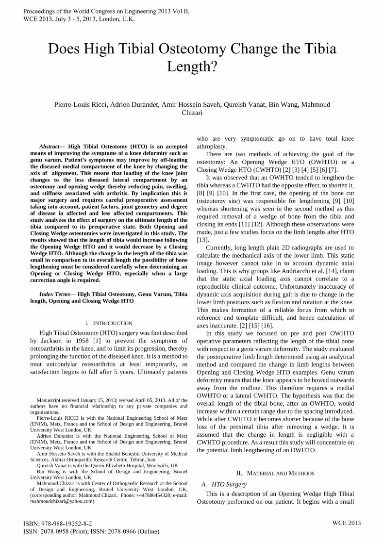
The Fig. 1 illustrates the modification of the tibia’s
geometry due to HTO with A and B points representing
respectively the beginning and the end of the tibia’s
mechanical axis. It is clear to see the mechanical axis of the
tibia lengthens as the initial distance AB becomes AB’
because B is shifting to B’ position.
Fig. 1. OWHTO performed on the tibia; original bone (a); OWHTO cut (b);
OWHTO (c). The Plateau-Ankle (PA) angle pre and post-operatively is also
shown on the picture.
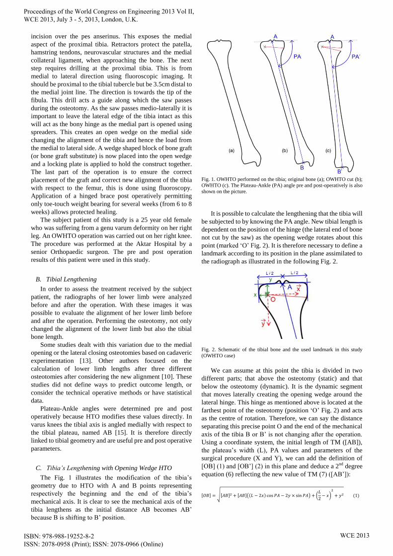
It is possible to calculate the lengthening that the tibia will
be subjected to by knowing the PA angle. New tibial length is
dependent on the position of the hinge (the lateral end of bone
not cut by the saw) as the opening wedge rotates about this
point (marked ‘O’ Fig. 2). It is therefore necessary to define a
landmark according to its position in the plane assimilated to
the radiograph as illustrated in the following Fig. 2.
Fig. 2. Schematic of the tibial bone and the used landmark in this study
(OWHTO case)
We can assume at this point the tibia is divided in two
different parts; that above the osteotomy (static) and that
below the osteotomy (dynamic). It is the dynamic segment
that moves laterally creating the opening wedge around the
lateral hinge. This hinge as mentioned above is located at the
farthest point of the osteotomy (position ‘O’ Fig. 2) and acts
as the centre of rotation. Therefore, we can say the distance
separating this precise point O and the end of the mechanical
axis of the tibia B or B’ is not changing after the operation.
Using a coordinate system, the initial length of TM ([AB]),
the plateau’s width (L), PA values and parameters of the
surgical procedure (X and Y), we can add the definition of
[OB] (1) and [OB’] (2) in this plane and deduce a 2nd
degree
equation (6) reflecting the new value of TM (7) ([AB’]):
[ ] √[ ] [ ](( ) ) (
)
( )
Proceedings of the World Congress on Engineering 2013 Vol II, WCE 2013, July 3 - 5, 2013, London, U.K.
ISBN: 978-988-19252-8-2 ISSN: 2078-0958 (Print); ISSN: 2078-0966 (Online)
WCE 2013

[OB] is joining O and B, respectively the endpoint of the
cut and the center of the ankle before the operation.
[ ] √[ ] [ ](( ) ) (
)
( )
[OB’] is the distance between O and B’, respectively the
endpoint of the cut and the center of the ankle after the
operation.
[OB] and [OB’] should have the same length as O is
considered as the center of the rotation and these two
distances represent the radius of this circle. We can therefore
deduce the following equations:
[ ] [ ] ( )
By replacing the respective values of [OB] and [OB’]
found in equation (1) and (2), we can reach these results and
express a 2nd
degree equation (6) reflecting the new value of
TM (7) ([AB’]):
( ) [ ] [ ] ( ) ( ) [ ] [ ] ( )
( ) [ ] [ ] ( )
( )
[ ] [ ][ ( ) ]
Values of b and c unknowns are expressed previously and
lead to the new value of the tibia’s length [AB’] (7):
( ) [ ] √
( )
D. Tibia’s Shortening with Closing Wedge HTO
The previous Fig. 3 shows the changes affecting the tibia
after CWHTO. Unlike the previous surgical procedure, this
requires a wedge of bone being removed. The gap created is
closed and fixed again with a plate and screws. Points A and
B represent the tibia’s mechanical axis. In this case, we
observe a shortening of the mechanical axis of the tibia as
length AB becomes AB’ after the intervention.
Once again, using the same parameters CW changes were
similarly evaluated this time for relative shortening of the
tibia. PA angles proved to be useful with respect to the hinge
defined again as point ‘O’ but modified as the osteotomy was
performed from the lateral side, as we can see in Fig. 4.
Fig. 3. CWHTO performed on the tibia; original bone (a); CWHTO cuts (b); CWHTO (c). The Plateau-Ankle (PA) angle pre and post-operatively is
shown on the picture.
Fig. 4. Schematic of the tibial bone and the used landmark in this study
(CWHTO case)
One more time we can assume the hypothesis introduced
in the previous paragraph which considers O as a hinge for
the rotation. This assumption lead to the same observations:
values [OB] (distance joining the end of the cut O and the
bottom extremity of the mechanical axis before the
operation) and [OB’] (same distance but considered after the
operation) stay equals to each other. Such as before, using the
Appropriate coordinate system, the initial length of TM
([AB]), the plateau’s width (L), PA values and parameters of
the surgical procedure (X and Y), we can reach the definition
of [OB] (8) and [OB’] (9) in order to find the 2nd
degree
equation EEE displaying the value of TM after the operation
EEE (AB’):
[ ] √[ ] [ ](( ) ) (
)
( )
[OB] is the value of the line linking the end of the two cuts
O and the bottom extremity of the mechanical axis before the
operation.
[ ] √[ ] [ ](( ) ) (
)
( )
[OB’] is similar to the definition here before but it
considers the case after the operation, this means O and B’
points.
Proceedings of the World Congress on Engineering 2013 Vol II, WCE 2013, July 3 - 5, 2013, London, U.K.
ISBN: 978-988-19252-8-2 ISSN: 2078-0958 (Print); ISSN: 2078-0966 (Online)
WCE 2013

Once again, because O is judged as the center of the
hinge, the distance joining O and the end of tibia’s
mechanical axis before and after the operation is unchanged:
[ ] [ ] ( )
Same reasoning as before in applied in this case:
replacing values of [OB] (8) and [OB’] (9) guides to the
researched 2nd
degree equation (13) introducing the new
value of TM (14) ([AB’]).
( ) [ ] [ ] ( )
( ) [ ] [ ] ( )
( ) [ ] [ ] ( )
( )
[ ] [ ][ ( ) ]
Values of b and c unknowns are expressed previously and
lead to the new value of the tibia’s length [AB’] (14):
( ) [ ] √
( )
III. RESULT AND DISCUSSION
This study evaluated the relative lengths of the tibia pre
and post OW and CW HTO surgery. The preoperative genu
varum knee showed a PA angle of 72.3° and reflected a
deformity close to 18.1° with respect to the mechanical axis
of the femur to the tibia. In order to restore a better alignment
of the tibia relative to the femur HTO surgery improved the
PA angle to 86.3° leading to an HKA angle of 178°.
Calculations for both the OWHTO and CWHTO cases were
performed. Evaluations were made for the relative changes in
mechanical axes and their resultant lengths.
For the OWHTO case, preoperative tibial length was
337.5 mm but after the intervention it reached 344.4 mm.
Opening wedge osteotomy increased the tibia by almost 7
mm by changing the initial 18.1° mechanical varus deformity
to a HKA angle of 178°. Clinically this degree of opening
was required to achieve the aim of the operation, that being to
off-load the diseased medial compartment onto the disease
free lateral compartment by changing the mechanical axis.
We also found that there was not just a link between the tibial
length and PA angle but there was a proportional relationship
between them. Table I reports the measures obtained from the
patient’s tibia pre and post operation and also shows the
difference from the operation.
The result of the tibial length in Table II assumes that the
patient obtained a closing wedge HTO.
TABLE I
TIBIAL LENGTH PRE AND POST OPENING WEDGE HTO
Measurements Pre Op Post Op
PA (°) 72.3 86.3
Cutting Angle (°) 0 14 Tibia length (mm) 337.5 344.4
TABLE II
TIBIAL LENGTH PRE AND POST CLOSED WAGE HTO
Measurements Pre Op Post Op
PA (°) 72.3 86.3 Cutting Angle (°) 0 14
Tibia length (mm) 337.5 337
IV. CONCLUSION
In conclusion, there was a change in tibia length after
opening and closed-wedge HTO. The change for OWHTO
was considerable while the tibial length change in CWHTO
was negligible. The tibia length increased 6.9 mm after the
opening wedge HTO and was reduced by 0.5 mm following
closed wedge HTO surgery. The osteotomy correction angle
was 14 degrees for both cases. The greater the correction in
axis the greater the change in the tibia length.
ACKNOWLEDGEMENTS
The authors are grateful to the head and research team of
Akhtar Orthopaedic Research Centre, Tehran, Iran, for
providing samples and facilities to carry on the experimental
research of this study.
REFERENCES
[1] J. JACKSON, “Osteotomy for Osteoarthritis of the Knee,” British
Journal of Bones and Joint Surgery, vol. 40B, p. 826, 1958.
[2] D. K. BAE, S. J. SONG and K. H. YOON, “Closed-Wedge High Tibial
Osteotomy using Computer-Assisted Surgery Compared to the
Conventional Technique,” British Journal of Bone and Joint Surgery, vol. 91, pp. 1164-1171, 2009.
[3] H. BITO, R. TAKEUCHI, K. KUMAGAI, M. ARATAKE, I. SAITO,
R. HAYASHI, Y. SASAKI, Y. AOTA and T. SAITO, “A Predictive Factor for Acquiring an Ideal Lower Limb Realignment after
Opening-Wedge High Tibial Osteotomy,” Knee Surgery and Sport
Traumatology Arthroscopy, vol. 17, pp. 382-389, 2009.
[4] F. GEBHARD, C. KRETTEK, T. HUFNER, P. A. GRUTZNER, U.
STOCKLE, A. B. IMHOFF, S. LORENZ, J. LJUNGQVIST and P.
KEPPLER, “Reliability of Computer-Assisted Surgery as an Intraoperative Ruler in Navigated High Tibial Osteotomy,” Archives of
Orthopeadics and Trauma Surgery, vol. 131, pp. 297-302, 2011.
[5] S. HANKEMEIER, P. MOMMSEN, C. KRETTEK, M. JAGODZINKI, J. BRAND, C. MEYER and R. MELLER, “Accuracy
of High Tibial Osteotomy: Comparison Between Open and
Closed-Wedge Techniaue,” Knee Surgery, Sport Traumatology, Arthroscopy, vol. 18, pp. 1328-1333, 2010.
[6] R. IORIO, A. VADALA, S. GIANNETTI, M. PAGNOTTELLI, P. DI
SETTE, F. CONTEDUCA and A. FERRETTI, “A Computer-Assisted
High Tibial Osteotomy: Preliminary Results,” Orthopedics, vol. 33
(10), pp. 82-86, 2010.
[7] K. A. JUNG, S. C. LEE, N. K. AHN, S. H. HWANG and C. H. NAM, “Radiographic Healing with Hemispherical Allogeneic Femoral Head
Bone Grafting for Opening-Wedge High Tibial Osteotomy,”
Arthroscopy, vol. 26, pp. 1617-1624, 2010.
[8] M. C. HARPER and S. T. CANALE, “Angulation Osteotomy: a
Trigonometric Analysis,” Clinical Orthopaedics and Related Research,
vol. 166, pp. 173-181, 1982.
[9] P. HERNIGOU and A. HAMDADOU, “Leg Length Changes After
Upper Tibial Osteotomy: Analysis of Different Preoperative Planning Methods,” Revue de Chirurgie Orthopédique et Réparatrice de
l'Appareil Moteur, vol. 88, pp. 68-73, 2002.
[10] W. M. MIHALKO and K. A. KRACKOW, “Preoperative Planning for Lower Extremity Osteotomies: an Analysis Using 4 Different Methods
and 3 Different Techniques,” The Journal of Arthroplasty, vol. 16, no.
3, pp. 322-239, 2001.
Proceedings of the World Congress on Engineering 2013 Vol II, WCE 2013, July 3 - 5, 2013, London, U.K.
ISBN: 978-988-19252-8-2 ISSN: 2078-0958 (Print); ISSN: 2078-0966 (Online)
WCE 2013

[11] S. HENKEMEIER, T. HUFNER, G. WANG, D. KENDOFF, J.
ZEICHEN, G. ZHENG and C. KRETTEK, “Navigated Open-Wedge
High Tibial Osteotomy: Advantages and Disadvantages Compared to the Conventional Techniaue in a Cadaver Study,” Knee urgery and
Sport Traumatology Arthroscopy, vol. 14, pp. 917-921, 2006.
[12] D. KENDHOFF, M. CITAK, A. PEARLE, M. J. GARDNER, S. HANKEMEIER, C. KRETTEK and T. HUFNER, “Influence of Lower
Limb Rotation in Navigated Alignment Analysis: Implications for High
Tibial Osteotomies,” Knee Surgery, Sport Traumatology and Arthroscopy, vol. 15, pp. 1003-1008, 2007.
[13] R. A. MAGNUSSEN, S. LUSTIG, G. DEMEY, P. NEYRET, P.
SERVIEN and E. SERVIEN, “The Effect of Medial Opening and Lateral Closing High Tibial Osteotomy on Leg Length,” American
Journal of Sports Medicine, vol. 39, no. 9, pp. 1900-1905, 2011.
[14] T. P. ANDRIACCHI, “Dynamics of Knee Malalignment,” The Orthopedic Clinics of North Anerica, vol. 25, pp. 395-403, 1994.
[15] D. COOKE, A. SCUDAMORE, J. LI, U. WYSS, T. BRYANT and P.
COSTIGAN, “Axial lower-limb alignment: comparison of knee geometry in normal volunteers and osteoarthritis patients,”
Osteoarthritis and Cartilage, vol. 5, pp. 39-47, 1997.
[16] G. J. VAN DE POL, N. VERDONSCHOT and A. VAN KAMPEN, “The Value of the Intra-Operative Clinical Mechanical Axis
Measurement in Open-Wedge Valgus High Tibial Osteotomies,” The
Knee, vol.19, no. 6, pp. 933-8, 2012.
[17] K. STOFFEL, G. STACHOWIAK and M. KUSTER, “Open Wedge
High Tibial Osteotomy: Biomechanical Investigation of the Modified
Arthres Osteotomy Plate (Puddu Plate) and the TomoFix Plate,” Clinical Biomechanics, vol. 19, no. 9, pp. 944-50, 2004.
[18] E. HOHMANN and A. BRYANT, “Closing or Opening Wedge High
Tibial Osteotomy: Watch Out for the Slope,” Operative Techniques in Orthopaedics, vol. XVII, no. 1, pp. 38-45, 2007.
[19] F. ZHIM, G. Y. LAFLAMME, H. VIENS, K. SAIDANE and L.
YAHIA, “Biomedichal Stability of High Tibial Opening Wedge Osteotomy: Internal Fixation Versus External Fixation,” Clinical
Bionechanics, vol. XX, no. 8, pp. 871-876, 2005.
[20] M. A. KUREMSKY, T. M. SCHALLER, C. C. H. HALL, B. A. ROEHR and J. L. MASONIS, “Comparison of Autograft vs Allograft
in Opening-Wedge High Tibial Osteotomy,” The Journal of
Arthroplasty, vol. XXV, no. 6, pp. 951-957, 2010.
[21] M. A. HUNT, A. G. SCHACHE, R. S. HINMAN and K. M.
CROSSLEY, “Varus thrust in medial knee osteoarthritis: quantification
and effects of different gait-related interventions using a single case study,” Arthritis Care and Research, vol. 63, no. 2, pp. 293-297, 2011.
[22] A. CHANGE, K. HAYES, D. DUNLOP, D. HURWITZ, J. SONG, S.
CAHUE, R. GENGE and L. SHARMA, “Thrust during anbulation and the progression of knee osteoarthritis,” Arthritis and Rheumatism, vol.
50, no. 12, pp. 3897-3903, 2004.
[23] D. PAPE and S. RUPP, “Preoperative Planning for High Tibial Osteotomies,” Operative Techniques in Orthopaedics, vol. 17, no. 1,
pp. 2 - 11, 2007.
Proceedings of the World Congress on Engineering 2013 Vol II, WCE 2013, July 3 - 5, 2013, London, U.K.
ISBN: 978-988-19252-8-2 ISSN: 2078-0958 (Print); ISSN: 2078-0966 (Online)
WCE 2013