x-ray, spectral and biological (antimicrobial and superoxide dismutase) studies of oxalato bridged...
TRANSCRIPT

JOURNAL OFInorganicBiochemistry
Journal of Inorganic Biochemistry 98 (2004) 231–237
www.elsevier.com/locate/jinorgbio
X-ray, spectral and biological (antimicrobialand superoxide dismutase) studies of oxalato bridged CuII–NiII
and CuII–ZnII complexes with pentamethyldiethylenetriamineas capping ligand
R.N. Patel a,*, Nripendra Singh a, K.K. Shukla a, U.K. Chauhan a,1,S. Chakraborty a,1, J. Nicl�os-Guti�errez b,2, A. Casti~neiras c,3
a Department of Chemistry, A.P.S. University Rewa 486 003, Madhya Pradesh, Indiab Departamento de Qu�ımica Inorg�anica, Facultad de Farmacia Universidad de Granada, Spain
c Departamento de Qu�ımica Inorg�anica, Facultad de Farmacia, Universidad de Santiago de Compostela, Spain
Received 11 June 2003; received in revised form 8 October 2003; accepted 9 October 2003
Abstract
X-band electron spin resonance (ESR) and electronic spectra of oxalatobridged heterodinuclear Cu–Ni and Cu–Zn com-
plexes, viz., [(PMDT)Cu–Ox–Ni(PMDT)](BPh4)2 � 2CH3CN and [(PMDT)Cu–Ox–Zn(PMDT)](BPh4)2 � 2CH3CN, where
PMDT¼ pentamethyldiethylenetriamine, Ox¼ oxalate ion have been described. Complex [(PMDT)Cu–Ox–
Ni(PMDT)](BPh4)2 � 2CH3CN has been structurally characterized. This complex crystallizes in the monoclinic space group, C2 (No.
5) with the unit parameters a ¼ 20:445ð4Þ �A, b ¼ 14:884ð3Þ �A, c ¼ 23:174ð5Þ �A, a ¼ 90�, b ¼ 102:693ð4Þ�, c ¼ 90�, V ¼ 6880ð2Þ �A3
and Z ¼ 4. The structure refined to R ¼ 0:0354 and Rw ¼ 0:0853 for 21,109 reflections with I > 2rðIÞ using 765 parameters, shows
the presence of a MN3O2 chromophore in a distorted trigonal-bipyramidal (TBP) heterometallic complex with oxalate dianion.
Taking with an equatorial Cu–O¼ 2.137(8) �A and an axial Cu–O¼ 1.961(6) �A coordination site at Cu(II) ion and equatorial Ni–
O¼ 2.178(7) �A and axial Ni–O¼ 1.994 (9) �A coordination site at Ni(II) ion. The Cu–Ni distance is 5.3532(9) �A and Cu–C2O4–Ni
unit is planar. The [(PMDT)Cu–Ox–Ni(PMDT)]2þ shows the ESR spectrum of the antiferromagnetic spin exchange with each
dinuclear delocalization of the unpaired electron over the unit and spin-doublet ground state which demonstrates the Cu–Ox–Ni
core. Antimicrobial and superoxide dismutase (SOD) activities of these complexes have also been measured.
� 2003 Elsevier Inc. All rights reserved.
Keywords: CuII–NiII/CuII–ZnII heterodinuclear complexes; Pentamethyldiethylenetriamine; Electron spin resonance; Crystal structure
1. Introduction
Synthetic dinuclear transition-metal complexes pro-
vide models for metalloprotein active sites and lend in-
* Corresponding author. Tel.: +91-07662-231373; fax: +91-07662-
230360.
E-mail address: [email protected] (R.N. Patel).1 School of Environment Biology, A.P.S. University, Rewa 486 003,
Madhya Pradesh, India.2 Departamento de Qu�ımica Inorg�anica, Facultad de Farmacia,
Universidad de Granada, E-18071 Granada, Spain.3 Departamento de Qu�ımica Inorg�anica, Facultad de Farmacia,
Universidad de Santiago de Compostela, E-15706 Santiago de
Compostela, Spain.
0162-0134/$ - see front matter � 2003 Elsevier Inc. All rights reserved.
doi:10.1016/j.jinorgbio.2003.10.003
sight toward the design of new catalysts. Dinuclearcomplexes are of extensive investigation owing to their
biological and industrial application [1]. Hetero dinu-
clear complexes particularly those comprising of cop-
per(II) and zinc(II) within the same ligand frame work
are gaining momentum due to their mimicking behav-
iour with active centre of copper zinc superoxide dis-
mutase [2–4]. Copper complexes have found possible
medical uses in the treatment of many diseases includingcancer [5,6]. It has been suggested that the anticancer
activity of some copper(II) complexes may be based on
their ability to inhibit DNA synthesis [5]. One further
possible mechanism of action that has been studied for
certain complexes involves the scavanging of superoxide

232 R.N. Patel et al. / Journal of Inorganic Biochemistry 98 (2004) 231–237
anion [5]. Some metal complexes may be expected to
be biologically active. The demand for new and better
antibacterial is driven by (a) the problem of bacte-
rial resistance and (b) the rate at which bacterial resis-
tance develops and shifts acakrate. The most alarmingaspect of this acceleration is the speed with which re-
sistance has spread among Gram positive organisms
(Pneumococci, Enterococci and Staphylococci). Many of
them release relatively large amounts of b-lactamase
into the surrounding medium and they can destroy the
b-lactamic antibiotics by hydrolysis of the b-lactamring,
this being the most prevalent mechanism of resistance
[5,6].Some oxalato-bridged [7–12] dinuclear complexes
have been reported but these are homometallic. There is
far less work published on heterodinuclear oxalato
bridged complexes. Recently a single heterodinuclear
oxalato bridged complex was reported by Yamada et al.
[9].
Therefore, in the course of our studies on syn-
thetic dinuclear transition-metal complexes [13–18], wehave synthesized and characterized two heterodinuclear
metal complexes [(PMDT)Cu–Ox–Zn(PMDT)](BPh4)2 �2CH3CN and [(PMDT)Cu–Ox–Ni(PMDT)](BPh4)2 �2CH3CN. Biological activities (antibacterial and super-
oxide dismutase) of these complexes have been studied.
The crystal structure of [(PMDT)Cu–Ox–Ni (PMDT)]-
(BPh4)2 � 2CH3CN is also presented.
2. Experimental
2.1. Materials
Pentamethyldiethylenetriamine (Aldrich), sodium
oxalate (s.d. fine chem.), copper perchlorate hexahy-
drate (Aldrich) were used as supplied. Other chemicalsused were of reagent grade.
2.2. Synthesis
2.2.1. Synthesis of [(PMDT)Cu–Ox–Ni(PMDT)]-
(BPh4)2 � 2CH3CN
A aqueous solution of Cu(ClO4)2 � 6H2O (0.005
mmol) and PMDT (0.015 mmol) and Na2C2O4 (0.005mmol) were mixed together and stirred well (solution A).
Similarly aqueous solution of Ni(ClO4)2 � 6H2O (0.005
mmol) and PMDT (0.015 mmol) were mixed and stirred
well (solution B). Solutions A and B were mixed together
and stirred well. Addition of aqueous solution of
NaBPh4 gave the desired dark blue product. The product
was washed, recrystallized in CH3CN solution and dried
in vacuo at room temperature. Yield: 65%. This hetero-dinuclear complex gives satisfactory microanalysis:
found: C, 66.93; H, 7.26; N, 8.65; Cu, 5.02 and Ni, 4.63.
Calc.: C, 67.65; H, 7.20; N, 8.77; Cu, 4.97; Ni, 4.59%.
2.2.2. Synthesis of [(PMDT)Cu–Ox–Zn(PMDT)]-
(BPh4)2 � 2CH3CN
This was also synthesized by the above-described
method. Zinc perchlorate hexahydrate was used as a
source of zinc. This heterodinuclear complex gives sat-isfactory microanalysis: found: C, 67.00; H, 7.24; N,
8.88; Cu, 4.95; Zn, 5.12. Calc.: C, 67.28; H, 7.16; N,
8.72; Cu, 4.94; Zn, 5.09%.
2.3. Physical measurements
2.3.1. Magnetic moment
Magnetic susceptibility measurements were recordedon a Gouy balance at room temperature. Hg[Co(NCS)4]
cobaltate(II) was used as a calibrant (16.44� 10�6 cgs
unit).
2.3.2. FAB mass spectra
FAB mass spectra were recorded on a JEOL SX 102/
DA 6000 mass spectrometer using Xenon (6 kV, 10 mA)
as the FAB gas. The accelerating voltage was 10 kV andthe spectra were recorded at room temperature (r.t.)
with m-nitrobenzyl alcohol as the matrix.
2.3.3. ESR spectra
ESR spectra of the complexes were recorded on a
Varian E-line Century Series Spectrometer with a dual
cavity at X-band radiation using 100 kHz modulation
frequency. The spectra were calibrated by using a sam-ple of tetracynoethylene (TCNE). Varian quartz tubes
were employed for taking ESR spectra of powder and
frozen solution.
2.3.4. Electronic spectra
The electronic spectra of the samples were run in
CH3CN and nujol on a Shimadzu UV–Vis 160 spec-
trophotometer.
2.3.5. Bio activity
SOD activity and antimicrobial (antibacterial) activ-
ities were evaluated using the following methods.
2.3.6. SOD activity
In vitro SOD activity was measured using alkaline
dimethyl sulphoxide (DMSO) as a source of superoxideradical (O�
2 ) and nitrobluetetrazolium (NBT) as scav-
enger [19,20].
2.3.7. Antimicrobial activity measurement
The in vitro antimicrobial (antibacterial) activities of
these complexes were tested using paper disc diffusion
method [21]. The chosen strains were G(+) Staphylo-
coccus and G()) Proteus vulgaris, Salmonella, Pseudo-monas sp. and Escherichia coli. The liquid medium
containing the bacterial subcultures was autoclaved for
20 min at 121 �C and at 15 lb pressure before inocula-
R.N. Patel et al. / Journal of Inorganic Biochemistry 98 (2004) 231–237 233
tion. The bacteria were then cultured for 24 h at 36 �C in
an incubator. Nutrient agar was poured in to a plate and
allowed to solidify. The test compounds (DMSO solu-
tions) were added dropwise to a 10 mm diameter filter
paper disc placed at the centre of each agar plate. Theplates were then kept at 5 �C for 1 h then transferred to
an incubator maintained at 36 �C. The width of the
growth inhibition zone around the disc was measured
after 24 h incubation. Four replicas were made for each
treatment.
2.3.8. Crystal structure determination
A blue prismatic crystal of [(PMDT)Cu–Ox–Ni(PMDT)](BPh4)2 � 2CH3CN was mounted on a glass
fiber and used for data collection. Crystal data were
collected at 293(2) K, using a Bruker SMART CCD
1000 diffractometer. Graphite monochromated Mo Ka
radiation (k ¼ 0:71073 �A) was used throughout. The
data were processed with SAINT [22] and corrected
for absorption using SADABS (transmissions factors:
1.000–0.663) [23]. The structure was solved by direct
methods using the program SHELXS-97 [24] and re-fined by full-matrix least-squares techniques against F 2
using SHELXL-97 [25]. Positional and anisotropic
atomic displacement parameters were refined for all
nonhydrogen atoms except for the C and N atoms of
two acetonitrile molecules (refined isotropically). Hy-
drogen atoms were placed geometrically and posi-
tional parameters were refined using a riding model.
Isotropic atomic displacement parameters for hydro-gen atoms were constrained to be 1.2 (1.5 for methyl
groups). The Flack X parameter (absolute structure
parameter) was calculated to be 0.00(5) for the present
structure and 0.99(5) for the inverted structure, thus
providing strong evidence that the absolute structure
has been assigned correctly [26]. Criteria of a satis-
factory complete analysis were the ratios of rms shift
to standard deviation less than 0.001 and no signifi-cant features in final difference maps. Atomic scat-
tering factors from ‘‘International Tables for
Crystallography’’ [27], molecular graphics from PLA-
TON [28] and SCHAKAL [29].
3. Results and discussion
3.1. Synthesis and molecular structure
By the following sequential routes the oxalato
bridged complexes have been synthesized:
PMDTþ ½CuðH2OÞ6�2þ ! ½ðPMDTÞCuðOH2Þ�2þ ð1Þ
½ðPMDTÞCuðOH2Þ�2þ þOx�
! ½ðPMDTÞCu–Ox�þ þH2O ð2Þ
½ðPMDTÞCu–Ox�þ þ ½ðPMDTÞZnðOH2Þ�2þ þOH�
! ½ðPMDTÞCu–Ox–ZnðPMDTÞ�2þ þ 2H2O ð3Þ
½ðPMDTÞCu–Ox�þ þ ½ðPMDTÞNiðOH2Þ�2þ þOH�
! ½ðPMDTÞCu–Ox–NiðPMDTÞ�2þ þ 2H2O ð4Þ
All the above-synthesized complexes gave satisfactory
elemental analysis. TheFABmass spectraof the dinuclear
complexes show the base peak atm=z 557 (Mþ). The FAB
mass spectra thus confirm the authenticity of the com-
plexes. The crystal structure of the complex [(PMDT)Cu–Ox–Ni(PMDT)](BPh4)2 � 2CH3CN consists of a discrete
[(PMDT)Cu–Ox–Ni(PMDT)]2þ, (BPh4)2 and CH3CN
units. The structure of the centrosymmetric complex
[(PMDT)Cu–Ox–Ni(PMDT)](BPh4)2 needle form was
determined by single crystal X-ray crystallographic
techniques. The crystallographic and refinement data
are given in Table 1 and bond angle and distances are
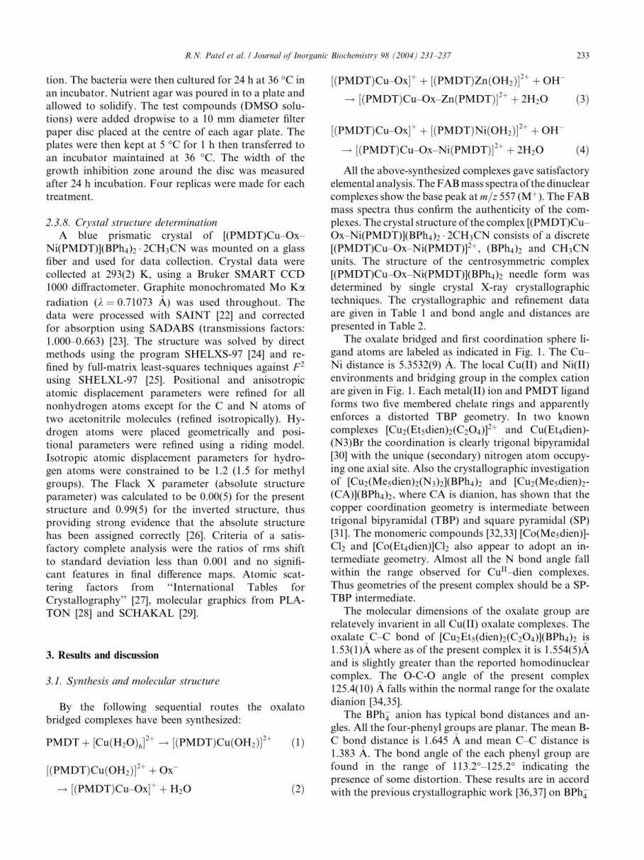
presented in Table 2.The oxalate bridged and first coordination sphere li-
gand atoms are labeled as indicated in Fig. 1. The Cu–
Ni distance is 5.3532(9) �A. The local Cu(II) and Ni(II)
environments and bridging group in the complex cation
are given in Fig. 1. Each metal(II) ion and PMDT ligand
forms two five membered chelate rings and apparently
enforces a distorted TBP geometry. In two known
complexes [Cu2(Et5dien)2(C2O4)]2þ and Cu(Et4dien)-
(N3)Br the coordination is clearly trigonal bipyramidal
[30] with the unique (secondary) nitrogen atom occupy-
ing one axial site. Also the crystallographic investigation
of [Cu2(Me5dien)2(N3)2](BPh4)2 and [Cu2(Me5dien)2-
(CA)](BPh4)2, where CA is dianion, has shown that the
copper coordination geometry is intermediate between
trigonal bipyramidal (TBP) and square pyramidal (SP)
[31]. The monomeric compounds [32,33] [Co(Me5dien)]-Cl2 and [Co(Et4dien)]Cl2 also appear to adopt an in-
termediate geometry. Almost all the N bond angle fall
within the range observed for CuII–dien complexes.
Thus geometries of the present complex should be a SP-
TBP intermediate.
The molecular dimensions of the oxalate group are
relatevely invarient in all Cu(II) oxalate complexes. The
oxalate C–C bond of [Cu2Et5(dien)2(C2O4)](BPh4)2 is1.53(1)�A where as of the present complex it is 1.554(5)�Aand is slightly greater than the reported homodinuclear
complex. The O-C-O angle of the present complex
125.4(10) �A falls within the normal range for the oxalate
dianion [34,35].
The BPh�4 anion has typical bond distances and an-
gles. All the four-phenyl groups are planar. The mean B-
C bond distance is 1.645 �A and mean C–C distance is1.383 �A. The bond angle of the each phenyl group are
found in the range of 113.2�–125.2� indicating the
presence of some distortion. These results are in accord
with the previous crystallographic work [36,37] on BPh�4
Table 1
Crystal data and structure refinement for [(PMDT)Cu–Ox–Ni(PMDT)](BPh4)2 � 2CH3CN
Empirical formula C72H92B2CuN8NiO4
Formula weight 1277.41
Temperature (K) 293(2)
Wavelength (�A) 0.71073
Crystal system, space group Monoclinic, C2 (No. 5)
Unit cell dimensions
a (�A) 20.445(4)
b (�A) 14.884(3)
c (�A) 23.174(5)
a (�) 90
b (�) 102.693(4)
c (�) 90
V (�A)3 6880(2)
Z, calculated density (Mg/m3) 4, 1.233
Absorption coefficient (mm�1) 0.636
F ð000Þ 2716
Crystal size (mm) 0.69� 0.50� 0.24
h range for data collection 0.90�–26.44�Limiting indices �246 h6 25, 06 k6 18, �286 l6 0
Reflections collected/unique 21,109/7341 [Rint ¼ 0:0427]
Completeness to h ¼ 26:44� 99.5%
Absorption correction SADABS
Maximum and minimum transmission 0.8624 and 0.6681
Refinement method Full-matrix least-squares on F2
Data/restraints/parameters 7341/1/765
Goodness-of-fit on F 2 1.044
Final R indices [I > 2rðIÞ] R1 ¼ 0:0354, wR2 ¼ 0:0853
R indices (all data) R1 ¼ 0:0639, wR2 ¼ 0:1039
Absolute structure parameter 0.00(5)
Largest difference peak and hole 0.273 and )0.368 e �A�3
234 R.N. Patel et al. / Journal of Inorganic Biochemistry 98 (2004) 231–237
compounds, which also indicates some amount of
crowding about Boron atom.
3.2. Magnetic moment
The room temperature magnetic moment values of
these complexes are also measured. At room tempera-
ture the magnetic moment value (1.68 B.M.) of the
compound [(PMDT)Cu–Ox–Ni(PMDT)]2þ is consider-
Table 2
Selected bond lengths (�A) and angles (�) for [(PMDT)Cu–Ox–Ni(PMDT)](B
Cu(1)–O(11) 1.961(6)
Cu(1)–N(12) 2.036(7)
Cu(1)–N(11) 2.050(8)
Cu(1)–N(13) 2.075(10)
Cu(1)–O(21) 2.137(8)
Cu(1)–Ni(1) 5.3532(9)
O(11)–Cu(1)–N(12) 177.2(4)
O(11)–Cu(1)–N(11) 93.5(3)
N(12)–Cu(1)–N(11) 87.2(3)
O(11)–Cu(1)–N(13) 92.3(3)
N(12)–Cu(1)–N(13) 85.6(4)
N(11)–Cu(1)–N(13) 145.3(4)
O(11)–Cu(1)–O(21) 81.7(3)
N(12)–Cu(1)–O(21) 100.8(3)
N(11)–Cu(1)–O(21) 106.2(4)
N(13)–Cu(1)–O(21) 108.5(4)
ably lower than spin only value for ST ¼ 1/2 resulting
from the spin coupling between Cu@S¼ 1/2 andS@Ni¼ 1. This magnetic behaviour is quite character-
istic of antiferromagnetic coupling between the para-
magnetic nickel(II) and copper(II) centers. Similar
magnetic behaviour is reported by Masami et al. [38]
and also by our school [39]. The room temperature value
(leff ¼ 1:91 B.M.) for [(PMDT)Cu–Ox–Zn(PMDT)]2þ
is in agreement with one spin (S ¼ 1=2) system.
Ph4)2 � 2CH3CN
Ni(1)–O(22) 1.973(6)
Ni(1)–N(23) 1.987(10)
Ni(1)–N(22) 1.994(9)
Ni(1)–N(21) 2.042(9)
Ni(1)–O(12) 2.178(7)
O(22)–Ni(1)–N(23) 93.6(3)
O(22)–Ni(1)–N(22) 177.8(4)
N(23)–Ni(1)–N(22) 86.2(4)
O(22)–Ni(1)–N(21) 92.6(3)
N(23)–Ni(1)–N(21) 146.9(4)
N(22)–Ni(1)–N(21) 86.4(4)
O(22)–Ni(1)–O(12) 80.7(3)
N(23)–Ni(1)–O(12) 109.4(4)
N(22)–Ni(1)–O(12) 101.5(3)
N(21)–Ni(1)–O(12) 103.7(4)
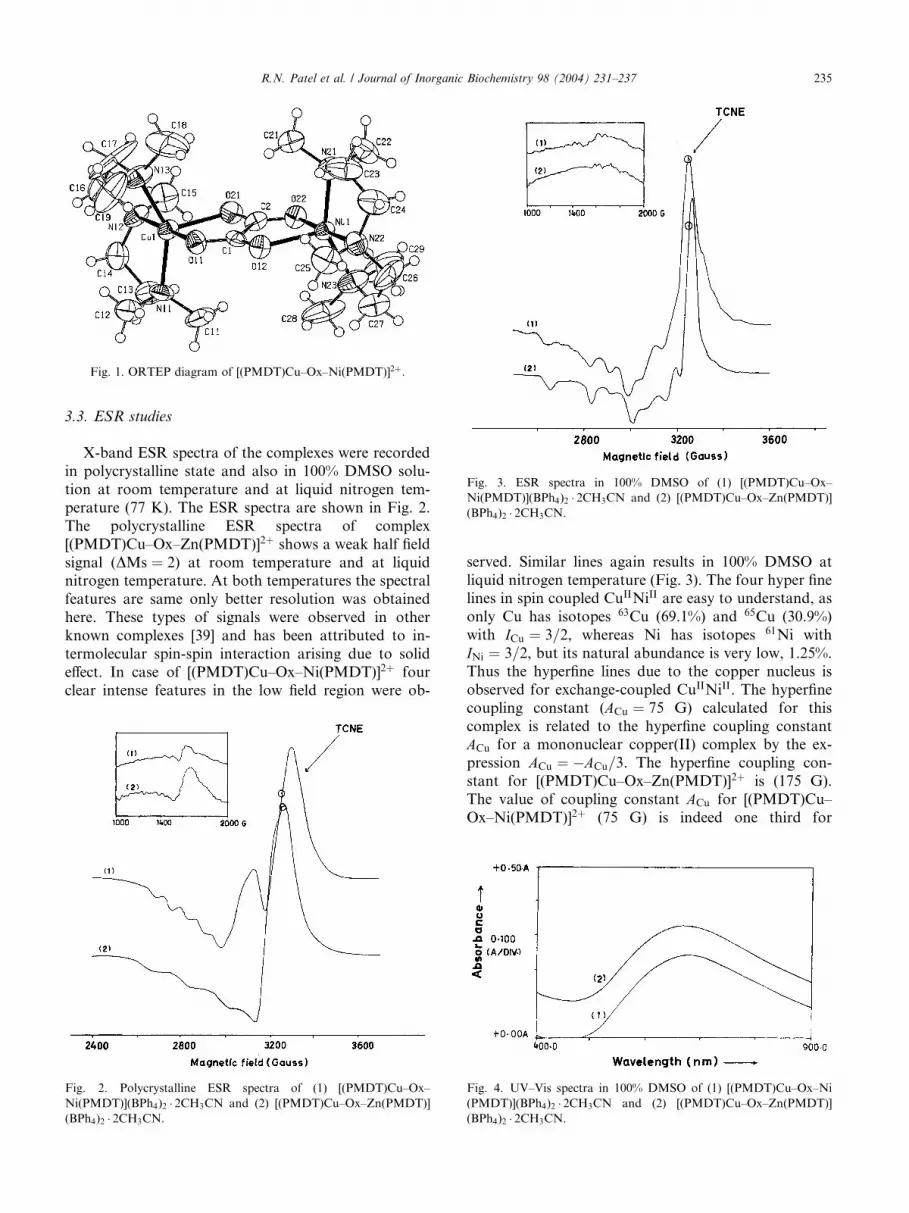
Fig. 3. ESR spectra in 100% DMSO of (1) [(PMDT)Cu–Ox–
Ni(PMDT)](BPh4)2 � 2CH3CN and (2) [(PMDT)Cu–Ox–Zn(PMDT)]
(BPh4)2 � 2CH3CN.
Fig. 1. ORTEP diagram of [(PMDT)Cu–Ox–Ni(PMDT)]2þ.
R.N. Patel et al. / Journal of Inorganic Biochemistry 98 (2004) 231–237 235
3.3. ESR studies
X-band ESR spectra of the complexes were recorded
in polycrystalline state and also in 100% DMSO solu-
tion at room temperature and at liquid nitrogen tem-
perature (77 K). The ESR spectra are shown in Fig. 2.
The polycrystalline ESR spectra of complex
[(PMDT)Cu–Ox–Zn(PMDT)]2þ shows a weak half field
signal (DMs ¼ 2) at room temperature and at liquidnitrogen temperature. At both temperatures the spectral
features are same only better resolution was obtained
here. These types of signals were observed in other
known complexes [39] and has been attributed to in-
termolecular spin-spin interaction arising due to solid
effect. In case of [(PMDT)Cu–Ox–Ni(PMDT)]2þ four
clear intense features in the low field region were ob-
Fig. 2. Polycrystalline ESR spectra of (1) [(PMDT)Cu–Ox–
Ni(PMDT)](BPh4)2 � 2CH3CN and (2) [(PMDT)Cu–Ox–Zn(PMDT)]
(BPh4)2 � 2CH3CN.
served. Similar lines again results in 100% DMSO at
liquid nitrogen temperature (Fig. 3). The four hyper finelines in spin coupled CuIINiII are easy to understand, as
only Cu has isotopes 63Cu (69.1%) and 65Cu (30.9%)
with ICu ¼ 3=2, whereas Ni has isotopes 61Ni with
INi ¼ 3=2, but its natural abundance is very low, 1.25%.
Thus the hyperfine lines due to the copper nucleus is
observed for exchange-coupled CuIINiII. The hyperfine
coupling constant (ACu ¼ 75 G) calculated for this
complex is related to the hyperfine coupling constantACu for a mononuclear copper(II) complex by the ex-
pression ACu ¼ �ACu=3. The hyperfine coupling con-
stant for [(PMDT)Cu–Ox–Zn(PMDT)]2þ is (175 G).
The value of coupling constant ACu for [(PMDT)Cu–
Ox–Ni(PMDT)]2þ (75 G) is indeed one third for
Fig. 4. UV–Vis spectra in 100% DMSO of (1) [(PMDT)Cu–Ox–Ni
(PMDT)](BPh4)2 � 2CH3CN and (2) [(PMDT)Cu–Ox–Zn(PMDT)]
(BPh4)2 � 2CH3CN.
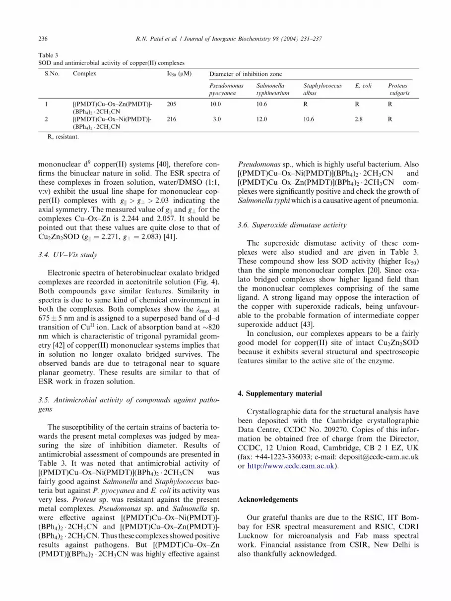
Table 3
SOD and antimicrobial activity of copper(II) complexes
S.No. Complex Ic50 (lM) Diameter of inhibition zone
Pseudomonas
pyocyanea
Salmonella
typhineurium
Staphylococcus
albus
E. coli Proteus
vulgaris
1 [(PMDT)Cu–Ox–Zn(PMDT)]-
(BPh4)2 � 2CH3CN
205 10.0 10.6 R R R
2 [(PMDT)Cu–Ox–Ni(PMDT)]-
(BPh4)2 � 2CH3CN
216 3.0 12.0 10.6 2.8 R
R, resistant.
236 R.N. Patel et al. / Journal of Inorganic Biochemistry 98 (2004) 231–237
mononuclear d9 copper(II) systems [40], therefore con-
firms the binuclear nature in solid. The ESR spectra of
these complexes in frozen solution, water/DMSO (1:1,
v:v) exhibit the usual line shape for mononuclear cop-
per(II) complexes with gk > g? > 2:03 indicating the
axial symmetry. The measured value of gk and g? for the
complexes Cu–Ox–Zn is 2.244 and 2.057. It should be
pointed out that these values are quite close to that ofCu2Zn2SOD (gk ¼ 2:271, g? ¼ 2:083) [41].
3.4. UV–Vis study
Electronic spectra of heterobinuclear oxalato bridged
complexes are recorded in acetonitrile solution (Fig. 4).
Both compounds gave similar features. Similarity in
spectra is due to same kind of chemical environment inboth the complexes. Both complexes show the kmax at
675� 5 nm and is assigned to a superposed band of d–d
transition of CuII ion. Lack of absorption band at �820
nm which is characteristic of trigonal pyramidal geom-
etry [42] of copper(II) mononuclear systems implies that
in solution no longer oxalato bridged survives. The
observed bands are due to tetragonal near to square
planar geometry. These results are similar to that ofESR work in frozen solution.
3.5. Antimicrobial activity of compounds against patho-
gens
The susceptibility of the certain strains of bacteria to-
wards the present metal complexes was judged by mea-
suring the size of inhibition diameter. Results ofantimicrobial assessment of compounds are presented in
Table 3. It was noted that antimicrobial activity of
[(PMDT)Cu–Ox–Ni(PMDT)](BPh4)2 � 2CH3CN was
fairly good against Salmonella and Staphylococcus bac-
teria but against P. pyocyanea and E. coli its activity was
very less. Proteus sp. was resistant against the present
metal complexes. Pseudomonas sp. and Salmonella sp.
were effective against [(PMDT)Cu–Ox–Ni(PMDT)]-(BPh4)2 � 2CH3CN and [(PMDT)Cu–Ox–Zn(PMDT)]-
(BPh4)2 � 2CH3CN.Thus these complexes showedpositive
results against pathogens. But [(PMDT)Cu–Ox–Zn
(PMDT)](BPh4)2 � 2CH3CN was highly effective against
Pseudomonas sp., which is highly useful bacterium. Also
[(PMDT)Cu–Ox–Ni(PMDT)](BPh4)2 � 2CH3CN and
[(PMDT)Cu–Ox–Zn(PMDT)](BPh4)2 � 2CH3CN com-
plexes were significantly positive and check the growth of
Salmonella typhiwhich is a causative agent of pneumonia.
3.6. Superoxide dismutase activity
The superoxide dismutase activity of these com-
plexes were also studied and are given in Table 3.
These compound show less SOD activity (higher Ic50)
than the simple mononuclear complex [20]. Since oxa-
lato bridged complexes show higher ligand field than
the mononuclear complexes comprising of the same
ligand. A strong ligand may oppose the interaction of
the copper with superoxide radicals, being unfavour-able to the probable formation of intermediate copper
superoxide adduct [43].
In conclusion, our complexes appears to be a fairly
good model for copper(II) site of intact Cu2Zn2SOD
because it exhibits several structural and spectroscopic
features similar to the active site of the enzyme.
4. Supplementary material
Crystallographic data for the structural analysis have
been deposited with the Cambridge crystallographic
Data Centre, CCDC No. 209270. Copies of this infor-
mation be obtained free of charge from the Director,
CCDC, 12 Union Road, Cambridge, CB 2 1 EZ, UK
(fax: +44-1223-336033; e-mail: [email protected] http://www.ccdc.cam.ac.uk).
Acknowledgements
Our grateful thanks are due to the RSIC, IIT Bom-
bay for ESR spectral measurement and RSIC, CDRI
Lucknow for microanalysis and Fab mass spectralwork. Financial assistance from CSIR, New Delhi is
also thankfully acknowledged.

R.N. Patel et al. / Journal of Inorganic Biochemistry 98 (2004) 231–237 237
References
[1] P.A. Vigato, S. Tamburini, D.E. Fenton, Coord. Chem. Rev. 106
(1990) 25.
[2] R.G. Birud, T.S. Srivastava, Inorg. Chim. Acta 179 (1991) 125.
[3] R.G. Birud, T.S. Srivastava, Inorg. Chim. Acta 173 (1990)
121.
[4] M. Zongwon, C. Dong, T. Wenxia, Y. Kaibei, L. Li, Polyhedron
11 (1992) 191.
[5] J.R.J. Sorenson, Chem. Br. 16 (1984) 1110.
[6] R.K. Gouch, T.W. Kensler, L.W. Oberlew, J.R.J. Sorenson,
Biochemical and Inorganic Copper Chemistry, vol. 1, Adenine,
New York, 1986, p. 139.
[7] S.J. Lippard, Oxo-bridged poly iron centres in biology and
chemistry, Angew Chem. Int. Ed. Engl. 27 (1988) 344.
[8] L.Jr. Que, A.E. True, Prog. Inorg. Chem. 38 (1990).
[9] Y. Yamada, M. Tanabe, Y. Miyashita, K. Okamoto, Polyhedron
22 (2003) 1407.
[10] A. Gartner, U. Weser, in: F. Vogtle, E. Weber (Eds.), Topics in
Current Chemistry, vol. 132, Springer, Berlin, 1986, p. 1.
[11] D. Darr, K.A. Zarilla, I. Fridovich, Arch. Biochem. Biophys. 258
(1987) 351.
[12] D.R. Williams, The Metals of Life, van Nostrand Reinhold,
London, 1971.
[13] R.N. Patel, K.B. Pandeya, Synth. React. Inorg. Met. Org. Chem.
29 (1999) 1733.
[14] R.N. Patel, K.K. Shukla, N. Singh, K.B. Pandeya, Indian
J. Chem. 41A (2002) 1369.
[15] R.N. Patel, K.B. Pandeya, J. Inorg. Biochem. 72 (1998) 109.
[16] R.N. Patel, S. Kumar, K.B. Pandeya, Spectrochim. Acta A 56
(2000) 2791.
[17] R.N. Patel, S. Kumar, K.B. Pandeya, P.V. Khadikar, Spectro-
chim. Acta A 58 (2002) 2961.
[18] R.N. Patel, S. Kumar, K.B. Pandeya, J. Inorg. Biochem. 89 (2002)
61.
[19] R.G. Bhirud, T.S. Shrivastava, Inorg. Chim. Acta 179 (1991) 125.
[20] R.N. Patel, K.B. Pandeya, Natl. Acad. Sci. Lett. 22 (2000) 201.
[21] D. Liu, K. Kwasniewska, Bull. Environ. Contam. Toxicol. 27
(1981) 289.
[22] Bruker, SMART and SAINT. Area Detector Control and
Integration Software, Bruker Analytical X-ray Instruments Inc.,
Madison, WI, USA, 1997.
[23] G.M. Sheldrick, SADABS. Program for Empirical Absorption
Correction of Area Detector Data, University of Goettingen,
Germany, 1997.
[24] G.M. Sheldrick, Acta Crystallogr. Sect. A 46 (1990) 467.
[25] G.M. Sheldrick, SHELXL-97. Program for the Refinement of
Crystal Structures, University of Goettingen, Germany, 1997.
[26] H.D. Flack, Acta Crystallogr. Sect. A 39 (1983) 876.
[27] International Tables for Crystallography, Kluwer Academic
Publishers, Dordrecht, The Netherlands C (1995).
[28] A.L. Spek, PLATON. A Multipurpose Crystallographic Tool,
Utrecht University, Utrecht, The Netherlands, 2002.
[29] E. Keller, SCHAKAL-97. A Computer Program for the Graphic
Representation of Molecular and Crystallographyc Models,
University of Freiburgi. Br., Germany, 1997.
[30] R.F. Ziolo, M. Allen, D.D. Titus, H.B. Gray, Z. Dori, Inorg.
Chem. 11 (1972) 3044.
[31] T.R. Felthouse, E.J. Laskowski, D.S. Bieksza, D.N. Hendrickson,
J. Chem. Soc. Chem. Commun. 777 (1976).
[32] M. Divaira, P.L. Orioli, Chem. Commun. 590 (1965).
[33] Z. Dori, R. Eisenberg, H.B. Gray, Inorg. Chem. 6 (1967) 483.
[34] D.J. Hodgson, J.A. Ibers, Acta Crystallogr. Sect. B 25 (1969) 469.
[35] A. Robbins, G.A. Jeffrey, J.P. Chesick, J. Donohue, F.A. Cotton,
B.A. Frenz, C.A.Murillo, Acta Crystallogr. Sect. B 31 (1975) 2395.
[36] D.M. Duggan, D.N. Hendrickson, Inorg. Chem. 13 (1974) 1911.
[37] E.J. Laskowski, D.N. Duggan, D.N. Hendrickson, Inorg. Chem.
14 (1975) 2449.
[38] M. Yonemura, Y. Matsumera, H. Furvtachi, M. Ohba, H.
Okawa, D.E. Fenton, Inorg. Chem. 36 (1997) 2711.
[39] R.N. Patel, Spectrochim. Acta A 59 (2003) 713.
[40] J. Mutler, K. Febix, C. Maichle, E. Lengfelder, J. Strahle, U.
Weser, Inorg. Chem. Acta 233 (1995) 11.
[41] C.M. Liu, R.G. Xiong, X.Z. You, Y.Z. Liu, Polyhedron 15 (1996)
4565.
[42] B.J. Hathaway, J. Chem. Soc. Dalton Trans. (1972) 1196.
[43] R.P. Bonomo, E. Conte, R. Marceli, A.M. Santoro, G.J. Tabi,
J. Inorg. Biochem. 53 (1994) 127.