what do we use: platelet-rich plasma or platelet-leukocyte gel
TRANSCRIPT
Platelet-Rich Plasma and Platelet Gel: A Review
Peter A.M. Everts, EKP, ECCP;* Johannes T.A. Knape, MD, PhD;†Gernot Weibrich, MD, DDS, PhD;‡, Jacques P.A.M. Schönberger, MD, PhD;§Johannes Hoffmann, PhD;¶ Eddy P. Overdevest, EKP, ECCP;* Henk A.M. Box, EKP, ECCP;*André van Zundert, MD, PhD, FRCA**
*Department of Extra Corporeal Blood Management, Catharina Hospital, Eindhoven, The Netherlands; †Department ofAnaesthesiology, Johannes Gutenberg University, Mainz, Germany; ‡Department of Prosthetic Surgery, Johannes GutenbergUniversity, Mainz, Germany; §Department of Cardiothoracic Surgery, Catharina Hospital, Eindhoven, The Netherlands;¶Department of General Laboratories, Catharina Hospital, Eindhoven, The Netherlands; and **Department of Anaesthesiology,University Medical Centre, Utrecht, The Netherlands
Abstract: Strategies to reduce blood loss and transfusion of al-logeneic blood products during surgical procedures are impor-tant in modern times. The most important and well-known au-tologous techniques are preoperative autologous predonation,hemodilution, perioperative red cell salvage, postoperativewound blood autotransfusion, and pharmacologic modulation ofthe hemostatic process. At present, new developments in thepreparation of preoperative autologous blood componenttherapy by whole blood platelet-rich plasma (PRP) and platelet-poor plasma (PPP) sequestration have evolved. This techniquehas been proven to reduce the number of allogeneic blood trans-fusions during open heart surgery and orthopedic operations.Moreover, platelet gel and fibrin sealant derived from PRP and
PPP mixed with thrombin, respectively, can be exogenously ap-plied to tissues to promote wound healing, bone growth, andtissue sealing. However, to our disappointment, not many well-designed scientific studies are available, and many anecdotic sto-ries exist, whereas questions remain to be answered. We there-fore decided to study perioperative blood management in moredetail with emphasis on the application and production of au-tologous platelet gel and the use of fibrin sealant. This reviewaddresses a large variety of aspects relevant to platelets, platelet-rich plasma, and the application of platelet gel. In addition, anoverview of recent animal and human studies is presented. Key-words: platelets, platelet-rich plasma, platelet gel, plateletgrowth factors, thrombin. JECT. 2006;38:174–187
Few hospitals in Europe routinely use autologous plate-let gel application techniques as part of a perioperativeblood management program. In the United States, an in-creasing number of clinicians tend to use platelet gel ap-plications in a variety of surgical settings, for both in andout of hospital surgery. The question of why this novel andpromising technique for the delivery of autologous growthfactors has not yet been adopted on a broader scale needsto be addressed. The main reason may be the lack ofconvincing scientific data that provide information ofwhether or not the use of platelet-rich plasma (PRP) andplatelet gels (PG) are appropriate in the clinical setting.
At Catharina Hospital (Eindhoven, Netherlands), westarted to use PG techniques in 2001 with a small group ofpatients undergoing complicated cardiac surgical proce-dures and in patients undergoing spinal fusion surgery.
This was carried out as an adjunct to the already existingperioperative blood management programs with appar-ently impressive clinical results.
The Department of Perioperative Blood Managementof the Catharina Hospital performs close to 1600 bloodmanagement procedures annually, of which 60% are re-lated to obtaining whole blood platelet to produce PRPfor the use of PG procedures. While its extended use isbased on positive clinical impressions and on clinical judg-ment, it still lacks a firm scientific basis. Therefore, clinicaltrials are required to answer questions on the efficacy,efficiency, and safety of the application of PRP and PGunder various surgical and medical conditions.
It is clear that a good understanding of the properpreparation and use of this specific blood managementtechnique is mandatory for clinicians to adequately evalu-ate results of its use and to avoid inconsistent results. Con-flicting data have been reported in clinical and experimen-tal research on the efficacy of PG treatment (1–5). Tounderstand how this arises, it is essential to be in posses-sion of the details of the preparation of PRP and PG.Knowledge of the following factors are of particular im-
Address correspondence to: Peter A.M. Everts, Catharina Hospital, dep.ECC and Blood Management, Michelangelolaan 2, 5623 EJ Eindhoven,The Netherlands. E-mail: [email protected] senior author has stated that authors have reported no material,financial or other relationship with any healthcare-related business orother entity whose products or services are discussed in this paper.
JECT. 2006;38:174–187The Journal of The American Society of Extra-Corporeal Technology
174
portance: the method of drawing blood, the quality of thePRP used, the platelet and growth factor counts, the PRPactivation, whether autologous or donor PRP was used,and the overall methodology. With respect to these issues,the clinician should be aware that data may sometimesseem to be conflicting in the eventual outcome.
This review addresses a variety of aspects pertaining tothe use of PG, including background on platelet activity,the pivotal role of platelets in hemostasis, soft tissue heal-ing, and bone growth, whole blood PRP production pro-cedure, platelet activation with thrombin, and a descrip-tion of the various actions of platelet-derived growth fac-tors (PDGFs). In addition, a discussion of the most recentclinical and experimental articles is presented with respectto these issues. Some safety issues including possible PGmitogenic effects are also addressed.
PLATELET ANATOMY AND FUNCTION
Platelets are small discoid blood cells (∼1–3 �m). Theaverage platelet count ranges from 1.5 to 3.0 × 10⁻⁵/mL ofcirculating blood, and the in vivo half-life time of plateletsis about 7 days. Platelets are formed from megakaryocytesand are synthesized in bone marrow by pinching off piecesof cytoplasm. Thereafter, platelets are extruded into thecirculation. Platelets have a ring of contractile microtu-bules (cytoskeleton) around their periphery, containingactin and myosin. Inside the platelet, a number of intra-cellular structures are present containing glycogen, lyso-somes, and two types of granules. These are known asdense granules, which contain ADP, ATP, serotonin, andcalcium, and �-granules, which contain clotting factors,growth factors, and other proteins. They are equippedwith an extensively invaginated membrane with an intri-cate canalicular system, which is in contact with the extra-cellular fluid (6). Normally, in the resting state, platelets
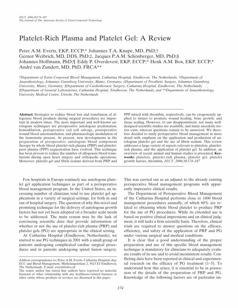
are nonthrombogenic and require a trigger before theybecome a potent and an active player in hemostasis andwound healing. On activation (e.g., by thrombin), theychange shape and develop pseudopodia, which promotesplatelet aggregation and subsequent release of the granulecontent through the open canalicular system (Figure 1).
PLATELET ACTIONS
Platelets and PG in HemostasisHemostasis is a balanced interaction of platelets, vascu-
lature, plasma clotting proteins, and low molecular weightsubstances. After an injury (e.g., by surgical trauma), themost important initial reactions leading to immediateblood coagulation are mainly mediated by platelets andblood vessel wall changes. In surgery, damaged blood ves-sel walls expose subendothelial collagen, binding von Wil-lebrand factor in the plasma, and subsequently changingthe structure so that the platelets can adhere to the bloodvessel wall. This process, known as platelet adhesion, actsthrough the glycoprotein Ib, and IIb/IIIa receptors thatare present in the platelet membrane. After this event,platelets become activated and aggregate. On activation,the platelet cytoskeleton changes from discoid to a spheri-cal shape with protruding pseudopods, which then spreadover injured tissues at the site of injury, a phenomenacalled platelet aggregation. After aggregation, the granu-lar contents are released through the canalicular system.Secreted serotonin probably assists in tissue vasoconstric-tion. ADP promotes release of granule contents fromother platelets and makes the platelets sticky, thus form-ing a hemostatic plug. Many other agents are able to causeplatelet aggregation and also to activate phospholipase A2
present in the platelet membrane. Subsequently, as a re-sult of the latter, membrane phospholipids release arachi-donic acid, which is converted into thromboxane A2 andalso causes platelet aggregation and platelet growth factor
Figure 1. Schematic overview of a restingand activated platelet. Normally plateletsare in a resting, nonactivated state. On ac-tivation (e.g., by thrombin), plateletschange their shape with the developmentof pseudopods to promote platelet aggre-gation and subsequent release of granulecontent through the open canalicular sys-tem (GP, glycoprotein).
175PLATELET-RICH PLASMA AND PLATELET GEL
JECT. 2006;38:174–187
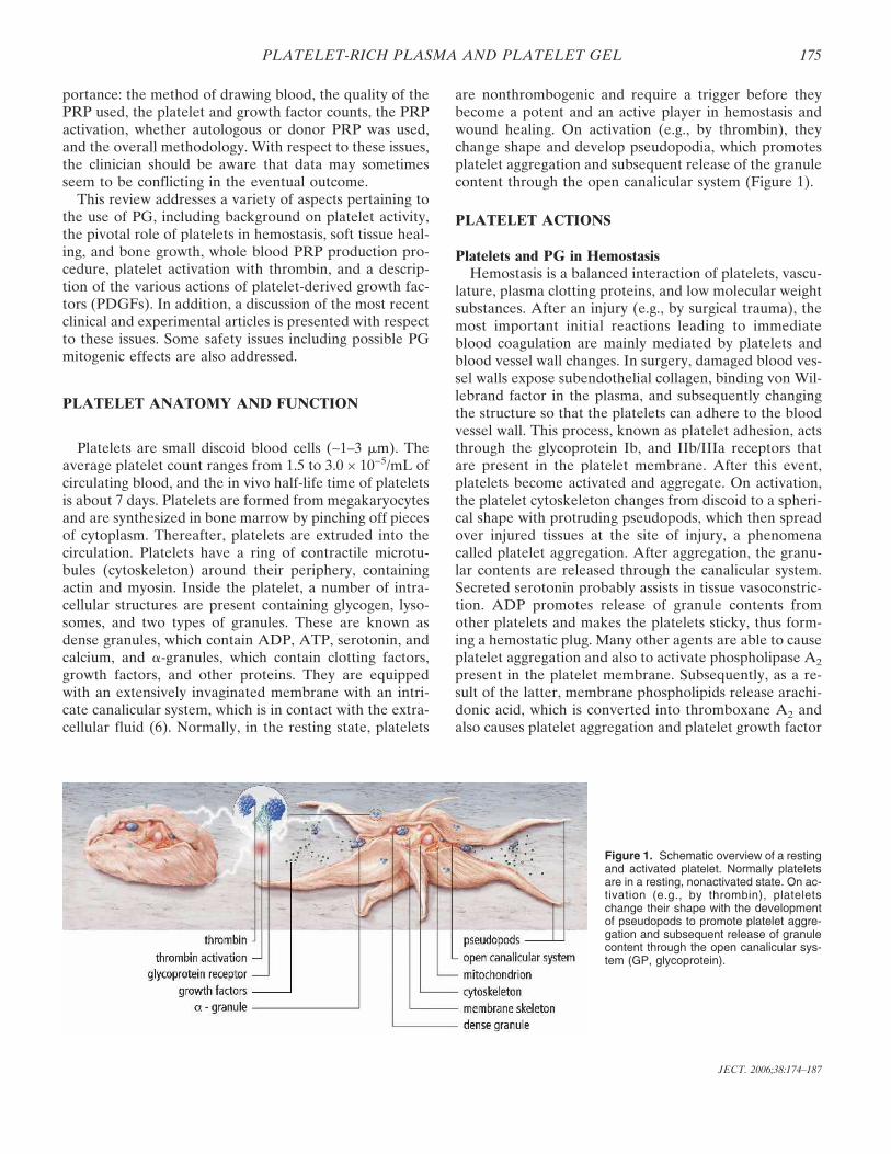
(PGF) release. Independent of thromboxane and ADP,another mechanism that causes platelet aggregation andplatelet granule release is induced by the presence ofthrombin. Thus, by these three mechanisms of plateletactivation, the platelet plug is extended in an attempt tostop blood loss from damaged vessels. Furthermore, thecoagulation system is activated by secreted and buddedparticles (7,8). The most well-understood platelet func-tion, at the onset of primary hemostasis, is the formationof a platelet plug. Thereafter, secondary hemostasis is ini-tiated with the activation of coagulation factors and theformation of a fibrin network that stabilizes the plateletplug (9). The final step is the activation of leukocytes in-vading the affected area with the release of cytokines,which activate the fibrinolytic system, leading ultimatelyto clot lysis (Figure 2). Because platelet �-granules secretePDGFs at the wound site almost at the instant of injury,repair of injured vasculature and tissue is directly initiatedwith the formation of new connective tissue and revascu-larization. Furthermore, the temporary formation ofplatelet and fibrin plugs at the wound site prevents theentry of microorganisms.
Based on the fundamental role of platelets in hemosta-sis, as discussed above, it may be hypothesized that exog-enously applied PG would contribute to a more effectivehemostatic condition of (surgical) wound surfaces, whereit attaches to tissues as a solid platelet plug. Stover et al.(10) prospectively evaluated the use of PG as a dural seal-ant in patients undergoing craniotomy or thoracolumbarprocedures and noted successful closure in 39 of 40 treatedpatients. Another therapeutic application is to use PG as awound sealant when it is sprayed by an aerosol techniqueover larger wound surfaces and suture lines in patientswho are at risk of postoperative wound leakage or fistulaformation. Furthermore, in patients who are at risk ofimpaired wound healing, such as diabetics, and thus at riskfor postoperative wound complications, a sprayed PG maydeliver a high concentration of PGF to the wound, thusboosting and supporting the natural healing process.
Platelets and PG in Wound HealingWound healing is a well-orchestrated and complex se-
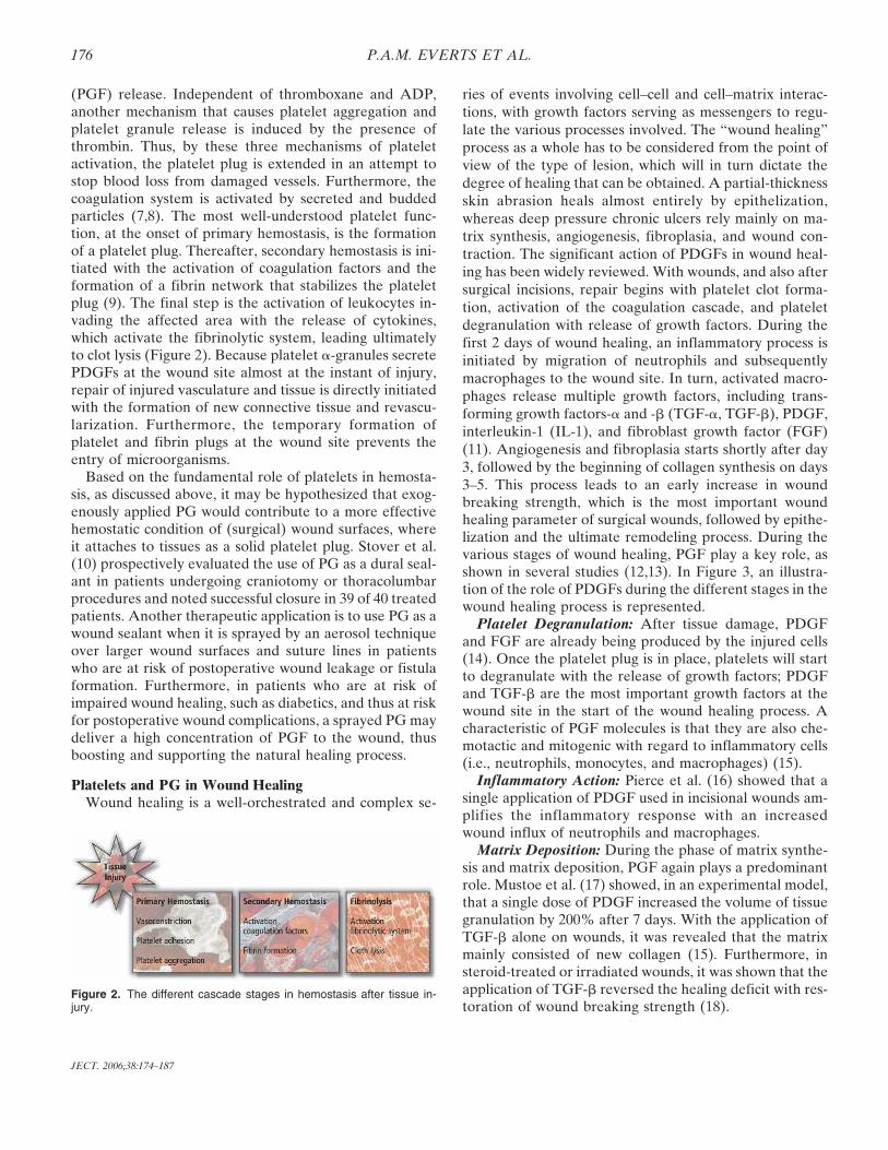
ries of events involving cell–cell and cell–matrix interac-tions, with growth factors serving as messengers to regu-late the various processes involved. The “wound healing”process as a whole has to be considered from the point ofview of the type of lesion, which will in turn dictate thedegree of healing that can be obtained. A partial-thicknessskin abrasion heals almost entirely by epithelization,whereas deep pressure chronic ulcers rely mainly on ma-trix synthesis, angiogenesis, fibroplasia, and wound con-traction. The significant action of PDGFs in wound heal-ing has been widely reviewed. With wounds, and also aftersurgical incisions, repair begins with platelet clot forma-tion, activation of the coagulation cascade, and plateletdegranulation with release of growth factors. During thefirst 2 days of wound healing, an inflammatory process isinitiated by migration of neutrophils and subsequentlymacrophages to the wound site. In turn, activated macro-phages release multiple growth factors, including trans-forming growth factors-� and -� (TGF-�, TGF-�), PDGF,interleukin-1 (IL-1), and fibroblast growth factor (FGF)(11). Angiogenesis and fibroplasia starts shortly after day3, followed by the beginning of collagen synthesis on days3–5. This process leads to an early increase in woundbreaking strength, which is the most important woundhealing parameter of surgical wounds, followed by epithe-lization and the ultimate remodeling process. During thevarious stages of wound healing, PGF play a key role, asshown in several studies (12,13). In Figure 3, an illustra-tion of the role of PDGFs during the different stages in thewound healing process is represented.
Platelet Degranulation: After tissue damage, PDGFand FGF are already being produced by the injured cells(14). Once the platelet plug is in place, platelets will startto degranulate with the release of growth factors; PDGFand TGF-� are the most important growth factors at thewound site in the start of the wound healing process. Acharacteristic of PGF molecules is that they are also che-motactic and mitogenic with regard to inflammatory cells(i.e., neutrophils, monocytes, and macrophages) (15).
Inflammatory Action: Pierce et al. (16) showed that asingle application of PDGF used in incisional wounds am-plifies the inflammatory response with an increasedwound influx of neutrophils and macrophages.
Matrix Deposition: During the phase of matrix synthe-sis and matrix deposition, PGF again plays a predominantrole. Mustoe et al. (17) showed, in an experimental model,that a single dose of PDGF increased the volume of tissuegranulation by 200% after 7 days. With the application ofTGF-� alone on wounds, it was revealed that the matrixmainly consisted of new collagen (15). Furthermore, insteroid-treated or irradiated wounds, it was shown that theapplication of TGF-� reversed the healing deficit with res-toration of wound breaking strength (18).
Figure 2. The different cascade stages in hemostasis after tissue in-jury.
176 P.A.M. EVERTS ET AL.
JECT. 2006;38:174–187
Collagen Production: Also important in wound healingis collagen production, which is initiated by the chemotac-tic and mitogenic actions of fibroblasts by FGF.
Epithelization: Topically applied epidermal growth fac-tor (EGF) leads to accelerated epithelization, as shown ina model by Nanney (19). In the beginning of the epithe-lization process, PDGF receptor genes were found, indi-cating that PDGF is also important during epithelization(20). During the last phase of wound healing, both FGFand PDGF increased contraction and remodeling time(21,22).
Based on the actions of the various PGF during thedifferent stages in the wound healing cascade, the use ofPG to stimulate wound repair is an interesting proposition(Figure 3). Compared to recombinant single growth factorapplications, PG has the supreme advantage that it offersmultiple synergistically working growth factors promotingmitogenesis of mesenchymal stem cells at the wound site(12,23–25).
Promising indications for topical PG applications mightbe for treatment of chronic nonhealing wounds and sup-portive healing after incisional wounds that occur, for ex-ample, in diabetic patients who are at risk of impairedwound healing. PG has been used successfully in woundcare patients to close chronic nonhealing (diabetic) ulcera(26,27). Margolis et al. (28) showed, in a large cohort ofpatients, that the application of the material released fromplatelets was more effective than standard care methods inwound healing. The treatment was even more effective in
patients with deeper wounds (28). Another interestingfinding in one study was the effect of PG had on thereduction of pain, an effect that is still not understood(29). In conclusion, there is sound evidence indicating thatthe use of PG in patients with chronic nonhealing woundscan be useful, and there is now a need to conduct clinicaltrials to study its effect on wound rehabilitation and earlierfunctional recovery in different surgical procedures.
Platelets and PG in Bone HealingBone is defined as a biological tissue composed of dy-
namically active cells that are integrated into a rigidframework. Bone cells consist of osteoblasts, osteoclasts,osteocytes, osteoprogenitor cells, and hematopoietic com-ponents (30). The bone healing process, whether in frac-ture repair or any given fusion model, is a delicate balancebetween bone deposition, resorption, and remodeling, andis influenced by numerous biochemical, biomechanical,cellular, and pathological mechanisms. During bone heal-ing, mature bone forming osteoblasts secrete growth fac-tors that are also present in platelets (31). Osteoclasts, incontrast, are bone-resorbing cells, a process controlled byhormonal and cellular mechanisms. Under normal circum-stances, the activity of osteoblasts and osteoclasts is inbalance.
In fracture repair and bone healing (i.e., callus forma-tion), platelets act as an exogenous source of growth fac-tors stimulating the activity of bone cells, based on their
Figure 3. Schematic illustration of the roleof PDGFs (numbers indicate the sequenceof actions) during the different stages ofthe wound healing process (VEGF, vascu-lar endothelial growth factor).
177PLATELET-RICH PLASMA AND PLATELET GEL
JECT. 2006;38:174–187
particular relevance to bone growth (32,33). As in woundhealing, bone fracture healing also incorporates the threestages of inflammation, proliferative repair, and remodel-ing. At bone fracture sites, platelets release PDGF, TGF-�, and EGF, providing an ideal system for the delivery ofgrowth factors to the injury site. The richest source ofTGF-� is found in platelets, bone, and cartilage. Two iso-forms, TGF-�1 and TGF-�2, are present in the platelets.TGF-�1 has the greatest potential for bone repair becauseboth chondrocytes and osteoblasts are enriched with re-ceptors for TGF-�1. In fact, TGF-� may contribute tobone healing at all stages (34,35). It has been shown that,with a combination of PGF, TGF-�, FGF, and EGF, anoptimum is created for the stimulation of differentiationand proliferation of osteoblasts to osteogenic cells (36,37).Similarly, proliferation was increased by the mitogenic ac-tion of PDGF in mesenchymal stem cell differentiationwhen TGF-� and EGF was added (38).
The ability of bone to heal is based on three concepts:osteogenesis, osteoinduction, and osteoconduction.
Osteogenesis is described as the ability to produce newbone and is determined by the presence of osteoprogeni-tor cells and osteogenic precursor cells in the area.
PGF are present in three of four stages during this bonehealing process (31).
Osteoinduction is defined as the ability to stimulatestem cells to differentiate into mature cells through thestimulation by local growth factors such as PDGF andTGF-� (39,40).
Osteoconduction is determined by the presence of ascaffold that allows for vascular and cellular migration andis usually achieved by the use of autologous harvestedbone (autograft), homologous graft materials (allograft),or artificial matrixes like demineralized bone (DMB), hy-droxyl apatite, tricalciumphosphate, and collagen (41). Inthe regulation and stimulation of these biochemical andcellular processes, PDGF plays a dominant role with re-gard to mitogenesis, chemotaxis, and stem cell differentia-tion. Recently, PRP has been successfully applied by sub-cutaneous administration in a diabetic femur fracturemodel. PRP normalized the early cellular proliferationand chondrogenesis, while improving the late mechanicalstrength (42).
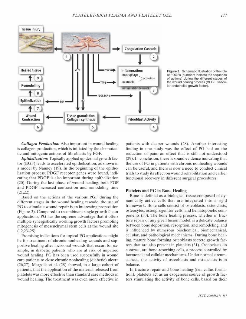
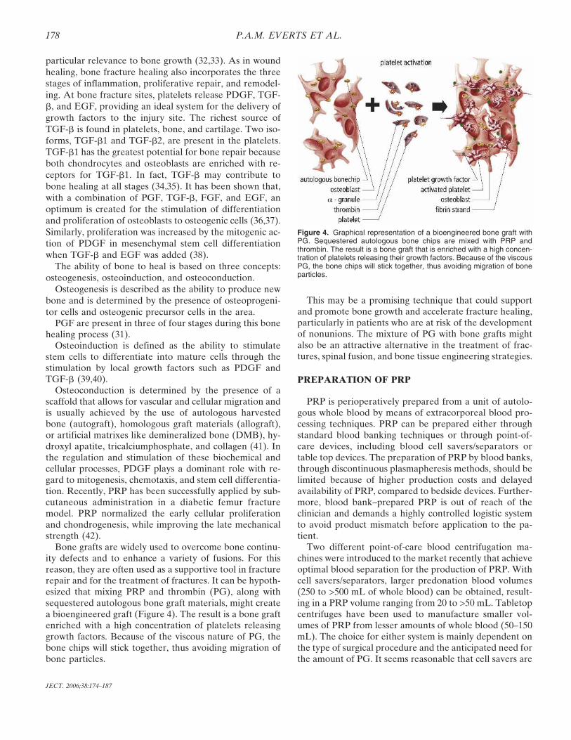
Bone grafts are widely used to overcome bone continu-ity defects and to enhance a variety of fusions. For thisreason, they are often used as a supportive tool in fracturerepair and for the treatment of fractures. It can be hypoth-esized that mixing PRP and thrombin (PG), along withsequestered autologous bone graft materials, might createa bioengineered graft (Figure 4). The result is a bone graftenriched with a high concentration of platelets releasinggrowth factors. Because of the viscous nature of PG, thebone chips will stick together, thus avoiding migration ofbone particles.
This may be a promising technique that could supportand promote bone growth and accelerate fracture healing,particularly in patients who are at risk of the developmentof nonunions. The mixture of PG with bone grafts mightalso be an attractive alternative in the treatment of frac-tures, spinal fusion, and bone tissue engineering strategies.
PREPARATION OF PRP
PRP is perioperatively prepared from a unit of autolo-gous whole blood by means of extracorporeal blood pro-cessing techniques. PRP can be prepared either throughstandard blood banking techniques or through point-of-care devices, including blood cell savers/separators ortable top devices. The preparation of PRP by blood banks,through discontinuous plasmapheresis methods, should belimited because of higher production costs and delayedavailability of PRP, compared to bedside devices. Further-more, blood bank–prepared PRP is out of reach of theclinician and demands a highly controlled logistic systemto avoid product mismatch before application to the pa-tient.
Two different point-of-care blood centrifugation ma-chines were introduced to the market recently that achieveoptimal blood separation for the production of PRP. Withcell savers/separators, larger predonation blood volumes(250 to >500 mL of whole blood) can be obtained, result-ing in a PRP volume ranging from 20 to >50 mL. Tabletopcentrifuges have been used to manufacture smaller vol-umes of PRP from lesser amounts of whole blood (50–150mL). The choice for either system is mainly dependent onthe type of surgical procedure and the anticipated need forthe amount of PG. It seems reasonable that cell savers are
Figure 4. Graphical representation of a bioengineered bone graft withPG. Sequestered autologous bone chips are mixed with PRP andthrombin. The result is a bone graft that is enriched with a high concen-tration of platelets releasing their growth factors. Because of the viscousPG, the bone chips will stick together, thus avoiding migration of boneparticles.
178 P.A.M. EVERTS ET AL.
JECT. 2006;38:174–187
used when both red cell salvage and PG application areboth indicated. In contrast, tabletop devices are used whenonly small amounts of PG are required during minimalblood loss surgical procedures. In Table 1, an overview ofthe currently available cell saver/separator devices isshown, and in Table 2, an overview of tabletop systems isshown.
PRP Preparation MethodologyIn the clinical standard setting, blood is drawn from the
median cubital vein. When a cell saver is used to manu-facture PRP, autologous whole blood is collected intostandard donor bags filled by gravity, not exceeding themaximum allowable predonation volume in relation to thecitrate volume in the blood bag. When tabletop devicesare used, the blood is carefully collected by aspirationtechniques into syringes, avoiding “negative pulling” to fillthe syringes quickly. The use of a needle diameter largerthan 17 gauge avoids trauma to the platelets during theblood draw. The autologous predonated blood is collectedin sufficient amounts of anticoagulation citrate dextrose-Asolution (ACD-A). In general, a ratio of 1 mL of ACD-Ato 7–8 mL of whole blood should be maintained. The as-pirated blood is gently agitated to thoroughly mix the an-ticoagulant with the blood.
Currently, most cell savers use a Latham (tapered) bowlinstead of a Baylor (straight) bowl, ranging in volume be-tween 50 and 225 mL. Furthermore, continuous autotrans-fusion systems, not using a bowl, can also be used to pre-pare PRP.
These sequester the whole blood in a semiautomaticcontrolled operating mode by centrifugation at 5600 rpm,separating the platelet-poor plasma (PPP) from the buffycoat layer and erythrocytes. The PPP volume is separatelycollected in a blood bag. Thereafter, centrifugation is
slowed down to 2400 rpm to obtain the buffy coat layerconsisting of PRP and leukocytes, which is collected in aseparate blood bag or syringe. After this procedure, theerythrocytes are also separately collected in a blood bag.The collected PPP and erythrocytes are reinfused duringsurgery at a time determined by the anesthesiologist. Thecollected PRP is used to prepare PG for application totissues.
With tabletop devices, a similar protocol of high- andlow-speed centrifugation is followed. Depending on thebrand of tabletop device, one may collect all blood com-ponents separately or collect only PRP. In those caseswhere no retransfusion of blood components is feasible,the PPP and erythrocytes are discarded.
Regardless of the type of PRP preparation method, theaim of working with whole blood is to prepare PRP witha platelet count in excess of the baseline platelet countvalues at the patient’s bedside.
PRP ActivationAlpha granules of the nonactivated platelets in the PRP
contain PGF and are thus nonfunctional, because they arenot released or in contact with the tissue. To initiate therelease of these growth factors, platelets must be acti-vated. Thrombin, the most potent platelet activator, willinduce immediate PGF release from the PRP in a dose-dependent fashion (43,44). In the United States, commer-cially available thrombin, derived from bovine plasma, isused as a “gold standard,” despite the fact that bovinethrombin has been associated some years ago with thedevelopment of antibodies to clotting factors V, XI, andthrombin, which had occasionally lead to life-threateningcoagulopathies (45). Alternatively, PRP can be activatedby autologous thrombin, produced with commerciallyavailable thrombin production kits, which either use au-
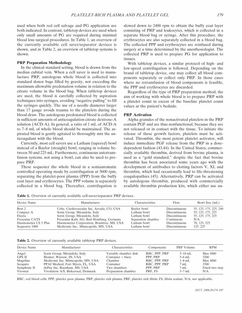
Table 1. Overview of currently available cell-saver/separator PRP devices.
Device Name Manufacturer Characteristics Flow Bowl Size (mL)
Brat 2 Cobe, Cardiovascular Inc, Arvada, CO, USA Baylor bowl Discontinuous 55, 125, 175, 225, 240Compact A Sorin Group, Mirandola, Italy Latham bowl Discontinuous 55, 125, 175, 225Electa Sorin Group, Mirandola, Italy Latham bowl Discontinuous 55, 125, 175, 225Fresenius CATS Fresenius Kabi AG, Bad Homburg, Germany Separation chamber Continuous N/AHaemonetics CS 5 Plus Haemonetics Corporation, Braintree, MS, USA Latham bowl Discontinuous 70, 125, 225Sequestra 1000 Medtronic Inc., Minneapolis, MN, USA Latham bowl Discontinuous 125, 225
Table 2. Overview of currently available tabletop PRP devices.
Device Name Manufacturer Characteristics Components PRP Volume RPM
Angel Sorin Group, Mirandola, Italy Variable chamber disk RBC, PPP, PRP 5–18 mL Max 4000GPS II Biomet, Warsaw, IN, USA Container + buoy PPP, PRP 5–6 mL 3200Magellan Medtronic Inc, Minneapolis, MN, USA Chamber RBC, PPP, PRP 1–8 mL Max 4000Secquire PPAI Medical, Fort Myers, FL, USA Container RBC, PPP, PRP 7 mL 3500Symphony II dePuy Inc, Raynham, MS, USA Two chambers PPP, PRP 7 mL Fixed two stepVivostat Vivolution A/S, Birkeroed, Denmark Preparation chamber PRF, FS 5–7 mL N/A
RBC, red blood cells; PPP, platelet poor plasma; PRP, platelet rich plasma; PRF, platelet rich fibrin; FS, fibrin scalant; N/A, not applicable.
179PLATELET-RICH PLASMA AND PLATELET GEL
JECT. 2006;38:174–187
tologous whole blood sequestered PPP or PRP (Table 3).Recently, Tsay et al. (46) reported that the use of a syn-thetic peptide that mimics thrombin known as peptide-6SFLLRN (TRAP). Activation with TRAP results in amore sustained release of the PGF with less PG retractionand higher PDGF-AB and TFG-� concentrations. Themechanism of this sustained release phenomenon is un-clear, but it may possibly be useful in the development andmaturation of platelet-enriched bone grafts and also intissue healing.
Mixing PRP with thrombin and calcium chloride to an-tagonize the anticoagulative effect of the citrate present inthe predonation blood bag will result in the activation ofthe platelet concentrate with the development of the vis-cous PG solution. Thereafter, the PG can be exogenouslyapplied with a syringe or as a solid clotted jelly mass ap-plied to soft tissues, bone, or synthetic bone.
From a surgical point of view, an “ideal” PG procedureis often defined as a procedure forming a platelet coagu-lum within 10 seconds. However, the formation of thecoagulum is merely a function of the activated fibrinogenconcentration, rather that the number of platelets.
PG Growth FactorsPGFs of the PG are peptides that promote cell prolif-
eration, differentiation, chemotaxis, and induce the migra-tion of various cells. Therefore, they play an importantrole in healing processes, as shown in several studies(47,48). We can classify growth factors into two groups:morphometric and mitogenic. The morphometric growthfactors, involved in bone growth, can turn undifferentiatedmultipotent mesenchymal stem cells (MSCs) into imma-ture and mature osteoprogenitor cells through the pres-ence of the so called bone morphogenic proteins (BMPs)(49). These BMP growth factors belong to the TGF-� su-per family, a growth factor that is also present in PRP.
Most of the PGF in the PRP have mitogenic actions thatincrease the population of healing cells by mitogenesis.However, the action depends on the presence of furtherdifferentiated MSCs.
PGF Receptor BindingAfter PG has been applied to tissues and the clot has
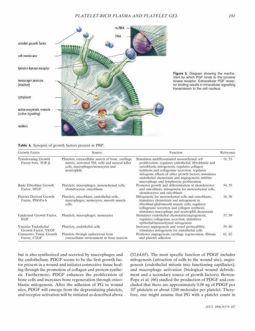
retracted and degranulated, PGF will be deposited in theextracellular matrix. Thereafter, during matrix degrada-tion, growth factors are released that interact and bindwith the platelet tyrosine kinase receptor (TKR), presentin the cell membranes of tissue cells (ligand–receptor in-teraction). The actual binding site is on the outer surfaceof the cell membrane. The TKR is a membrane spanningprotein that extends into the cytoplasm of the cell. Aftergrowth factor interaction with the external part of theTKR, activation of (inactive) messenger proteins in thecytoplasm occurs. The activated TKR cytoplasmic tail nowserves as a binding site for the messenger proteins. Anactivated protein is generated through a signalling cascadethat is capable of entering the cell nucleus, where it trig-gers the genes responsible for controlling cell division.Subsequently, transcription of mRNA is induced, produc-ing a biological response that initiates the cascades thatinduce tissue repair and regeneration (Figure 5) (50,51).
Growth factors seem to have three different modes ofaction. They may act in a paracrine manner, where thegrowth factors are secreted by one cell stimulating a neigh-boring second cell. Second, they can act in an autocrinemanner, where the cell releases factors act on itself, in-creasing its own activity. Third, in an endocrine manner,where growth factors may influence a cell that is differentin phenotype from the original cell and is located at aremote anatomical site. Because of the unique modes ofaction, growth factors are capable of inducing effects onmultiple cell types and may therefore provoke a series ofcellular functions in different tissues (52,53).
The next paragraph gives some background informationon two of the most well-described factors and on a re-cently evaluated growth factor present in PRP. In Table 4,a synopsis of the most well-known PRP growth factors isprovided along with a description of the source and spe-cific function, (16,54–63).
PDGFPDGF is a glycoprotein with a molecular weight of ap-
proximately 30 kd and with two disulphide-bondedpolypeptides, referred to as A and B chains. There arethree isoforms, PDGF-AA, -BB, and -AB (57,59). PDGFis not only found in the dense �-granules of the platelet
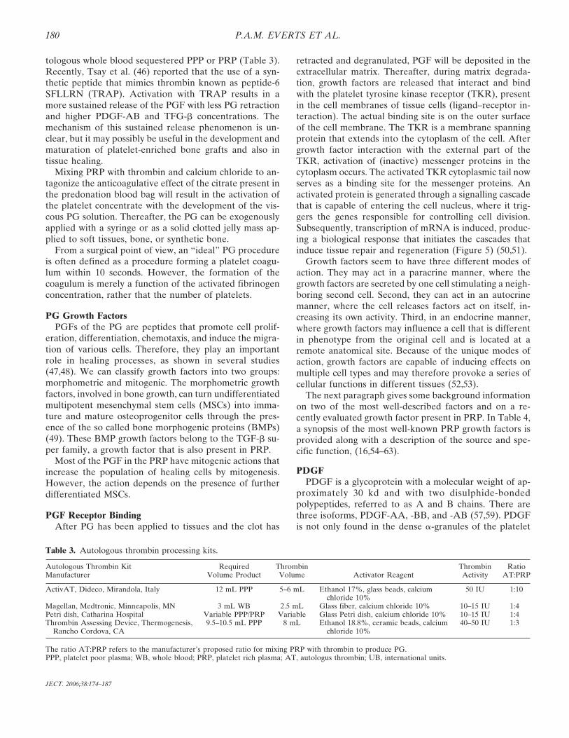
Table 3. Autologous thrombin processing kits.
Autologous Thrombin KitManufacturer
RequiredVolume Product
ThrombinVolume Activator Reagent
ThrombinActivity
RatioAT:PRP
ActivAT, Dideco, Mirandola, Italy 12 mL PPP 5–6 mL Ethanol 17%, glass beads, calciumchloride 10%
50 IU 1:10
Magellan, Medtronic, Minneapolis, MN 3 mL WB 2.5 mL Glass fiber, calcium chloride 10% 10–15 IU 1:4Petri dish, Catharina Hospital Variable PPP/PRP Variable Glass Petri dish, calcium chloride 10% 10–15 IU 1:4Thrombin Assessing Device, Thermogenesis,
Rancho Cordova, CA9.5–10.5 mL PPP 8 mL Ethanol 18.8%, ceramic beads, calcium
chloride 10%40–50 IU 1:3
The ratio AT:PRP refers to the manufacturer’s proposed ratio for mixing PRP with thrombin to produce PG.PPP, platelet poor plasma; WB, whole blood; PRP, platelet rich plasma; AT, autologus thrombin; UB, international units.
180 P.A.M. EVERTS ET AL.
JECT. 2006;38:174–187
but is also synthesized and secreted by macrophages andthe endothelium. PDGF seems to be the first growth fac-tor present in a wound and initiates connective tissue heal-ing through the promotion of collagen and protein synthe-sis. Furthermore, PDGF enhances the proliferation ofbone cells and increases bone regeneration through osteo-blastic mitogenesis. After the adhesion of PG to woundsites, PDGF will emerge from the degranulating platelets,and receptor activation will be initiated as described above
(52,64,65). The most specific function of PDGF includesmitogenesis (attraction of cells to the wound site), angio-genesis (endothelial mitosis into functioning capillaries),and macrophage activation (biological wound debride-ment and a secondary source of growth factors). Bowen-Pope et al. (66) studied the production of PDGF and con-cluded that there are approximately 0.06 ng of PDGF per106 platelets or about 1200 molecules per platelet. There-fore, one might assume that PG with a platelet count in
Figure 5. Diagram showing the mecha-nism by which PGF binds to the tyrosinekinase receptor. Extracellular PGF recep-tor binding results in intracellular signallingtransmission to the cell nucleus.
Table 4. Synopsis of growth factors present in PRP.
Growth Factor Source Function Reference
Transforming GrowthFactor-beta, TGF-�
Platelets, extracellular matrix of bone, cartilagematrix, activated TH1 cells and natural killercells, macrophages/monocytes andneutrophils
Stimulates undifferentiated mesenchymal cellproliferation; regulates endothelial, fibroblastic andosteoblastic mitogenesis; regulates collagensynthesis and collagenase secretion; regulatesmitogenic effects of other growth factors; stimulatesendothelial chemotaxis and angiogenesis; inhibitsmacrophage and lymphocyte proliferation
16, 53
Basic Fibroblast GrowthFactor, bFGF
Platelets, macrophages, mesenchymal cells,chondrocytes, osteoblasts
Promotes growth and differentiation of chondrocytesand osteoblasts; mitogenetic for mesenchymal cells,chondrocytes and osteoblasts
54, 55
Platelet Derived GrowthFactor, PDGFa-b
Platelets, osteoblasts, endothelial cells,macrophages, monocytes, smooth musclecells
Mitogenetic for mesenchymal cells and osteoblasts;stimulates chemotaxis and mitogenesis infibroblast/glial/smooth muscle cells; regulatescollagenase secretion and collagen synthesis;stimulates macrophage and neutrophil chemotaxis
16, 56
Epidermal Growth Factor,EGF
Platelets, macrophages, monocytes Stimulates endothelial chemotaxis/angiogenesis;regulates collagenase secretion; stimulatesepithelial/mesenchymal mitogenesis
57, 58
Vascular EndothelialGrowth Factor, VEGF
Platelets, endothelial cells Increases angiogenesis and vessel permeability,stimulates mitogenesis for endothelial cells
59, 60
Connective Tissue GrowthFactor, CTGF
Platelets through endocytosis fromextracellular environment in bone marrow
Promotes angiogenesis, cartilage regeneration, fibrosisand platelet adhesion
61, 62
181PLATELET-RICH PLASMA AND PLATELET GEL
JECT. 2006;38:174–187
excess of 3- to 5-fold the baseline level would have a pro-found effect on both wound healing and bone regenera-tion.
TGF-�TGF-� is the name given to a group of proteins of mo-
lecular weight approximately 25 kd that are involved inthe formation and development of many tissues (67).TGF-� is part of a super family to which BMP also belong.In humans, three subtypes of TGF-� are present, butTGF-�1 and TGF-�2 seem to be the most important withregard to general connective tissue repair and bone regen-eration (68,69). TGF-� is found predominantly in plate-lets, which account for 95% of the total, while some is alsofound in macrophages in a latent form. TGF-� has aninhibitory effect on cell growth of many tissues, except forMSCs, where proliferation is enhanced. The other func-tions of TGF-� are to promote chemotaxis and mitogen-esis of fibroblasts and osteoblastic precursor cells, whichwill later differentiate into mature osteoblasts, and also tostimulate osteoblast deposition at the collagen matrix ofthe tissue wound healing and bone matrix regions (70).TGF-� acts both in an autocrine and paracrine fashion,making it a growth factor with long-term healing and boneregeneration capabilities (71). Some concern on the use ofTGF-� has been muted by Dieudonne et al. (72), whostudied its the effect of on osteoclastic bone resorption inan experimental setting. They concluded that low concen-trations have a stimulatory effect on bone cell prolifera-tion, whereas at higher concentrations, proliferation issuppressed.
In PRP, both PDGF and TGF-� are present, implyingthat a mixture of combinations of growth factors will al-ways be present at tissue sites. This unavoidable effectseems to be beneficial toward tissue healing because vari-ous results are reported on the synergistic effect of differ-ent growth factors (23,24,50).
Connective Tissue Growth FactorVery recently, Kubota et al. (62) described a new PGF
known as connective tissue growth factor (CTGF). Plate-lets adhere to CTGF at injured tissue wound sites, whereit is overexpressed along with the platelet coagulation pro-cess. In their experiments, they showed that nonactivatedplatelets contain considerable amounts of CTGF that isreleased by activated PRP. It was also shown that theCTGF content in platelets is more than 20-fold higherthan any other PGF and that CTGF endorses angiogeneticactivity, cartilage regeneration, and fibrosis. Cicha et al.(63) showed that CTGF is expressed in bone marrow cells,but not by platelet-producing megakaryocytes, suggestingthat the total amount of CTGF in platelets is the result ofendocytosis from the extracellular environment in bonemarrow. CTGF might be considered as an importantmember of the PGF family.
PG STUDIES
Animal StudiesThere is a large variety of animal studies on PG research
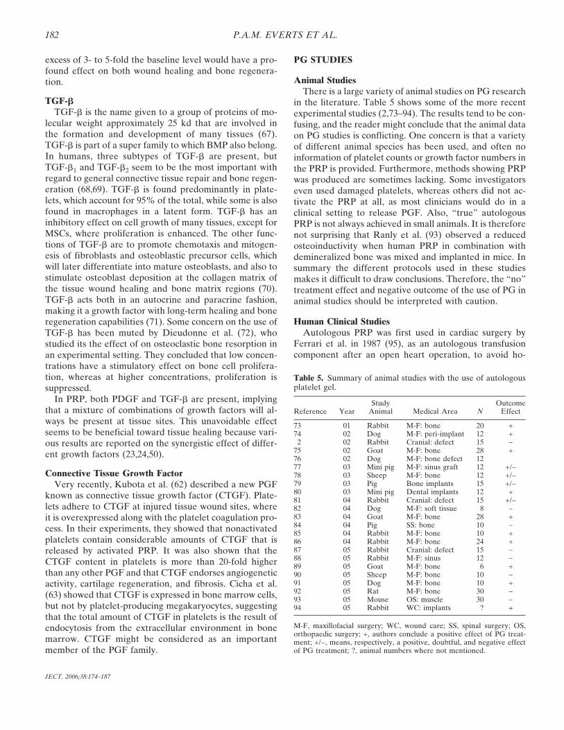
in the literature. Table 5 shows some of the more recentexperimental studies (2,73–94). The results tend to be con-fusing, and the reader might conclude that the animal dataon PG studies is conflicting. One concern is that a varietyof different animal species has been used, and often noinformation of platelet counts or growth factor numbers inthe PRP is provided. Furthermore, methods showing PRPwas produced are sometimes lacking. Some investigatorseven used damaged platelets, whereas others did not ac-tivate the PRP at all, as most clinicians would do in aclinical setting to release PGF. Also, “true” autologousPRP is not always achieved in small animals. It is thereforenot surprising that Ranly et al. (93) observed a reducedosteoinductivity when human PRP in combination withdemineralized bone was mixed and implanted in mice. Insummary the different protocols used in these studiesmakes it difficult to draw conclusions. Therefore, the “no”treatment effect and negative outcome of the use of PG inanimal studies should be interpreted with caution.
Human Clinical StudiesAutologous PRP was first used in cardiac surgery by
Ferrari et al. in 1987 (95), as an autologous transfusioncomponent after an open heart operation, to avoid ho-
Table 5. Summary of animal studies with the use of autologousplatelet gel.
Reference YearStudy
Animal Medical Area NOutcome
Effect
73 01 Rabbit M-F: bone 20 +74 02 Dog M-F: peri-implant 12 +
2 02 Rabbit Cranial: defect 15 −75 02 Goat M-F: bone 28 +76 02 Dog M-F: bone defect 1277 03 Mini pig M-F: sinus graft 12 +/−78 03 Sheep M-F: bone 12 +/−79 03 Pig Bone implants 15 +/−80 03 Mini pig Dental implants 12 +81 04 Rabbit Cranial: defect 15 +/−82 04 Dog M-F: soft tissue 8 −83 04 Goat M-F: bone 28 +84 04 Pig SS: bone 10 −85 04 Rabbit M-F: bone 10 +86 04 Rabbit M-F: bone 24 +87 05 Rabbit Cranial: defect 15 −88 05 Rabbit M-F: sinus 12 −89 05 Goat M-F: bone 6 +90 05 Sheep M-F: bone 10 −91 05 Dog M-F: bone 10 +92 05 Rat M-F: bone 30 −93 05 Mouse OS: muscle 30 −94 05 Rabbit WC: implants ? +
M-F, maxillofacial surgery; WC, wound care; SS, spinal surgery; OS,orthopaedic surgery; +, authors conclude a positive effect of PG treat-ment; +/−, means, respectively, a positive, doubtful, and negative effectof PG treatment; ?, animal numbers where not mentioned.
182 P.A.M. EVERTS ET AL.
JECT. 2006;38:174–187
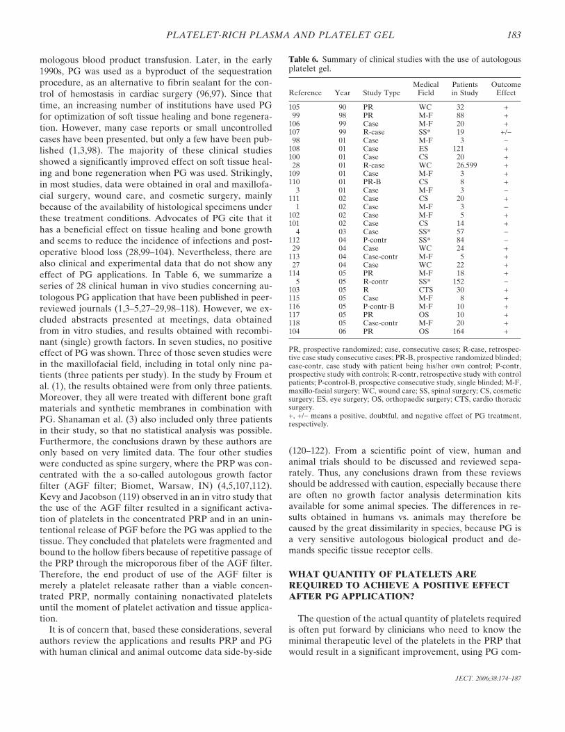
mologous blood product transfusion. Later, in the early1990s, PG was used as a byproduct of the sequestrationprocedure, as an alternative to fibrin sealant for the con-trol of hemostasis in cardiac surgery (96,97). Since thattime, an increasing number of institutions have used PGfor optimization of soft tissue healing and bone regenera-tion. However, many case reports or small uncontrolledcases have been presented, but only a few have been pub-lished (1,3,98). The majority of these clinical studiesshowed a significantly improved effect on soft tissue heal-ing and bone regeneration when PG was used. Strikingly,in most studies, data were obtained in oral and maxillofa-cial surgery, wound care, and cosmetic surgery, mainlybecause of the availability of histological specimens underthese treatment conditions. Advocates of PG cite that ithas a beneficial effect on tissue healing and bone growthand seems to reduce the incidence of infections and post-operative blood loss (28,99–104). Nevertheless, there arealso clinical and experimental data that do not show anyeffect of PG applications. In Table 6, we summarize aseries of 28 clinical human in vivo studies concerning au-tologous PG application that have been published in peer-reviewed journals (1,3–5,27–29,98–118). However, we ex-cluded abstracts presented at meetings, data obtainedfrom in vitro studies, and results obtained with recombi-nant (single) growth factors. In seven studies, no positiveeffect of PG was shown. Three of those seven studies werein the maxillofacial field, including in total only nine pa-tients (three patients per study). In the study by Froum etal. (1), the results obtained were from only three patients.Moreover, they all were treated with different bone graftmaterials and synthetic membranes in combination withPG. Shanaman et al. (3) also included only three patientsin their study, so that no statistical analysis was possible.Furthermore, the conclusions drawn by these authors areonly based on very limited data. The four other studieswere conducted as spine surgery, where the PRP was con-centrated with the a so-called autologous growth factorfilter (AGF filter; Biomet, Warsaw, IN) (4,5,107,112).Kevy and Jacobson (119) observed in an in vitro study thatthe use of the AGF filter resulted in a significant activa-tion of platelets in the concentrated PRP and in an unin-tentional release of PGF before the PG was applied to thetissue. They concluded that platelets were fragmented andbound to the hollow fibers because of repetitive passage ofthe PRP through the microporous fiber of the AGF filter.Therefore, the end product of use of the AGF filter ismerely a platelet releasate rather than a viable concen-trated PRP, normally containing nonactivated plateletsuntil the moment of platelet activation and tissue applica-tion.
It is of concern that, based these considerations, severalauthors review the applications and results PRP and PGwith human clinical and animal outcome data side-by-side
(120–122). From a scientific point of view, human andanimal trials should to be discussed and reviewed sepa-rately. Thus, any conclusions drawn from these reviewsshould be addressed with caution, especially because thereare often no growth factor analysis determination kitsavailable for some animal species. The differences in re-sults obtained in humans vs. animals may therefore becaused by the great dissimilarity in species, because PG isa very sensitive autologous biological product and de-mands specific tissue receptor cells.
WHAT QUANTITY OF PLATELETS AREREQUIRED TO ACHIEVE A POSITIVE EFFECTAFTER PG APPLICATION?
The question of the actual quantity of platelets requiredis often put forward by clinicians who need to know theminimal therapeutic level of the platelets in the PRP thatwould result in a significant improvement, using PG com-
Table 6. Summary of clinical studies with the use of autologousplatelet gel.
Reference Year Study TypeMedical
FieldPatientsin Study
OutcomeEffect
105 90 PR WC 32 +99 98 PR M-F 88 +
106 99 Case M-F 20 +107 99 R-case SS* 19 +/−
98 01 Case M-F 3 −108 01 Case ES 121 +100 01 Case CS 20 +
28 01 R-case WC 26.599 +109 01 Case M-F 3 +110 01 PR-B CS 8 +
3 01 Case M-F 3 −111 02 Case CS 20 +
1 02 Case M-F 3 −102 02 Case M-F 5 +101 02 Case CS 14 +
4 03 Case SS* 57 −112 04 P-contr SS* 84 −
29 04 Case WC 24 +113 04 Case-contr M-F 5 +
27 04 Case WC 22 +114 05 PR M-F 18 +
5 05 R-contr SS* 152 −103 05 R CTS 30 +115 05 Case M-F 8 +116 05 P-contr-B M-F 10 +117 05 PR OS 10 +118 05 Case-contr M-F 20 +104 06 PR OS 164 +
PR, prospective randomized; case, consecutive cases; R-case, retrospec-tive case study consecutive cases; PR-B, prospective randomized blinded;case-contr, case study with patient being his/her own control; P-contr,prospective study with controls; R-contr, retrospective study with controlpatients; P-control-B, prospective consecutive study, single blinded; M-F,maxillo-facial surgery; WC, wound care; SS, spinal surgery; CS, cosmeticsurgery; ES, eye surgery; OS, orthopaedic surgery; CTS, cardio thoracicsurgery.+, +/− means a positive, doubtful, and negative effect of PG treatment,respectively.
183PLATELET-RICH PLASMA AND PLATELET GEL
JECT. 2006;38:174–187
pared with standard treatments. At present, not manydata are available to answer this question directly, andonly indirect information exists. In 1998, Marx et al. (99)performed the first study showing a significant improve-ment in mandibular continuity defects when PRP wasmixed with autogenous bone grafts. Their PRP containedthree to four times higher platelet count compared withbaseline values, although the average PRP platelet countfound in their patients was just below 8 × 10⁵/�L, a num-ber that is lower than in most other studies. Nevertheless,they observed a significantly faster radiographic matura-tion and histomorphometrically denser bone regeneration.Nowadays, the latest separation devices produce PRPplatelet counts in excess of 6–10 times the baseline plateletcount values. Manufacturers tend to interpret a high plate-let concentration as a quality performance indicator oftheir separation devices, regardless of the fact that thesehigh concentrations may not be necessary or might evencontribute to a negative outcome. Weibrich et al. (86)observed an advantageous effect with platelet concentra-tions of approximately 106/�L. Furthermore, they statethat higher concentrations might have a paradoxically in-hibitory effect.
Haynesworth et al. (123) studied the response of PRPon cellular mechanisms of adult human MSCs (ahMSCs)in vitro. In soft tissue and bone healing, ahMSCs are es-sential components for the repair processes (124,125). Itwas shown that release of PRP PGF stimulates the migra-tion and proliferation of ahMSCs in a PRP concentration-dependent manner. A significant cellular response oc-curred with a 4- to 5-fold increase of platelet count com-pared with the baseline platelet count. In another study,Liu et al. (126) showed that the fibroblast proliferationand type I collagen production were augmented by a 4- to5-fold increase in the PRP platelet count.
With these studies, it was shown that a PRP plateletcount of approximately 106/�L is likely, which is in thetherapeutically effective range, because in most patients, awhole blood platelet count between 1.5 and 3 × 10⁵/�L isfound. A PRP platelet count with a four to five timeshigher baseline value seems to be adequate to achievesignificant outcome using a PG application.
SAFETY ISSUES
Patients who are considered to be candidates for a PGapplication must undergo a minor hematological evalua-tion to exclude blood disorders or platelet dysfunction.The authors feel the following are relative contraindica-tions for PG application: a platelet count less than 105/�L,a hemoglobin level less than 10 g/dL, presence of a tumorin the wound bed or metastatic disease, and other activeinfections. PRP preparation and PG production is safelyexecuted by certified perfusionists or other health care
professionals who have been trained in proper asepticpheresis and transfusion techniques, complying with gen-erally accepted safety requirements. Any concerns of im-munogenic reactions or disease transmission such as HIVand hepatitis that exist with homologous blood productsare eliminated because PRP is produced from autologousblood.
As discussed earlier, the use of bovine thrombin shouldbe decrease, because there are high quality and safer al-ternatives available for activating PRP.
To our knowledge, no wound infections after PG appli-cations have been reported, although the preparation ofPG demands many processing steps, and thus theoretical-ly, there is the possibility of contamination (119).
Some of the commercial available autologous thrombinkits require the use of ethanol. The safety of using a smallamount of ethanol in the PG on nerves was studied in ananimal model by de Somer et al. (127). It was concludedthat the myelin sheaths were normal in appearance, withno axonal swelling and no collagen necrosis caused by theethanol.
Despite the fact that PGF has mitogenic properties,there is no evidence that the growth factors in PG promotetumor growth or that they are involved in carcinogenesis(128,129). Furthermore, Scott and Pawson (130) showedthat growth factors act on cell membranes and not on thecell nucleus and that PGF activates normal rather thanabnormal gene expression. However, the effect of PG dur-ing tumor surgery should be studied before using it underthese circumstances.
CONCLUSIONS
Platelets are unique blood elements initiating hemosta-sis and healing processes. Therefore, the potential of au-tologous PG growth factor applications are numerous.PRP contains a high concentration of platelets, which canbe activated to form PG and to release PGF for therapeu-tic use. Data from human and animal studies provide bothdirect and indirect evidence that PGF plays a considerablerole in tissue regenerative processes. Nevertheless, someuncertainty is present, and some clinicians remain skepti-cal of the clinical benefits of PG and are uncertain aboutthe ideal biological setting (e.g., percentage of vital bonecells, volume of PRP) for the application of the PG.Therefore, randomized controlled trials are required tostudy the potential of the use of PG and to provide ma-terial for sound clinical decision-making in the near future.
ACKNOWLEDGMENTS
The authors thank J. Derwall and E. Lemmens en I. van Hezikfrom www.dlgraphics.nl, Kerkrade, The Netherlands, for prepa-ration of the illustrations.
184 P.A.M. EVERTS ET AL.
JECT. 2006;38:174–187
REFERENCES
1. Froum SJ, Wallace SS, Tarnow DP, et al. Effect of platelet richplasma on bone growth and osseointegration in human maxillarysinus grafts: Three bilateral case reports. Int J Periodont RestorDent. 2002;22:45–49.
2. Aghaloo TL, Moy PK, Freymiller EG. Investigations of platelet richplasma in rabbit cranial defects. A pilot study. J Oral MaxillofacSurg. 2002;60:1176–81.
3. Shanaman R, Filstein MR, Danesh-Meyer MJ. Localized ridge aug-mentation using GBR and platelet rich plasma: Three case reports.Int J Periodont Restor Dent. 2001;21:343–55.
4. Weiner BK, Walker M. Efficacy of autologous growth factors inlumbar intertransverse fusions. Spine. 2003;28:1968–71.
5. Carreon LY, Glassman SD, Anekstein Y, Puno RM. Platelet gel(AGF) fails to increase fusion rates in instrumented posterolateralfusions. Spine. 2005;30:243–7.
6. Zucker-Franklin C. Megakaryocytes and platelets. In: Zucker-Franklin D, Greaves MF, Grossi CE, Marmont AM, eds. Atlas ofBlood Cells. Philadelphia, PA: Lea & Febiger; 1998; 621
7. Sixma JJ, Sakariassen KS, Beeser-Visser NH, et al. Adhesion ofplatelets to human artery subendothelium: Effects of factor VIII-von Willebrand factor of various multimerc composition. Blood.1984;63:128–39.
8. Fox JEB. The platelet cytoskeleton. In: Verstraete M, Vermylen J,Lijnen R, Arnout J, eds. Thrombosis and Haemostasis. Leuven,Belgium: Leuven University Press; 1987; 175.
9. Dhall TZ, Shah GA, Ferguson IA, et al. Fibrin network structure:modification by platelets. Thromb Haemostas. 1983;49:42–6.
10. Stover EP, Siegel LC, Hood PA, et al. Intraoperatively preparedplatelet gel as an alternative to fibrin glue in dermal wound repair.Transfusion. 1996;36(suppl):S46.
11. McGrath MH. Peptide growth factors and wound healing. Clin PlastSurg. 1990;17:421–32.
12. Hunt TK. Basic principles of wound healing. J Trauma. 1990;30:S122–8.
13. Robson MC. Growth factors as wound healing agents. Curr OpinBiotechnol. 1991;2:863–7.
14. McNeil PL. Cell wounding and healing. Am Sci. 1991;79:222–35.15. Cromack DT, Pierce GF, Mustoe TA. TGF-� and PDGF medicated
tissue repair: Identifying mechanisms of action using impaired andnormal models of wound healing. In: Barbul A, Caldwell M, Eagl-stein W, Hunt T, Marshall D, Pines E, Skower G, eds. Clinical andExperimental Approaches to Dermal and Epidermal Repair: Nor-mal and Chronic Wounds. New York: Wiley Liss; 1991; 359–73.
16. Pierce GF, Mustoe TA, Altrock BW, Deuel TF, Thomason A. Roleof platelet-derived growth factor in wound healing. J Cell Biochem.1991;45:319–26.
17. Mustoe TA, Pierce GF, Morisima C, Deuel TF. Growth factor in-duced acceleration of tissue repair through direct and inductiveactivities in a rabbit dermal ulcer model. J Clin Invest. 1991;87:694–703.
18. Cromack DT, Porras-Reyes B, Mustoe TA. Current concepts inwound healing: growth factor and macrophage interaction. JTrauma. 1990;30:S129–33.
19. Nanney LB. epidermal and dermal effects of epidermal growth fac-tor during wound repair. J Invest Dermatol. 1990;94:624–9.
20. Antoniades HN, Galanopoulos T, Neville-Golden J, Kiritsy CP,Lynch SE. Injury induces in vivo expression of platelet-derivedgrowth factor (PDGF) and PDGF receptor mRNAs in skin epider-mal cells and PDGF mRNA in connective tissue fibroblasts. ProcNatl Acad Sci USA. 1991;88:565–9.
21. Stenberg BD, Phillips LG, Hokanson JA, Heggers JF, Robson MC.Effects of bFGF on the inhibition of contraction caused by bacteria.J Surg Res. 1991;50:47–50.
22. Sprugel KH, Greenhalgh DG, Murray MJ, Ross R. Platelet derivedgrowth factor and impaired wound healing. In: Barbul A, CaldwellM, Eaglstein W, Hunt T, Marshall D, Pines E, Skower G, eds.Clinical and Experimental Approaches to Dermal and EpidermalRepair: Normal and Chronic Wounds. New York: Wiley Liss; 1991;327–40.
23. Lynch SE, Nixon JC, Colvin RB, Antoniades HN. Role of platelet-derived growth factor in wound healing: synergistic effects withother growth factors. Proc Natl Acad Sci USA. 1987;84:7696–700.
24. Brown RL, Breeden MP, Greenhalg DG. PDGF and TGF-alphaact synergistically to improve wound healing in the genetically dia-betic mouse. J Surg Res. 1994;56:562–70.
25. Kells AF, Coats SR, Schwartz HS, Hoover RL. TGF-beta andPDGF act synergistically in affecting the growth of human osteo-blast-enriched cultures. Connect Tissue Res. 1995;31:117–24.
26. Mazzuco L, Medici D, Serra M, et al. The use of autologous plateletgel to treat difficult-to-heal wounds: a pilot study. Transfusion.2004;44:1013–8.
27. Henderson JL, Cupp CL, Ross EV, et al. The effects of autologousplatelet gel on wound healing. Ear Nose Throat J. 2003;82:598–602.
28. Margolis DJ, Kantor J, Santanna J, Strom BL, Berlin JA. Effec-tiveness of platelet releasate for the treatment of diabetic neuro-pathic foot ulcers. Diabetes Care. 2001;24:483–8.
29. Crovetti G, Martinelli G, Issi M, et al. Platelet gel for healing cu-taneous chronic wounds. Transfus Apher Sci. 2004;30:145–51.
30. Canalis E, McCarthy TL, Centrella M. Growth factorsand cytokinesin bone cell metabolism. Annu Rev Med. 1991;42:17–24.
31. Slater M, Patava J, Kingham K, Mason RS. Involvement of plateletsin stimulating osteogenic activity. J Orthop Res. 1995;13:655–63.
32. Bolander ME. Regulation of fracture repair by growth factors. ProcSoc Exp Biol Med. 1992;200:165–70.
33. Thiede MA, Smock SL, Petersen DN, Grasser WA, Nishimoto SK,Thompson DD. Production of osteocalcin by platelets: a potentiallyimportant link of platelet action in bone turnover. J Bone MinerRes. 1993;8:S147–51.
34. Robey PG, Young KC, Flanders KC, et al. Osteoblasts synthesizeand respond to transforming growth factor-type beta (TGF-beta) invitro. J Cell Biol. 1987;105:457–63.
35. Bourquie WT, Gross M, Hall BK. Expression of four growth factorsduring frcture repair. Int J Dev Biol. 1993;37:573–9.
36. Kasperk CH, Wergedal JE, Mohan S, Long DL, Lau KH, BaylinkDJ. Interaction of growth factors present in bone matrix with bonecells: effects on DNA synthesis and alkaline phosphatise. GrowthFactors. 1990;3:147–58.
37. Katagiri T, Lee T, Takeshima H, Suda H, Omura S. Transforminggrowth factor-beta modiulates proliferation and differentiation ofmouse clonal osteoblastic MC3T3-E1 cells depending on theirmaturation stages. Bone Miner. 1990;11:285–93.
38. Piche JE, Graves DT. Study of the growth factor requirements ofhuman bone-derived cells: a comparison with human fibroblasts.Bone. 1989;10:131–8.
39. Kalfas IH. Principles of bone healing. Neurosurg Focus. 2001;10:1–8.
40. Solheim E. Growth factors in bone. Int. Orthop. 1998;22:410–416.41. Helm GA. Bone graft substitutes for the promotion of spinal ar-
throdesis. Clin Neurosurg. 2005;52:250–255.42. Gandhi A, Dumas C, O’Conner JP, Parsons JR, Lin SS. The effects
of local platelet delivery on diabetic fracture healing. Bone. 2006;38:540–6.
43. Lacoste E, Martineau I, Gagnon G. Platelet concentrates: effects ofcalcium and thrombin on endothelial cell proliferation and growthfactor release. J Periodontol. 2003;74:1498–507.
44. Chouhan VD, De La Cadena RA, Nagaswani C, Weisel JW, KajaniM, Rao AK. Simultaneous occurrence of human antibodies di-rected against fibrinogen, thrombin, and factor V following expo-sure to bovine thrombin: effects on blood coagulation, protein Cactivation and platelet function. Thromb Haemost. 1997;77:343–9.
45. Zehnder JL, Leung LLK. Development of antibodies to thrombinand factor V with recurrent bleeding in a patient exposed to topicalbovine thrombin. Blood. 1990;76:2011–6.
46. Tsay RC, Burke A, Eisig SB, Lu HH, Landesberger R. Differentialgrowth factor retention by platelet-rich plasma composites. J OralMaxillofac Surg. 2005;63:521–8.
47. Giannoble WV. Periodontal tissue engineering by growth factors.Bone. 1996;S19:S23–37.
185PLATELET-RICH PLASMA AND PLATELET GEL
JECT. 2006;38:174–187
48. Giannoble WV, Hernandez RA, Finkelman RA, et al. Comparitiveeffects of platelet-derived growth factor-BB and insulin-like growthfactor I, individually and in combination, on periodontal regenera-tion in Macaca fascicularis. J Periodontal Res. 1996;31:301–12.
49. Hock JM, Canalis E. Platelet-derived growth factor enhances bonecell replication, but not differentiated function of osteoblasts. En-docrinology. 1994;134:301–12.
50. Schliephake H. Bone growth factors in maxillofacial skeletal recon-struction. Int J Oral Maxillofac Surg. 2002;31:469–84.
51. Antoniades HN, Williams LT. Human platelet-derived growth fac-tor: structure and functions. Fed Proc. 1983;81:2396–400.
52. Trippel SB, Coutts RD, Einhorn TA, Mundy GR, Rosenfeld RG.Growth factors as theurapeutic agents. J Bone Joint Surg Am. 1996;78:1272–86.
53. Bames GL, Kostenuik PJ, Gerstenfeld LC, Einhorn TA. Growthfactor regulation of fracture repair. J Bone Miner Res. 1999;14:1805–15.
54. Rosier RN, O’Keefe RJ, Hicks DG. The potential role of trans-forming growth factor beta in fracture healing. Clin Orthop.1998;(355 Suppl):S294–300.
55. Wang JS. Basic fibroblastic growth factor for stimulation of boneformation in osteoinductive or conductive implants. Acta OrthopScand. 1996;269:1–33.
56. Friesel RE, Maciag T. Molecular mechanisms of angiogenesis: fi-broblast growth factor signal transduction. FASEB J. 1995;9:919–25.
57. Canalis E, McCarthy TL, Centrella M. Effects of platelet-derivedgrowth factor on bone formation in vitro. J Cell Physiol. 1989;140:530–7.
58. Steenfos HH. Growth factors and wound healing. Scand J PlastReconstr Hand Surg. 1994;28:95–105.
59. Martin P, Hopkinson-Woolley J, McClusky J. Growth factors andcutaneous wound repair. Prog Growth Factor Res. 1992;4:25–44.
60. Rhee JS, Black M, Schubert U, et al. The functional role of bloodplatelet components in angiogenesis. Thromb Haemost. 2004;92:394–402.
61. Hom DB, Maisel RH. Angiogenic growth factors: Their effects andpotential in soft tissue wound healing. Ann Otol Rhinol Laryngol.1992;101:349–54.
62. Kubota S, Kawata K, Yanagita T, Doi H, Kitoh T, Takigawa M.Abundant retention and release of connective tissue growth factor(CTGF/CCN2) by platelets. J Biochem (Tokyo). 2004;136:279–82.
63. Cicha I, Garlichs CD, Daniel WG, Goppelt-Struebe M. Activatedhuman platelets release connective tissue growth factor. ThrombHaemost. 2004;91:755–60.
64. Lieberman JR, Daluiski A, Einhorn TA. The role of growth factorsin the repair of bone. J Bone Joint Surg Am. 2002;84:1032–44.
65. Heldin CH, Miyazono K, ten Dijke P. TGF-beta signalling from cellmembrane to nucleus through SMAD proteins. Nature. 1997;390:465–71.
66. Bowen-Pope DF, Vogel A, Ross R. Production of platelet-derivedgrowth factor-like molecules reduced expression of platelet-derivedgrowth factor receptors accompany transformation by a wide spec-trum of agents. Proc Natl Acad Sci USA. 1984;81:2396–400.
67. Gao J, Symons AL, Bartold PM. Expression of transforming growthfactor-beta 1 (TGF-�1) in the developing periodontium of rats. JDent Res. 1998;77:1708–16.
68. Roberts AB, Spron MB. Physiological actions and clinical applica-tions of transforming growth factor –beta (TGF-beta). Growth Fac-tors. 1993;8:1–9.
69. Miyazono K, ten Dijke P, Ichiyo H, Heldin CH. Receptors fortransforming growth factor-bata. Adv Immunol. 1994;55:181–220.
70. Mohan S, Baylink DJ. Bone growth factors. Clin Orthop Rel Res.1991;263:30–43.
71. Beck LS, De Guzman L, Lee WP, Yvette XU, Siegel MW, AmentoEP. One systemic administration of transforming growth factor-beta 1 reverses age or glucocorticoid-impaired wound healing. JClin Invest. 1993;92:2841–9.
72. Dieudonne SC, Foo P, van Zoeken EJ, Burger EH. Inhibiting andstimulating effects of TGF-�1 on osteoclastic bone resorption infetal mouse bone organ cultures. J Bone Miner Res. 1991;6:479–87.
73. Kim ES, Park EJ, Choung PH. Platelet concentration and its effecton bone formation in calvarial defects: an experimental study inrabbits. J Prosthet Dent. 2001;86:428–33.
74. Kim SG, Chung CH, Kim YK, Park JC, Lim SC. Use of particulatedentin-plaster of Paris combination with/without platelet-richplasma in the treatment of bone defect around implants. Int J OralMaxillofac Surg. 2002;17:86–94.
75. Fennis JP, Stoelinga PJ. Jansen. JA. Mandibular reconstruction: aclinical and radiographic animal study on the use of autogenousscaffolds and platelet-rich plasma. Int J Oral Maxillofac Implants.2002;31:281–6.
76. Kim SG, Kim WK, Park JC, Kim HJ. A comparative study ofosseointegration of Avana implants in demineralised freeze-driedbone alone or with platelet-rich plasma. J Oral Maxillofac Surg.2002;60:1018–25.
77. Furst G, Gruber R, Tangl S, et al. Sinus grafting with autogenousplatelet-rich plasma and bovine hydroxyapetite. A histomorpho-metric study in minipigs. Clin Oral Implants Res. 2003;14:500–8.
78. Jakse N, Tangl S, Gilli R, et al. Influence of PRP on autogenoussinus grafts. An experimental study on sheep. Clin Oral ImplantsRes. 2003;14:578–83.
79. Schegel KA, Kloss FR, Kessler P, Schultze-Mosgau S, Nkenke E,Wiltfang J. Bone conditioning to enhance implant osseointegration:an experimental study in pigs. Int J Oral Maxillofac Implants. 2003;18:505–11.
80. Zechner W, Tangl S, Tepper G, et al. Influence of platelet-richplasma on osseous healing of dental implants: a histologic and his-tomorphometric study in minipigs. Int J Oral Maxillofac Implants.2003;18:15–22.
81. Aghaloo TL, Moy PK, Freymiller EG. Evaluation of platelet-richplasma in combination with anorganic bovine bone in the rabbitcranium: a pilot study. Int J Oral Maxillofac Implants. 2004;19:59–65.
82. Choi BH, Im CJ, Huh JY, Suh JJ, Lee SH. Effects of platelet-richplasma on bone regeneration in autogenous bone graft. Int J OralMaxillofac Surg. 2004;33:56–9.
83. Fennis JP, Stoelinga PJ, Jansen JA. Mandibular reconstruction: ahistological and histomorphometric study on the use of autogenousscaffolds, particulate cortico-cancellous bone grafts and platelet richplasma in goats. Int J Oral Maxillofac Surg. 2004;33:48–55.
84. Li H, Zou X, Xue Q, Egund N, Lind M, Bunger C. Anterior lumbarinterbody fusion with carbon fiber cage loaded with bioceramicsand platelet-rich plasma. An experimental study on pigs. Eur SpineJ. 2004;13:354–8.
85. Yazawa M, Ogata H, Kimura A, Nakajima T, Mori T, Watanabe N.Basic studies on the bone formation ability by platelet rich plasmain rabbits. J Craniofac Surg. 2004;15:439–46.
86. Weibrich G, Hansen T, Kleis W, Buch R, Hitzler WE. Effect ofplatelet concentration in platelet-rich plasma on peri-implant boneregeneration. Bone. 2004;34:665–71.
87. Aghaloo TL, Moy PK, Freymiller EG. Evaluation of platelet-richplasma in combination with freeze-dried bone in the rabbit cra-nium. A pilot study. Clin Oral Implants Res. 2005;16:250–7.
88. Butterfield KJ, Bennett J, Gronowicz G, Adams D. Effects of plate-let-rich plasma with autogenous bone graft for maxillary sinus aug-mentation in a rabbit model. J Oral Maxillofac Surg. 2005;63:370–6.
89. Fennis JP, Stoelinga PJ, Merkx MA, Jansen JA. Reconstruction ofthe mandible with a poly (D,L-lactide) scaffold, autogenous corti-cocancellous bone graft, and autogenous platelet-rich plasma: Ananimal experiment. Tissue Eng. 2005;11:1045–53.
90. Grageda E, Lozada JL, Boyne PJ, Caplanis N, McMillan PJ. Boneformation the maxillary sinus by using platelet-rich plasma: an ex-perimental study in sheep. J Oral Implantol. 2005;31:2–17.
91. Kovacs K, Velich N, Huszar T, Fenyves B, Suba Z, Szabo G. His-tomorphometric and densitometric evaluation of the effects ofplatelet-rich plasma on the remodeling of beta-tricalcium phos-phate in beagle dogs. J Craniofac Surg. 2005;6:150–4.
92. Pryor ME, Polimeni G, Koo KT, et al. Analysis of rat calvariadefects implanted with a platelet-rich plasma preparation: histologicand histometric observations. J Clin Periodontol. 2005;32:966–72.
186 P.A.M. EVERTS ET AL.
JECT. 2006;38:174–187
93. Ranly DM, McMillan J, Keller T, et al. Platelet-derived growthfactors inhibits demineralized bone matrix-induced intramuscularcartilage and bone formation. A study of immunocompromisedmice. J Bone Joint Surg Am. 2005;87:2052–64.
94. Sclafani AP, Romo T III, Ukrainsky G, et al. Modulation of woundresponse and soft tissue ingrowth in synthetic and allogeneic im-plants with platelet concentrate. Arch Facial Plast Surg. 2005;7:163–9.
95. Ferrari M, Zia S, Valbonesi M, et al. A new technique for hemodi-lution, preparation of autologous platelet-rich plasma and intraop-erative blood salvage in cardiac surgery. Int J Artif Org. 1987;10:47–50.
96. Tawes RL, Sydorak GR, DuVall TB. Autologous fibrin glue: Thelast step in operative hemostasis. Am J Surg. 1994;168:120–2.
97. Oz MC, Jeevanandam V, Smith CR, et al. Autologous fibrin gluefrom intraoperatively collected platelet-rich plasma. Ann ThoracSurg. 1992;53:530–1.
98. Anitua E. The use of plasma-rich growth factors (PRGF) in oralsurgery. Pract Proced Aesthet Dent. 2001;13:487–93.
99. Marx RE, Carlson ER, Eichstaedt RM, et al. Platelet-rich-plasma:growth factor enhancement for bone grafts. Oral Surg Oral MedOral Pathol Oral Radiol Endod. 1998;85:638–46.
100. Man D, Plosker H, Winland-Brown JE. The use of autologousplatelet-rich plasma (platelet gel) and autologous platelet-poorplasma (fibrin glue) in cosmetic surgery. Plast Reconstr Surg. 2001;107:229–37.
101. Valbonesi M, Giannini G, Migliori F, Dalla Costa R, Galli A. Therole of autologous fibrin-platelet glue in plastic surgery: a prelimi-nary report. Int J Artif Organs. 2002;25:334–8.
102. Robiony M, Polini F, Costa F, Politi M. Osteogenesis distractionand platelet-rich plasma for bone restoration of the severely atro-phic mandible: preliminary results. J Oral Maxillofac Surg. 2002;60:630–5.
103. Englert SJ, Estep TH, Ellis-Stoll CC. Autologous platelet gel ap-plications during cardiovascular surgery: effect on wound healing. JExtra Corpor Technol. 2005;37:148–52.
104. Everts PAM, Devilee RJJ, Brown-Mahoney CH. Platelet gel andfibrin sealant reduce allogenic blood transfusions and in total kneearthroplasty. Acta Anaesth Scand. 2006;50:539–599.
105. Knighton DR, Cirisi K, Fiegel VD, Schumerth S, Butler E, Cerra F.Stimulation of repair in chronic, non healing, cutaneous ulcers usingplatelet-derived wound healing formula. Surg Gynecol Obstet.1990;170:56–60.
106. Anitua E. Plasma rich in growth factors: preliminary results of usein the preparation of future sites for implants. Int J Oral MaxillofacImplants. 1999;14:529–35.
107. Lowery GL, Kulkarni S, Pennisi AE. Use of autologous growthfactors in lumbal spinal fusion. Bone. 1999;(25 Suppl):S47–50.
108. Blumenkranz MS, Ohana E, Shaikh S, et al. Adjuvant methods inmacular hole surgery: intraoperative plasma-thrombin mixture andpostoperative fluid-gas exchange. Ophthalmic Surg Lasers. 2001;32:198–207.
109. Pertrungaro PS. Using platelet-rich plasma to accelerate soft tissuematuration in esthetic periodontal surgery. Compend Contin EducDent. 2001;22:729–34.
110. Powell DM, Chang E, Farrior EH. Recovery from deep-planerhytidectomy following unilateral wound treatment with autologousplatelet gel: a pilot study. Arch Facial Plast Surg. 2001;3:245–50.
111. Adler SC, Kent KJ. Enhancing wound healing with growth factors.Facial Plast Surg Clin North Am. 2002;10:129–46.
112. Castro FP Jr. Role of activated growth factors in lumbar spinalfusions. J Spinal Disord Tech. 2004;17:380–4.
113. Giannini G, Mauro V, Agostino T, Gianfranco B. Use of autolo-gous fibrin-platelet glue and bone fragments in maxillofacial sur-gery. Transfus Apher Sci. 2004;30:139–44.
114. Camargo PM, Lekovic V, Weinlaender M, Vasilic N, MadzarevicM, Kenney EB. A reentry study on the use of bovine porous bonemineral, GTR, and platelet-rich plasma in the regenerative treat-ment of intrabony defects in humans. Int J Periodontics RestorativeDent. 2005;25:49–59.
115. Merkx MA, Fennis JP, Verhagen CM, Stoelinga PJ. Reconstructionof the mandible using preshaped 2.3 mm titanium plates, autog-enous particulate cortico-cancellous bone grafts and platelet richplasma: a report on eight patients. Int J Oral Maxillofac Surg. 2004;33:733–9.
116. Kassolis JD, Reynolds MA. Evaluation of the adjunctive benefits ofplatelet-rich plasma in subantral sinus augmentation. J CraniofacSurg. 2005;16:280–7.
117. Savarino L, Cenni E, Tarabusi C, et al. Evaluation of bone healingenhancement by lyophilized bone grafts supplemented with plateletgel: A standardized methodology in patients with tibial osteotomyfor genu varus. J Biomed Mater Res B Appl Biomater. 2006;76:364–72.
118. Steigmann M, Garg AK. A comparative study of bilateral sinus liftsperformed with platelet-rich plasma alone versus alloplastic graftmaterial reconstituted with blood. Implant Dent. 2005;14:261–6.
119. Kevy SV, Jacobson MS. Comparison of methods for point of carepreparation of autologous platelet gel. J Extra Corpor Technol.2004;36:28–35.
120. Freymiller EG, Aghaloo TL. Platelet rich plasma: ready or not? JOral Maxillofac Surg. 2004;62:484–8.
121. Sanchez AR, Sheridan PJ, Kupp LI. Is platelet rich plasma theperfect enhancement factor? A current review. Int J Oral Maxillo-fac Surg. 2003;18:93–103.
122. Marx RE. Platelet rich plasma: evidence to support its use. J OralMaxillofac Surg. 2004;62:489–96.
123. Haynesworth SE, Kadiyala S, Liang LN, et al. Mitogenic stimula-tion of human mesenchymal stem cells by platelet release suggest amechanism for enhancement of bone repair by platelet concen-trates. Presented at the 48th Annual Meeting of the OrthopedicResearch Society, Dallas, TX, February 2002.
124. Nakagawa H, Akita S, Fukui M, Fujii T, Akino K. Human mesen-chymal stem cells successfully improve skin-substitute wound heal-ing. Br J Dermatol. 2005;153:29–36.
125. Caplan AI. Review: mesenchymal stem cells: cell-based reconstruc-tive therapy in orthopedics. Tissue Eng. 2005;7:1198–211.
126. Liu Y, Kalem A, Risto O, Wahlstrom O. Fibroblast proliferationdue to exposure to a platelet concentrate in vitro is pH dependent.Wound Repair Regen. 2002;5:336–340.
127. De Somer F, De Brauwer V, Vandekerckhove M, Ducatelle R,Uyttendaele D, Van Nooten G. Can autologous thrombin with arest fraction of ethanol be used safely for activation of concentratedautologous platelets applied on nerves? Eur Spine J. 2006;15:501–505.
128. Landesberger R, Moses M, Karpatkin M. Risks of using plateletrich plasma. J Oral Maxillofac Surg. 1998;56:1116–7.
129. Martinez-Gonzales JM, Cano-Sanchez J, Gonzalo-Lafuente JC,Campo-Trapero J, Esparza-Gomez G, Seoane J. Do ambulatory-use platelet-rich plasma (PRP) concentrates present risks? MedOral. 2002;7:375–90.
130. Scott JD, Pawson T. Cell communication: The inside story. Sci Am.2000;282:72.
187PLATELET-RICH PLASMA AND PLATELET GEL
JECT. 2006;38:174–187