viral hemorrhagic septicemia virus, ichthyophonus hoferi, and other causes of morbidity in pacific...
TRANSCRIPT
DISEASES OF AQUATIC ORGANISMS Dis Aquat Org Published February 26
Viral hemorrhagic septicemia virus, Ichthyophonus hoferi, and other causes of morbidity in Pacific herring Clupea pallasi
spawning in Prince William Sound, Alaska, USA
Gary D. Marty'l: Ellen F. ~reiberg', Theodore R. Meyers2, John Wilcock3. Thomas B. ~ a r v e r ~ , David E . Hinton1
'Department of Anatomy, Physiology, and Cell Biology, School of Veterinary Medicine, University of California, One Shields Avenue, Davis, California 95616, USA
*Alaska Department of Fish and Game, Juneau Fish Pathology Laboratory, PO Box 25526, Juneau, Alaska 99802, USA 3Alaska Department of Fish and Game, PO Box 669. Cordova, Alaska 99574. USA
-Department of Population Health and Reproduction. School of Veterinary Medicine. University of California. Davis, California 95616. USA
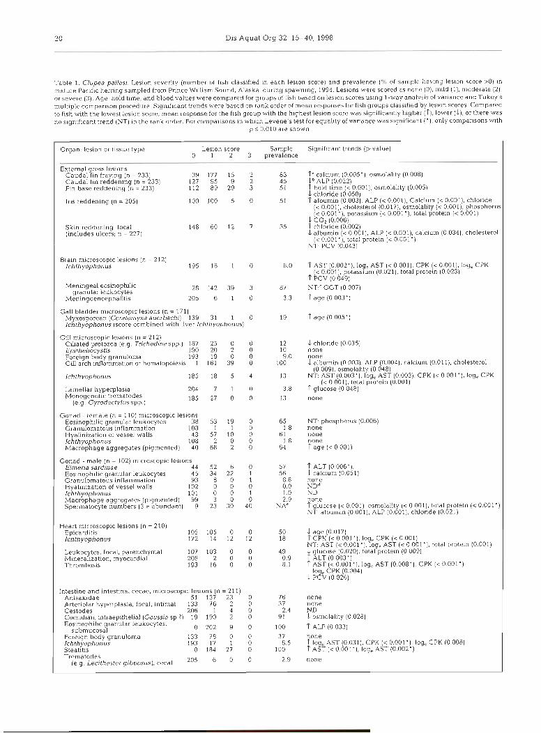
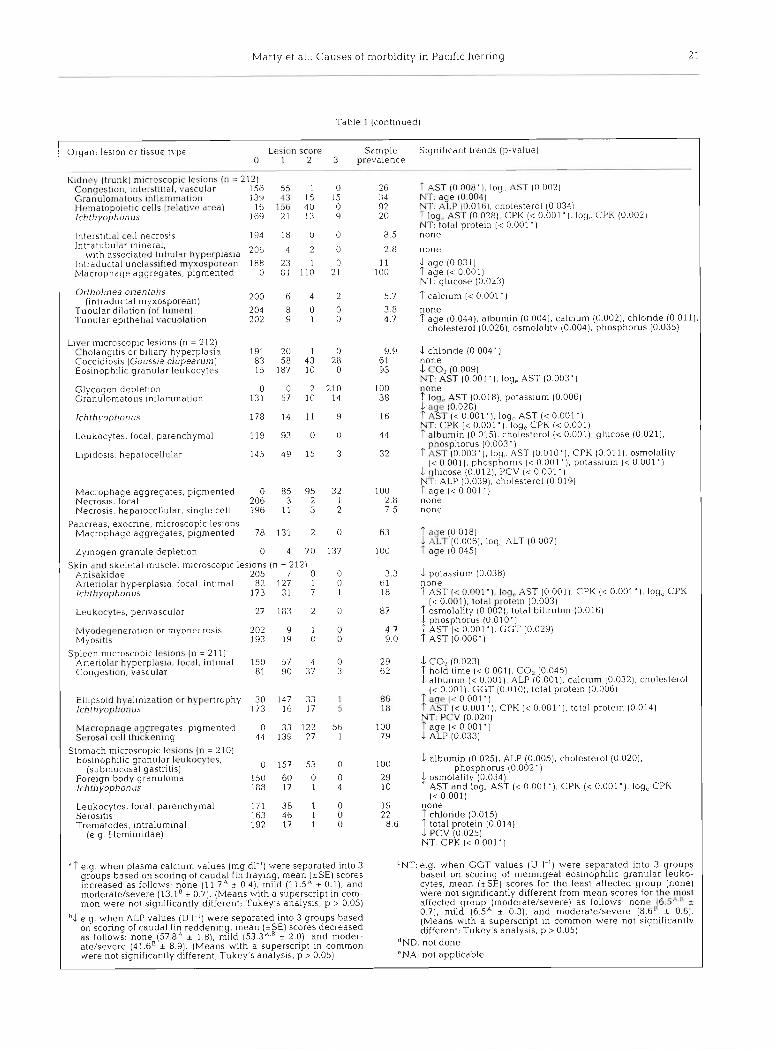
ABSTRACT: Pacific herring Clupea pallasi populations in Prince William Sound, Alaska, USA, declined from an estimated 9.8 X lo7 kg in 1992 to 1.5 X 10' kg in 1994. To determine the role of disease in population decline, 233 Pacific herring from Prince William Sound were subjected to complete necropsy during April 1994. The North American strain of viral hemorrhagic septicemia virus (VHSV) was isolated from 11 of 233 fish (4 .7%) . VHSV was significantly related to myocardial mineralization. hepatocellular necrosis, submucosal gastritis, and meningoencephalitis. Ichthyophonus hoferi infected 62 of 212 (29%) fish. I. hoferi infections were associated with severe, disseminated, granulomatous inflammation and with increased levels of plasma creatine phospholunase (CPK) and aspartate amino- transferase (AST). I. hoferi prevalence in 1994 was more than double that of most previous years (1989 to 1993). Plasma chemistry values significantly greater (p < 0.01) in males than females included albu- min, total protein, cholesterol, chloride, glucose, and potassium; only alkaline phosphatase was signif- icantly greater in females. Hypoalbuminemia was relatively common in postspawning females; other risk factors included VHSV and moderate or severe focal skin reddening. Pacific herring had more than 10 species of parasites, but they were not associated with significant lesions. Two of the parasites have not previously been described: a renal intraductal myxosporean (11 % prevalence) and an intestinal coccidian (91 % prevalence). Transmission electron microscopy of a solitary mesenteric lesion revealed viral particles consistent with lymphocystis virus. No fish had viral erythrocytic necrosis (VEN). Preva- lence of external gross lesions and major parasites was not related to fish age, and fish that were year- lings at the time of the 1989 'Exxon Valdez' oil spill (1988 year class) had no evidence of increased &sease prevalence.
KEY WORDS: Clupea pallasi . 'Exxon Valdez' . Histopathology . Hypoalbuminemia . Ichthyophonus hoferi . Pacific herring . Plasma chemistries . Viral hemorrhagic septicemia virus (VHSV)
INTRODUCTION
Pacific herring Clupea pallasi are among the most abundant fish species in coastal regions of the North Pacific, where they are important for conlrnercial and
subsistence fishing and as prey for many marine fish, birds, and mammals. In Prince William Sound (PWS), Alaska, Pacific herring normally support 5 commercial fisheries, with an average annual ex-vessel value of $8.3 d l i o n . Roe fisheries, the most valuable, are har- vested in April just before spawning. Pacific herring in PWS first spawn when 3 or 4 yr old; they rarely live
O Inter-Research 1998 Resale of full article not permitted
Dis Aquat Org 32: 15-40, 1998
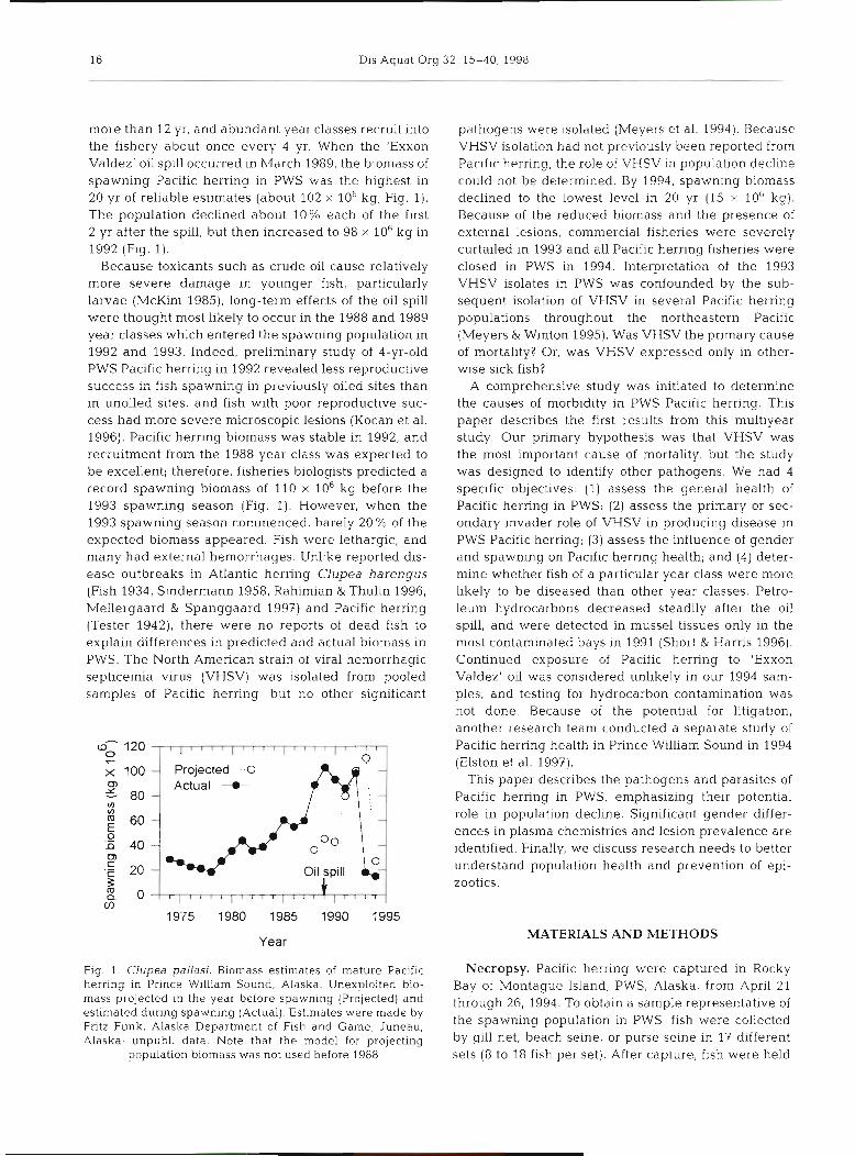
more than 12 yr, and abundant year classes recruit into the fishery about once every 4 yr. When the 'Exxon Valdez' oil spill occurred in h,larch 1989, the biomass of spawning Pacific herring in PWS was the highest in 20 yr of reliable estimates (about 102 X lob kg; Fig. 1). The population declined about 10% each of the first 2 yr after the spill, but then increased to 98 X 106 kg in 1992 (Fig. 1).
Because toxicants such as crude oil cause relatively more severe damage in younger fish, particularly larvae (McKirn 1985), long-term effects of the oil spill were thought most likely to occur in the 1988 and 1989 year classes which entered the spawning population in 1992 and 1993. Indeed, preliminary study of 4-yr-old PWS Pacific herring in 1992 revealed less reproductive success in fish spawning in previously oiiea sites than in unoiled sites, and fish with poor reproductive suc- cess had more severe microscopic lesions (Kocan et al. 1996). Pacific herring biomass was stable in 1992, and recruitment from the 1988 year class was expected to be excellent; therefore, fisheries biologists predicted a record spawning biomass of 110 X 10-g before the 1993 spawning season (Fig. 1). However, when the 1993 spawning season commenced, barely 20 % of the expected biomass appeared. Fish were lethargic, and many had external hemorrhages. Unlike reported dis- ease outbreaks in Atlantic herring Clupea harengus (Fish 1934, Sindermann 1958, Rahimian & Thulin 1996, Mellergaard & Spanggaard 1997) and Pacific herring (Tester 1942), there were no reports of dead fish to explain differences in predicted and actual biomass in PWS. The North American strain of viral hemorrhagic septicemia virus (VHSV) was isolated from pooled samples of Pacific herring, but no other significant
- Projected --0
Actual --e -
Year
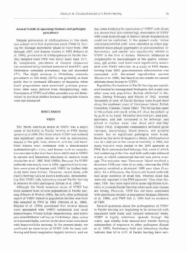
Fig. 1. Clupea pallasi. Biomass estimates of mature Pacific herring in Prince William Sound, Alaska. Unexploited bio- mass projected in the year before spawning (Projected) and estimated during spawning (Actual). Estimates were made by Fritz Funk, Alaska Department of Fish and Game, Juneau, Alaska; unpubl. data. Note that the model for projecting
population biomass was not used before 1988
pathogens were isolated (Meyers et al. 1994). Because VHSV isolation had not previously been reported from Pacific herring, the role of VHSV in population decline could not be determined. By 1994, spawning biomass declined to the lowest level in 20 yr (15 X 10' kg). Because of the reduced biomass and the presence of external lesions, commercial fisheries were severely curtailed in 1993 and all Pacific herring fisheries were closed in PWS in 1994. Interpretation of the 1993 VHSV isolates in PWS was confounded by the sub- sequent isolation of VHSV in several Pacific herring populations throughout the northeastern Pacific (Meyers & Winton 1995). Was VHSV the primary cause of mortality? Or, was VHSV expressed only in other- wise sick fish?
A comprehensive study was initiated to determine the causes of morbidity in PWS Pacific herring. This paper describes the first results from this multiyear study. Our primary hypothesis was that VHSV was the most important cause of mortality, but the study was designed to identify other pathogens. We had 4 specific objectives: (1) assess the general health of Pacific herring in PWS; (2) assess the primary or sec- ondary invader role of VHSV in producing disease in PWS Pacific herring; (3) assess the influence of gender and spawning on Pacific herring health; and (4) deter- mine whether fish of a particular year class were more likely to be diseased than other year classes. Petro- leum hydrocarbons decreased steadily after the oil spill, and were detected in mussel tissues only in the most contaminated bays in 1991 (Short & Harris 1996). Continued exposure of Pacific herring to 'Exxon Valdez' oil was considered unlikely in our 1994 sam- ples, and testing for hydrocarbon contamination was not done. Because of the potential for litigation, another research team conducted a separate study of Pacific herring health in Prince William Sound in 1994 (Elston et al. 1997).
This paper describes the pathogens and parasites of Pacific herring in PWS, emphasizing their potential role in population decline. Significant gender differ- ences in plasma chemistries and lesion prevalence are identified. Finally, we discuss research needs to better understand population health and prevention of epi- zootics.
MATERIALS AND METHODS
Necropsy. Pacific herring were captured in Rocky Bay of Montague Island, PWS, Alaska, from April 21 through 26, 1994. To obtain a sample representative of the spawning population in PWS, fish were collected by gill net, beach seine, or purse seine in 17 different sets (8 to 18 fish per set). After capture, fish were held
Marty et al . : Causes of m o r b i d ~ t y In Pacific h e r r ~ n g l. 7
in plastic containers filled with about 100 1 of seawater for no more than 4 h before processing. In groups of 2, herring were anesthetized in tricaine methane sul- fonate (Finquelo), weighed and measured (standard length), and a scale was removed for age determina- tion. Each fish was assigned a unique identifying number. Several diagnostic procedures were done as part of complete necropsy and subsequent analysis on each of 233 fish:
(1) External lesions-scored as none (0), mild ( l) , moderate (2), or severe (3). Also, each fish was as- signed a summary 'external lesion score' equal to the most severe score for fin base reddening, caudal fin reddening, or focal skin reddening.
(2) Blood-about 1.5 m1 of blood was drawn from the caudal vein into 3 m1 syringes containing 0.1 m1 of sodium heparin (10 000 IU ml-l). A capillary tube was filled and centrifuged (5500 X g for 5 min) for determi- nation of packed cell volume (PCV). A blood smear was made and air-dried. Remaining blood was cen- trifuged (13 600 X g for 5 min) and resultant plasma was frozen for storage until analysis.
Osmolality was analyzed on a Micro Osmometer Model 3MO-plus (Advanced Instruments, Norwood, MA, USA) using 20 p1 of plasma. All other analyses were done using about 200 p1 of sample in a Monarch- plus analyzer (Instrumentation Laboratories, Lexing- ton, MA, USA) calibrated and run at a stabilized 25°C. Plasma was analyzed for total protein (biuret method), albumin (bromocresol green method), and CO2 (enzy- matic method). Instrumentation Laboratories sub- strates were used to analyze calcium, cholesterol, glucose, phosphorus, total bilirubin, alkaline phos- phatase (ALP), alanine aminotransferase (ALT), aspar- tate aminotransferase (AST), and creatine phosphoki- nase (CPK). Sigmam (St. Louis, MO, USA) substrates were used to analyze gamma glutamyltransferase (GGT). Ion-selective electrodes were used to analyze sodium, potassium, and chloride. Blood smears were stained with Diff-Quik (Dade Diagnostics, Inc., Aquada, Puerto Rico) and 30 fields ( 1 0 0 0 ~ ) were exam- ined for cytoplasmic inclusions of viral erythrocytic necrosis (VEN).
(3) Virus isolation-head kidney and spleen from each fish were pooled in a plastic bag and shipped on ice to the Alaska Department of Fish and Game Fish Pathology Laboratory in Juneau, Alaska. Skin lesions, if present, were sampled and bagged separately for individual virus assay. Propagation of EPC cell lines, media formulation, and tissue preparation for cell line inoculation were as described by Meyers et al. (1994).
(4) Tissue preservation-samples of gill, liver, gonad, spleen, trunk kidney, gastrointestinal tract, heart, skin, skeletal muscle, and brain were fixed in 10% neutral buffered formalin.
(5) Bacterial isolation-for fish with moderate or severe external lesions; kidney tissues were aseptically inoculated onto trypticase soy agar (TSA) and plates were incubated at 25°C for at least 5 d.
(6) Kidney parasite identification-a touch prepara- tion of kidney (junction of head and trunk kidney) was air-dried, stained with Diff-Quik, and examined for pansporoblasts of the myxosporean Ortholinea orien- talis; extent of infection was scored as for external lesions.
(7) Organ weights-liver and gonad were weighed. (8) Herring worms (Anisakidae)-larvae in the peri-
toneal cavity were counted. Histopathologic analysis. Tissues from 233 Pacific
herring were sent to the University of California, Davis, and randomly assigned an individual histo- pathology number for blind study. Tissues from 21 her- ring had been inadvertently put in water rather than fixative. Therefore, data on histopathology reflect the 212 herring that were adequately fixed. After routine paraffin processing, tissue blocks were sectioned at 5 pm and stained with hematoxylin and eosin. Lesions were scored using a 4-point scale as none (0), mild (l), moderate (2), or severe (3). For quality control, auto- lysis and artifact in each organ were scored on the same 4-point scale. Ranking of lesions was often based on the number of structures (e.g. Ichthyophonus rest- ing spores) per lOOx field; the 100x field was examined through a 10x objective lens and a 10x ocular lens on an Olympus binocular light microscope. Differentia- tion of severity scores for each lesion was based on written criteria and 'type specimen' examples. Not all scores were used for each lesion, because many lesions had no examples that were 'severe'. After all organs were examined and lesions scored, data were re- arranged by necropsy number and subjected to statis- tical analysis.
Transmission electron microscopy. After determin- ing that 2 fish had gross and microscopic lesions con- sistent with lymphocystis virus, more detailed analysis was needed to confirm the presence of viral particles in the lesions. A stained histological section of one sus- pect cell was removed from the glass slide and pro- cessed for transn~ission electron microscopy as previ- ously described (Meyers et al. 1990).
Statistical analysis. The primary hypothesis was that fish with lesions were different from fish without lesions. The association of categorical variables (e.g. none, mild, moderate, and severe) with continuous variables (e.g. CPK values) was determined using l-way analysis of variance (l-way ANOVA). For example, the CPK values for fish with a liver Ichthyophonus score of zero were compared to CPK values in fish with mild, moderate, and severe hepatic Ichthyophonus. When necessary, cate- gories were combined to ensure that each group had at
18 Dis Aquat Org 32: 15-40, 1998
least 6 fish. Category-specific means and standard errors were calculated for each continuous variable and compared using Tukey's Studentized range method. Levene's test was used to evaluate the homogeneity of variance assumption for the ANOVA.
The association between 2 selected categorical vari- ables (e.g. Ichthyophonus scores versus scores for hepatic focal necrosis) was evaluated using chi-square methods for categorical data analysis; comparisons were considered valid only if individual expected cell frequencies were > l . Odds ratios were calculated for standard (2 X 2) 2-way contingency tables only. To m/easure the strength of the linear relationship be- tween 2 continuous variables, the correlation coeffi- cient r was calculated.
In the initid1 univariate analysis, some plasma chemistries were significantly associated with several lesions or other variables. In selected cases, multiple regression analysis was used to model a multifactor ANOVA, examining the relationships between the dependent variable (e.g. plasma albumin) and associ- ated variables (e.g. focal skin reddening, splenic con- gestion, and VHSV). Lesion scores were forced into a multiple regression equation using stepwise regres- sion to determine their joint impact in the prediction of the dependent variable (e.g. albumin level), while con- trolling for gender, gonad weight, hold time, and length. Criteria used for inclusion of variables in the evaluation included significance in the univariate analysis and postulated association of the equation variable with the dependent variable. Length was used rather than age or weight for 2 reasons: (1) length was more normally distributed than was age; and (2) length was more consistent in spawning fish than was weight.
To determine i f certain age classes of fish were more likely to be infected by certain parasites, the association of fish age with common parasites was evaluated using the chi-square test for homogeneity. Fish were grouped into 3 categories for analysis: < 5 yr old, 5 or 6 yr old, or >6 yr old. Regardless of severity of infestation, fish with a given parasite were classified as positive, and fish with- out the parasite were classified as negative.
For all analyses, comparisons were considered sig- nificant when p < 0.05 and highly significant when p < 0.01. For this report, use of the term 'prevalence' refers to the sample prevalence.
RESULTS
External gross lesions
The summary external lesion score was moderate or severe in 47 of 233 fish (20%), and several of these fish concurrently had more than one lesion graded as mod- erate or severe. Seven of 233 (3.0 %) had ulcers (scored as severe focal skin reddening; Table 1, Fig. 2A, B). Some ulcers penetrated to underlying bone and one ulcer perforated into the peritonea1 cavity, resulting in adhesions of viscera to the body wall. External lesions were significantly associated with several microscopic lesions. For example, increased scores for focal skin reddening were associated with increased scores for gill arch inflammation or hematopoiesis, submucosal gastritis, intestinal mesenteric steatitis, and renal hematopoietic cells. By comparison, scores for hepatic parenchyma1 leukocytes decreased as scores for focal skin reddening increased. The relationship among other gross lesions and histologic lesions were not consistent.
Because of the lack of published information on normal Pacific herring gross and microscopic anatomy, some findings were scored without knowledge of whether they were in fact lesions. Iris reddening is a good example. The inferior margin of the iris has a blood vessel about 3 mm long and 0.5 mm in diameter. Iris reddening occurred when the vessel contained enough blood to be detected by gross observation (Fig. 2C). Scores for iris reddening were assigned as follows: no reddening (0); reddening was limited to the primary vessel (1); reddening extended beyond the margins of the primary vessel, probably due to con- gestion of connecting venules (2); and reddening involved the entire iris (3). No fish had severe iris reddening, and mild iris reddening probably was normal. Several lesions were more prevalent in fish with no iris reddening than in fish with mild or moderate iris reddening (Table 2). For example, branchial ciliated protozoa and meningoencephalitis were more likely in fish with no iris reddening. Also, mean albumin and total protein were significantly lower in fish with no iris reddening than in fish with mild iris reddening (albumin, 0.46 vs. 0.54 g dl-'; total protein, 2.0 vs 2.3 g dl-l).
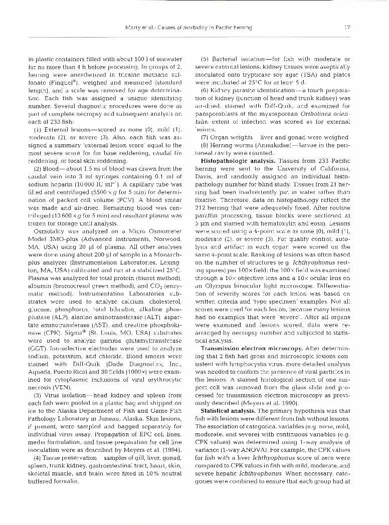
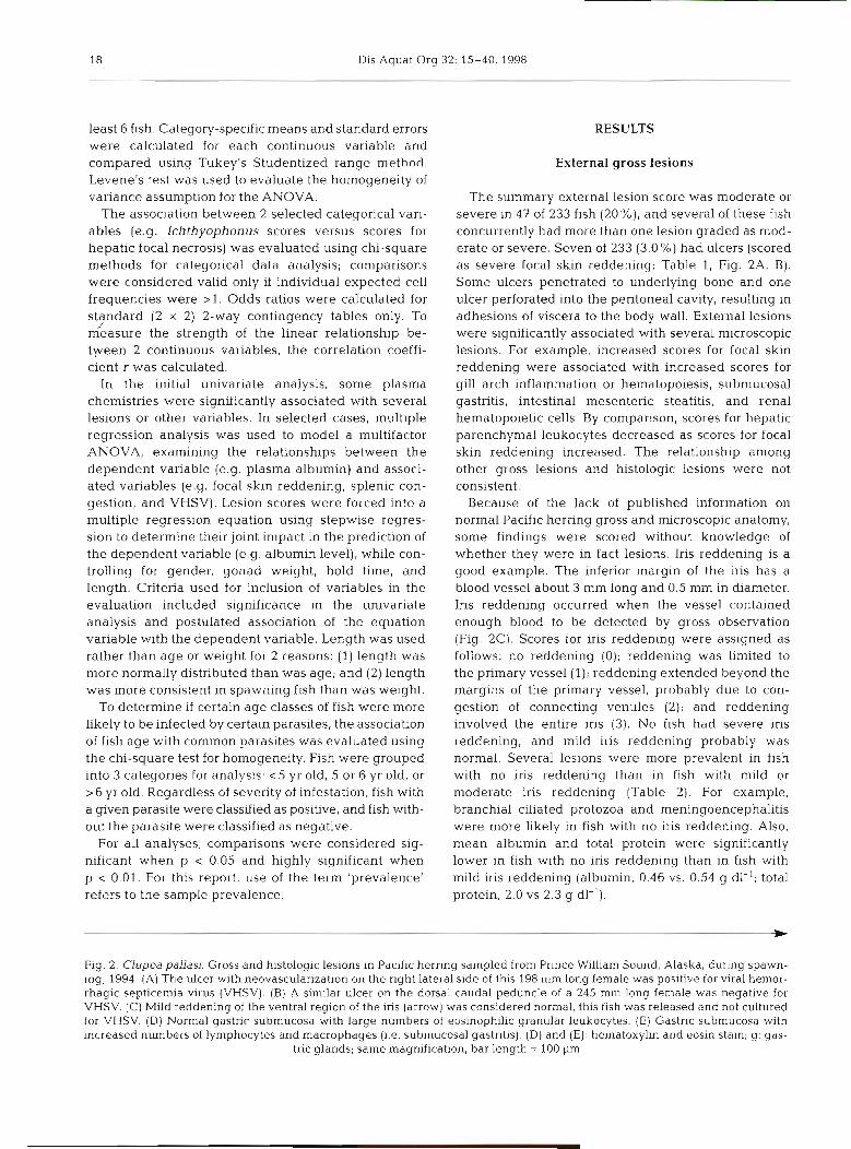
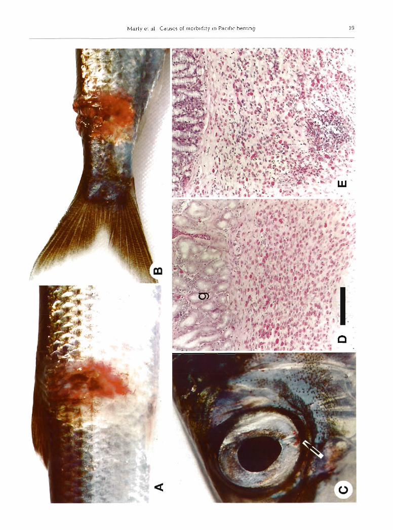
Fig. 2. Clupea pallasi. Gross and histologic les~ons in Pacific herr~ng sampled from Prince William Sound, Alaska, during spawn- ing, 1994. (A) The ulcer with neovascularization on the right lateral side of this 198 mm long female was positive for viral hemor- rhagic septicemia virus (VHSV). (B) A similar ulcer on the dorsal caudal peduncle of a 245 mm long female was negative for VHSV. (C) Mild reddening of the ventral region of the iris (arrow) was considered normal; this fish was released and not cultured for VHSV. (D) Normal gastric submucosa with large numbers of eosinophilic granular leukocytes. (E) Gastric submucosa with increased numbers of lymphocytes and macrophages (i.e. submucosal gastritis). (D) and (E) : hematoxylin and eosin stain; g: gas-
tric glands; same magnification, bar length = 100 pm
2 2 Dis Aquat Org 32: 15-40, 1998
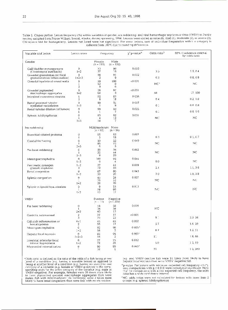
Table 2 Clupea pallas) Leslon frequency (%) w ~ t h ~ n vanables of gender, Ins r edden~ng and viral hemorrhaglc sept~cemla vlrus (VHSV) rn Pacdlc hernng sampled from Prmce Wllham Sound, Alaska, dunng spawning, 1994 Leslons were scored as none (0), m ~ l d ( l ) , moderate (21, or severe (3) Chl-square test for homogene~ty Les~ons not llsted were not slgntflcant For some les~ons sum of l n d ~ v ~ d u a l f requenc~es wth ln a category IS
d~fferent from 100% duc to roundlng dlfferences
Vanable and leslon Les~on score Frequency X 2 p-value Odds ratlo" 95% Conf~dence ~nterval for odds ratlo
Gender Female Male ( n = 1 1 0 ) ( n = 1 0 2 )
Gall bladder myxosporeans 0 7 3 90 (Ceratonlyxa aue rhach~) 1 +2 27 10
Gonadal granulomas (or focal 0 98 9 1 granulomatous ~nflammatlon) 1+2+3 2 9
Gonadal hyallnlzed vessel walls 0 39 100 1 52 0 2 9 0
Gonadal plgmented 0 36 97 macrophage aggregates 1+2 64 3
Intestinal mesentenc steatltls 1 92 82 2 8 18
Renal prox~rnal tubular 0 99 91 e p ~ t h e l ~ a l vacuolat~on 1 +2 1 9
Renal tubular d l la t~on (of lumen) 0 99 93 1 1 7
S p l e n ~ c Ichthyophonus 0 83 82 1 4 12
2+3 14 7
Ins r e d d e n ~ n g M~ld/moderate None (n = 93) (n = 94)
Branchal c l l~ated protozoa 95 82 5 18
Caudal fln frayulg 10 22 85 73
6 5 Fin base reddening 59 36
32 44 9 20
Men~ngoencephah t~s 100 96 0 4
Pancreatic zymogen 27 44 granule deple t~on 73 56
Renal congestion 67 80 33 20
Splenlc congestion 45 28 35 52 20 20
Sp len~c e lhpso~d h y a l ~ n ~ z a t ~ o n 9 23 78 60 13 17
VHSV Pos~tlve Negatlve (n = l l ) (n = 200)
Fln base reddening 0 18 50 0 005 1 36 38 NC
2+3 4 5 12 Gas tn t~s submucosal 2 27 77 <O 001
3 73 23 9 1 2 3 36 G111 arch ~nf larnmat~on or 0+1 45 83 0 002
hematopo~es~s 2 55 17 5 7 1 6 20 Menlngoencephallt~s 0 82 98 0 005'
1 +2 18 2 8 7 1 5 , 5 1 Hepabc focal necrosls 0 82 98 0 002'
1+2+3 18 2 11 1 8, 68 lntest~nal arter~olar focal 0 27 75 0 012
~ n t ~ m a l hyperplas~a 1 +2 73 35 5 0 1 3 19 Myocard~al mlnerah7atlon 0 90 99 0 003'
1 10 1 2 2 1 3 380
"Odds ratlo IS deflned as the ratlo of the odds of a flsh belng at one Ing and VHSV poslt~ve flsh were 11 tlmes more llkely to have level of a condi t~on ( e g h a v ~ n g a scorable lesion) as opposed to h e p a t ~ c focal necrosls than were VHSV-negat~ve f ~ s h belng a t another level of a c o n d ~ t ~ o n ( e g havlng no leslon) for one category of a vanable (e g female or VHSV-pos~t~vel to the corre
b p value For l e s~ons w ~ t h mlnlmum expected cell frequency < l ( ' 1 spondlng odds for the other category of the vanable (e g male or only compansons wlth p < 0 010 were cons~dered s~gn l f~can t Note
VHSV-negative) For example females were 58 t ~ m e s more hkely that for compansons w t h a low expected cell frequency, the odds
to have plgrnented gonadal macrophage aggregates than were ratlo has a w ~ d e confidence Interval
males flsh with mlld/moderate Ins reddening were 2 tlmes more CNC odds ratlos were not calculated for les~ons wlth more than 2 l~ke ly to have renal congestion than were flsh wlth no Ins redden- groups ( e g s p l e n ~ c lchthyophonus)
Marty et al.: Causes of mol .bidity in Pacific herring 23
Ichthyophonus hoferi
All organs contained Ichthyophonus hoferi (hereafter referred to as Ichthyophonus) (Table l ) , and the multi- nucleate resting spore stage was the most common form. Morphology of Ichthyophonus and the host reaction were similar to those reported in infections in Atlantic herring (Daniel 193313, Sindermann 1970). Most resting spores were surrounded by a rim of fibro- blasts and maturing collagenous connective tissue, but some were surrounded by activated macrophages. Severe granulomatous inflammation, common in the heart, was usually associated with developing spores (Fig. 3C). Occasionally, resting spores had burst and released multinucleate endospores (Fig. 4A). A con- sistent scoring system was used for Ichthyophonus in each organ: score = 0 (no Ichthyophonus); score = 1 (< 1 resting spore per lOOx field); score = 2 (21 but < 3 resting spores per lOOx field, but inflammation was limited to a thin rim of fibrous connective tissue); score = 3 (21 resting spore per lOOx field, with prominent granulomatous inflammation, or 1 3 resting spores per lOOx field, regardless of the amount of inflammation).
Granulomatous inflammation associated with Ich- thyophonus had to be differentiated from other forms of macrophage aggregates. Pigmented macrophage aggregates at least 60 pm in diameter were common in liver, spleen, and kidney. Pigment varied from yellow- brown (Fig. 5A, B) to green-brown, but aggregates did not contain melanin. Pigmented macrophage aggre- gates were more common in older fish, and some aggregates were as large as 300 pm in diameter (Fig. 5B) . Aggregates of nonpigmented activated macrophages were classified as nonspecific granulo- matous inflammation (Fig. 5C). Granulomatous inflam- mation was composed of activated macrophages with pale eosinophilic cytoplasm. Activated macrophages sometimes infiltrated and expanded foci of pigmented macrophage aggregates. Small numbers of lympho- cytes and eosinophilic granular leukocytes were scat- tered throughout foci of granulomatous inflammation.
Lesions associated with Ichthyophonus occurred in 62 of 212 (29%) fish, but no single organ had greater than 21 % prevalence (Fig. 6). Prevalence of Ichthyo- phonus in skin and skeletal muscle was the second highest after kidney, but most cases in slun and skele- tal muscle were mild (31 of 39, 79%). By comparison, prevalence of Ichthyophonus in the heart was similar to that in skin and skeletal muscle, but relatively few cases in the heart were mild (14 of 38, 37 %).
A sum-Ichthyophonus (sumICH) score was calcu- lated for each fish by adding the individual Ichthyo- phonus scores from all 10 organs for that particular fish. For example, Ichthyophonus scores in organs of fish #l06 included spleen (score = 2) , kidney (score = l ) ,
and a combined score for skin and skeletal muscle (score = l), but the other 7 organs had no Ichthyo- phonus (score = 0); therefore, the sumICH score for fish #l06 was 4 . Because the maximum Ichthyophonus score for each organ was 3 (severe), the maximum possible sumICH score for a fish was 30. The highest actual score was 24. SumICH scores significantly in- creased with increased severity of several internal lesions, but sumICH scores were not associated with any external lesions. Several lesions were significantly associated with greater sumICH scores: cardiac throm- bosis, gastric foreign body, gastric focal parenchyma1 leukocytes, hepatic eosinophilic granular leukocytes, intestinal foreign body granuloma, intestinal mesen- teric steatitis, and skeletal myositis. Note that Levene's test for equality of variances was significant for all comparisons except skeletal myositis.
Association of Ichthyophonus scores with plasma chemistries was variable (Table l), but AST and CPK, enzymes con~monly used in mammalian medicine as part of the evaluation of general health, were signifi- cantly associated with lchthyophonus scores in every organ (univariate ANOVA). Increases in CPK in mam- mals result from disruption in muscle cell membranes (Willard et al. 1989). By comparison, AST is present in significant quantities in mitochondria of hepatocytes, muscle, erythrocytes, and other blood-rich organs. The most common causes of increased AST in small domes- tic mammals are hepatic disease, muscular disease (inflammation or necrosis), and hemolysis (Willard et al. 1989).
The significant increase in CPK and AST in every or- gan was inconsistent with distribution of these enzymes in nlarnrnals. Therefore, multiple regression analysis was used to model a multifactor ANOVA, examining the linear relationships between the dependent variable CPK (or AST) and Ichthyophonus lesion scores in 9 organs (brain, gill, heart, intestine, kidney, liver, skin/ skeletal muscle, spleen, and stomach). Gonad scores were not analyzed because only 3 gonads contained Ich- thyophonus. For CPK, brain Ichthyophonus status, gender, and gonad weight were the only significant predictors when all organs were included in the multiple regression equation. For AST, renal Ichthyophonus status and gonad weight were the significant predictors; however, in the final model, predicted values for AST decreased when a fish had renal Ichthyophonus.
As a relative measure of the severity of Ichthyo- phonus in individual organs, a mean sumICH score was computed as follows for each organ: all fish with Ichthyophonus in an organ were selected, their sumICH scores were totaled, and this sum of sumICH scores was divided by the number of fish in which the organ was infected. For example, of 212 kidneys ex- amined, 43 had Ichthyophonus; the mean sumICH
C., 3 &,A,
Marty et al.: Causes of morbidity in Pacific herring
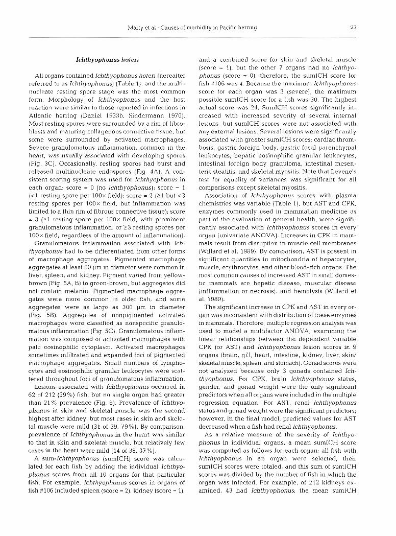
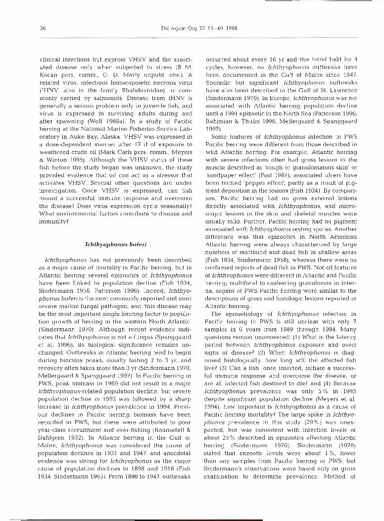
Fig. 3. Clupea pallasi. Internal parasites of Pacific herring sampled from Prince William Sound, Alaska, during spawning. 1994; hematoxylin and eosin stain. (A) The myxosporean Ceralomyxa auerbachi in the gall bladder lumen; despite large numbers of organisms, inflammation in the gall bladder wall is minimal; bar length = 80 pm. Inset: trophozoites and maturing spores (arrow points to polar capsules); bar in larger print is 30 pm long at inset magnification. (B) Several stages of an unclassified coccidian (Goussia sp.?) in the apical margin of epithel~al cells of intestinal cecae. Note different stages of development (arrows) and lack of inflammation; bar length = 30 pm. (C) Forms of Ichthyophonus in the heart include multinucleate rest~ng spores with minimal inflammation (arrows), remnants of ruptured resting spores (r) with small endospores, and developing spores surrounded by
severe granulomatous inflammation (g); bar length = 400 pm
score for those 43 fish was 9.4; by comparison, the mean sumICH score for the 17 fish with brain Ichthyophonus was 14.2. Generally, organs with the lowest Ichthyophonus prevalence (e.g, brain) had the highest mean sumICH scores (Fig. 6).
Sheets of mononuclear cells within gill arches were significantly associated with VHSV infection (Table 2). Gill arches normally contained scattered mononuclear cells that had densely basophilic nuclei and relatively scant basophilic cytoplasm (Fig. ?A). Not all cells could be identified, but they included mature inflammatory cells and hematopoietic cells in various stages of devel-
VHSV opment. In 39 fish, these mononuclear cells were more abundant, but the cells did not alter tissue architecture (Fig. 7B). Eleven of 233 Pacific herring (4.7 %) were positive
for VHSV. Virus was isolated from 7 of 233 spleen- Meningoencephalitis was significantly associated kidney pools and from 5 of 15 skin lesions. One fish with VHSV infection (Table 2), and eosinophilic had VHSV isolated from both the spleen-kidney pool meningitis was marginally associated with VHSV and a skin lesion. Several lesions and alterations in blood chemistries were associated with VHSV infec- tion (Tables 2 & 3). Among external lesions, fin base
infection (p = 0.06). In the brain, meninges usually con- tained 2 to 25 eosinophilic granular leukocytes in at least one 400x field, but normal meninges did not
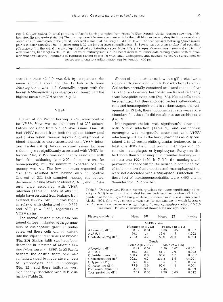
reddening was significantly associated with VHSV in- contain macrophages or lymphocytes. Forty-two fish fection. Also, VHSV was significantly associated with had more than 25 eosinophilic granular leukocytes in focal skin reddening (p = 0.03, chi-square test for at least one 400x f~eld. In 7 fish, the meninges and homogeneity), but the minimum expected cell fre- perivascular space within the neuropile contained foci quency was < l . The low minimum expected cell of inflammation (lymphocytes and macrophages) that frequency resulted from having only 11 positive were not associated with Ichthyophonus infection, but fish out of 233 fish sampled. Among chemistries, these foci of meningoencephalitis were <400 pm in decreased plasma levels of albumin, ALP, and choles- diameter in all but one fish. terol were associated with VHSV infection ( T ~ ~ ~ ~ 3) , of albumin Table 3. Clupea pallasi. Plasma chemistry values that were significantly differ-
ent (p < 0.05) based on status of viral hemorrhagic septicemia virus (VHSV) or might have leakage gender. Paclfic herring were sampled during spawning in Prince Williarn Sound, external lesions. Albumin was highly Alaska, 1994. One-way analysis of variance; for comparisons in which Levene's correlated with cholesterol (r = 0.895) test for equality of variance was significant ( ' ) , only comparisons with p 5 0.010
and ALP (r = 0.587) regardless of are shown. Plasma chemistries not shown were not significant
VHSV status. The normal gastric submucosa con- p-value
tained diffuse infiltrates of large num- VHSV status bers of eosinophilic granular leuko-
0.36 0.05 0.007 cytes. but these cells did not extend 0.002
into the adjacent muscularis or mucosa 221.4 4.7 156.9 21.0 0 003 . -
(Fig. 2D). Similar infiltrates have been 1 Gender Female (n = 11 7)
0.47 0.02 59.3 2.1
160.4 0.9 202.1 6.3
5.6 0.2 75.9 2.6
2.13 0.10 2.14 0.06
described in intestine of Atlantic her- ring (Morrison et al. 1986). In 53 Pacific herring, the gastric submucosa also contained small to moderate numbers
Male (n = 116) 0.56 0.02 <0.001
51.1 1.6 0.002 165.6 1.2 0.001 ' 234.8 6.6 <O 001
6.5 0.2 0 004 90.0 4.3 0.001
2.45 0.11 0.029 2.30 0.05 0.042
*lbumin (g dl-') ALP (U I-') Chloride (rnmol 1 - 1 ) Cholesterol (rng dl-l)
fection (Table 2).
of lymphocytes and macrophages (Fig. 2E), and these infiltrates were significantly associated with VHSV in-
CO2 (mrnO1 Glucose (mg dl-l) Potassium (mmoll-~) Total protein (g dl-l)
26 Dis Aquat Org 32: 15-40,1998
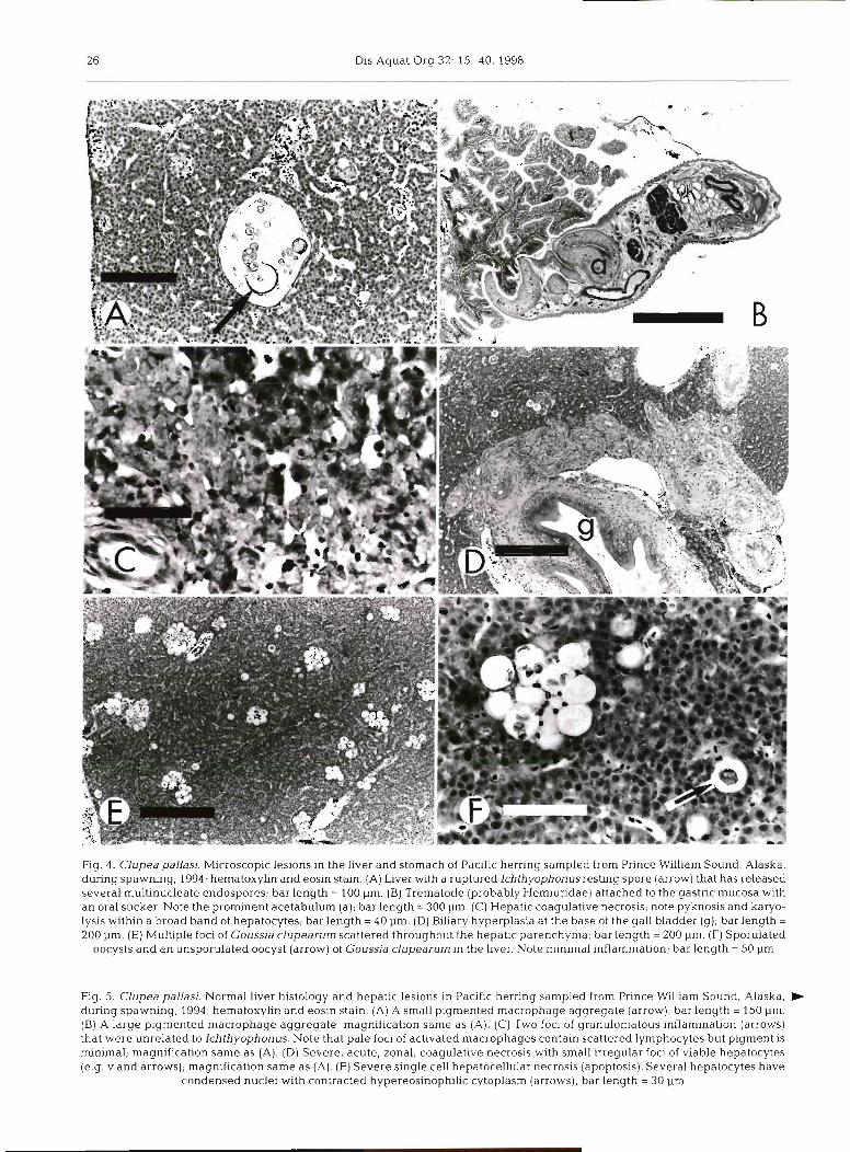
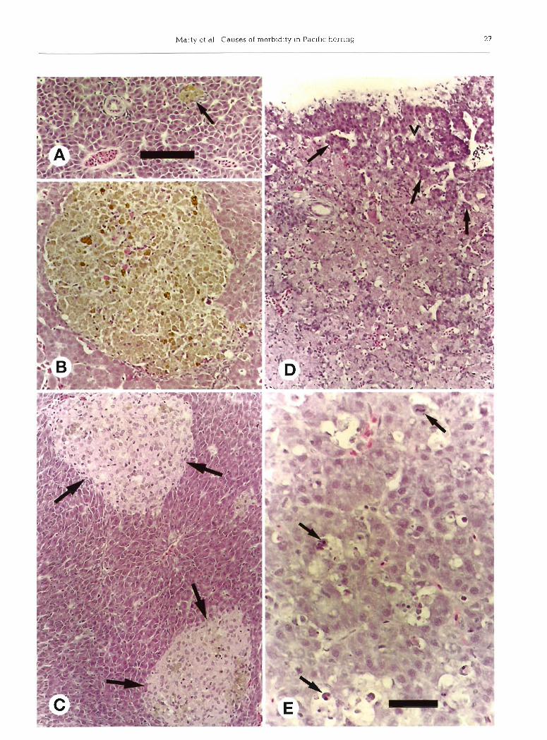
Fig. 4. Clupea pallad. Microscopic lesions in the liver and stomach of Pacific herring sampled from Prince William Sound, Alaska, during spawning, 1994; hematoxylin and eosin stain. (A) Llver with a ruptured Ichthyophonus resting spore (arrow) that has released several multinucleate endospores; bar length = 100 pm. (B) Trematode (probably Herniuridae) attached to the gastric mucosa with an oral sucker. Note the prominent acetabulurn (a]; bar length = 300 pm. (C) Hepatlc coagulative necrosis; note pyknosis and karyo- lysis within a broad band of hepatocytes; bar length = 40 pm. (D) Biliary hyperplasia at the base of the gall bladder (g); bar length = 200 pm. (E) Multiple foci of Goussia clupearum scattered throughout the hepatic parenchyma; bar length = 200 pm. (F) Sporulated
oocysts and an unsporulated oocyst (arrow) of Goussla clupearum In the Ilver. Note minimal inflammation; bar length = 50 pm
Fig. 5. Clupea pallasi. Normal liver histology and hepatic lesions in Paclflc herring sampled from Prince William Sound, Alaska, b during spawning, 1994; hernatoxylin and eosln staln. (A) A small pigmented macrophage aggregate [arrow); bar length = 150 pm. (B) A large pigmented macrophage aggregate; rnagnificatlon same as (A). (C) Two foci of granulomatous inflammation (arrows) that were unrelated to lchthyophonus. Note that pale foci of activated rnacrophages contain scattered lymphocytes but pigment is minimal; magnification same as (A). (D) Severe, acute, zonal, coagulative necrosis with small irregular foci of viable hepatocytes (e.g. v and arrows); magnification same as (A). (E) Severe single cell hepatocellular necrosis (apoptosis). Several hepatocytes have
condensed nuclei with contracted hypereosinophilic cytoplasm (arrows); bar length = 30 pm
28 Dis Aquat Org 32: 15-40, 1998
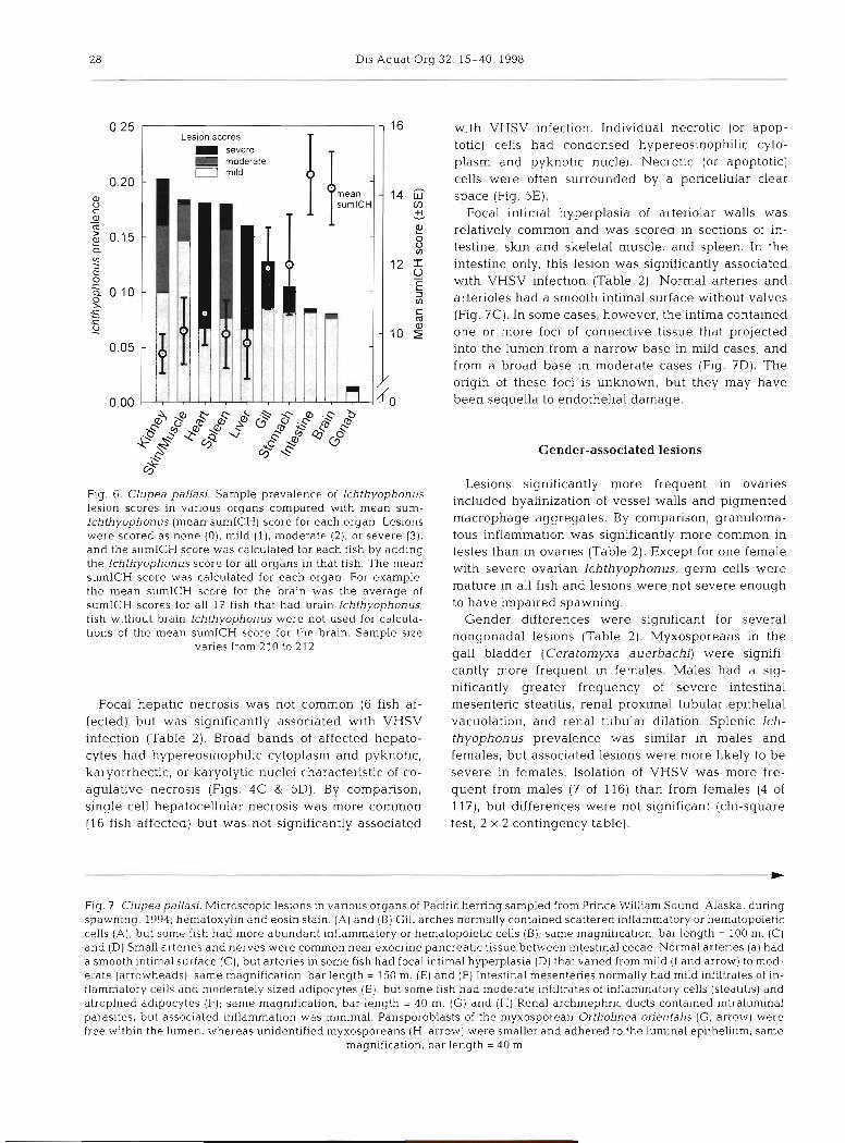
Les~on scores severe moderate I rnmlld
mean
I
n
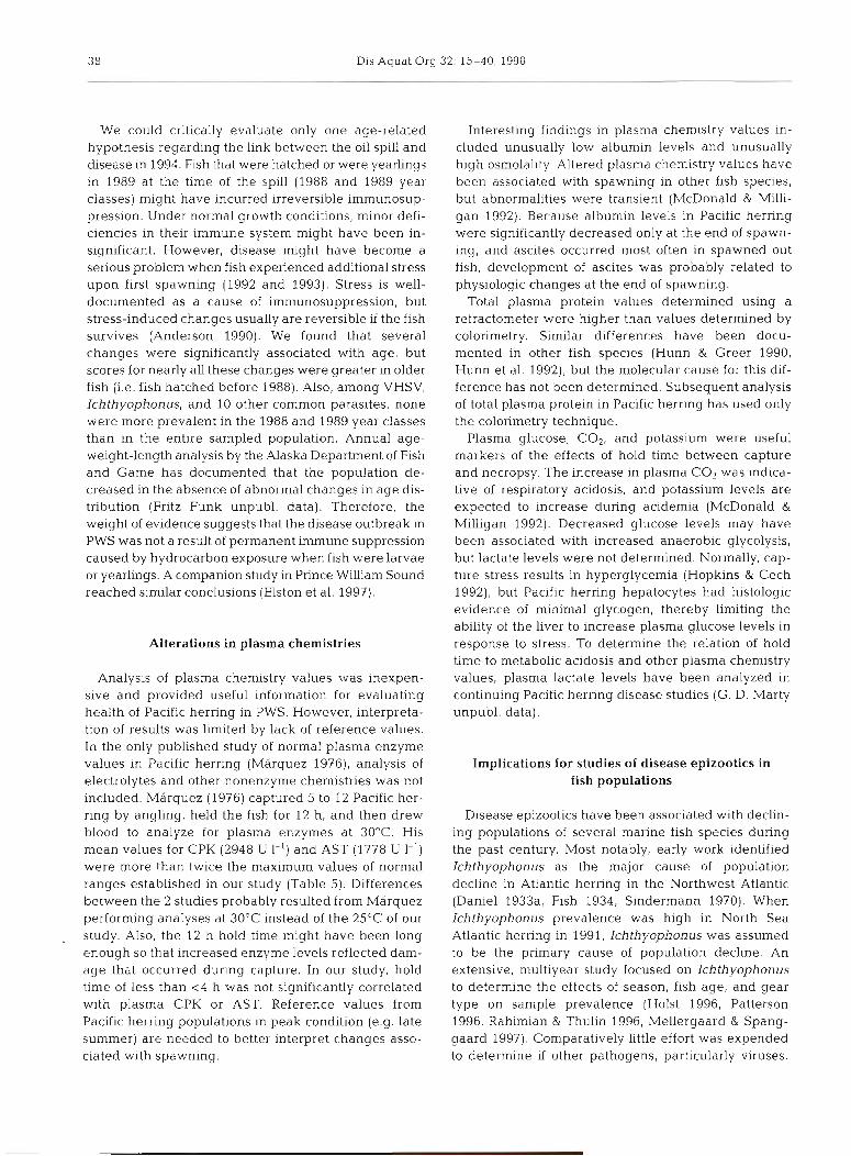
Fig. 6. Clupea pallasj. Sample prevalence of lchthyophonus lesion scores in various organs compared with mean sum- Ichthyophonus (mean sumICH) score for each organ. Lesions were scored as none (0), mild ( l) , moderate (2), or severe (3). and the sumICH score was calculated for each fish by adding the Ichthyophonus score for all organs in that fish. The mean sumlCH score was calculated for each organ. For example, the mean sumICH score for the brain was the average of sumICH scores for all I f fish that had brain Ichthyophonus; fish without brain Ichthyophonus were not used for calcula- tions of the mean sumICH score for the brain. Sample size
varies from 210 to 212
Focal hepatic necrosis was not common (6 fish af- fected) but was significantly associated with VHSV infection (Table 2). Broad bands of affected hepato- cytes had hypereosinophilic cytoplasm and pyknotic, karyorrhectic, or karyolytic nuclei characteristic of co- agulative necrosis (Figs. 4C & 5D). By comparison, single cell hepatocellular necrosis was more common (16 fish affected) but was not significantly associated
with VHSV infection. Individual necrotic (or apop- totic) cells had condensed hypereosinophilic cyto- plasm and pyknotic nuclei. Necrotic (or apoptotic) cells were often surrounded by a pencellular clear space (Fig. SE).
Focal intimal hyperplasia of arteriolar walls was relatively common and was scored in sections of in- testine, skin and skeletal muscle, and spleen. In the intestine only, this lesion was significantly associated with VHSV infection (Table 2 ) . Normal arteries and arterioles had a smooth intimal surface without valves (Fig. ?C). In some cases, however, the intima contained one or more foci of connective tissue that projected into the lumen from a narrow base in mild cases, and from a broad base in moderate cases (Fig. ?D). The origin of these foci is unknown, but they may have been sequella to endothelial damage.
Gender-associated lesions
Lesions significantly more frequent in ovaries included hyalinization of vessel walls and pigmented macrophage aggregates. By comparison, granuloma- tous inflammation was significantly more common in testes than in ovaries (Table 2). Except for one female with severe ovarian Ichthyophonus, germ cells were mature in all fish and lesions were not severe enough to have impaired spawning.
Gender differences were significant for several nongonadal lesions (Table 2). Myxosporeans in the gall bladder (Ceratomyxa auerbachi) were signifi- cantly more frequent in females. Males had a sig- nificantly greater frequency of severe intestinal mesenteric steatitis, renal proximal tubular epithelia1 vacuolation, and renal tubular dilation. Splenic Ich- thyophonus prevalence was similar in males and females, but associated lesions were more likely to be severe in females. Isolation of VHSV was more fre- quent from males (7 of 116) than from females (4 of 1 l?), but differences were not significant (chi-square test, 2 X 2 contingency table).
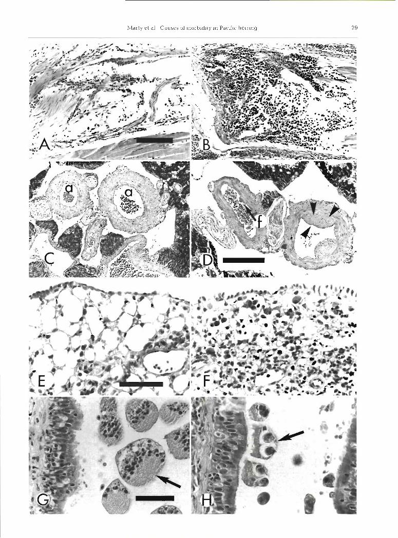
Fig. 7 Clupea pallasi. Microscopic lesions in various organs of Pacific herring sampled from Prince William Sound, Alaska, during spawning. 1994; hematoxylin and eosin stain. (A) and (B) Gill arches normally contained scattered inflammatory or hematopoietic cells (A), but some fish had more abundant inflammatory or hematopoietic cells (B); same magnification, bar length = 100 m. (C) and (D) Small arteries and nerves were common near exocrine pancreatic tissue between intestinal cecae. Normal arteries (a) had a smooth intimal surface (C), but arteries in some fish had focal intimal hyperplasia (D) that varied from mild (f and arrow) to mod- erate (arrowheads); same magnification, bar length = 150 m. (E) and (F) Intestinal mesenteries normally had mild infiltrates of In- flammatory cells and moderately slzed adipocytes (E), but some fish had moderate infiltrates of inflammatory cells (steatitis) and atrophied adipocytes (F); same magnification, bar length = 40 m. (G) and (H) Renal archinephric ducts contained intraluminal parasites, but associated inflammation was minimal. Pansporoblasts of the myxosporean Orthofinea onentalis (G, arrow) were free within the lumen, whereas unidentified myxosporeans (H, arrow) were smaller and adhered to the lumina1 epithelium; same
magnification, bar length = 40 m
30 Dis Aquat Org 32: 15-40, 1998
Intestinal mesenteric steatitis involved peritoneal fat herring worms than did fish without renal interstitial throughout the mesenteries of the viscera. Lipid vol- cell necrosis. ume of adipocytes varied from moderately abundant to minimal. In moderate cases of steatitis, lipid volume was often less than the volume of adipocyte nuclei Lymphocystis virus (Fig. ?F). Inflammatory infiltrates included macro- phages, lymphocytes, and eosinophilic granular leuko- Two Pacific herring had internal lesions consistent cytes. All fish had at least some inflammatory cells with lymphocystis virus, but the skin of these fish was within the peritoneal fat (Fig. ?E), but 19 males and normal. Affected fish had 1 or 2 spherical, white foci, 8 females had more than 30% of the volume of pen- each about 2 mm in diameter. One focus was in the toneal fat infiltrated by inflammatory cells. The cause cranial part of the peritoneal cavity, and the other focus of these inflammatory infiltrates was not determined. expanded the intestinal mesenteries. Histologcally, each
Proximal renal tubular epithelium was considered white focus was composed of a single hypertrophic vacuolated if intracytoplasmic clear spaces were larger fibroblast. The affected fibroblast had a multilayered, than adjacent nuclei. Kidneys from 9 males and 1 12 pm thick, hyaline capsule, with abundant granular female contained vacuolated tubular epithelial cells, basophilic cytoplasm, and a large nucleus (500 pm in but in only one case (a male) were more than 20% of diameter) with vacuolated and marginated chromatin the proximal tubular epithelial cells affected. Renal (Fig. BA, B). The infected fibroblast was not associated tubules were considered dilated when lumina1 diame- with any inflammatory cells. Ultrastructurally, the cyto- ter was more than twice the thickness of tubular plasm contained abundant icosahedral viral particles, epithelial cells. Kidneys from 7 males and 1 female each about 200 nm in diameter, with an electron-dense contained dilated tubules, but in no cases were more viroplasm (Fig. BC). The ultrastructural features of the than 50% of the tubules dilated. Causes for these tubu- virus are characteristic of lymphocystis virus. lar changes are unknown. Although pansporoblasts of the renal myxosporean Ortholinea orientalis some- times nearly filled archinephric ducts (Fig. ?G), only Other potential pathogens one of 44 cases was associated with dilated tubules, i.e. Ortholinea orientalis was not associated with dilated No significant bacterial pathogens were isolated, tubules. and none of the blood smears had evidence of VEN.
In addition to these lesions, gender differences were Ulcers often contained variable amounts of granulation significant for several plasma chemistries (Table 3). tissue with a surface layer of filamentous bacteria; Compared to males, females had significantly lower however, culture results indicated that the bacteria values for albumin, chloride, cholesterol, COz, glucose, had not spread to the kidney. potassium, and total protein, and significantly higher Pacific herring had 12 other parasites, most of which values for ALP. Gender differences were not signifi- were associated with few lesions. These parasites in de- cant for other plasma chemistries. scending order of prevalence included: (1) an intestinal
coccidian (Goussia sp.?) that has not previously been described, 91 %; (2) a coccidian in the liver, Goussia
Intraperitoneal herring worms (Anisakidae) (Eimeria) clupearurn, 61 %; (3) a testicular coccidian, 57 % of males; (4) a myxosporean in renal tubules, Or-
All 233 Pacific herring contained larval parasites of tholinea orientalis, 1 9 % ; ( 5 ) a myxosporean in the gall the family Anisakidae within their peritoneal cavities. bladder, Ceratomyxa auerbachi, 19%; (6 ) branchial No attempt was made to differentiate species (e.g. monogenetic trematodes Gyrodactylus spp., 13%; Anisakis vs Contracecum), and parasite morphology (7) branchial ciliated protozoans, probably Trichodina and inflammatory response were consistent with pre- and Cryptokaryon spp., 12%; ( 8 ) unclassified renal in- vious descriptions (Hauck & May 1977). Herring worm traductal myxosporean (?), 11 %; (9) branchial Epithe- numbers were significantly greater in females than in liocystis, 10%; (10) gastric intraluminal trematodes, males, and numbers significantly increased with in- e.g. Hemiuridae, 8.6%; ( I l ) intestinal trematodes, e.g. creasing severity of several lesions. For example, fish Lecithaster gibbosus, 2.9%; and (12) intestinal ces- with more severe hepatic cholangitis or biliary hyper- todes, 2.4 %. Infestation with these branchial and gas- plasia (Fig. 4D) had increased numbers of herring trointestinal parasites did not significantly alter plasma worms. Also, increased numbers of intraperitoneal chemistry values or inflammatory changes. Anisakidae were associated with increased scores for Morphologic features and distribution of the intestinal Anisakidae in the liver, intestine, and skeletal muscle. coccidian were very similar to descriptions of Goussia Fish with renal interstitial cell necrosis had fewer zarnowskii in the 3-spined stickleback Gasterosteus
Marty et al.: Causes of morbidity in Pacific herring 3 1
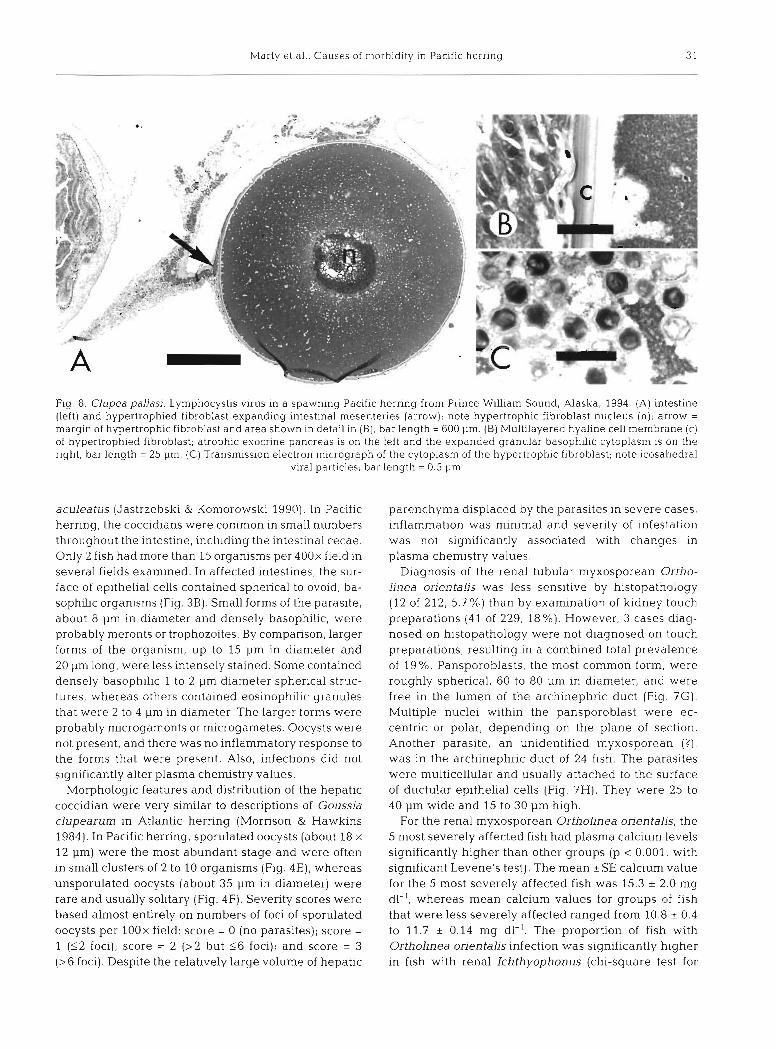
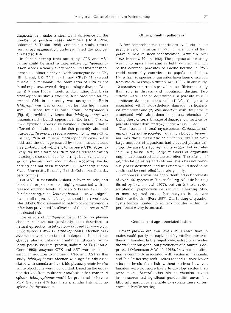
Fig. 8. Clupea pallasi. Lymphocystis virus in a spawning Pacific herring from Prince Willia~n Sound, Alaska, 1994. (A) intest~ne (left) and hypertrophied fibroblast expanding intestinal mesenteries (arrow); note hypertrophic fibroblast nucleus (n); arrow = margin of hypertrophic fibroblast and area shown in detail in (B); bar length = 600 pm. (B) Multilayered hyaline cell membrane (c) of hypertrophied fibroblast; atrophic exocrine pancreas is on the left and the expanded granular basophilic cytoplasm is on the right; bar length = 25 pm. (C) Transmission electron micrograph of the cytoplasm of the hypertrophic fibroblast; note icosahedral
viral particles; bar length = 0.5 pm
aculeatus (Jastrzebski & Komorowski 1990). In Pacific parenchyma displaced by the parasites in severe cases, herring, the coccidians were common in small numbers inflammation was minimal and severity of infestation throughout the intestine, including the intestinal cecae. was not significantly associated with changes in Only 2 fish had more than 15 organisms per 400x field in plasma chemistry values. several fields examined. In affected intestines, the sur- Diagnosis of the renal tubular myxosporean Ortho- face of epithelial cells contained spherical to ovoid, ba- linea orientalis was less sensitive by histopathology sophilic organisms (Fig. 3B). Small forms of the parasite, (12 of 212, 5.7 %) than by examination of kidney touch about 8 pm in diameter and densely basophilic, were preparations (41 of 229, 18%). However, 3 cases diag- probably meronts or trophozoites. By comparison, larger nosed on histopathology were not diagnosed on touch forms of the organism, up to 15 pm in diameter and preparations, resulting in a combined total prevalence 20 pm long, were less intensely stained. Some contained of 19 %. Pansporoblasts, the most common form, were densely basophilic 1 to 2 pm diameter spherical struc- roughly spherical, 60 to 80 pm in diameter, and were tures, whereas others contained eosinophilic granules free in the lumen of the archinephric duct (Fig. ?G) . that were 2 to 4 pm in diameter. The larger forms were Multiple nuclei within the pansporoblast were ec- probably rnicrogamonts or microgametes. Oocysts were centric or polar, depending on the plane of section. not present, and there was no inflammatory response to Another parasite, an unidentified myxosporean ( ? ) ,
the forms that were present. Also, infections did not was in the archinephric duct of 24 fish. The parasites significantly alter plasma chemistry values. were multicellular and usually attached to the surface
Morphologic features and distribution of the hepatic of ductular epithelial cells (Fig. 7H). They were 25 to coccidian were very similar to descriptions of Goussia 40 pm wide and 15 to 30 pm high. clupearum in Atlantic herring (Morrison & Hawkins For the renal myxosporean Ortholinea orientalis, the 1984). In Pacific herring, sporulated oocysts (about 18 X 5 most severely affected fish had plasma calcium levels 12 pm) were the most abundant stage and were often significantly higher than other groups (p < 0.001, with in small clusters of 2 to 10 organisms (Fig. 4E), whereas significant Levene's test). The mean *SE calcium value unsporulated oocysts (about 35 pm in diameter) were for the 5 most severely affected fish was 15.3 * 2.0 mg rare and usually solitary (Fig. 4F). Severity scores were dl-l, whereas mean calcium values for groups of fish based almost entirely on numbers of foci of sporulated that were less severely affected ranged from 10.8 + 0.4 oocysts per lOOx field: score = 0 (no parasites); score = to 11.7 * 0.14 mg dl-l. The proportion of fish with 1 ($2 foci); score = 2 (>2 but 1 6 foci); and score = 3 Ortholinea orientalis infection was significantly higher (>6 foci). Despite the relatively large volume of hepatic in fish with renal Ichthyophonus (chi-square test for
32 Dis Aquat Org 32: 15-40, 1998
homogeneity). Infection with the renal intraductal par- asite (probably a myxosporean) was not significantly associated with any changes in plasma chemistries or any other renal lesions.
The gall bladder sometimes contained large num- bers of the myxosporean Ceratomyxa auerbachi (Fig. 3A). Most common were forms that were roughly spherical, multicellular, and 15 to 30 pm in diameter with 1 to 6 nuclei. Less common were spindle-shaped forms that were 50 to 80 pm long, 15 to 20 pm in dia- meter, and had pale eosinophilic to vacuolated cyto- plasm. Sections of the elongate structures often con- tained 1 or 2 spherical structures (spores?), about ? pm in diameter, that stained intensely eosinophilic. Severe infestations sometimes had mild mononuclear inflam- mation in the idrrlina propria of the gall biadder, but infestations were not significantly associated with liver lesions or with changes in plasma chemistries.
Age-associated changes
The most consistent age-related change was increased severity of pigmented macrophage aggregates in older fish. Indeed, age-related changes were significant in all organs in which pigmented macrophage aggregates were scored: exocrine pancreas, liver, ovary, spleen, and trunk kidney (Table 1). Lesion scores that significantly increased with age included meningoencephalitis, epi- carditis, renal tubular epithelia1 vacuolation, pancreatic zymogen granule depletion, and splenic ellipsoid hyalinization (Table 1). Interestingly, in the liver, scores for increased granulomatous inflammation were sig- nificantly associated with decreased age.
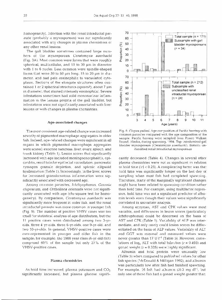
Among common parasites, Ichthyophonus, Goussia clupearurn, and Ortholinea orientalis were not signifi- cantly associated with age (chi-square test for homo- geneity). By comparison, Ceratomyxa auerbachi was significantly more frequent in older fish, and the renal intraductal parasite was more common in younger fish (Fig. 9). The number of positive VHSV cases was too small for statistical analysis of age distribution, but the 11 positive cases were distributed among two 3-yr- olds, three 4-yr-olds, three 6-yr-olds, one 9-yr-old, and two 10-yr-olds. In general, VHSV-positive cases were over-represented in younger and older fish in the sample; for example, the 1988 year class (6-yr-old fish) comprised 60% of the sample but only 27% of the VHSV-positive cases.
Plasma chemistries
As hold time increased, plasma potassium and CO2 significantly increased, but plasma glucose signifi-
Total sample (n = 212)
unclassified renal intraductal myxosporea
3 4 5 6 7 8 9 1 0 1 1 Age (years)
Fig. 9. Clupea pallasi. Age composition of Pacific herring with common parasites compared with the age composition of the sample. Pacific herring were sampled from Prince Wllliam Sound, Alaska, during spawning, 1994. Top: intraluminal gall bladder myxosporean (Ceratomyxa auerbachi). Bottom: un-
classified renal intraductal myxosporean
cantly decreased (Table 4). Changes in several other plasma chemistries were not as significant in relation to hold time (Irl i 0.25). A complicating factor was that hold time was significantly longer on the last day of sampling when most fish had completed spawning. Therefore, many of the marginally significant changes might have been related to spawning condition rather than hold time. For example, using multifactor regres- sion, hold time was not a significant predictor of albu- min levels even though their values were significantly correlated in univariate analysis.
Among enzymes, AST and CPK values were most variable, and differences in lesion scores (particularly Ichthyophonus) could be discerned on the basis of AST and CPK (Table 1). Variability of ALP was inter- mediate, and only rarely could lesion scores be differ- entiated on the basis of ALP values. Variability of ALT and GGT was minimal and measured values were never greater than 17 U 1-' (Table 5). However, corre- lations of log, ALT with total bilirubin (r = 0.493) and gonad weight (r = 0.335) were highly significant.
Albumin and total protein were unusually low (Table 5) when compared to published values for other fish species (McDonald & Milligan 1992), and albumin was particularly low after fish had finished spawning. For example, 16 fish had albumin 50.3 mg dl-l, but only one of these fish had a gonad weight greater than
Marty et al.: Causes of morbidity in Pacific herring 33
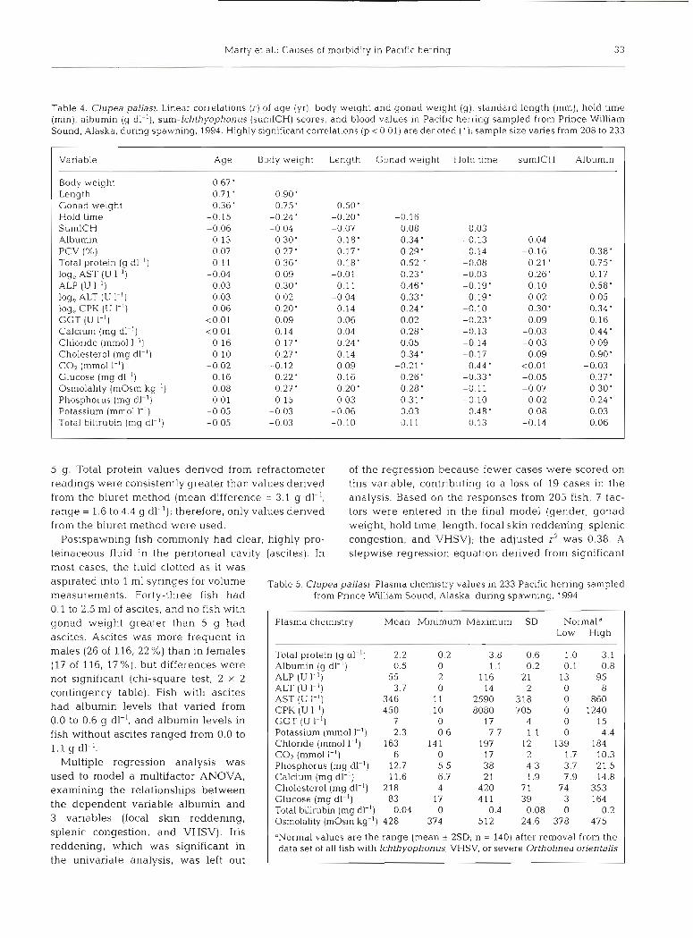
Table 4. Clupea pallasi. Linear correlations (r) of age (yr), body weight and gonad weight (g), standard length (mm), hold time (min), albumin (g dl-l), sum-lchthyophonus (sumICH) scores, and blood values in Pacific herring sampled from Prince William Sound, Alaska, during spawning, 1994. Highly significant correlations (p < 0.01) are denoted ( ' ) ; sample size varies from 208 to 233
Variable Age Body weight Length Gonad weight Hold time sumlCH Albumin
Body weight 0.67' Length 0.71' 0.90' Gonad weight 0.36' 0.75' 0.50' Hold time -0.15 -0.24' -0.20' -0.16 SumlCH -0.06 -0.04 -0.07 0.08 0.03 Albumin 0.13 0.30' 0.18' 0.34 ' -0.13 0.04 PCV (%) 0.07 0.27' 0.17' 0.29' -0.14 -0.16 0.38' Total protein (g dl-l) 0.11 0.36' 0.18' 0.52 ' -0.08 0.21 ' 0.75' log, AST (U I-') -0.04 0.09 -0.01 0.23 ' -0.03 0.26' 0.17 ALP (U I - ' ) 0.03 0.30' 0.11 0.46' -0.19' 0.10 0.58' log, ALT (U 1.') -0.03 0.02 -0.04 0.33' 0.19' 0.02 0.05 log, CPK (U I-') 0.06 0.20' 0.14 0.24 ' -0.10 0.30' 0.34 ' GGT (U I-') <0.01 0.09 0.06 0.02 -0.23' 0.09 0.16 Calcium (mg dl-l) <0.01 0.14 0.04 0.28' -0.13 -0.03 0.44 ' Chloride (mm01 1.') 0.16 0.17' 0.24' 0.05 -0.14 -0.03 0.09 Cholesterol (mg dl-l) 0.10 0.27' 0.14 0.34 ' -0.17 0.09 0.90' COz (mm01 I-') -0.02 -0.12 -0.09 -0.21' 0.44 ' <0.01 -0.03 Glucose (mg dl-l) 0.16 0.22' 0.16 0.26' -0.33' -0.05 0.37' Osmolality (mOsm kg-') 0.08 0.27 ' 0.20' 0.28' -0.11 -0.07 0.30' Phosphorus (mg dl-l) 0.01 0.15 0.03 0.31 ' -0.10 0.02 0.24 ' Potassium (mm01 1.') -0.05 -0.03 -0.06 0.03 0.48' 0.08 0.03 Total billrubin (mg dl-l) -0.05 -0.03 -0.10 0.11 0.13 -0.14 0.06
5 g. Total protein values derived from refractometer readings were consistently greater than values derived from the biuret method (mean difference = 3.1 g dl-', range = 1.6 to 4.4 g dl-'); therefore, only values derived from the biuret method were used.
Postspawning fish commonly had clear, highly pro- teinaceous fluid in the peritonea1 cavity (ascites). In
of the regression because fewer cases were scored on this variable, contributing to a loss of 19 cases in the analysis. Based on the responses from 205 fish, 7 fac- tors were entered in the final model (gender, gonad weight, hold time, length, focal skin reddening, splenic congestion, and VHSV); the adjusted r2 was 0.38. A stepwise regression equation derived from significant
most cases, the fluid clotted as it was into m1 Table 5. Clupea pallasi. Plasma chemistry values in 233 Pacific herring sampled
measurements. Forty-three fish had from Prince William Sound, Alaska, during spawning, 1994
0.1 to 2.5 m1 of ascites, and no fish with I I
gonad weight greater than 5 g had ascites. Ascites was more frequent in males (26 of 116, 22%) than in females ( l? of 116, l ? % ) , but differences were not significant (chi-square test, 2 X 2 contingency table). Fish with ascites had albumin levels that varied from 0.0 to 0.6 g dl-l, and albumin levels in fish without ascites ranged from 0.0 to l . l g dl-l.
Multiple regression analysis was used to model a multifactor ANOVA, examining the relationships between the dependent variable albumin and 3 variables (focal skin reddening, splenic congestion, and VHSV). Iris reddening, which was significant in the univariate analysis, was left out
Plasma chemistry Mean Minimum Maximum SD Normald Low High
Total protein (g dl-l) 2.2 0.2 3.8 0.6 1.0 3.1 Albumin (g dl-l) 0.5 0 1.1 0.2 0.1 0.8 ALP (U 1-l) 55 2 116 21 13 95 ALT (U 1-l) 3.7 0 14 2 0 8 AST (U I-') 346 11 2590 318 0 860 CPK (U 1-l) 450 10 8080 705 0 1240 GGT (U I-') 7 0 17 4 0 15 Potassium (mm01 1-l) 2.3 0.6 7.7 1.1 0 4.4 Chloride (mm01 I-') 163 14 1 1 97 12 139 184 CO2 (mm01 l-l) 6 0 17 2 1.7 10.3 Phosphorus (mg dl-l) 12.7 5.5 38 4.3 3.7 21.5 Calcium (mg dl-') 11.6 6.7 21 1.9 7.9 14.8 Cholesterol (mg dl-l) 218 4 420 71 74 353 Glucose (mg dl-l) 83 17 4 11 39 3 164 Total bfirubin (mg dl-l) 0.04 0 0.4 0.08 0 0.2 Osmolality (mOsm kg-') 428 374 512 24.6 378 475
"Normal values are the range (mean * 2SD; n = 140) after removal from the data set of all fish with Ichthyophonus, VHSV, or severe Ortholinea orientalis
34 Dis Aquat Org 32: 15-40, 1998
factors only was used to quantify the contribution of each variable to albumin levels (g dl-'). The constant (0.21 CJ dl-') is altered as follows:
gender male =
gender female =
gonad weight (g) = VHSV-negative =
VHSV-positive =
focal skin reddening, none = focal skin reddening, mild = focal skin reddening,
moderate/severe = splenic congestion, none = splenic congestion, mild = splenic congestion,
moderatekevere =
+0.114 +0.000 + 0.0045 X (gonad wt) +0.047 -0.047 + 0.098 -0.006
For example, a male (+0.114) with a gonad weight of 10 g (+0.045) that was VHSV negative (+0.047) and had no focal skin reddening (+0.098) and mild splenic congestion (-0.008) would be expected to have a plasma albumin level of 0.51 g dl-l. The pre- dicted plasma albumin level in a similar male with moderate focal skin reddening would decrease to 0.30 g dl-l.
Like albumin, scores for several lesions and other variables could be differentiated on the basis of PCV, and PCV was significantly associated with several plasma chemistries (Tables 1 & 4). Multiple regression analysis was used to model a multifactor ANOVA, examining the relationships between PCV and 7 vari- ables. Based on the results from 186 fish, 12 factors were entered in the final model (gender, gonad weight, hold time, length, osmolality, focal skin red- dening, splenic Ichthyophonus, renal hematopoietic cells, hepatic lipidosis, cardiac thrombosis, gastric trematodes, and VHSV); the adjusted r2 was 0.29.
Because of the potential that dehydration could effect PCV, osmolality was added as a controlling variable. A steptvise regression equation derived from significant factors only was used to quantify the contribution of each variable to PCV ( X ) . The constant (51.14 %) is altered as follows:
gender male = +2.30 gender female = + 0.00 gonad weight (g)= +0.1513x (gonad wt) osmolality (mOsm/kg) = -0.0433 X (osmolality) hepatic lipidosis, none = + 1.49 hepatic lipidosis, mild = -0.7 1 hepatic lipidosis,
moderatekevere = -0.77 splenir Ichthyophonus, none = + 1.50 splenic Ichth yophonus, mild = -2.43 splenic Ichthyophonus,
moderatehevere =
renal hematopoietic cells, none =
renal hematopoietic cells, mild =
renal hematopoietic cells, moderate =
gastric trematodes, none = +1.87 gastric trematodes,
-1.87 mild/moderate =
For example, a male (+2.30) with a gonad weight of 10 g (+1.51), osmolality of 425 mOsm kg-' (-18.40), no hepatic lipidosis (+1.49), no splenic Ichthyophonus (+ 1.50), mild renal hematopoietic cells (+ 1.63), and mild gastric trematodes (- 1.87) would be expected to have a PCV of 39.3%. By comparison, a similar male with no renal hematopoietic cells and mild splenic Ichthyophonus would have a predicted PCV of 31.5%.
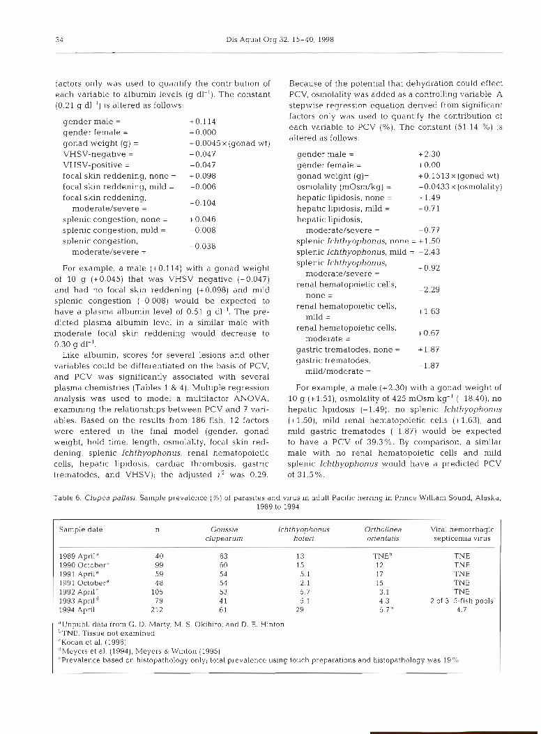
Table 6. Clupea pallasi. Sample prevalence (%) of parasites and virus in adult Pacific herring in Prince William Sound, Alaska, 1989 to 1994
Sample date n Goussia Ichthyophonus Ortholinea Vlral hemorrhagic clupearurn hoferi orientalis septicemia virus
1989 April " 40 63 13 TNE" TNE 1990 Octoberd 99 60 15 12 TNE 1991 Aprilo 59 54 5.1 17 TNE 1991 Octoberd 4 8 54 2.1 15 TNE 1992 Aprilr 105 53 5.7 3.1 TNE 1993 ~ p r i l ~ 79 4 1 5.1 4.3 2 of 3 5-fish pools 1994 April 212 6 1 29 5.7" 4.7
dUnpubl, data from G. D. Marty, M. S Okihiro, and D. E Hinton 'TNE: Tissue not examined 'Kocan et al. (1996) "eyers et al. (1994), Meyers & Winton (1995) 'Prevalence based on histopathology only; total prevalence using touch preparations and histopathology was 19%
h4arty e t a1 . Causes of morbidity in Pac~f ic herrlng 3 5
Annual trends in spawning biomass and pathogen prevalence
Sample prevalence of Ichthyophonus in this study was almost twice that of previous years (Table 6 ) . Dur- ing the damage assessment phase of study from 1989 through 1992, and disease studies in 1993 (Meyers et al. 1994), prevalence of Ichthyophonus in Pacific her- ring sampled from PWS was never more than 1570. By comparison, prevalence of Goussia clupearum has remained fairly constant between 41 and 63 %, and Ortholinea orientalis prevalence has not exceeded 17%. The slight increase in Ortholinea orientalis prevalence in this study (19%) was probably a t least partly due to increased efficiency of diagnosis when touch preparations were examined; previous preva- lence data were derived from histopathology only. Prevalence of VHSV and other parasites was not deter- mined in previous studies because appropriate tissues were not examined.
DISCUSSION
VHSV
The North American strain of VHSV was a major cause of morbidity in Pacific herring in PWS during spawning in 1994. Fish from which VHSV was isolated had significant gross lesions as well as microscopic lesions in the gills, liver, stomach, arteries, and heart. Most lesions were consistent with a disseminated endotheliotrophic virus, and lesions such as coagula- tive necrosis in the liver have been attributed to VHSV in natural and laboratory infections in rainbow trout (Amlacher et al. 1980, Wolf 1988b). Because the VHSV outbreak was nearly over in 1994, opportunities to con- firm association of lesions with VHSV by further field study have been limited. However, recent study with Pacific herring fulfilled Koch's postulates, demonstrat- ing that VHSV kills laboratory-reared Pacific herring in absence of other pathogens (Kocan et al. 1997).
Although the North American strain of VHSV has been isolated from several populations of Pacific her- ring (Meyers & Winton 1995), the only other published report of VHSV linked to population decline was from fish sampled in PWS in 1993 (Meyers e t al. 1994). Meyers et al. (1994) postulated that several lesions were associated with VHSV: subdermal and renal hemorrhages, kidney tubule degeneration, and active reticuloendothelial cell foci in the kidneys. Also, active reticuloendothelial cell foci in the liver were associated with hepatocellular necrosis. In the present study, we confirmed a n association of VHSV with fin base red- dening and focal coagulative hepatic necrosis, and we
had some evidence for association of VHSV with ulcers (i.e. severe focal skin reddening). Association of VHSV with renal hemorrha.ge or kidney tubule degeneration could not be confirmed. In the present study, 'active reticuloendothelial cells' were classified as either p ~ g - mented macrophage aggregates or granulomatous in- flammation, and neither was significantly related to VHSV in the liver or kidney. However, infiltrates of lymphocytes or macrophages in the gastric submu- cosa, gill arches, and brain were significantly associ- ated with VHSV infection. In a study of PWS Paciflc herring from 1992, granulomatous inflammation was associated with decreased reproductive success (Kocan et al. 1996), but based on our results w e cannot attribute these lesions to VHSV.
Population fluctuations in Pacific herring a re consid- ered normal by management biologists, but in only one other case was population decline attributed to dis- ease. During February and March of 1942, 'several thousands of tons' of Pacific herring were found dead along the southeast coast of Vancouver Island, British Columbia, Canada (Tester 1942). 'The dying fish came to the surface and could, while still alive, be picked up by gulls or by hand.' Mortality involved pre- and post- spawners, and fish continued to be lethargic and school in shallow water near shore until mid-May (Tester 1942). Diagnostic examination included gross necropsy, bacteriology, blood smears, and parasite screen, but no significant pathogens were found. Based on this level of diagnostic detail, Ichthyophonus can be ruled out a s the cause of mortality in 1942, but many features were similar to the 1993 epizootic in PWS. Both outbreaks had lethargic fish, some of which had reddening of the fins, and both outbreaks followed a year in which commercial harvest was above aver- age. The epizootic near Vancouver Island involved a dominant 1938 year class (4-yr-olds), whereas the PWS epizootic involved a dominant 1988 year class (5-yr- olds). As a difference, the Vancouver Island outbreak had large numbers of dead fish, whereas dead fish were not reported in the PWS epizootic. One other dis- ease, VEN, has been reported to cause significant mor- tality in juvenile Pacific herring when such year classes are strong. However, VEN has not been associated with significant decline in population biomass (Meyers et al. 1986), and PWS fish in 1994 had no evidence of VEN.
Several questions about the pathogenesis of VHSV in Pacific herring a re beginning to be answered with continued field study and focused laboratory study. VHSV is highly infectious, spreads through the water, and readily kills disease-free Pacific herring independent of exposure to other pathogens (Kocan et al. 1997). Preliminary field and laboratory studies indicate that 10 to 15% of Pacific herring have sub-
36 Dis Aquat Org 32: 15-40, 1998
clinical infections but express VHSV and the associ- ated disease only when subjected to stress (R. M. Kocan pers. comm., G . D. Marty unpubl obs.). A related virus, infectious hematopoietic necrosis virus (IHNV, also in the family Rhabdoviridae), is com- monly carried by salmonids. Disease from IHNV is generally a serious problem only in juvenile fish, and virus is expressed in surviving adults during and after spawning (Wolf 1988a). In a study of Pacific herring at the National Marine Fisheries Service Lab- oratory in Auke Bay, Alaska, VHSV was expressed in a dose-dependent manner after 17 d of exposure to weathered crude oil (Mark Carls pers. comm., Meyers & Winton 1995). Although the VHSV status of these fish before the study began was unknown, the study provided evidence that oil can act as a stressor that activates VHSV. Several other questions are under investigation. Once VHSV is expressed, can fish mount a successful immune response and overcome the disease? Does virus expression cycle seasonally? What environmental factors contribute to disease and immunity?
Ichthyophonus hoferi
Ichthyophonus has not previously been described as a major cause of mortality in Pacific herring, but in Atlantic herring several epizootics of Ichthyophonus have been linked to population decline (Fish 1934, Sindermann 1958, Patterson 1996). Indeed, Ichthyo- phonus hoferiis the most commonly reported and most severe marine fungal pathogen, and 'this disease may be the most important single limiting factor to popula- tion growth of herring in the western North Atlantic' (Sindermann 1970). Although recent evidence indi- cates that Ichthyophonus is not a fungus (Spanggaard et al. 1996), its biological significance remains un- changed. Outbreaks in Atlantic herring tend to begin during biomass peaks, usually lasting 2 to 3 yr, and recovery often takes more than 3 yr (Sindermann 1970, Mellergaard & Spanggaard 1997). In Pacific herring in PWS, peak biomass in 1989 did not result in a major Ichthyophonus-related population decline, but severe population decline in 1993 was followed by a sharp increase in Ichthyophonus prevalence in 1994. Previ- ous declines in Pacific herring biomass have been recorded in PWS, but these were attributed to poor year-class recruitment and over-fishing (Rounsefell &
Dahlgren 1932). In Atlantic herring in the Gulf of Maine, Ichthyophonus was considered the cause of population declines in 1931 and 1947, and anecdotal evidence was strong for Ichthyophonus as the major cause of population declines in 1898 and 1916 (Fish 1934, Sindermann 1965). From 1898 to 1947, outbreaks
occurred about every 16 yr and this trend held for 4 cycles; however, no Ichthyophonus outbreaks have been documented in the Gulf of Maine since 1947. Sporadic but significant Ichthyophonus outbreaks have also been described in the Gulf of St. Lawrence (Sindermann 1970). In Europe, Ichthyophonus was not associated with Atlantic herring population decline until a 1991 epizootic in the North Sea (Patterson 1996, Rahimian & Thulin 1996, Mellergaard & Spanggaard 1997).
Some features of Ichthyophonus infection in PWS Pacific herring were different from those described in wild Atlantic herring. For example, Atlantic herring with severe infections often had gross lesions in the muscle described as 'rough or granulomatous skin' or 'sandpaper effect' (Post 1987); associated ulcers have been termed 'pepper effect', partly as a result of pig- ment deposition in the lesions (Fish 1934). By compari- son, Pacific herring had no gross external lesions directly associated with Ichthyophonus, and micro- scopic lesions in the skin and skeletal muscles were usually mild. Further, Pacific herring had no pigment associated with Ichthyophonus resting spores. Another difference was that epizootics in North American Atlantic herring were always characterized by large numbers of moribund and dead fish in shallow areas (Fish 1934, Sindermann 1958), whereas there were no confirmed reports of dead fish in PWS. Not all features of Ichthyophonus were different in Atlantic and Pacific herring; multifocal to coalescing granulomas in inter- nal organs of PWS Pacific herring were similar to the descriptions of gross and histologic lesions reported in Atlantic herring.
The epizootiology of Ichthyophonus infection in Pacific herring in PWS is still unclear with only 7 samples in 6 years from 1989 through 1994. Many questions remain unanswered: (1) What is the latency period between Ichthyophonus exposure and overt signs of disease? (2) When Ichthyophonus is diag- nosed histologically, how long will the affected fish live? (3) Can a fish, once infected, initiate a success- ful immune response and overcome the disease, or are all infected fish destined to die? and (4) Because Ichthyophonus prevalence was only 5 % in 1993 despite significant population decline (Meyers et al. 1994), how important is lchthyophonus as a cause of Pacific herring mortality? The large spike in Ichthyo- phonus prevalence in this study (29%) was unex- pected, but was consistent with infection levels of about 25 % described in epizootics affecting Atlantic herring (Sindermann 1970). Sindermann (1970) stated that enzootic levels were about l % , lower than any samples from Pacific herring in PWS, but Sindermann's observations were based only on gross examination to determine prevalence. Method of
hdarty et al.. Causes of morbidity in Pacific herring 37
diagnosis can make a significant difference in the number of positive cases identified (Holst 1994, Rahimian & Thulin 1996), and in our study, results from gross examination underestimated the number of infected fish.
In Pacific herring from our study, CPK and AST values could be used to differentiate Ichthyophonus lesion scores in nearly every organ. Creatine phospho- kinase is a dimeric enzyme with isoenzyme types CK, (BB, brain), CK2 (MB, heart), and CK3 (MM, skeletal muscle). In mammals, the brain form of CPK is not found in plasma, even during neurologic disease (Dun- can & Prasse 1986); therefore, the finding that brain Ichthyophonus status was the best predictor for in- creased CPK in our study was unexpected. Brain Ichthyophonus was uncommon, but the high mean sumICH score for fish with brain Ichthyophonus (Fig. 6) provided evidence that Ichthyophonus was disseminated when it appeared in the brain. That is, if Ichthyophonus was disseminated sufficiently that it affected the brain, then the fish probably also had muscle I ~ h t h y o p h o ~ u s severe enough to increase CPK. Further, 79% of muscle Ichthyophonus cases were mild, and the damage caused by these muscle lesions was probably not sufficient to increase CPK. Alterna- tively, the brain form of CPK might be released during neurologic disease in Pacific herring. Isoenzyme analy- sis on plasma from Ichthyophonus-positive Pacific herring has not been successful (C. Kennedy, Simon Fraser University, Burnaby, British Columbia, Canada, pers comm.).
For AST in mammals, lesions in liver, muscle, and blood-rich organs are most highly associated with in- creased enzyme levels (Duncan & Prasse 1986). For Pacific herring, renal Ichthyophonus status was signif- icant in all regressions, but spleen and heart were not. Most likely, the disseminated nature of Ichthyophonus infections prevented localization of the source of AST in infected fish.
The effects of Ichthyophonus infection on plasma chemistries have not previously been described in natural epizootics. In laboratory-exposed rainbow trout Oncorhynchus mykiss, Ichthyophonus infection was associated with anemia and leukopenia, but did not change plasma chloride, creatinine, glucose, osmo- larity, potassium, total protein, sodium, or T4 (Rand & Cone 1990); enzymes CPK and AST were not mea- sured. In addition to increased CPK and AST in this study, Ichthyophonus infection was significantly asso- ciated with anemia and variable plasma protein levels; white blood cells were not counted. Based on the equa- tion derived from multifactor analysis, a fish with mild splenic Ichthyophonus would be predicted to have a PCV that was 4 % less than a similar fish with no splenic Ichthyophonus.
Other potential pathogens
A few comprehensive reports are available on the prevalence of parasites in Pacific herring, and their potential role in stock identification (Arthur & Arai 1980, Moser & Hsieh 1992). The purpose of our study was not to repeat these studies, but to determine which of the common parasites of Pacific herring in PWS could potentially contribute to population decline. More than 30 species of parasites have been described from Pacific herring (Arthur & Arai 1980). In our study, 10 parasites occurred in prevalences sufficient to study their role in disease and population decline. Two criteria were used to determine if a parasite caused significant damage to the host: (1) Was the parasite associated with histopathologic damage, particularly inflammation? and (2) Was infection with the parasite associated with alterations in plasma chemistries? Using these criteria, linkage of damage to infections by parasites other than Ichthyophonus was not clear.
The intraductal renal myxosporean Ortholinea ori- entalis was not associated with morphologic lesions, nor was there metastatic calcification, but fish with large numbers of organisms had elevated plasma cal- cium. Because the kidney is one organ that excretes calcium (Dacke 1979), large numbers of organisms might have impaired calcium excretion. The relation of intraductal parasites and calcium levels has not previ- ously been described, and this effect would need to be confirmed by controlled laboratory study.
Lymphocystis virus has been identified in fibroblasts of over 150 species of fish, including Atlantic herring (listed by Lawler et al. 1977), but this is the first de- scription of lymphocystis virus in Pacific herring. Also. in most reported cases, lymphocystis lesions are limited to the skin (Post 1987). Our finding of lympho- cystis lesions limited to solitary nodules within the peritonea1 cavity is unusual.
Gender- and age-associated lesions
Lower plasma albumin levels in females than in males could partly be explained by vitellogenin syn- thesis in females. In the hepatocyte, estradiol activates the vitellogenin gene, but production of albumin is de- pressed (Mommsen & Walsh 1988). Low plasma albu- min is commonly associated with ascites in mammals, and Pacific herring with ascites tended to have lower albumin levels than fish without ascites; however, females were not more Likely to develop ascites than were males. Several other plasma chemistries and lesion scores had significant gender differences, but little information is available to explain these differ- ences in Pacific herring.
38 Dis Aquat Org 32: 15-40, 1998
We could critically .evaluate only one age-related hypothesis regarding the link between the oil spill and disease in 1994. Fish that were hatched or were yearlings in 1989 at the time of the spill (1988 and 1989 year classes) might have incurred irreversible immunosup- pression. Under normal growth conditions, minor defi- ciencies in their immune system might have been in- significant. However, disease might have become a serious problem when fish experienced additional stress upon first spawning (1992 and 1993). Stress is well- documented as a cause of immunosuppression, but stress-induced changes usually are reversible if the fish survives (Anderson 1990). We found that several changes were significantly associated with age, but scores for nearly all these changes were greater in older fish (i.e. fish hatched before 1988). Also, among VHSV, Ichthyophonus, and 10 other common parasites, none were more prevalent in the 1988 and 1989 year classes than in the entire sampled population. Annual age- weight-length analysis by the Alaska Department of Fish and Game has documented that the population de- creased in the absence of abnormal changes in age dis- tribution (Fritz Funk unpubl. data). Therefore, the weight of evidence suggests that the disease outbreak in PWS was not a result of permanent immune suppression caused by hydrocarbon exposure when fish were larvae or yearlings. A companion study in Prince William Sound reached similar conclusions (Elston et al. 1997).
Alterations in plasma chemistries
Analysis of plasma chemistry values was inexpen- sive and provided useful information for evaluating health of Pacific herring in PWS. However, interpreta- tion of results was limited by lack of reference values. In the only published study of normal plasma enzyme values in Pacific herring (Marquez 1976), analysis of electrolytes and other nonenzyme chemistries was not included. Marquez (1976) captured 5 to 12 Pacific her- ring by angling, held the fish for 12 h, and then drew blood to analyze for plasma enzymes at 30°C. His mean values for CPK (2948 U 1-l) and AST (1778 U 1-l) were more than twice the maximum values of normal ranges established in our study (Table 5). Differences between the 2 studies probably resulted from Marquez performing analyses at 30°C instead of the 25°C of our
. study. Also, the 12 h hold time might have been long enough so that increased enzyme levels reflected dam- age that occurred during capture. In our study, hold time of less than <4 h was not significantly correlated with plasma CPK or AST. Reference values from Pacific herring populations in peak condition (e.g. late summer) are needed to better interpret changes asso- ciated with spawning.
Interesting findings in plasma chemistry values in- cluded unusually low albumin levels and unusually high osmolality. Altered plasma chemistry valucs have been associated with spawning in other fish species, but abnormalities were transient (McDonald & Milli- gan 2992). Because albumin levels in Pacific herring were significantly decreased only at the end of spawn- ing, and ascites occurred most often in spawned out fish, development of ascites was probably related to physiologic changes at the end of spawning.
Total plasma protein values determined using a refractometer were higher than values determined by colorimetry. Similar differences have been docu- mented in other fish species (Hunn & Greer 1990, Hunn et al. 1992), but the molecular cause for this dif- ference has not been determined. Subsequent analysis of total plasma protein in Pacific herring has used only the colorimetry technique.
Plasma glucose, COz, and potassium were useful markers of the effects of hold time between capture and necropsy. The increase in plasma CO2 was indica- tive of respiratory acidosis, and potassium levels are expected to increase during acidemia (McDonald &
Milligan 1992). Decreased glucose levels may have been associated with increased anaerobic glycolysis, but lactate levels were not determined. Normally, cap- ture stress results in hyperglycemia (Hopkins & Cech 1992), but Pacific herring hepatocytes had histologic evidence of minimal glycogen, thereby limiting the ability of the liver to increase plasma glucose levels in response to stress. To determine the relation of hold time to metabolic acidosis and other plasma chemistry values, plasma lactate levels have been analyzed in continuing Pacific herring disease studies (G. D. Marty unpubl. data).
Implications for studies of disease epizootics in fish populations
Disease epizootics have been associated with declin- ing populations of several marine fish species during the past century. Most notably, early work identified Ichthyophonus as the major cause of population decline in Atlantic herring in the Northwest Atlantic (Daniel 1933a, Fish 1934, Sindermann 1970). When Ichthyophonus prevalence was high in North Sea Atlantic herring in 1991, Ichthyophonus was assumed to be the primary cause of population decline. An extensive, multiyear study focused on Ichthyophonus to determine the effects of season, fish age, and gear type on sample prevalence (Holst 1996, Patterson 1996, Rahimian & Thulin 1996, Mellergaard & Spang- gaard 1997). Comparatively little effort was expended to determine if other pathogens, particularly viruses,
Marty et al.: Causes of morbidity in Pacific herring 39
were contributing to the epizootic. The recent isolation of the European strain of VHSV from Atlantic herring (Dixon et al. 1997) introduces a new hypothesis into the interpretation of the Ichthyophonus findings. Was Ich- thyophonus the primary cause of the epizootic de- scribed in Atlantic herring? Or, did an increase in Ich- thyophonus prevalence follow an outbreak of VHSV that went undetected?
In other epizootics, a broader range of diagnostic techniques was used to identify the source of the epi- zootic. For dying striped bass Morone saxatilis sam- pled from an estuary, histopathology was combined with plasma chemistry and toxicant analysis, but virus isolation was not attempted and the cause of death was not determined (Young et al. 1994). For dying pilchard Sardinops sagax, a herpesvirus was consistently iden- tified on histological and ultrastructural analysis of the gills of sick fish (Hyatt et al. 1997, Whittington et al. 1997), but the virus could not be cultured in vitro (study was limited by lack of pilchard cell lines for virus isolation). Underlying causes of these epizootics and the Pacific herring epizootic are not fully under- stood (Meyers & Winton 1995). However, through com- prehensive pathological examination, combined with focused laboratory study, we have shown that signifi- cant pathogens and risk factors can be identified and many variables can be ruled out as significant causes of population decline. Study of disease in PWS Pacific herring has been expanded to include a reference site and semiannual study through spring of 1998. De- creasing prevalence of VHSV and Ichthyophonus has been accompanied by an increasing fish population. Future papers will detail the results of these studies.
Acknowledgements. We thank J Vansant, D. Branshaw, D. C. Phipps, and C. T. Stack for finding and capturing the fish, and W. Widmann for sectioning tissues. Corrine R. Davis per- formed half of the necropsies and all gonad h~stopathology. Adam Moles helped identify the parasites, and Joseph R. Sul- livan and Barbara S. Washburn reviewed the project design. The research described in this paper was supported by the 'Exxon Valdez' Oil Spill Trustee Council through contracts with the Alaska Department of Flsh and Game. However, the findings and conclusions presented by the authors are their own and do not necessarily reflect the views or position of the Trustee Council.
LITERATURE CITED
Amlacher E, Ude J , Rudolph C, Ernst G (1980) Direct electron microscopical visualization of the presumptive virus of viral haemorrhayic septicaemia (VHS) in the rainbow trout Salmo gairdneri kchardson and additional histopathological and haematological observations. J Fish Dis 355-62
Anderson DP (1990) Immunological indicators: effects of envi- ronmental stress on immune protection and disease out- breaks. Am Fish Soc Symp 8:38-50
Arthur JR. Arai HP (1980) Studies on the parasites of Pacific herring (Clupea harengus pallasi Valenciennes): survey results. Can J Zoo1 58:64-70
Dacke CG (1979) Calcium regulation in sub-mammalian vertebrates. Academic Press, New York
Daniel GE (1933a) Studies on Ichthyophonus hoferi, a para- sitlc fungus of the herring, Clupea harengus. I. The para- site as it is found in the herring. Am J Hyg, Baltimore 17:262-276
Danlel GE (1933b) Studies on Ichthyophonus hofen', a para- sitic fungus of the herring, Clupea harengus. 11. The gross and microscopic lesions produced by the parasite. Am J Hyg, Baltimore 1?:491-501
Dixon PF, Feist S, Kehoe E, Parry L, Stone DM, Way K (1997) Isolation of viral haemorrhagic septicaemia virus from Atlantic herring Clupea harengus from the English Chan- nel. Dis Aquat Org 30231-89
Duncan JR, Prasse KW (1986) Veterinary laboratory medicine. Iowa State University Press, Ames, IA
Elston RA, Drum AS, Pearson NTH, Parker K (1997) Health and condition of Pacific herring, Clupea pallasi from Prince William Sound, Alaska, 1994. Dis Aquat Org 31 109-126
Fish FF (1934) A fungus disease of fishes of the Gulf of Maine Parasitol 26:l-16
Hauck AK, May EB (1977) Histopathologic alterations associ- ated with Anisakis larvae in Pacific herring from Oregon. J Wildl Dis 13:290-293
Holst JC (1994) The precision of two macroscopic screening procedures relatlve to a microscopic procedure for screen- ing of the fungus Ichthyophonus hoferi in hernng (Clupea harengus L.). Fish Res (Amst) 19:187-190
Holst J C (1996) Estimating the prevalence of Ichthyophonus hoferi (Plehn and Mulsow) in a herring stock (Clupea harengus L.): observed effects of sampling gear, target school density and migration Fish Res 28:85-97
Hopkins TE, Cech J J J r (1992) Physiological effects of captur- ing striped bass in gill nets and fyke traps. Trans Am Fish SOC 121:819-822
Hunn JB, Greer IE (1990) Colorirnetric and refractometer estimates of total plasma protein in striped bass, Morone saxatilis (Walbaum). J Fish Biol36:617-618
Hunn JB, Mriedmeyer RH, Greer IE, Grady AW (1992) Blood chemistry of laboratory-reared golden trout. J Aquat Anim Health 4:218-222
Hyatt AD, Hine PM, Jones JB, Whittington RJ, Kearns C, Wise TG, Crane MS, Willlams LM (1997) Epizootlc mortality in the pilchard Sardinops sagax neopilchardus in Australia and New Zealand in 1995. 11. Identification of a herpes- virus within the gill epithelium. Dis Aquat Org 28~17-29
Jastrzebski M, Komorowski Z (1990) Light and electron microscopic studies on Goussia zarnowskii (Jastrzebskl. 1982): an intestinal coccidium parasitizing the three- spined stickleback, Gasterosteus aculeatus. J Fish Dis 13: 1-24
Kocan R, Bradley M, Elder N, Meyers TK, Batts W, Winton J (1997) The North American strain of viral hemorrhqgic septicemia virus is highly pathogenic for laboratory reared Pacific herring (Clupea pallasi). J Aquat Anim Health 9: 239-290
Kocan RM, Marty GD, Okihiro MS, Brown ED, Baker TT (1996) Reproductive success and histopathology of indi- vidual Prince Wllliam Sound herring 3 years after the 'Exxon Valdez' oil spill. Can J Fish Aquat Sci 53:2388-2393
Lawler AR, Ogle JT, Donnes C (1977) Dascyllus spp: new hosts for lymphocystis, and a list of recent hosts. J Wildl Dis 133:307-311
Dis Aquat Org 32: 15-40, 1998
Marquez ED (1976) A comparison of glutamic-oxalacetate transaminase, lactate dehydrogenase and creatine phos- phokinase activities in non-spawning, pre-spawning, and spawning pink salmon. Comp Biochem Physiol 54B: 121-123
McDonald DG, Milligan CL (1992) Chemical properties of the blood. In: Hoar WS, Randall DJ, Farrell AP (eds) Fish phys- iology, Vol XIIb, The cardiovascular system. Academic Press, San Diego, CA, p 55-133
McKim JM (1985) Early life stage toxicity tests. In: Rand GM, Petrocelli SR (eds) Fundamentals of aquatic toxicology. Hemisphere, Washington, DC, p 58-95
Mellergaard S. Spanggaard B (1997) An Ichthyophonus hofen epizootic in herring in the North Sea, the Skagerrak, the Kattegat and the Baltic Sea. Dis Aquat Org 28:191-199
Meyers TR, Hauck AK, Blankenbeckler WD, Minicucci T (1986) First report of viral erythrocytic necrosis in Alaska, USA, associated with epizootic mortality in Pacific her- nng, Clupea harangrrs pa.llasi (Valenciennes). J Fish Dis 9:479-491
~Meyers TR, Short S, Eaton W (1990) Summer mortalities and incidental parasitisms of cultured Pacific oysters in Alaska. J Aquat Anim Health 2:172-176
Meyers TR, Short S, Lipson K, Batts WN, Winton JR, Wilcock J , Brown E (1994) Association of viral hemorrhagic sep- ticemia virus with epizootic hemorrhages of the skin in Pacific herring Clupea harengus pallasi from Prince William Sound and Kodiak Island, Alaska, USA. Dis Aquat Org 19:27-37
Meyers TR, Winton JR (1995) Viral hemorrhagic septicemia virus in North America. Annu Rev Fish Dis 5:3-24
Mommsen TP, Walsh PJ (1988) Vitellogenesis and oocyte assembly. In: Hoar WS, Randall DJ (eds) Fish physiology, Vol XIa, The physiology of developing fish. Academic Press, San Diego, CA, p 347-406
Morrison CM, Hawkins WE (1984) Coccidians in the liver and testis of the herring Clupea harengus L. Can J Zoo1 62: 480-493
Morrison CM, McClelland G, Cornick J , Marcogliese D (1986) Parasites and disease of some marine finfish of Nova Scotia. Can Tech Rep Fish Aquat Sci 1424:l-36
Moser M. Hsieh J (1992) Biological tags for stock separation in Pacific herring Clupea harengus pallasi in California. J Parasitol 78:54-60
Patterson KR (1996) Modelling the impact of disease-induced mortahty in an exploited population: the outbreak of the fun- gal parasite Ichthyophonus hoferiin the North Sea herring (Clupea harengus). Can J Fish Aquat Sci 53:2870-2887
Editorial responsibility: Jo-Ann Leong, Corvallis, Oregon, USA
Post G (1987) Textbook of fish health. TFH Publications, Nep- tune City. NJ
Rahimian H, Thulin J (1996) Epizootiology of Ichthyophonus hofen in hernng populations off the Swedish west coast. Dis Aquat Org 27:187-195
Rand TG, Cone DG (1990) Effects of Ichthyophonus hoferi on condition indices and blood chemistry of experimentally infected rainbow trout (Oncorhynchusmykiss). J Wildl Dis 26:323-328
Rounsefell GA, Dahlgren EH (1932) Fluctuations in the sup- ply of herring, Clupea pallasii, in Prince William Sound, Alaska. Bull US Bur Fish No 9 47:263-291
Short JW, Harris PM (1996) Petroleum hydrocarbons in caged mussels deployed in Prince William Sound after the 'Exxon Valdez' oil spill. Am Fish Soc Symp 18:29-39
Sindermann CJ (1958) An epizootic in Gulf of St. Lawrence fishes. Trans N Am Wildl Conf 23:349-360
Sindermann CJ (1965) Effects of environment on several dis- eases of herr'lng from the western North Atl~intic. Spec Pub1 Intern Comm Northw At1 Fish 6:603-610
Sindermann CJ (1970) Principal diseases of marine fish and shellfish. Academlc Press, New York
Spanggaard B, Skouboe P, Rossen L, Taylor JW (1996) Phylo- genet~c relationships of the intercellular fish pathogen lchthyophonus hofen and fungi, choanoflagellates and the rosette agent. Mar Biol 126:109-115
Tester AL (1942) Herring mortality along the south-east coast of Vancouver Island. Fish Res Board Can, Prog Rep Pac Coast Stn 52:ll-15
Whittington RJ, Jones JB, Hine PM, Hyatt AD (1997) Epi- zootic mortality in the pilchard Sardinops sagax neo- pilchardus In Australia and New Zealand in 1995. I. Pathology and epizootiology. Dis Aquat Org 28: 1- 16
Willard MD, Tvedten H, Turnwald GH (1989) Small animal clinical diagnosis by laboratory methods. WB Saunders Company. Philadelphia, PA
Wolf K (1988a) Infectious hematopoietic necrosis. In: Wolf K (ed) Fish viruses and fish viral diseases. Cornell University Press, Ithaca, NY, p 83-114
Wolf K (1988b) Viral hemorrhagic septicemia. In: Wolf K (ed) Fish viruses and fish viral diseases. Cornell University Press, Ithaca, NY, p 217-249
Young G, Brown CL, Nishioka RS, Folmar LC, Andrews M, Cashman JR, Bern HA (1994) Histopathology, blood chemistry, and physiological status of normal and mori- bund striped bass (Morone saxatilis) involved in summer mortality ('die-off') in the Sacramento-San Joaquin delta of California. J Fish Biol44:491-512
Submitted: August 14, 1997; Accepted: October 31, 1997 Proofs received from author(s): February 12, 1998