vaccine -pig 2007
TRANSCRIPT

A
sbttorvwdvnt©
K
1
epSosdmt
0d
Available online at www.sciencedirect.com
Vaccine 25 (2007) 7806–7817
Reduction of foot-and-mouth disease (FMD) virus load in nasalexcretions, saliva and exhaled air of vaccinated pigs
following direct contact challenge
S. Parida ∗, L. Fleming, Y. Oh, M. Mahapatra, P. Hamblin, J. Gloster,C. Doel, S. Gubbins, D.J. Paton
Institute for Animal Health, Pirbright Laboratory, Ash Road, Pirbright, Surrey GU24 0NF, United Kingdom
Received 6 July 2007; received in revised form 12 August 2007; accepted 19 August 2007Available online 17 September 2007
bstract
In future, a policy of “vaccinate-to-live” may be included in the repertoire of foot-and-mouth disease (FMD) control measures and inupport of this approach, we have investigated the hypothesis that vaccine-induced reduction in virus replication and excretion from pigs cane correlated to the severity of clinical signs of FMD by measuring excretion of virus in natural secretions and aerosols. The other aims ofhis study were to verify the existence of sub-clinical infection in vaccinated pigs, to evaluate the correlation between this and seroconversiono foot-and-mouth disease virus (FMDV) non-structural protein antibodies and to re-examine the occurrence of FMDV persistence in thero-pharynx of pigs. Therefore, pigs were vaccinated (O1 Manisa) and challenged (O1 UKG) in a manner calculated to produce a broadange of clinical outcomes and were monitored for a minimum of another 33 days post-challenge. Eighty-one percent of the early (10 daysaccinated) challenged pigs and 25% of the late (29 days vaccinated) challenged pigs were clinically infected and all other vaccinated pigsere sub-clinically infected. Although vaccination could not provide complete clinical or virological protection, it reduced the severity of theisease, virus excretion and production of non-structural FMDV antibodies in vaccinated and subsequently infected pigs. As hypothesised,
accine-induced reduction of virus replication and excretion was found to be correlated to the severity of clinical disease. RNA copies, buto live virus was detected from the pharyngeal and soft palate tissues of a minority of vaccinated and infected pigs beyond the acute stage ofhe infection.2007 Elsevier Ltd. All rights reserved.
act cha
rsp
roth
eywords: FMD virus excretion; FMD emergency vaccination; Direct cont
. Introduction
Foot-and-mouth disease (FMD) is a highly contagious dis-ase caused by FMD virus (FMDV), an apthovirus within theicornavirus family that infects all cloven hoofed animals [1].usceptible livestock may be infected with FMDV by directr indirect contact with infected animals. When infected andusceptible animals are in close proximity, aerial transfer of
roplets and droplet nuclei is probably the most commonode of transmission [2]. Though pigs are less susceptibleo FMDV infection by the airborne route when compared to
∗ Corresponding author. Tel.: +44 1483 232441; fax: +44 1483 232448.E-mail address: [email protected] (S. Parida).
v[tvud
264-410X/$ – see front matter © 2007 Elsevier Ltd. All rights reserved.oi:10.1016/j.vaccine.2007.08.058
llenge
uminants, they excrete more airborne FMD virus [2,3]. Con-equently, a common pattern of airborne FMD spread is fromigs to cattle, sheep and goats downwind [4,5].
Since the 2001 FMD outbreak in the UK, there has beenenewed interest in Europe to use vaccination as a meansf reducing reliance on culling of animals. Consequently,he new European council Directive 2003/85/EC on FMDas made provision for vaccination and the use of post-accination serosurveillance to detect sub-clinical infection6]. The extent of reduction of virus load in natural secre-
ions such as saliva and nasal fluids and in exhaled air fromaccinated and subsequently infected pigs in comparison tonvaccinated infected pigs are crucial parameters for pre-icting the likelihood of spread from animal to animal on
cine 25
afipbio
bbtvc[tFeivfttcorbos
iovvcosUami
2
2
2sd
2
as
obvci
2
icohcTks
2
ulvctFmfcEsswh
2
p
2
pfsswBTwRh
S. Parida et al. / Vac
n affected farm as well as airborne FMDV spread from pigarms to cattle, sheep and goat farms. The degree of vaccine-nduced clinical protection afforded to pigs may correlate torotection against virus replication and excretion and it woulde useful to know if this so, as clinical signs are relatively eas-ly observed and could then be used to predict the likelihoodf virus shedding and spread of disease.
Long-term sub-clinical infection with FMDV has alsoeen demonstrated in ruminants in which live virus coulde detected within the oro-pharynx beyond 28 days of infec-ion and these viral “carriers” [7,8] may be found amongstaccinated and subsequently challenged animals [7–9]. Sincearriers may be considered a risk for transmitting infection10], they must be identified by post-vaccination surveillanceo substantiate freedom from infection [6,11] to regain theMD-free status for the purpose of international trade. How-ver, in cattle it has been seen that though the virus could besolated up to 57 [14] or 98 [15] days post-challenge fromaccinated and challenged animals, introducing naı̈ve cattleor direct contact with these carrier animals could not transmithe disease. Pigs are considered to clear virus rapidly and noto become carriers [10], although two publications [12,13]ontest this. A proper investigation of virus persistence inro-pharyngeal fluid (collected by probang cup) and in pha-yngeal tissues of pig after 28 days post-challenge has noteen reported in the literature, possibly due to the difficultyf maintaining infected pigs due to the severity of clinicaligns in this species.
To address these questions, a vaccination challenge studyn pigs was designed and carried out so as to produce a rangef clinical outcomes and allow testing of the hypotheses thataccinated pigs can become sub-clinically infected and thataccine-induced reduction in virus replication and excretionan be correlated to the severity of clinical signs of FMD. Thepportunity was also taken to evaluate the production of non-tructural antibodies to FMDV in sub-clinically affected pigs.se of vaccine to ameliorate the clinical effects of challenge
lso enabled us to keep some of the pigs alive after experi-ental challenge and to examine the persistence of FMDV
n the oro-pharynx before and after death.
. Materials and methods
.1. Animals
Thirty-six large white × landrace pigs initially weighing0–25 kg were used for immunisation and challenge in thistudy. All animals were housed in disease-secure accommo-ation at IAH, Pirbright.
.2. Immunisation and challenge protocol
Twenty-four pigs were initially housed and vaccinated inclean isolation unit in which FMDV is not handled and
ubsequently exposed to FMDV in a challenge unit, at 10
iwpp
(2007) 7806–7817 7807
r 29 days post-vaccination. Vaccination was done with fullovine doses of 1/1 antigen payload of O1 Manisa oil adju-ant vaccine with a previously determined 50% potency forattle (PD50) of 18. Control and donor pigs were also housedn the clean unit until moved for challenge.
.2.1. Challenge group one (Gr-1)Sixteen pigs vaccinated 10 days previously were housed
n two pens of eight animals along with two unvaccinatedontrol pigs per pen. All twenty pigs were challenged by 9 hf direct contact with two donor infected pigs per pen thatad been inoculated intradermally in the heel bulb at 48 h pre-hallenge with 0.2 ml of pig passaged FMDV O UKG (105.7
CID50). After the challenge, the donors were separated andilled and the unvaccinated pigs were removed and housedeparately.
.2.2. Challenge group two (Gr-2)The procedure was as for Gr-1 except that a single pen was
sed containing eight pigs vaccinated 29 days before chal-enge with two unvaccinated control pigs. Out of a total of 24accinates and six control pigs, eight vaccinates and all of theontrols had to be killed humanely on ethical grounds, withinhe first week of challenge, due to the onset of severe signs ofMD. Shedding of hooves was considered as end point ter-ination. The remaining 16 vaccinated pigs were monitored
or at least 57 days post-vaccination. Rectal temperatures andlinical scores were recorded for up to 9 days post-challenge.levated temperature more than 39.5 ◦C and congestion ofkin in the inter-digital space and coronary band region werecored as 1 whereas fresh lesions on the tongue, snout or feetere scored as 2. Severe lesions were scored as 3 whereasealed lesions were scored as 1.
.3. Challenge virus
Challenge virus O UKG FMDV 34/2001 was prepared asreviously described [16].
.4. Sample collection and processing
Heparinised and clotted blood, saliva, nasal and oro-haryngeal fluids and exhaled air samples were collectedrom the pigs for detection of virus and/or antibodies. Small-terilised cotton buds were used to collect nasal and salivaecretions daily up to 16 days post-challenge and thereaftereekly intervals in 1 ml of PBS or 0.5 ml of Trizol (GibcoRL) for virus isolation and real-time RT-PCR, respectively.o detect viraemia, 0.2 ml of heparinised blood was mixedith 0.3 ml of lysis buffer (Roche) for analysis by real-timeT-PCR and stored at −70 ◦C together with 1 ml of untreatedeparinised blood for virus isolation. Oro-pharyngeal flu-
ds were collected from the upper oesophagus and pharynxith a probang sampling cup at 28, 33, 41 and 48 daysost-challenge from the Gr-1 pigs and at 29 and 33 daysost-challenge from the Gr-2 pigs. The probang cup used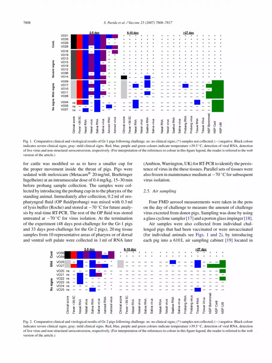
7808 S. Parida et al. / Vaccine 25 (2007) 7806–7817
F allengei nd greeo ion of thv
ftsIblsposuoasa
(tav
2
ova
Fiov
ig. 1. Comparative clinical and virological results of Gr-1 pigs following chndicates severe clinical signs; gray: mild clinical signs. Red, blue, purple af live virus and non-structural seroconversion, respectively. (For interpretatersion of the article.)
or cattle was modified so as to have a smaller cup forhe proper movement inside the throat of pigs. Pigs wereedated with meloxicam (Metacam® 20 mg/ml, Boehringerngelheim) at an intramuscular dose of 0.4 mg/kg, 15–30 minefore probang sample collection. The samples were col-ected by introducing the probang cup in to the pharynx of thetanding animal. Immediately after collection, 0.2 ml of oro-haryngeal fluid (OP fluid/probang) was mixed with 0.3 mlf lysis buffer (Roche) and stored at −70 ◦C for future analy-is by real-time RT-PCR. The rest of the OP fluid was storedntreated at −70 ◦C for virus isolation. At the termination
f the experiment (48 days post-challenge for the Gr-1 pigsnd 33 days post-challenge for the Gr-2 pigs), 20 mg tissueamples from 10 representative areas of pharynx or of dorsalnd ventral soft palate were collected in 1 ml of RNA laterl(e
ig. 2. Comparative clinical and virological results of Gr-2 pigs following challengendicates severe clinical signs; gray: mild clinical signs. Red, blue, purple and greef live virus and non-structural seroconversion, respectively. (For interpretation of thersion of the article.)
. ns: no clinical signs; (*) samples not collected; (−) negative. Black colourn colours indicate temperature >39.5 ◦C, detection of viral RNA, detectione references to colour in this figure legend, the reader is referred to the web
Ambion, Warrington, UK) for RT-PCR to identify the persis-ence of virus in the these tissues. Parallel sets of tissues werelso frozen in maintenance medium at −70 ◦C for subsequentirus isolation.
.5. Air sampling
Four FMD aerosol measurements were taken in the pensn the day of challenge to measure the amount of challengeirus excreted from donor pigs. Sampling was done by usingglass cyclone sampler [17] and a porton glass impinger [18].
Air samples were also collected from individual chal-enged pigs that had been vaccinated or were unvaccinatedfor individual animals see Figs. 1 and 2), by introducingach pig into a 610 L air sampling cabinet [19] located in
. ns: no clinical signs; (*) samples not collected; (−) negative. Black colourn colours indicate temperature >39.5 ◦C, detection of viral RNA, detectione references to colour in this figure legend, the reader is referred to the web

cine 25
ad
2
cfivtma
2
Mu[utfoTb
2
iMet(tIE
2
rp(Psat
2
FFEC
2
aav
2
saspwc
asnboo
vtu
3
3
sUddioiacdmIlBn
to
S. Parida et al. / Vac
separate pen. Measurements of virus/ RNA copy were asescribed [4,20,21].
.6. Virus isolation
Samples for virus isolation were inoculated onto BTYells [22] in roller drums. Antigen ELISA was used to con-rm the presence of FMD virus in cultures. For isolation ofirus from air samples, a similar protocol was followed afteritrating samples in a 10-fold dilution series such that 3 BTY
onolayer tubes were inoculated with neat (undiluted), 10−1
nd 10−2 dilutions of air samples.
.7. RNA extraction from liquid samples
Total nucleic acid was extracted from liquid samples withagNA pure LC total nucleic acid isolation kits (Roche)
sing an automated nucleic acid robotic work station (Roche)24]. QIAamp® MinElute® Virus Spin kits (Qiagen) weresed for RNA extraction from air samples according tohe manufacturer’s instructions. Briefly, RNA was extractedrom 200 �l of the original samples and a final volumef 18 �l RNA was recovered at the end of the process.his material was used for real-time RT-PCR as describedelow.
.8. RNA extraction from tissues
Twenty milligrams of tissues were homogenized by plac-ng them in 600 �l of tissue lysis buffer (Roche) in Lyzing
atrix D tubes (Q-Biogene, Cambridge, UK) and homog-nizing them at 6500 revolutions per minute for 45 s,hree times in a FastPrep FP120 homogenizing machineQ-Biogene). RNA was extracted and eluted in 50 �l elu-ion buffer using the MagNA Pure LC RNA isolation kitII (Roche) with an automated robot as described above.xtracted RNA was stored at −80 ◦C until used.
.9. Real-time RT-PCR
Viral RNA in samples was reverse transcribed [23] usingandom hexamers and quantified by real-time RT-PCR usingrimers and a probe from the internal ribosomal entry siteIRES) of FMDV O UKG 34/01[24]. A Stratagene MX4000CR machine was used. The c-DNAs obtained from the tis-ues were reanalysed for real-time PCR by using the probend primers from the 3D region of FMDV [25,26] followinghe above described methods without use of standards.
.10. Non-structural protein serology
Sera were examined for antibody against non-structural
MDV proteins by three commercial tests, i.e. Cedi testMDV-NS, Cedi-Diagnostics [27]; the UBI FMDV NSPLISA, United Biochemical Incorporated [28] and theHEKIT-FMD-3ABC, Bommeli Diagnostics [29].op
t
(2007) 7806–7817 7809
.11. Virus neutralising antibody test (VNT)
Titres of neutralising antibodies against FMDV O1 Manisand O1 UKG viruses were measured by micro-neutralisationssay as described in the OIE Manual of Diagnostic Tests andaccines [30].
.12. Statistics
The RNA copy numbers in different groups (control,evere signs, mild signs and no signs) of pigs were comparedt each time point using a Kruskal–Wallis test and, whereignificant differences (P < 0.05) were found, using multi-le contrasts. Animals vaccinated 10 days before challengeere also compared with those vaccinated 29 days before
hallenge, irrespective of the severity of clinical signs.All vaccinated and control pigs (n = 30) were regrouped
ccording to severity of infection (controls, vaccinated withevere signs, vaccinated with mild signs and vaccinated witho signs) and a Fisher exact test was used to detect differencesetween the proportion of pigs in each group for which RNAr virus was detected in the nasal fluids, saliva and aerosol,r which were positive by NSP Bommeli test.
The results of the virus neutralisation (VN) test for animalsaccinated 10 days prior to challenge were compared withhose vaccinated 29 days prior to challenge at each time pointsing a Wilcoxon rank sum test.
. Results
.1. Development of clinical FMD
The six needle challenged donor pigs in both the groupshowed clinical disease within 1–2 days post-infection.nvaccinated control pigs succumbed to disease within 1–2ays of contact (Tables 1 and 2). The majority (n = 13) ofirectly challenged vaccinated pigs of Gr-1 were clinicallynfected at the same time as their controls within 1–2 daysf contact challenge (Tables 1 and 2). However, the clin-cal scoring in these vaccinated clinically infected pigs waslways lower than the control pigs (Table 1) except one unvac-inated animal (VO28). According to degree of severity ofisease, the vaccinated pigs were regrouped as severe (n = 8),ild (n = 5) and no signs (n = 3) (Tables 1 and 2, Figs. 1 and 2).
ncrease of temperature was observed on the first day of chal-enge and 1 day thereafter in Gr-1 vaccinated pigs (Table 2).y the fourth day of challenge temperatures had returned toormal in all animals except VO12 and 19.
Out of eight vaccinated pigs in group two (Gr-2), onlywo (VO26 and 27) were clinically affected with mild signs,bserved on or after the third day of contact challenge; a delay
f 1–2 days compared to clinically affected Gr-1 vaccinatedigs.High temperatures preceded the development of lamenesshat was associated with congestion in the inter-digital space

7810 S. Parida et al. / Vaccine 25 (2007) 7806–7817
Table 1Total clinical scoring of vaccinated and unvaccinated pigs (Gr-1 and -2) following direct contact challenge
Category Animal no. Group 1 Animal no. Group 2
0 dpc 1 dpc 2 dpc 3 dpc 4 dpc 0 dpc 1 dpc 2 dpc 3 dpc 4 dpc
Unvaccinated (control) VO31 0 3 17 15 16 VO58 0 3 13 12b
VO30 0 1 13 14 16 VO59 0 3 13 12b
VO29 0 0 13 13 16VO28 0 1 10 7 7
VaccinatedSevere VO16 0 3 10 10 14
VO19 0 1 6 12 13VO13 0 2 8 9 13VO15 0 0 7 7 9VO18 0 0 2 5 9VO12 0 0 5 5 8VO06 0 1 4 4 7VO14 0 3 3 5 6
Mild signs VO09 0 2 2 4 4 VO26 0 0, 1a 1a 3VO17 0 0 2 3 4 VO27 0 0, 1a 3 3VO10 0 1 1 2 2VO11 0 2 2 1 2VO08 0 0 1 1 2
No signs (Protected) VO04 0 0 0 0 0 VO20 0 0 0 0 0VO05 0 0 0 0 0 VO21 0 0 0 0 0VO07 0 0 0 0 0 VO22 0 0 0 0 0
VO23 0 0 0 0 0VO24 0 0 0 0 0VO25 0 0 0 0 0
Clinical scoring was carried out by considering the following. Rectal temperature more than 39.5 ◦C, congestion of skin in inter-digital space and coronaryb snout, ew dpc: da
aht
3b
twb(nffcRnucofnd
gv2r
f1ddd(hntbFccvb
and, healed lesions in each foot were scored as 1; fresh lesions in tongue,ere scored as 3. Total scoring was calculated by adding individual scores.a The clinical scoring is due to lameness without showing any lesions.b The animal was killed.
nd on the coronary band. Vesicular lesions appeared a fewours later. Foot lesions appeared before those in the mouth,ongue and snout and were more severe.
.2. Detection of virus/genome in nasal, saliva andlood samples
Detection of virus/genome, as determined by virus isola-ion and/or RT-PCR on nasal fluids, saliva or blood samples,as evident in all of the unvaccinated and vaccinated pigs ofoth the groups (Figs. 1 and 2, Table 3). From one animalVO24) in Gr-2, no viral RNA was recovered from eitherasal (Table 3) or saliva swabs, but live virus was isolatedrom blood (data not shown). From nasal swabs collectedrom the Gr-1 pigs, virus could be isolated up to day 3 afterhallenge, irrespective of vaccination status whereas viralNA was detected for longer; up to 13 days in one vacci-ated pig (VO4, Table 3). Although virus could be isolatedp to 3 days after challenge from nasal swabs from both theontrol pigs in Gr-2, no virus was recovered from nasal swabs
f vaccinated animals (Fig. 2). Viral RNA was detected lessrequently in nasal swabs of Gr-2 vaccinated pigs than theasal swabs of Gr-1 vaccinated pigs and the frequency ofetection of viral RNA was found to be decreased in both theg
nA
ach foot were scored as 2; severe lesions in tongue, snout and in each footys post-challenge.
roups after the first week of challenge (Table 3). However,iral RNA from nasal swab samples was detected at 11 and1 days post-challenge in vaccinated pigs VO22 and VO25,espectively (Table 3).
The similar quantities of viral RNA initially detectedrom nasal swabs of all vaccinated and control pigs in Gr-
(no significant difference on day of challenge, P > 0.05)emonstrated that similar amounts of virus had been inhaleduring the first 4 h of contact challenge (Fig. 3). By theay after challenge, only significant differences were foundP = 0.04), with the vaccinated, mild clinical signs groupaving a significantly higher copy number than the vacci-ated, clinically protected group. At 3 days post-challengehe unvaccinated group had a significantly higher copy num-er than the vaccinated, clinically protected group (P = 0.02).inally, significant differences were found at 4 days post-hallenge (P = 0.02), but were not confirmed by the multipleontrast analysis. Viral RNA copy number was reduced in allaccinated animals irrespective of severity of clinical signsy 4 days post-challenge, except for the unvaccinated control
roup where the RNA copy number was increased (Fig. 3).Again, there were no significant differences in RNA copyumbers at 0 day post-challenge (P > 0.05) in Gr-2 pigs.lthough a higher copy number of RNA was detected in

S. Parida et al. / Vaccine 25 (2007) 7806–7817 7811
Table 2Rectal temperature of vaccinated and unvaccinated pigs (Gr-1 and -2) following direct contact challenge
Category Animal no. Group 1 Animal no. Group 2
0 dpc 1 dpc 2 dpc 3 dpc 4 dpc 0 dpc 1 dpc 2 dpc 3 dpc 4 dpc
Unvaccinated (control) VO31 38.6 39.6 40.6 38.4 38.5b VO58 38 38.8 40 40.5a
VO30 38.7 40 40.5 39.1 38.8b VO59 38.4 38.7 40.3 40a
VO29 38.6 38.6 39.9 37.5 38.6b
VO28 38.7 38.7 40.5 38.5 38.5b
VaccinatedSevere VO16 38.2 40.7 39.5 38.5 38.9b
VO19 38.5 38.8 40.4 40.3 39.7c
VO13 38.5 39.8 39.6 40.1 38.4b
VO15 38.6 40 40 39.9 38.6c
VO18 38.4 39.9 39.9 40.2 38.9c
VO12 38.5 40 40.3 40.3 39.2c
VO06 38.6 40.7 39.5 38.4 38.2c
VO14 38.5 39.7 40.1 40.5 38.5d
Mild signs VO09 38.6 39.8 40 38.4 38.8 VO26 38.5 39.2 39 38.7 38.5VO17 38.5 40.2 40.2 38.7 38.8 VO27 38.3 38.3 40 40 38.6VO10 38.5 39.9 39.5 37.5 38.3VO11 38.6 39.3 40.2 38.7 38.5VO08 38.4 39.6 40 37.7 38.2
No signs (Protected) VO04 37.9 40 38.5 37.5 37.9 VO20 38 39.4 38.5 38.7 38.3VO05 38 39.4 39.5 38.5 38 VO21 38.2 38.2 38.8 38.5 39VO07 38.6 39 39 39 39.3 VO22 38.2 38.4 38.7 38.3 38.8
VO23 38 38 38.8 39 38.7VO24 38.5 38.9 38.6 38 38.9VO25 38.5 39.5 39.2 39.1 39.5
a Indicates the animal was killed on 3 dpc.
tGicw
FR
s
b Indicates the animal was killed on 4 dpc.c Indicates the animal was killed on 5 dpc.d Indicates the animal was killed on 6 dpc.
he nasal swabs of control animals than of the vaccinated
r-2 pigs (Table 3), no significant differences (P > 0.05)n RNA copy numbers were found at 1 or 3 days post-hallenge. At 2 days post-challenge, significant differencesere found (P = 0.04), with the unvaccinated group having a
ig. 3. Mean FMDV RNA copy number detected over time by real-timeT-PCR from cotton bud samples collected from nose of Gr-1 pigs.
cg
v2wnn1
fttp(cRd1avaa
ignificantly higher copy number than both the vaccinated,linically protected and the vaccinated, mild clinical signsroups.
When comparing the viral RNA copy number for animalsaccinated 10 days prior to challenge with those vaccinated9 days prior to challenge, significant differences (P < 0.01)ere found at 2 and 4 days post-challenge, with the copyumber for Gr-1 vaccinated pigs higher than for Gr-2 vacci-ated pigs. No significant differences (P > 0.1) were found atand 3 days post-challenge.Live FMD virus could not be detected in saliva samples
rom vaccinated animals irrespective of clinical signs beyondhe second day after challenge except on the fourth day fromwo pigs in Gr-2 from a vaccinated and clinically affectedig (VO27) and a vaccinated clinically protected pig (VO21)Fig. 2). Beyond 4 days after challenge, all vaccinated clini-ally protected animals in Gr-1 and 2 were negative for viralNA in saliva samples (Figs. 1 and 2). RNA copy/virus wasetected only in vaccinated clinically affected animals up toweek after challenge in Gr-1 pigs where as up to 4 days
fter challenge in Gr-2 vaccinated pigs. On two occasions,iral RNA could be detected from convalescent saliva swabst 21 days post-challenge and 28 days post-challenge fromnimals VO22 (data not shown) and VO26 (Fig. 2), respec-

7812 S. Parida et al. / Vaccine 25 (2007) 7806–7817
Table 3RNA copy number calculated in real-time PCR per one cotton bud sample from the nose of different groups of pigs
R mals froa RNA inb
twn
3
ilcgv
P2idccv(wn
pa(d7oa(
3(c
2wwo
NA copy numbers are expressed in log10 copies per cotton swab. *The aninimals were killed. †Indicates the animal is found positive for detection oflood.
ively. The proportion of animals detected positive in PCRas less in saliva samples than from the nasal samples (dataot shown).
.3. Detection of virus/genome in exhaled air samples
Air sampling within the challenge pens followed by virussolation and real-time RT-PCR revealed a consistent chal-enge in the two experiments. Total output over the 9 h ofhallenge was 5.5 log TCID50 virus and 7.1 log copies of viralenome from the two donor pigs for Gr-1 and 5.4 log TCID50irus and 6.7 log copies of viral genome for Gr-2.
For Gr-1, individual pig sampling using a cabinet and aorton sampler detected viable virus in both control pigs atdays post-challenge (dpc) at levels of 4.4 log TCID50/24 h
n pig VO28 and 6.1 log TCID50/24 h in pig VO31. RT-PCRetected 7.0, 7.8, 6.2 log RNA copies/24 h of FMD virus inontrol pig VO28 on days 2–4 post-challenge and 7.8 logopies/24 h at 2 dpc only from control pig VO31. In contrast,
iable virus was detected only from one of the vaccinated pigsVO16-severely affected), whilst 3.6 log RNA copies/24 here detected at day 2 post-challenge from another vacci-ated and subsequently infected pig (VO5).GwRr
m Gr 2 pigs, dpc indicates days post-challenge, gray shadings indicates thesaliva swab sample. ††The animal was found virus isolation positive from
For Gr-2, viable virus was detected in one of the controligs (VO58) at day 2 post-challenge (4.2 log TCID50/24 h)nd on days 1 and 2 post-challenge in the other control pigVO59: 5.2, 4.2 log TCID50/24 h). RNA copies of virus wereetected in only one control pig (VO59) on 1 and 2 dpc (7.4,.3 log copies/24 h). No viable virus was detected from anyf the vaccinated pigs, whilst RNA copies were detectedt 4 dpc from one vaccinated clinically affected pig VO275.4 log TCID50/24 h).
.4. Detection of virus/genome in oro-pharyngeal fluidprobang) and tissues for identification of FMD virusarriers
Oro-pharyngeal fluids obtained by probang cup on or after8 days of challenge up to termination of the experimentere analysed by virus isolation and RT-PCR. No virus/RNAas obtained from cell culture/real-time RT-PCR from anyf the samples except on one occasion from one of the
r-1 animals (VO17) where viral RNA (2.37E+04 copies)as found by real-time PCR at 28 days post-challenge. TheNA extraction and RT-PCR was repeated twice with similaresults.

cine 25 (2007) 7806–7817 7813
vltV(G(p(ao(AHtlcrcfvr
3n
vabdaep
FpN(
Fui(
aptacTp(
(ip
S. Parida et al. / Vac
Extracted RNA from tissues of pharynx and dorsal andentral soft palate of both Gr-1 and Gr-2 vaccinated chal-enged pigs (n = 16) were used for real-time RT-PCR usinghe primers and probe from the IRES region of the FMDV.iral RNA was detected from only four pigs. Viral RNA
9.63E + 02) was detected from the dorsal soft palate of oner-1 pig (VO4), killed at 48 days post-challenge. Viral RNA
1.14E + 03 and 1.25E + 03) was also detected from the naso-harynx of two vaccinated and clinically affected Gr-2 pigsVO26 and 27), killed at 33 dpc. Viral RNA (1.54E + 01nd 1.86E + 01) was also detected in the dorsal soft palatef both of these pigs. Viral RNA was detected at 33 dpc4.4E + 01) from dorsal soft palate of a fourth animal (VO25).ll RT-PCR tests were repeated three times with same results.owever, we were not able to recover live virus from any of
hese tissues when extracts from frozen tissues were inocu-ated repeatedly, at least with two further passages in BTYells. When the tissue-derived RNA or c-DNA samples weree-examined with another set of primers and a probe spe-ific for the 3D region of FMDV, no amplified product wasound for these samples whereas a strong positive signal (CTalue of 22) was obtained for positive control material. Sameesults were also obtained on repetition of this experiment.
.5. Detection of clinical and sub-clinical infection byon-structural antibody assay
All three tests detected antibodies to FMDV NSPs inaccinated and clinically affected pigs (Figs. 1, 2 and 4)nd all seven animals were scored positive at some stagey both the Cedi and UBI tests. The UBI test started to
etect infection from 4 days post-challenge and scored allffected pigs positive from 13 days post-challenge up to thend of the experiment except on one occasion at 33 daysost-challenge (Fig. 4). Eighty-six percent of the clinicallyig. 4. Detection of non-structural antibodies in clinically infected recoveredigs of Gr-1 and Gr-2. (�) indicates % of positive pigs detected in BommeliSP test, (©) indicates % of positive pigs detected in Cedi NSP test and�) indicates % of positive pigs detected in UBI NSP test.
Hdt3cwb
3
sfGded
wtipat
ig. 5. Detection of sub-clinical infection in vaccinated and challenged pigssing various non-structural antibody assays. (�) % of positive pigs detectedn Bommeli NSP test, (©) % of positive pigs detected in Cedi NSP test and�) % of positive pigs detected in UBI NSP test.
ffected animals were detected by the Cedi test at 6 daysost-challenge and as for the UBI test 100% of samples ini-ially scored positive from 13 days post-challenge. Howeverfter 21 days, the detection rate fell and by 33 days post-hallenge only 57% of affected pigs scored positive (Fig. 4).he Bommeli test detected 29% of affected pigs at 13 daysost-challenge, peaking at 43% at 21 days post-challengeFig. 4).
The Bommeli test did not detect any sub-clinical infectionFigs. 1, 2 and 5). With the UBI test, detection of sub-clinicalnfection was first possible at 2 days post-challenge (11%ositive) and peaked at 6 days post-challenge (66%, Fig. 5).owever, some non-specificity was observed in this assayuring the pre-challenge period (Fig. 4). The Cedi test startedo detect sub-clinical infection at 6 dpc and detected up to3% of sub-clinical infection. On the 40th and 47th day ofhallenge all three protected but sub-clinically infected pigsere detected by the UBI test whereas only two were detectedy the Cedi test (data not shown).
.6. Virus neutralising antibody
Serum antibody responses against O1 Manisa were mea-ured by virus neutralisation test before and after challengeor vaccinated animals in both groups of pigs (Fig. 6). Ther-2 pigs had on average more neutralising antibody on theay of challenge as well as after challenge throughout thexperiment. Differences were significant (P < 0.05) on theay of challenge, and at 21 and 28 days post-challenge.
When the clinically protected and clinically affected pigsere regrouped for their mean VNT titre separately within
he groups, VNT titres were higher on the day of challenge
n clinically protected animals than in clinically affectedigs, particularly in group two pigs (Fig. 7). Immediatelyfter challenge, the clinically affected pigs had higher VNTitres than the clinically protected animals due to anamnestic
7814 S. Parida et al. / Vaccine 25 (2007) 7806–7817
Fig. 6. Mean neutralising (O1 Manisa) antibody titres in vaccinated pigsfollowing challenge. The pigs were vaccinated with full bovine dose ofO1 Manisa oil adjuvant vaccine and challenged with O UKG 2001 on day10 (Gr-1) and day 29 (Gr-2) post-vaccination. (�) Mean neutralising (O1Manisa) antibody titres for Gr-1 pigs and (©) mean neutralising (O1 Manisa)antibody titres for Gr-2 pigs.
Fig. 7. Mean neutralising (O1 Manisa) antibody titres in Gr-2 vaccinatedclinically infected and clinically protected pigs following challenge. The pigswere vaccinated with full bovine dose of O1 Manisa oil adjuvant vaccine andchallenged with O UKG 2001 on day 29 post-vaccination. Gray bars indicatemac
ra
3r
atauinc
Table 4Establishment of correlation between vaccine-induced reduction in FMDvirus replication and excretion with severity of clinical signs
N RNA N virus S RNA S virus A RNA A virus B NSP
Signs P/T P/T P/T P/T P/T P/T P/TControl 6/6 5/6 6/6 6/6 4/4 4/4 NASevere 8/8 6/8 8/8 6/8 0/2 1/2 NAMild 7/7 2/7 7/7 3/7 1/3 0/3 4/7No signs 5/9 0/9 6/9 1/9 1/5 0/5 0/9
PN
arslawbdcvmv(cstfNct
4
aIiwpsovolwTvtv
ean neutralising (O1 Manisa) antibody titres for clinically protected pigsnd black bars indicate mean neutralising (O1 Manisa) antibody titres oflinically infected pigs.
esponses, which declined later in comparison to protectednimals (Fig. 7).
.7. Correlation of vaccine-induced reduction in viraleplication and excretion with severity of clinical signs
All vaccinated and control pigs (n = 30) were regroupedccording to the severity of clinical signs (unvaccinated con-rols, vaccinated with severe signs, vaccinated with mild signsnd vaccinated with no signs) and a Fisher exact test was
sed to detect differences between the proportion of pigsn each group for which RNA or virus was detected in theasal fluids, saliva and aerosol within the first 5 days ofhallenge, or which were positive by Bommeli NSP testssfs
/T: no. of positives/total no. of samples tested, NA: sample not available,: nasal, S: saliva, A: aerosol and B: Bommeli.
fter 27 days post-challenge (Table 2). The Bommeli testesults were used for this purpose since the test had a lowensitivity allowing discrimination between extensive andow level replication of virus. As control animals as wells infected animals with severe signs were killed within 1eek of challenge, seroconversion rate was only comparedetween pigs with mild or no signs of disease. The mainifference found between unvaccinated controls and the vac-inated severely infected group was in recovery of aerosolisedirus: always control groups excreted more. However, theild signs and no signs groups had a lower rate of infectious
iral recovery from nasal and saliva swabs, whilst no signsub-clinically infected/protected) group had the lowest indi-ators of viral RNA/ virus excretion and replication (NSPeroconversion). The proportion of pigs which were posi-ive in each group (Table 4) differed significantly (P < 0.05)or all the tests (nasal, saliva and aerosol RNA or virus, andSP test); animals in the control group or with more severe
linical signs were more likely to be positive for any of theests.
. Discussion
Pigs are important ‘amplifiers’ of FMDV because of thebundance of infectious material excreted in their breath [31].ntensification of agricultural systems has led to a massivencrease in the size and density of pig populations whichhen juxtaposed with other susceptible livestock providesotential for large scale and rapid spread of FMDV [32]. Thistudy is aimed to define the efficacy of a single applicationf vaccine in providing protection to pigs from infection,irus replication, virus excretion and clinical disease. Usef a semi-heterologous 9 h direct contact challenge simu-ated a ‘worse-case’ scenario encounter with FMDV, fromhich extrapolations can be made to less severe challenges.he reduction in virus replication and excretion afforded byaccination was quantified and shown to be correlated tohe severity of clinical signs of FMD. The study also pro-ided insights into the ability to detect infection, particularlyub-clinical infection in vaccinated pigs, by means of NSP
erology; an important approach in substantiating freedomrom infection in a post-vaccination situation. Finally, thetudy found evidence of viral RNA but not infectious virus
cine 25
ii
pdlvtbrrasvpawu
O1woondeedacvwiidnipos[dciqvpRtp
cipa
mp1aatpnitont(talcfdosarmg
pKoidtrddFdcctdtqt
lrrpgd
S. Parida et al. / Vac
n the oro-pharynx of vaccinated pigs recovered from acutenfection.
In our severe challenge model, vaccination could not fullyrotect pigs against clinical disease even when challenge waselayed until 29 days later. However, the disease seen wasess severe compared to that in pigs challenged 10 days afteraccination, and vaccinated pigs were less severely affectedhan controls. Although vaccinated pigs all appeared to haveecome infected to some degree, vaccination significantlyeduced excretion of live virus, which is likely to reduce theisk of onward viral transmission to other naı̈ve, or vaccinatednimals. Even better protection might be observed after lessevere FMDV challenge, e.g. following indirect exposure toirus in the field after animal movement restrictions are inlace [33,34], demonstrated protection against clinical signss early as 4 days after vaccination, when vaccinated pigsere exposed for 1–4 h to indirect/direct contact challengesing infected donor pigs.
Eble et al. [35,36] challenged three groups of pigs withTaiwan virus 7 days after vaccination with O Taiwan or
4 days after vaccination with either O1 Manisa or O Tai-an vaccines. The challenge was done by inoculating halff the vaccinated pigs in each group of 10, giving a chancef direct contact challenge for the other half of the vacci-ated pigs in each group. None of their pigs vaccinated 14ays prior to challenge developed generalised FMD, whereasven challenge at 29 days post-vaccination resulted in gen-ralised FMD in two out of eight pigs in our study. Possibly,irect heel-bulb inoculation of pigs with FMDV representedless severe challenge than direct exposure for 9 h to unvac-inated donor pigs exhibiting clinical disease. In additioniral strain, dose, age of animals might have influence forhich it is difficult to compare the results between the exper-
ments. Eble et al. also found that five inoculated and threen-contact pigs developed FMD in their group vaccinated 7ays prior to challenge. Assuming that 2 or 3 days would beeeded before the inoculated pigs were ready to transmit, then-contact pigs would by then have been at around 10 daysost-vaccination, as for Gr-1 of our own study, in which 9 hf direct contact challenge was able to induce disease in aimilar proportion of animals (13 out of 16 pigs). Eble et al.36] reported that the titres of virus in OP swabs from their 7ay vaccinated pigs were no less than from their unvaccinatedontrols. However, due to the lack of aerosol measurementsn their experiments and the different way of collecting anduantifying virus in the oro-pharynx, a direct comparison ofiral excretion between the two studies cannot be made. Theresent study is the first to quantify viral RNA by real-timeT-PCR in the excretions of vaccinated challenged pigs and
o analyse the excretion of virus in exhaled air after use ofurified high potency emergency vaccine.
Some controversy exists over whether pigs can be FMDV
arriers [12,13,34]. Due to the prior application of vaccinen this study, we were able to keep at least 16 vaccinatedigs up to 33–47 days after virus challenge. Alexandersen etl. [20] reported that pharynx, tonsil and soft palate are thetlac
(2007) 7806–7817 7815
ajor sites for FMD virus replication in pigs during the earlyhase of infection from where they were able to detect 104 to06 TCID50 equiv./g. However, their study only lasted 4 daysfter infection of unvaccinated pigs. To find out whether pigsre carriers of FMD virus or not beyond 28 days after infec-ion, oro-pharyngeal fluids and tissues were collected fromigs after 28 days post-challenge. Though live virus couldot be recovered in these samples, viral RNA was recoveredn some tissues from four vaccinated challenged pigs by real-ime RT-PCR using a probe and primers from the IRES regionf the virus. Two of the four animals were from the vacci-ated and clinically affected group of pigs (VO26 and 27) andwo were from vaccinated and sub-clinically infected animalsVO4 and 25). A saliva sample of the pig VO26 was also posi-ive for viral RNA at 28 days after challenge. Viral RNA waslso obtained at 13 days post-challenge from pig VO4 (itsatest detection in Gr-1 pigs) and in VO25 at 21 days afterhallenge. However, as live virus was never recovered eitherrom oro-pharyngeal fluid (probang) or from tissues after 28ays post-challenge, these animals do not meet the definitionf FMD carrier animals [7], which supports the non-carriertatus of pigs [16]. RNA recovery could not be confirmed by3D real-time PCR that is at least as sensitive as the IRES
eal-time PCR [26]. Possibly, the IRES region RNA might beore resistant to degradation than certain other parts of the
enome due to its high degree of structure.NSP tests have been widely evaluated in cattle but not
igs. In unpublished studies on FMD in four vaccinated Hongong pig herds [37] we observed that serological evidencef infection was only found in groups of animals with heal-ng lesions, whereas neighbouring groups without signs ofisease were seronegative for NSP antibody. This led uso conclude that either infection following vaccination wasarely sub-clinical or else, if it occurred, did not lead toetectable NSP seroconversion. However, this study clearlyemonstrated that, sub-clinical infections can occur in pigs.urthermore, seroconversion in NSP tests was related to theegree of virus replication and excretion and the severity oflinical signs. As seen earlier in cattle [38], no single NSP testould detect infection in all pigs, but combining two or moreests could increase specificity and sensitivity. The failure toetect NSP antibody in the study of [36] may be attributed tohe low level of viral replication in the vaccinated and subse-uently FMDV inoculated animals that could not efficientlyransmit infection to vaccinated in-contact pigs.
In our study, the rise in neutralising antibody after chal-enge of both the groups could be attributed to an anamnesticesponse to virus replication or a maturing primary adaptiveesponse to vaccination since vaccinated and unchallengedigs were not available for comparison. Comparing the tworoups, the mean VNT titre of the Gr-2 pigs (vaccinated for 29ays) had significantly more neutralising antibody titre than
he Gr-1 pigs (vaccinated for 10 days) on the day of chal-enge and therefore, there was a correlation between VN titrend both clinical and virological protection. Although vac-ination could not provide complete clinical or virological
7 cine 25
ptvvw
A
(ISnc
R
[
[
[
[
[
[
[
[
[
[
[
[
[
[
[
[
[
[
[
[
[
[
[
[
816 S. Parida et al. / Vac
rotection, it reduced the severity of the disease, virus excre-ion and production of non-structural FMDV antibodies inaccinated and subsequently infected pigs. As hypothesised,accine-induced reduction of virus replication and excretionas found to be correlated to the severity of clinical disease.
cknowledgements
This work was supported financially by Defra, UKProject SE1122) and from the European Commission (FMD-mprocon project of the EU 6th Framework Programme,SPE-CT-2003-503603). Thanks are due to Dr. Paul Bar-et for participating in the discussion for the designing of thehallenge.
eferences
[1] Thomson G. Foot and mouth disease: facing the new dilemmas. RevSci Technol 2002;21(3):425–8.
[2] Alexandersen S, Zhang Z, Donaldson AI, Garland AJ. The patho-genesis and diagnosis of foot-and-mouth disease. J Comp Pathol2003;129(1):1–36.
[3] Sellers RF, Parker J. Airborne excretion of foot-and-mouth diseasevirus. J Hyg (Lond) 1969;67(4):671–7.
[4] Alexandersen S, Donaldson AI. Further studies to quantify the dose ofnatural aerosols of foot-and-mouth disease virus for pigs. EpidemiolInfect 2002;128(2):313–23.
[5] Donaldson AI, Alexandersen S. Predicting the spread of foot and mouthdisease by airborne virus. Rev Sci Technol 2002;21(3):569–75.
[6] Annonymous. Council Directive 2003/85/EC on Community mea-sures for the control of foot-and-mouth disease repealing Directive85/511/EEC and Decisions 89/531/EEC and 96/665/EEC and amend-ing Directive 92/46/EEC: Official Journal of the European Union L3062003; 2003.
[7] Salt JS. The carrier state in foot and mouth disease—an immunologicalreview. Br Vet J 1993;149(3):207–23.
[8] Sutmoller P, McVicar JW, Cottral GE. The epizootiological impor-tance of foot-and-mouth disease carriers. I. Experimentally producedfoot-and-mouth disease carriers in susceptible and immune cattle. ArchGesamte Virusforsch 1968;23(3):227–35.
[9] Kitching RP. Identification of foot and mouth disease virus carrierand subclinically infected animals and differentiation from vaccinatedanimals. Rev Sci Technol 2002;21(3):531–8.
10] Alexandersen S, Zhang Z, Donaldson AI. Aspects of the persistenceof foot-and-mouth disease virus in animals—the carrier problem.Microbes Infect 2002;4(10):1099–110.
11] Anonymous. OIE. 2005. Foot and mouth disease. In: Commission OS,editor. Terrestrial animal health code. 14th ed. 12 rue de Prony, 75017Paris, France.
12] Yadin H, Chai D. Surveillance of FMD in wild animals in Israel InResearch group of the standing technical committee, European com-mission for the control of foot and mouth disease, 1994.
13] Mezencio JM, Babcock GD, Kramer E, Brown F. Evidence forthe persistence of foot-and-mouth disease virus in pigs. Vet J1999;157(3):213–7.
14] Golde WT, Pacheco JM, Duque H, Doel T, Penfold B, Ferman GS, et
al. Vaccination against foot-and-mouth disease virus confers completeclinical protection in 7 days and partial protection in 4 days: use inemergency outbreak response. Vaccine 2005;23(50):5775–82.15] Parida S, Anderson J, Cox SJ, Barnett PV, Paton DJ. Secretory IgA asan indicator of oro-pharyngeal foot-and-mouth disease virus replication
[
[
(2007) 7806–7817
and as a tool for post-vaccination surveillance. Secretory IgA as an indi-cator of oro-pharyngeal foot-and-mouth disease virus replication and asa tool for post-vaccination surveillance. Vaccine 2006;24(8):1107–16.
16] Alexandersen S, Brotherhood I, Donaldson AI. Natural aerosol trans-mission of foot-and-mouth disease virus to pigs: minimal infectiousdose for strain O1 Lausanne. Epidemiol Infect 2002;128(2):301–12.
17] Errington FP, Powell EO. A cyclone separator for aerosol sampling inthe field. J Hyg (Lond) 1969;67(3):387–99.
18] May KR, Harper GJ. The efficiency of various liquid impinger samplersin bacterial aerosols. Br J Ind Med 1957;14(4):287–97.
19] Gibson CF, Donaldson AI. Exposure of sheep to natural aerosols offoot-and-mouth disease virus. Res Vet Sci 1986;41(1):45–9.
20] Alexandersen S, Oleksiewicz MB, Donaldson AI. The early pathogene-sis of foot-and-mouth disease in pigs infected by contact: a quantitativetime-course study using TaqMan RT-PCR. J Gen Virol 2001;82(Pt4):747–55.
21] Snowdon WA. Growth of foot-and mouth disease virus in monolayercultures of calf thyroid cells. Nature 1966;210(5040):1079–80.
22] Ferris NP, Dawson M. Routine application of enzyme-linkedimmunosorbent assay in comparison with complement fixation for thediagnosis of foot-and-mouth and swine vesicular diseases. Vet Micro-biol 1988;16(3):201–9.
23] Zhang ZD, Alexandersen S. Detection of carrier cattle and sheep persis-tently infected with foot-and-mouth disease virus by a rapid real-timeRT-PCR assay. J Virol Methods 2003;111(2):95–100.
24] Quan M, Murphy CM, Zhang Z, Alexandersen S. Determinants ofearly foot-and-mouth disease virus dynamics in pigs. J Comp Pathol2004;131(4):294–307.
25] Callahan JD, Brown F, Osorio FA, Sur JH, Kramer E, Long GW, etal. Use of a portable real-time reverse transcriptase-polymerase chainreaction assay for rapid detection of foot-and-mouth disease virus. JAm Vet Med Assoc 2002;220(11):1636–42.
26] King DP, Ferris NP, Shaw AE, Reid SM, Hutchings GH, GiuffreAC, et al. Detection of foot-and-mouth disease virus: compar-ative diagnostic sensitivity of two independent real-time reversetranscription-polymerase chain reaction assays. J Vet Diagn Invest2006;18(1):93–7.
27] Sorensen KJ, Madsen KG, Madsen ES, Salt JS, Nqindi J, Mackay DK.Differentiation of infection from vaccination in foot-and-mouth diseaseby the detection of antibodies to the non-structural proteins 3D, 3ABand 3ABC in ELISA using antigens expressed in baculovirus. ArchVirol 1998;143(8):1461–76.
28] Shen F, Chen PD, Walfield AM, Ye J, House J, Brown F,et al. Differentiation of convalescent animals from those vacci-nated against foot-and-mouth disease by a peptide ELISA. Vaccine1999;17(23–24):3039–49.
29] Bruderer U, Swam H, Haas B, Visser N, Brocchi E, Grazioli S, et al.Differentiating infection from vaccination in foot-and-mouth-disease:evaluation of an ELISA based on recombinant 3ABC. Vet Microbiol2004;101(3):187–97.
30] Anonymous. OIE. 2004. Foot and mouth disease. In: Manual of Stan-dards for Diagnostic Tests and Vaccines. 5th ed. 12 rue de Prony, 75017Paris, France, 2004.
31] Sellers RF, Herniman KA, Gumm ID. The airborne dispersal of foot-and-mouth disease virus from vaccinated and recovered pigs, cattle andsheep after exposure to infection. Res Vet Sci 1977;23(1):70–5.
32] Donaldson AI, Doel TR. Foot-and-mouth disease: the risk for GreatBritain after 1992. Vet Rec 1992;131(6):114–20.
33] Barnett PV, Cox SJ, Aggarwal N, Gerber H, McCullough KC. Furtherstudies on the early protective responses of pigs following immuni-sation with high potency foot and mouth disease vaccine. Vaccine2002;20(25–26):3197–208.
34] Salt JS, Barnett PV, Dani P, Williams L. Emergency vaccination ofpigs against foot-and-mouth disease: protection against disease andreduction in contact transmission. Vaccine 1998;16(7):746–54.
35] Eble PL, de Bruin MG, Bouma A, van Hemert-Kluitenberg F, DekkerA. Comparison of immune responses after intra-typic heterologous and

cine 25
[
[
2006.
S. Parida et al. / Vac
homologous vaccination against foot-and-mouth disease virus infectionin pigs. Vaccine 2006;24(9):1274–81.
36] Eble PL, Bouma A, de Bruin MG, van Hemert-Kluitenberg F, vanOirschot JT, Dekker A. Vaccination of pigs two weeks before infec-
tion significantly reduces transmission of foot-and-mouth disease virus.Vaccine 2004;22(11–12):1372–8.37] Paton D, Sammin D, Dyrting K, Wong Mary, Fleming Lucy,Verin Blesilda et al. Comparative evaluation of serological testsfor the detection of foot-and-mouth disease virus infection in
[
(2007) 7806–7817 7817
vaccinated pigs. Open session of the research group of thestanding technical committee of the European Commission forthe control of Foot and mouth Disease, Oct, Paphos, Cyprus,
38] Brocchi E, Bergmann IE, Dekker A, Paton DJ, Sammin DJ, Greiner M,et al. Comparative evaluation of six ELISAs for the detection of anti-bodies to the non-structural proteins of foot-and-mouth disease virus.Vaccine 2006;24(47–48):6966–79.