vaccination against gip for the treatment of obesity
TRANSCRIPT

University of ZurichZurich Open Repository and Archive
Winterthurerstr. 190
CH-8057 Zurich
http://www.zora.uzh.ch
Year: 2008
Vaccination against GIP for the treatment of obesity
Fulurija, A; Lutz, T A; Sladko, K; Osto, M; Wielinga, P Y; Bachmann, M F; Saudan, P
Fulurija, A; Lutz, T A; Sladko, K; Osto, M; Wielinga, P Y; Bachmann, M F; Saudan, P (2008). Vaccination againstGIP for the treatment of obesity. PLoS ONE, 3(9):3163.Postprint available at:http://www.zora.uzh.ch
Posted at the Zurich Open Repository and Archive, University of Zurich.http://www.zora.uzh.ch
Originally published at:PLoS ONE 2008, 3(9):3163.
Vaccination against GIP for the treatment of obesity
Abstract
BACKGROUND: According to the WHO, more than 1 billion people worldwide are overweight and atrisk of developing chronic illnesses, including cardiovascular disease, type 2 diabetes, hypertension andstroke. Current therapies show limited efficacy and are often associated with unpleasant side-effectprofiles, hence there is a medical need for new therapeutic interventions in the field of obesity. Gastricinhibitory peptide (GIP, also known as glucose-dependent insulinotropic polypeptide) has recently beenpostulated to link over-nutrition with obesity. In fact GIP receptor-deficient mice (GIPR(-/-)) wereshown to be completely protected from diet-induced obesity. Thus, disrupting GIP signaling represents apromising novel therapeutic strategy for the treatment of obesity. METHODOLOGY/PRINCIPALFINDINGS: In order to block GIP signaling we chose an active vaccination approach using GIPpeptides covalently attached to virus-like particles (VLP-GIP). Vaccination of mice with VLP-GIPinduced high titers of specific antibodies and efficiently reduced body weight gain in animals fed a highfat diet. The reduction in body weight gain could be attributed to reduced accumulation of fat.Moreover, increased weight loss was observed in obese mice vaccinated with VLP-GIP. Importantly,despite the incretin action of GIP, VLP-GIP-treated mice did not show signs of glucose intolerance.CONCLUSIONS/SIGNIFICANCE: This study shows that vaccination against GIP was safe andeffective. Thus active vaccination may represent a novel, long-lasting treatment for obesity. Howeverfurther preclinical safety/toxicology studies will be required before the therapeutic concept can beaddressed in humans.
Vaccination against GIP for the Treatment of ObesityAlma Fulurija1, Thomas A. Lutz2, Katja Sladko1, Melania Osto2, Peter Y. Wielinga2, Martin F. Bachmann1,
Philippe Saudan1*
1 Cytos Biotechnology AG, Schlieren, Switzerland, 2 Institute of Veterinary Physiology and Center of Integrative Human Physiology, Vetsuisse Faculty, University of Zurich,
Zurich, Switzerland
Abstract
Background: According to the WHO, more than 1 billion people worldwide are overweight and at risk of developingchronic illnesses, including cardiovascular disease, type 2 diabetes, hypertension and stroke. Current therapies show limitedefficacy and are often associated with unpleasant side-effect profiles, hence there is a medical need for new therapeuticinterventions in the field of obesity. Gastric inhibitory peptide (GIP, also known as glucose-dependent insulinotropicpolypeptide) has recently been postulated to link over-nutrition with obesity. In fact GIP receptor-deficient mice (GIPR2/2)were shown to be completely protected from diet-induced obesity. Thus, disrupting GIP signaling represents a promisingnovel therapeutic strategy for the treatment of obesity.
Methodology/Principal Findings: In order to block GIP signaling we chose an active vaccination approach using GIPpeptides covalently attached to virus-like particles (VLP-GIP). Vaccination of mice with VLP-GIP induced high titers of specificantibodies and efficiently reduced body weight gain in animals fed a high fat diet. The reduction in body weight gain couldbe attributed to reduced accumulation of fat. Moreover, increased weight loss was observed in obese mice vaccinated withVLP-GIP. Importantly, despite the incretin action of GIP, VLP-GIP-treated mice did not show signs of glucose intolerance.
Conclusions/Significance: This study shows that vaccination against GIP was safe and effective. Thus active vaccination mayrepresent a novel, long-lasting treatment for obesity. However further preclinical safety/toxicology studies will be requiredbefore the therapeutic concept can be addressed in humans.
Citation: Fulurija A, Lutz TA, Sladko K, Osto M, Wielinga PY, et al. (2008) Vaccination against GIP for the Treatment of Obesity. PLoS ONE 3(9): e3163. doi:10.1371/journal.pone.0003163
Editor: Alessandro Bartolomucci, University of Parma, Italy
Received December 11, 2007; Accepted August 15, 2008; Published September 9, 2008
Copyright: � 2008 Fulurija et al. This is an open-access article distributed under the terms of the Creative Commons Attribution License, which permitsunrestricted use, distribution, and reproduction in any medium, provided the original author and source are credited.
Funding: Most of the work was carried out and financed by Cytos Biotechnology AG.
Competing Interests: A. Fulurija, K. Sladko, P. Saudan, and M.F. Bachmann are employees of Cytos Biotechnology AG and hold stocks or stock options in thecompany.
* E-mail: [email protected]
Introduction
Obesity has become one of the leading health problems
worldwide. The global obesity epidemic results from a combina-
tion of genetic susceptibility, increased availability of high-energy
foods and decreased requirement for physical activity in modern
society [1]. Obesity and excess weight are major risk factors for
chronic diseases, including type II diabetes, cardiovascular
diseases, gastrointestinal disorders and certain forms of cancer.
Importantly, body weight reduction in the range of 10% is
associated with significant improvements in a wide range of co-
morbid conditions [2–4]. Currently approved anti-obesity drugs
show only limited efficacy, generally facilitating no more than a 5–
10% reduction of body weight and are often associated with
unpleasant side-effect profiles [5–7]. To date the only treatment
leading to substantial, sustained body weight loss is bariatric
surgery. However, this intervention is associated with between
1.5% and 4.5% mortality during the first three month following
surgery [8]. Hence there is a major medical need for the
development of new anti-obesity drugs. In the past decade our
knowledge of gut hormones and their central role in the control of
food intake and energy balance has substantially improved [9–11].
This increased understanding has led to the identification of new
potential targets for pharmaceutical intervention.
Gastric inhibitory peptide, also known as glucose-dependent
insulinotropic polypeptide (GIP) is one of these peptide hormones.
GIP is a 42 amino acid, gastrointestinal polypeptide released from
duodenal and jejunal K-cells after ingestion of nutrients and has
been shown to facilitate the disposal of both glucose and fat [12].
GIP acts rapidly on pancreatic b-cells to stimulate the release of
insulin thus ensuring prompt uptake of glucose into the tissue. In
addition, GIP aids fat deposition and triglyceride accumulation in
adipocytes. Specifically, GIP has been shown to promote
triglyceride clearance from the circulation [13,14], a process
partly mediated by its ability to stimulate lipoprotein lipase activity
[15]. Moreover, GIP receptors are expressed on adipocytes [16]
consistent with a direct role of GIP on these cells. Recently, GIP
receptor-deficient mice (GIPR2/2) were shown to be completely
protected from diet-induced obesity [17]. Likewise, recent studies
demonstrated that treatment with a GIP-receptor antagonist led to
reduced weight gain in mice fed a high fat diet and weight loss in
obese mice [18–20]. Hence, disrupting GIP signaling represents a
promising, novel therapeutic strategy for the treatment of obesity.
The induction of GIP-specific, neutralizing antibodies through
vaccination is a particularly attractive possibility, given that the
blockade of GIP would be long-lasting. We have previously shown
that antigens displayed on highly repetitive viral surfaces can break
B cell tolerance [21] and epitopes displayed on the surface of virus-
PLoS ONE | www.plosone.org 1 September 2008 | Volume 3 | Issue 9 | e3163

like particles (VLPs) are able to efficiently induce self-specific
antibody responses in mice and humans [22–26]. In this study, we
show that vaccination against GIP prevents excessive body weight
gain in rodents fed a high fat diet and induces increased weight loss
in obese mice. Hence, active vaccination may represent an
attractive and convenient new therapy for the treatment of obesity.
Results
Vaccination against GIP results in high levels of GIP-specific antibodies
To overcome GIP-specific B cell unresponsiveness, we cova-
lently coupled peptides consisting of the first 15 amino acids of
mature GIP to the highly repetitive surface of bacteriophage QbVLPs [24,27] (Figure 1A). The resulting vaccine was named Qb-
GIP. Mice were immunized s.c. with 100 mg of Qb-GIP,
formulated in saline, on days 0, 14, 28 and 42. GIP-specific
antibody titers were determined at regular intervals. After a single
immunisation, high GIP-specific antibody titers were induced.
Antibody levels further increased with subsequent injections and
were maintained over a period of at least three months (Figure 1B).
These results demonstrate that Qb-GIP could overcome immu-
nological tolerance resulting in the induction of high GIP-specific
antibody titers. Since the N-terminal peptide used in Qb-GIP
shares approximately 50% homology with GLP-1 and oxyntomo-
dulin, sera from vaccinated mice were analysed for cross-reactivity
with these hormones in an inhibition ELISA. While pre-
incubation with GIP efficiently prevented binding of GIP-specific
antibodies to plate-coated GIP, neither GLP-1 nor oxyntomodulin
inhibited binding of anti-GIP sera to GIP, demonstrating that the
induced antibody response is highly specific (Figure 1C). Consid-
ering that the first two N-terminal amino acids of GIP are rapidly
cleaved by dipeptidyl peptidase (DPP)-IV [28] in vivo we wanted to
test whether the induced immune response by our vaccine also
recognizes the N-terminally cleaved form of GIP which has been
shown to be a partial antagonist of GIP in vitro [29]. To test this, a
competition ELISA with GIP coated plates was performed with
sera from Qb-GIP immunised mice pre-incubated either with the
peptide used for immunisation (GIP1–15) or an N-terminally
truncated version of the peptide (GIP3–15). As shown in
Figure 1D, GIP1–15 efficiently competed serum binding to plate
coated GIP, whereas GIP3–15 failed to do so even at very high
concentrations. These results demonstrate that with the vaccine
used here, most of the immune response is directed towards an
intact N-terminus of GIP.
Measurement of GIP concentration in serum revealed that Qb-
GIP immunized mice displayed significantly higher GIP levels
than Qb immunized control mice (data not shown) most likely due
to stabilization of GIP by the induced antibody response. Hence
we set out to test whether the induced antibodies could still
neutralise physiological concentrations of GIP in vivo. To this end
Qb-GIP immunised animals and control animals were challenged
i.v. with 1 ng of I125-GIP and 30 minutes after the injection
animals were culled, serum collected and antibody bound and free
I125-GIP determined. As shown in Figure 1E, in vaccinated mice
roughly 85% of the radiolabeled GIP was antibody bound whereas
only 4% were found associated with the antibody fraction in naı̈ve
mice. This result demonstrates that the induced antibody response
can efficiently sequester GIP in vivo. In similar experiments
performed with I125-GLP-1 or I125-Oxyntomodulin only back-
ground levels of the radiolabelled ligands were found associated
with the antibodies, further demonstrating that the induced
antibody response is highly specific for GIP (Figure S1). Having
demonstrated that the induced antibodies efficiently bound GIP in
the serum we tested whether specific antibodies would prevent the
interaction of GIP with its receptor. Hence, CHOK1 cells
expressing the human GIP receptor were generated and used for
in vitro receptor binding studies. Increasing amounts of purified
IgGs from Qb-GIP or Qb immunised mice were incubated with a
fixed amount of I125-GIP and added to receptor expressing cells.
After overnight incubation at 4uC receptor bound GIP was
determined. As shown in Figure 1F the induced GIP specific
antibodies efficiently prevented GIP binding to its receptor. In
contrast, purified IgG from Qb immunized mice did not influence
GIP receptor binding. Only very low levels of unspecific binding
(,50 cpm) of GIP to the parental CHOK1 cells were observed in
the presence or absence of purified IgG from Qb-GIP or Qbimmunised mice (data not shown). Taken together these results
show that vaccination with Qb-GIP induces highly specific
antibodies which can sequester GIP and prevent its binding to
the GIP receptor.
Vaccination against GIP protects against diet-inducedobesity
Having established that Qb-GIP induces a strong antibody
response which can efficiently sequester GIP we wanted to
investigate whether vaccination against GIP could reduce body
weight gain in rodents. Adult, female mice were immunized with
Qb-GIP or control Qb VLPs and placed on a high fat diet (35%
fat w/v). As shown in Figure 2A, Qb-GIP-vaccinated animals
displayed significantly reduced body weight gain compared to Qb-
vaccinated animals. In fact, 4 months after the first vaccination,
Qb-GIP-treated mice had gained 8 g less weight than control
animals. This corresponds to a 35% reduction in weight gain from
start of the experiment. Next, body composition in these animals
was analyzed by dual energy X-ray absorption scan (DEXAscan)
on day 142. Whereas Qb-vaccinated control mice displayed a fat
content of 47%, the fat content of Qb-GIP vaccinated mice was
34%. Hence, the body fat content in Qb-GIP vaccinated mice was
reduced by 28% (Figure 2B). In contrast, lean body mass was
unaffected. Consequently, the reduction in body weight gain
observed in Qb-GIP-vaccinated mice was exclusively due to
decreased fat accumulation. The difference in body weight gain
and fat accumulation was also macroscopically very evident
(Figure 2C). Since GIP is a self molecule produced by K-cells, we
wanted to rule out an auto-inflammatory reaction in the gut as
cause for the reduced body weight gain observed in these animals.
Histological evaluation of gut sections revealed no evidence of
inflammation (Figure S2). We further tested the effect of
vaccination against GIP on age-related body weight gain in
females fed a standard rodent diet (4% fat w/v). Both Qb-GIP-
and Qb-vaccinated mice showed a similar age-related increase in
body weight (Figure 2D). Hence, vaccination against GIP
specifically prevents excessive body weight gain in rodents fed a
high fat diet.
Vaccination against GIP increases energy expenditureTo further elucidate why animals vaccinated against GIP gained
less body weight, food intake, physical activity and energy
expenditure were measured after 4 months on a high fat diet.
Qb-GIP-vaccinated mice showed significantly higher energy
expenditure compared to control mice in both the dark and the
light phase (Figure 3A). This can best be explained by an increase
in basal metabolism, since resting metabolic rate was significantly
higher in Qb-GIP-vaccinated animals (Figure 3B) and no
significant increase in physical activity was observed (Figure 3C).
Moreover, Qb-GIP-vaccinated animals displayed a lower respira-
tory quotient (RQ) throughout the experimental period, indicative
Vaccination against Obesity
PLoS ONE | www.plosone.org 2 September 2008 | Volume 3 | Issue 9 | e3163
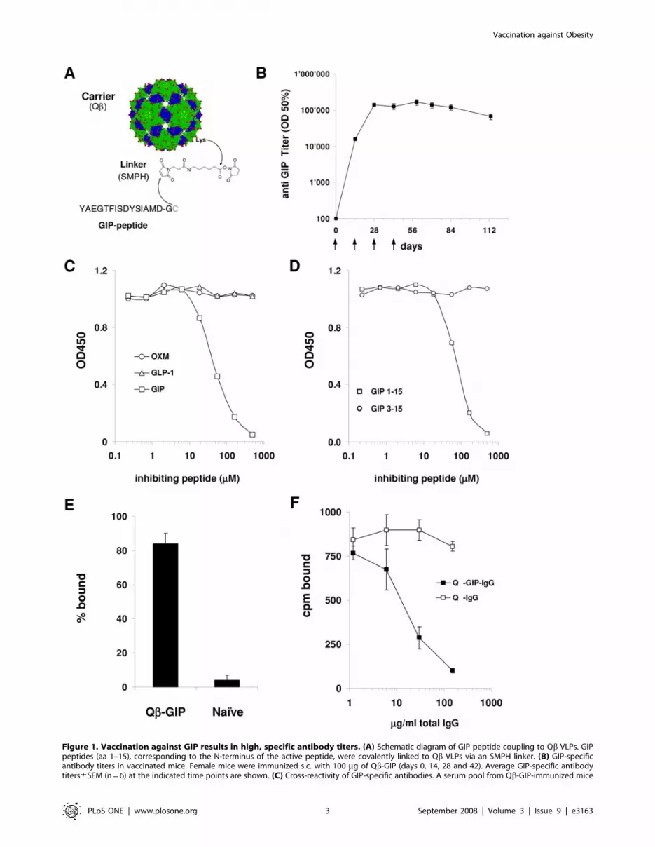
Figure 1. Vaccination against GIP results in high, specific antibody titers. (A) Schematic diagram of GIP peptide coupling to Qb VLPs. GIPpeptides (aa 1–15), corresponding to the N-terminus of the active peptide, were covalently linked to Qb VLPs via an SMPH linker. (B) GIP-specificantibody titers in vaccinated mice. Female mice were immunized s.c. with 100 mg of Qb-GIP (days 0, 14, 28 and 42). Average GIP-specific antibodytiters6SEM (n = 6) at the indicated time points are shown. (C) Cross-reactivity of GIP-specific antibodies. A serum pool from Qb-GIP-immunized mice
Vaccination against Obesity
PLoS ONE | www.plosone.org 3 September 2008 | Volume 3 | Issue 9 | e3163

Figure 2. Vaccination against GIP protects against diet-induced obesity. (A) Body weight gain in immunized mice. Female mice wereimmunized (days 0, 14, 28, 42 and 133) with 100 mg of Qb-GIP or Qb VLPs and placed on a high fat diet (35% fat w/v). The average body weight+/2SEM (n = 6) is shown. Body weight gain was was significanty reduced in Qb-GIP- compared to Qb VLP-immunized animals from day 70 onwards(two way ANOVA F(1,80) = 18.55, p,0.0001). (B) Body composition of mice shown in (A) was measured by DEXAscan on day 142. Average total bodymass, lean and fat tissue mass+/2SEM (n = 6) are shown. A significant reduction in fat content (p = 0.01) was observed between the Qb-GIP- and Qb-vaccinated group as determined by t-test. DEXAscan images of one representative animal per group are shown. (C) Macroscopic analysis of miceafter 142 days of treatment with Qb-GIP or Qb VLPs. A representative mouse from each group from the experiment described in Figure 3A–B isshown. (D) Body weight gain in immunized mice on a standard rodent diet. Female mice were immunized (days 0, 14, 28, 42 and 112) with Qb-GIP orQb VLPs and fed a standard diet (4% fat w/v) during the whole experiment. Average body weights+/2SEM (n = 5) are shown. No significant differencebetween the two experimental groups was observed as determined (two way ANOVA F(1/88) = 0.81, p = 0.6751).doi:10.1371/journal.pone.0003163.g002
was incubated with increasing concentrations of GIP, GLP-1 or oxyntomodulin (OXM). The amount of free antibody was quantified by ELISA. (D)Recognition of the N-terminus of full length GIP. A serum pool from Qb-GIP-immunized mice was preincubated either with GIP1–15 or GIP3–15 andfree antibodies quantified by ELISA. (E) Sequestration of GIP in vivo. Qb-GIP immunized mice or naı̈ve mice were challenged i.v. with 1 ng of I125-GIP.30 minutes later the amount of antibody-bound GIP was determined. The percentage of antibody bound GIP6SEM (n = 4) is shown. (F) Antibodymediated blocking of GIP binding to its receptor. I125-GIP was incubated with purified total IgG from Qb-GIP or Qb immunized mice and added toCHOK1-GIPR cells and bound GIP determined after an overnight incubation at 4uC. The final concentration of GIP was 20 ng/ml and theconcentration of total IgG is shown on the x-axis. Error bars represent standard deviations from triplicates.doi:10.1371/journal.pone.0003163.g001
Vaccination against Obesity
PLoS ONE | www.plosone.org 4 September 2008 | Volume 3 | Issue 9 | e3163

Figure 3. Vaccination against GIP increases energy expenditure and metabolic rate. Indirect calorimetry in immunized mice. Female micewere immunized (days 0, 14, 28, 42 and 125) with Qb-GIP (n = 8) or Qb VLPs (n = 10) and placed on a high fat diet. Indirect calorimetry was performedon half of the group on day 128 and on the other half on day 139. Combined data from these measurements are shown. (A) Oxygen consumption(VO2). The left panel shows average oxygen consumption+/2SEM. Qb-GIP-vaccinated animals display statistically, significantly increased VO2(p,0.0001) over the 24 h period. Average oxygen consumption+/2SEM during the dark and light phase is shown on the right. VO2 was significantlyincreased in Qb-GIP-vaccinated animals compared to Qb controls in both the dark (p = 0.02) and light phase (p = 0.02). (B) Resting metabolic rate(RMR). RMR was increased in Qb-GIP-vaccinated animals compared to Qb VLP controls (p = 0.05). (C) Physical activity was determined by measuringbeam brakes over a 24 h period. No significant differences were observed between the two experimental groups. (D) Respiratory quotient (RQ) wasmeasured for 24 hours during the dark and light phase. Average RQ6SEM is shown. RQ is defined as VCO2 (L)/VO2 (L). The difference observedbetween the two experimental groups did not reach statistical significance. (E) Food intake. Food intake was monitored over three consecutive dayafter the energy expenditure experiment. Average daily food intake in mg/g body weight+/2SEM (n = 5 are shown). No statistically significantdifference was observed between the experimental groups (p.0.05). All statistical analyses were performed by two-sided t-tests.doi:10.1371/journal.pone.0003163.g003
Vaccination against Obesity
PLoS ONE | www.plosone.org 5 September 2008 | Volume 3 | Issue 9 | e3163
of preferential burning of fat in the treated group. However, the
observed difference in RQ did not reach statistical significance
(Figure 3D). No differences in food intake were observed between
the experimental groups determined during three consecutive days
after the energy expenditure experiment (Figure 3E). Taken
together these data indicate that the reduced body weight gain in
Qb-GIP-vaccinated mice fed a high fat diet was rather due to
higher energy expenditure than lower energy intake or increased
activity.
Glucose homeostasis is unaffected after vaccinationagainst GIP
Since GIP is a key incretin, it was necessary to test whether
vaccination against GIP may disturb glucose homeostasis. First,
non-fasted and fasted blood glucose levels were measured at
regular intervals in vaccinated mice on a high fat diet. No
significant differences in blood glucose levels were observed
between Qb-GIP- and Qb-vaccinated animals (Figure 4A–B),
suggesting that vaccination against GIP does not interfere with
glucose homeostasis. To further explore overall blood glucose
levels, serum samples from vaccinated mice were collected at bi-
weekly intervals and the fructosamine content determined.
Fructosamine levels provide a retrospective reading of blood
glucose for the 2 weeks prior to measurement [30,31]. As shown in
Figure 4C, no significant difference between the Qb-GIP- and
Qb-vaccinated mice was seen. Similar observations were made for
HbA1c levels, which provide a retrospective picture of average
blood glucose levels up to 3 months before analysis (Figure 4D).
Taken together, these results demonstrate that active vaccination
against GIP does not result in increased blood glucose levels. To
further elucidate the consequences of ablation of GIP on glucose
homeostasis, vaccinated mice fed a high fat diet were subjected to
oral glucose tolerance tests (OGTT). OGTT were performed at
the end of the experiment on day 142 in Qb-GIP- and Qb-
immunized mice. Glucose was eliminated in both experimental
groups with similar kinetics, indicating that glucose tolerance was
not impaired by vaccination against GIP (Figure 4E). Additionally,
OGTT were performed at monthly intervals. As shown in
Figure 4F, no difference in oral glucose tolerance was observed
between Qb-GIP- and Qb VLP-immunized mice throughout the
experiment. Taken together, these findings suggest that active
vaccination against GIP prevents excessive body weight gain and
adiposity without altering glucose homeostasis.
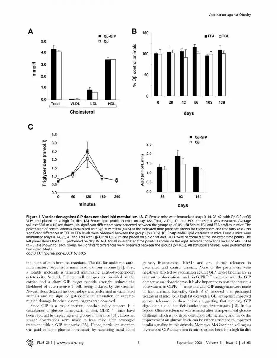
Vaccination against GIP does not interfere with lipidmetabolism
To elucidate the effects of vaccination against GIP on lipid
metabolism, serum lipid profiles were determined in vaccinated
animals fed a high fat diet 4 months after the first injection. No
significant differences were observed in total cholesterol, HDL,
LDL or vLDL in Qb-GIP-immunized mice compared to control
mice (Figure 5A). Likewise, triglyceride and free fatty acid profiles
were monitored at regular interval in vaccinated and control
animals placed on a high fat diet. As shown in Figure 5B, no
significant difference between the two experimental groups was
observed. Since GIP is known to promote triglyceride clearance
from the circulation [13,14], we examined whether postprandial
lipid clearance was affected in vaccinated animals. Oral lipid
tolerance tests (OLTT) were performed in vaccinated mice fed a
high fat diet on days 36, 93 and 163. Olive oil was administered by
oral gavage and the TGL concentration in the blood determined
at the indicated time points. Lipids were eliminated with similar
kinetics in both experimental groups, indicating that TGL
elimination was not impaired by vaccination against GIP
(Figure 5C)
Discussion
Here we describe a potential new treatment for obesity based on
immunoneutralization of GIP, a gut hormone that has recently been
shown to link over-nutrition to obesity [17]. GIP is a particularly
attractive target since it is a peripheral hormone released into the
circulation, where it can be easily captured by antibodies. Our
approach is based on active vaccination using VLPs displaying GIP
peptides on their surface. The highly repetitive display of peptides
together with the strong T-helper cell epitopes provided by the VLP
allowed for self tolerance to be overcome and led to a potent
antibody response against GIP. Detailed analysis revealed that the
induced antibodies were highly specific, since they showed no cross-
reactivity with the closely related peptide hormones, GLP-1 and
oxyntomodulin. As anticipated from studies in GIPR2/2 mice, Qb-
GIP vaccination resulted in reduced body weight gain and reduced
fat accumulation in mice fed a high fat diet. In contrast, active
vaccination against GIP did not affect normal age-related body
weight gain in mice fed a standard rodent diet. These findings are in
accordance with previous findings in GIPR2/2 mice showing little
[17] or no difference [32] in age-related body weight gain in mice
fed normal chow. Interestingly in the study shown here, the weight
of Qb-GIP immunized mice started to diverge from the control
mice, only roughly 6 weeks after high titers had been reached
around 70 days after the first immunization. This apparent delayed
response is best explained by the observation that mice fed normal
chow display a similar weight gain as mice fed a high fat diet during
the first 10 weeks of the experiment (week 8–18). Only after 10–12
weeks animals fed a high fat diet start to significantly gain more
weight and become obese. Similarly, Miyawaki and colleagues
observed divergence of animals fed a high fat diet compared to
animals fed a normal chow after only relatively late after 10–12
weeks of diet at an age of 18 weeks [17]. Hence, these observations
suggest that only after prolonged high fat feeding excess fat in the
diet is stored in the form of adipose tissue. Taken together these
observations suggest that GIP maximizes the accumulation of fat
tissue when energy rich food is consumed. This is of particular
interest, since one of the major reasons for the increased incidence
of obesity in humans is the unlimited access and consumption of
energy rich food. The reduced body weight gain is best explained by
the observed increase in energy expenditure, which was indepen-
dent of physical activity, since no changes were observed between
treated and control animals for this parameter. Although, at the end
of the experiment no differences in food intake were observed, we
cannot rule out a difference in food intake between the experimental
groups early in the experiment which might account for parts of the
differences in weight gain observed. In contrast to our findings,
Hansotia et al. recently reported increased energy expenditure in
single and double incretin receptor knockout mice, which could
partly be attributed to increased locomotor activity in the dark
phase [32]. Like the present study, they also observed an increase in
resting metabolic rate in GIPR2/2 mice. Thus, GIP appears to
affect energy expenditure by reducing resting metabolic rate and
activity. The fact that we saw a trend with no statistically significant
effect on locomotor activity, but found significant differences in
resting metabolic rate, might be explained by different GIP
thresholds needed to exert these effects. In the case of active
vaccination against GIP, where GIP is unlikely to be completely
blocked, resting metabolic rate is mostly affected.
While beneficial in reducing body weight gain, vaccination
against GIP, a self-antigen, may raise concerns associated with the
Vaccination against Obesity
PLoS ONE | www.plosone.org 6 September 2008 | Volume 3 | Issue 9 | e3163
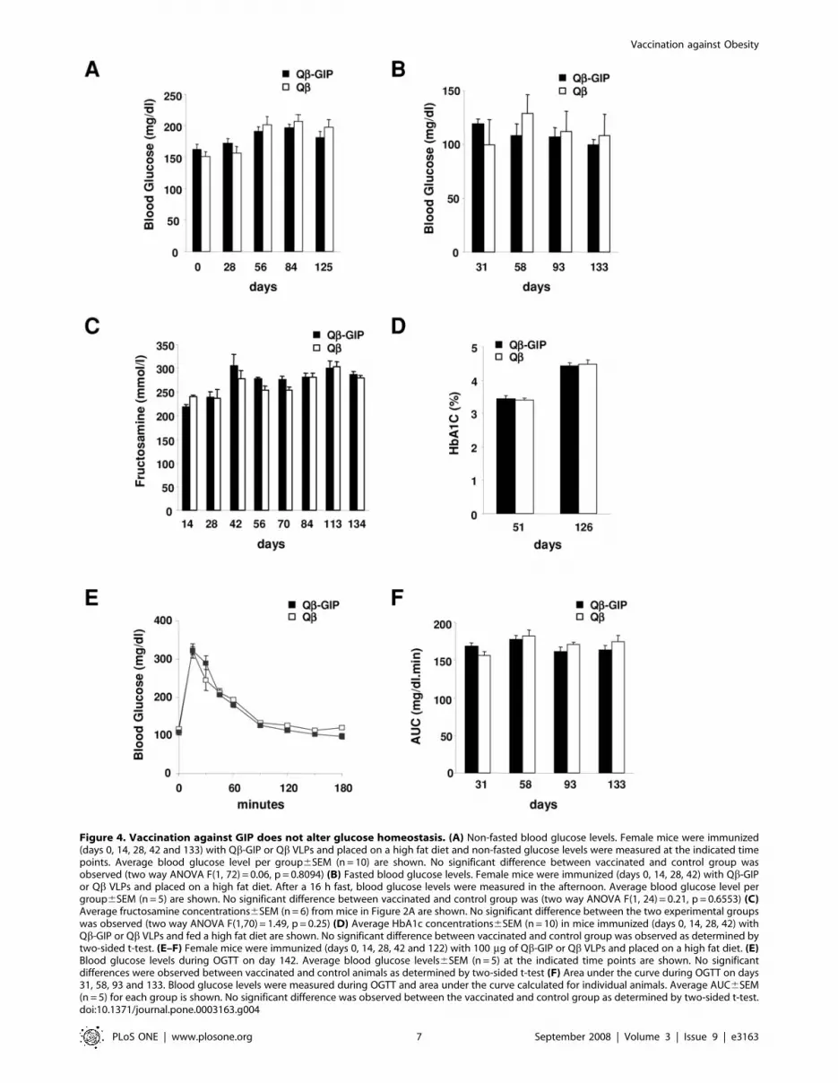
Figure 4. Vaccination against GIP does not alter glucose homeostasis. (A) Non-fasted blood glucose levels. Female mice were immunized(days 0, 14, 28, 42 and 133) with Qb-GIP or Qb VLPs and placed on a high fat diet and non-fasted glucose levels were measured at the indicated timepoints. Average blood glucose level per group6SEM (n = 10) are shown. No significant difference between vaccinated and control group wasobserved (two way ANOVA F(1, 72) = 0.06, p = 0.8094) (B) Fasted blood glucose levels. Female mice were immunized (days 0, 14, 28, 42) with Qb-GIPor Qb VLPs and placed on a high fat diet. After a 16 h fast, blood glucose levels were measured in the afternoon. Average blood glucose level pergroup6SEM (n = 5) are shown. No significant difference between vaccinated and control group was (two way ANOVA F(1, 24) = 0.21, p = 0.6553) (C)Average fructosamine concentrations6SEM (n = 6) from mice in Figure 2A are shown. No significant difference between the two experimental groupswas observed (two way ANOVA F(1,70) = 1.49, p = 0.25) (D) Average HbA1c concentrations6SEM (n = 10) in mice immunized (days 0, 14, 28, 42) withQb-GIP or Qb VLPs and fed a high fat diet are shown. No significant difference between vaccinated and control group was observed as determined bytwo-sided t-test. (E–F) Female mice were immunized (days 0, 14, 28, 42 and 122) with 100 mg of Qb-GIP or Qb VLPs and placed on a high fat diet. (E)Blood glucose levels during OGTT on day 142. Average blood glucose levels6SEM (n = 5) at the indicated time points are shown. No significantdifferences were observed between vaccinated and control animals as determined by two-sided t-test (F) Area under the curve during OGTT on days31, 58, 93 and 133. Blood glucose levels were measured during OGTT and area under the curve calculated for individual animals. Average AUC6SEM(n = 5) for each group is shown. No significant difference was observed between the vaccinated and control group as determined by two-sided t-test.doi:10.1371/journal.pone.0003163.g004
Vaccination against Obesity
PLoS ONE | www.plosone.org 7 September 2008 | Volume 3 | Issue 9 | e3163
induction of auto-immune reactions. The risk for undesired auto-
inflammatory responses is minimized with our vaccine [33]. First,
a soluble molecule is targeted minimizing antibody-dependent
cytotoxicity. Second, T-helper cell epitopes are provided by the
carrier and a short GIP target peptide strongly reduces the
likelihood of auto-reactive T-cells being induced by the vaccine.
Nevertheless, detailed histopathology was performed in vaccinated
animals and no signs of gut-specific inflammation or vaccine-
related damage in other visceral organs was observed.
Since GIP is a major incretin, another safety concern is a
disturbance of glucose homeostasis. In fact, GIPR2/2 mice have
been reported to display signs of glucose intolerance [34]. Likewise,
similar observations were made in lean mice after prolonged
treatment with a GIP antagonist [35]. Hence, particular attention
was paid to blood glucose homeostasis by measuring basal blood
glucose, fructosamine, HbA1c and oral glucose tolerance in
vaccinated and control animals. None of the parameters were
negatively affected by vaccination against GIP. These findings are in
contrast to observations made in GIPR2/2 mice and with the GIP
antagonist mentioned above. It is also important to note that previous
observations in GIPR2/2 mice and with GIP antagonists were made
in lean animals. Recently, Gault et al. reported that prolonged
treatment of mice fed a high fat diet with a GIP antagonist improved
glucose tolerance in these animals suggesting that reducing GIP
signaling could be beneficial under these circumstances [18]. In this
reports Glucose tolerance was assessed after intraperitoneal glucose
challenge which is not dependent upon GIP signaling and hence the
improvement on glucose levels can be rather attributed to improved
insulin signaling in this animals. Moreover McClean and colleagues
investigated GIP antagonism in mice that had been fed a high fat diet
Figure 5. Vaccination against GIP does not alter lipid metabolism. (A–C) Female mice were immunized (days 0, 14, 28, 42) with Qb-GIP or QbVLPs and placed on a high fat diet. (A) Serum lipid profile in mice on day 122. Total, vLDL, LDL and HDL cholesterol was measured. Averagevalues6SEM (n = 10) are shown. No significant differences were observed between the groups (p.0.05). (B) Serum TGL and FFA profiles in mice. Thepercentage of control animals immunized with Qb VLPs6SEM (n = 5) at the indicated time point are shown for triglycerides and free fatty acids. Nosignificant differences in TGL or FFA levels were observed between the groups (p.0.05). (C) Postprandial lipid clearance in mice. Female mice wereimmunized (days 0, 14, 28, 41 and 126) with Qb-GIP or Qb VLPs and placed on a high fat diet. OLTT were performed at the indicated time points. Theleft panel shows the OLTT performed on day 36. AUC for all investigated time points is shown on the right. Average triglyceride levels or AUC6SEM(n = 5) are shown for each group. No significant differences were observed between the groups (p.0.05). All statistical analyses were performed bytwo sided t-tests.doi:10.1371/journal.pone.0003163.g005
Vaccination against Obesity
PLoS ONE | www.plosone.org 8 September 2008 | Volume 3 | Issue 9 | e3163

for 160 days prior to the treatment with a GIP antagonist.
Interestingly in these mice they found that a 50 day treatment with
(Pro3)GIP led to weight loss, and significant improvement of glucose
tolerance after both, i.p. challenge and feeding, suggesting that in
severely obese mice antagonism of GIP is even beneficial for glucose
homeostasis [20]. Hence, removal of GIP signaling appears to affect
glucose homeostasis differently depending on the nutritional state of
the animals. Whereas GIPR2/2 animals show glucose intolerance
on normal chow, the more recent work of McClean and colleagues
show improvement of these parameters with their antagonists in
severely obese mice after prolonged high fat feeding. Here these
parameters were investigated in mice fed a high fat diet and most of
our measurement were performed in between those two extreme
situations. Hence, in view of these observations, the lack of notable
effect on oral glucose tolerance in our experiments may not be that
unexpected. Alternatively the differences observed here may be due
to an incomplete neutralization of GIP by induced antibodies, still
allowing for partial signaling to occur.
Investigations of lipid metabolism revealed no changes in
vLDL-, LDL- and HDL-cholesterol concentrations in the serum of
Qb-GIP-vaccinated mice. Likewise, triglyceride and free fatty acid
levels, as well as postprandial lipid clearance were not changed in
vaccinated animals. Taken together, this study shows that active
immunization against GIP leads to a strong reduction in body
weight gain in mice on a high fat diet and without deteriorating
blood glucose or lipid homeostasis.
Moreover in a preliminary experiment performed in obese male
mice, suggests that active vaccination against GIP not only
prevents excessive weight gain in animals fed a high fat diet but
can also enhance weight loss in obese mice (Figure S3).
The role of GIP in glucose and lipid metabolism is well
documented in several animal species [12,36]. Likewise, the incretin
activity of GIP and GLP1 is well established in humans [37–39].
Based on these observations DPP-IV inhibitors have been
developed and drugs like Sitagliptin and Vildagliptin are now on
the market as a novel class of type II diabetes drugs [40–42]. These
antagonist, prevent the specific cleavage of the incretin hormones
thereby increasing their half-lives and leading to increased insulin
secretion. Interestingly, GIP has been reported to have a reduced
incretin effect in type II diabetic patients whereas the insulinotropic
effect of GLP is preserved in this patient population [43–45]. These
findings suggest on one hand that the major effect of DPP-IV
antagonist is mediated by the stabilization of GLP1 and on the other
hand that elimination of GIP should not have a major impact on
glucose homeostasis in type II diabetic patients. However, whereas
the incretin effect of GIP has been described in detail in humans
further investigations will be required to understand the potential
role of GIP in linking over-nutrition with obesity in man. Hence,
provided that further studies demonstrate a role of GIP in linking
over nutrition to the development of obesity in humans, and further
preclinical safety studies do not reveal toxic effects of concern, we
believe that active vaccination against GIP may have the potential
to be a convenient therapy for the treatment of obesity.
Materials and Methods
Animals8 week old, C57BL/6 mice (,20 g) were purchased from Harlan
Netherlands (Horst, The Netherlands). Animals were housed in a
pathogen free facility and were allowed to acclimatize for 2 weeks
prior to immunization. In all experiments mice were caged in groups
with the exception of the period during which energy expenditure
and food intake was determined. The therapeutic experiment shown
in Figure S3 was performed with male mice, all other experiments
were performed with female mice. Unless otherwise indicated
animals were fed a high fat diet (35% fat w/v (Diet 2127), Provimi
Kliba, Switzerland), ad libitum, for the duration of the experimental
period and had free access to water. Experiments were in accordance
with Swiss Federal Veterinary Office (BVET) guidelines.
Vaccine productionGIP fragment 1–15 including a GC linker sequence fused to the
C-terminus of the GIP fragment (YAEGTFISDYSIAMDGC) was
chemically synthesized according to standard procedures and
coupled to Qb VLPs as previously described [46,47]. Briefly, A
solution of 1 ml of 2.0 mg/ml Qb VLPs in 20 mM Hepes,
150 mM NaCl pH 7.2 was reacted for 30 minutes with 67.2 ml of
a 50 mM stock solution of SMPH (Pierce) in DMSO at 25uC. The
reaction solution was subsequently dialyzed twice for 2 hours
against 2 L of 20 mM Hepes, 150 mM NaCl, pH 7.2 at 4uC.
Then 1 ml derivatized Qb VLP was reacted with 286 ml of a
10 mM peptide solution for 2 hours at 20uC in 20 mM Hepes,
150 mM NaCl, pH 7.2. The coupling reactions were then
centrifuged at 13 000 rpm for 5 minutes and the supernatants
were collected and dialyzed once for 2 hours and then overnight
against 2 L of 20 mM Hepes, 150 mM NaCl, pH 7.2 at 4uC. The
covalent chemical coupling of GIP peptides to Qb VLPs was
assessed by SDS-PAGE using 12% Nu-PAGE gels (Invitrogen).
Coomassie blue stained gels of the coupling reaction demonstrated
the appearance of bands with molecular weights corresponding to
those predicted for GIP peptides covalently linked to QbVLPs
Coupling bands corresponding to one, two, three or four peptides
coupled per subunit could be identified. Coupling efficiency [i.e.
mol Qb-GIP/mol Qb monomer (total)] was estimated, by
densitometric analysis of the Coomassie blue stained SDS-PAGE,
to be between 1.6–2.2 GIP fragments per Qb monomer.
Immunization and in vitro assaysAnimals were immunized subcutaneously with 100 mg (mice) or
300 mg (rats) of Qb-GIP or Qb VLPs, diluted in 200 ml PBS, at
the indicated time points. GIP-specific antibody titers were
determined by ELISA according to standard protocols using
porcine GIP (Bachem #H-6220) at a concentration of 2.5 mg/ml
for coating. The ELISA titer is defined as the reciprocals of the
serum dilution needed to reach half the maximum optical density
at saturation. For competition ELISAs, a serum pool from Qb-
GIP-immunized mice was diluted 1:2500 and incubated with
increasing concentrations of porcine GIP (Bachem #H-6220),
murine GLP-1 (Bachem #H6795) or murine oxyntomodulin
(Bachem #H6058). After overnight incubation, the amount of free
antibody was quantified in a GIP-specific ELISA. For in vivo
binding experiments, Qb-GIP-immunized and naı̈ve mice were
challenged intravenously with 1 ng I125-GIP (Bachem H-5016)
and 30 minutes after challenge animals were sacrificed and serum
collected by cardiac puncture. Total IgG was pulled down from
serum samples with protein G beads (Amersham, #17-0618-02).
Protein G beads associated and free radioactivity was measured
and the percentage antibody bound GIP calculated.
Generation of CHOK1 cells over-expressing the GIP
receptor and binding studies. The human GIP receptor was
amplified by RTPCR from a 3T3 cell line expressing the human
GIP receptor (a kind gift from Dr. Jens Holst) using primer
ATTTAATTAAGGCGCGCCACCATG ACTA CCTCTCCG-
ATCC as forward and AATTAATTAACTCGAGCT AGCAGT-
AACTTTCCAAC TCC as reverse primer. The PCR product was
then cloned into pBP [48]. The resulting construct was named pBP-
GIPR. VSV G pseudotyped retroviruses were made by co-
transfection of pBP-GIPR with pVPack-GP and pVPack-VSV-G
Vaccination against Obesity
PLoS ONE | www.plosone.org 9 September 2008 | Volume 3 | Issue 9 | e3163
(Stratagene) according to manufacturer’s instruction. 48 h after
transfection supernatants were collected and added to CHOK1
cells. One day later cells were passaged under puromycin selection
(10 mg/ml). Transduced cell populations were than cultivated under
selective pressure. For binding assays, 16105 CHOK1-GIPR/well
were seeded 2 days prior to binding assays in 24 well plates in each
well. Blocking assays were performed in a final volume of 500 ml.
I125-GIP (20 ng/ml) was incubated in the presence of the indicated
amounts of purified total IgG form Qb-GIP or Qb immunized mice
in binding buffer (DMEM, 1% BSA, 10 mM Hepes ph 7.4) for 1 h
at 4uC. This solution was than added to adherent cells and the cells
were incubated over night at 4uC. Cells were then washed with
binding buffer, resuspended in 500 ml 0.1 M NaOH and
transferred to 1.5 ml scintillation fluid and bound GIP
determined with a b-counter.
Body weight and body composition analysisAnimals were weighed using a high precision scale. Body
composition was determined by dual energy X-ray absorption
scan (Piximus Series Densitometer, GE Medical Systems,
Madison, USA) at the ICS (Illkirch, France).
Glucose and lipid homeostasisTotal blood glucose, cholesterol, TGL, HDL, LDL, vLDL and
FFA levels were measured after a 16 h fast. BGL were determined
using a glucometer (Accu-Chek Aviva, Roche). Cholesterol, TGL
and FFA levels were determined by enzymatic assays on an
Olympus AU400 automated laboratory work station at the ICS
(Illkirch, France). Lipid profiles were determined by F.P.L.C.
(Dionex). HbA1c levels were determined from whole blood using
the A1cNow monitoring kit (Metrika #0520105). For OGTT,
mice were fasted for 16 h and then 2 g/kg body weight glucose in
water was administered by oral gavage. BGL were determined at
the indicated time points. For OLTT, mice were fasted for 16 h
and then administered 8.35 ml/g olive oil by oral gavage. TGL
levels were measured from whole blood using the CardioChek
P.A. analyzer (PTS Inc.).
Energy expenditure experimentsEnergy expenditure, activity and respiratory quotient (RQ) were
measured for 24 hours (dark and light phase) by using two open-
circuit calorimetry systems (Integra system, AccuScan Instruments
Inc., Columbus OH). Mice were allowed to adapt to the metabolic
cages for 5 days. For measurements of oxygen consumption (VO2)
and carbon dioxide production (VCO2), mice were placed in air-tight
respiratory cages that were continuously ventilated with a flow rate of
about 1 l/min. For each cage, air was sampled for 20 seconds at
2 min intervals. RQ was defined as VCO2 (L)/VO2 (L). RMR was
calculated by taking the average of the 3 lowest VO2 readings in each
experimental group during the light phase. Physical activity was
determined by measuring beam breaks over a 24 h period. Physical
activity was monitored by 3 arrays of 16 infrared light beam sensors
and then converted into distance travelled in cm. Data were analyzed
with AccuScan Integra ME software.
HistopathologyTissue samples were fixed in 4% paraformaldehyde, sectioned
and stained with H&E according to standard methods. Histolog-
ical appraisal was made by a qualified veterinary pathologist.
Statistical analysisFor the analysis of body weight, blood glucose and fructosamine
data two-way ANOVA were used. All other statistical analyses
were made using a two-sided student’s t-test. Areas under the
curve for the OGTT were determined using Prism Graphpad
software.
Supporting Information
Figure S1 No cross reaction of Qb-GIP induced antibodies with
GLP1 and OXM in vivo. Qb-GIP immunized mice. mice were
challenged i.v. with 1 ng of I125-GIP, I125OXM or I125 GLP1.
As a control naı̈ve mice were challenged with 1 ng of I125-GIP.
30 minutes later the amount of antibody-bound GIP, OXM or
GLP1 was determined. The percentage of antibody bound GIP,
OXM or GLP-16SEM (n = 4) is shown. Whereas most of the
injected GIP was found associated with Antibodies only back-
ground levels of OXM or GLP1 were found associated with the
antibody fraction in Qb-GIP immunised mice.
Found at: doi:10.1371/journal.pone.0003163.s001 (0.03 MB EPS)
Figure S2 Vaccination against GIP does not induce inflamma-
tion in the GIT. Female, C57BL/6 mice were immunized
subcutaneously with 100 mg of Qb-GIP or Qb VLP on days 0,
14, 28 and 42. Animals were fed a high fat diet (35% fat w/v) from
the start of the experiment. Mice were sacrificed on day 99. Tissue
samples were fixed, sectioned and stained with H&E according to
standard methods. One representative mouse from each group is
shown. Histological analysis of the sample by a pathologist did not
reveal any signs of inflammation in the gut in Qb-GIP immunised
animals compared to Qb treated control animals. Similar results
were obtained when animals were culled either on day 36 or 142
and analyzed by a pathologist.
Found at: doi:10.1371/journal.pone.0003163.s002 (15.05 MB
EPS)
Figure S3 Vaccination against GIP results in weight loss in
obese male mice. Male mice were fed a high fat diet for 4 month.
By this time all animals were severely obese and had reached
weights between 45–50 g. The animals were then immunized
(days 0, 14, 36, 50 and 119) with Qb-GIP or Qb VLPs and kept on
a high fat diet (35% fat w/v). Fat content in the diet was reduced
to 20% fat (w/v) from day 42 onwards. Average changes in body
weight6SEM (n = 10) are shown. Qb-GIP treated animals lost
significantly more weight than Qb VLP-immunized animals from
day 70 onwards (two way ANOVA F(1,162) = 9.82, p = 0.0057).
Found at: doi:10.1371/journal.pone.0003163.s003 (0.05 MB EPS)
Acknowledgments
We thank Dr Paula Grest (Institute for Veterinary Pathology, University of
Zurich) for histopathological evaluation of tissue samples and imaging and
Dr. Jens Holst for providing a 3T3 cell line expressing the human GIP
receptor and Dr. Lidia Ivanova for the help with the cloning of the GIP
receptor.
Author Contributions
Conceived and designed the experiments: AF TAL MO PYW MFB PS.
Performed the experiments: AF KS MO. Analyzed the data: AF TAL KS
MO PYW MFB PS. Wrote the paper: AF TAL PYW MFB PS.
References
1. Kopelman PG (2000) Obesity as a medical problem. Nature 404: 635–
643.
2. Blackburn G (1995) Effect of degree of weight loss on health benefits. Obes Res 3
Suppl 2: 211s–216s.
Vaccination against Obesity
PLoS ONE | www.plosone.org 10 September 2008 | Volume 3 | Issue 9 | e3163

3. Bosello O, Armellini F, Zamboni M, Fitchet M (1997) The benefits of modest
weight loss in type II diabetes. Int J Obes Relat Metab Disord 21 Suppl 1:S10–13.
4. Goldstein DJ (1992) Beneficial health effects of modest weight loss. Int J Obes
Relat Metab Disord 16: 397–415.5. Greenway FL, Caruso MK (2005) Safety of obesity drugs. Expert Opin Drug Saf
4: 1083–1095.6. Ioannides-Demos LL, Proietto J, McNeil JJ (2005) Pharmacotherapy for obesity.
Drugs 65: 1391–1418.
7. Leung WY, Thomas GN, Chan JC, Tomlinson B (2003) Weight managementand current options in pharmacotherapy: orlistat and sibutramine. Clin Ther 25:
58–80.8. Flum DR, Salem L, Elrod JA, Dellinger EP, Cheadle A, et al. (2005) Early
mortality among Medicare beneficiaries undergoing bariatric surgical proce-dures. Jama 294: 1903–1908.
9. Heijboer AC, Pijl H, Van den Hoek AM, Havekes LM, Romijn JA, et al. (2006)
Gut-brain axis: regulation of glucose metabolism. J Neuroendocrinol 18:883–894.
10. Huda MS, Wilding JP, Pinkney JH (2006) Gut peptides and the regulation ofappetite. Obes Rev 7: 163–182.
11. Yamada Y, Seino Y (2004) Physiology of GIP–a lesson from GIP receptor
knockout mice. Horm Metab Res 36: 771–774.12. Kieffer TJ (2003) GIP or not GIP? That is the question. Trends Pharmacol Sci
24: 110–112.13. Ebert R, Nauck M, Creutzfeldt W (1991) Effect of exogenous or endogenous
gastric inhibitory polypeptide (GIP) on plasma triglyceride responses in rats.Horm Metab Res 23: 517–521.
14. Wasada T, McCorkle K, Harris V, Kawai K, Howard B, et al. (1981) Effect of
gastric inhibitory polypeptide on plasma levels of chylomicron triglycerides indogs. J Clin Invest 68: 1106–1107.
15. Eckel RH, Fujimoto WY, Brunzell JD (1979) Gastric inhibitory polypeptideenhanced lipoprotein lipase activity in cultured preadipocytes. Diabetes 28:
1141–1142.
16. Yip RG, Boylan MO, Kieffer TJ, Wolfe MM (1998) Functional GIP receptorsare present on adipocytes. Endocrinology 139: 4004–4007.
17. Miyawaki K, Yamada Y, Ban N, Ihara Y, Tsukiyama K, et al. (2002) Inhibitionof gastric inhibitory polypeptide signaling prevents obesity. Nat Med 8: 738–742.
18. Gault VA, McClean PL, Cassidy RS, Irwin N, Flatt PR (2007) Chemical gastricinhibitory polypeptide receptor antagonism protects against obesity, insulin
resistance, glucose intolerance and associated disturbances in mice fed high-fat
and cafeteria diets. Diabetologia 50: 1752–1762.19. Irwin N, McClean PL, O’Harte FP, Gault VA, Harriott P, et al. (2007) Early
administration of the glucose-dependent insulinotropic polypeptide receptorantagonist (Pro3)GIP prevents the development of diabetes and related
metabolic abnormalities associated with genetically inherited obesity in ob/ob
mice. Diabetologia 50: 1532–1540.20. McClean PL, Irwin N, Cassidy RS, Holst JJ, Gault VA, et al. (2007) GIP
receptor antagonism reverses obesity, insulin resistance and associated metabolicdisturbances induced in mice by prolonged consumption of high fat diet.
Am J Physiol Endocrinol Metab 293: E1746–1755.21. Bachmann MF, Rohrer UH, Kundig TM, Burki K, Hengartner H, et al. (1993)
The influence of antigen organization on B cell responsiveness. Science 262:
1448–1451.22. Ambuhl PM, Tissot AC, Fulurija A, Maurer P, Nussberger J, et al. (2007) A
vaccine for hypertension based on virus-like particles: preclinical efficacy andphase I safety and immunogenicity. J Hypertens 25: 63–72.
23. Chackerian B, Lowy DR, Schiller JT (1999) Induction of autoantibodies to
mouse CCR5 with recombinant papillomavirus particles. Proc Natl AcadSci U S A 96: 2373–2378.
24. Jegerlehner A, Tissot A, Lechner F, Sebbel P, Erdmann I, et al. (2002) Amolecular assembly system that renders antigens of choice highly repetitive for
induction of protective B cell responses. Vaccine 20: 3104–3112.
25. Rohn TA, Jennings GT, Hernandez M, Grest P, Beck M, et al. (2006)Vaccination against IL-17 suppresses autoimmune arthritis and encephalomy-
elitis. Eur J Immunol 36: 2857–2867.26. Sonderegger I, Rohn TA, Kurrer MO, Iezzi G, Zou Y, et al. (2006)
Neutralization of IL-17 by active vaccination inhibits IL-23-dependentautoimmune myocarditis. Eur J Immunol 36: 2849–2856.
27. Kozlovska TM, Cielens I, Dreilinna D, Dislers A, Baumanis V, et al. (1993)
Recombinant RNA phage Q beta capsid particles synthesized and self-
assembled in Escherichia coli. Gene 137: 133–137.
28. Deacon CF (2004) Circulation and degradation of GIP and GLP-1. Horm
Metab Res 36: 761–765.
29. Gault VA, Parker JC, Harriott P, Flatt PR, O’Harte FP (2002) Evidence that the
major degradation product of glucose-dependent insulinotropic polypeptide,
GIP(3–42), is a GIP receptor antagonist in vivo. J Endocrinol 175: 525–533.
30. Johnson RN, Metcalf PA, Baker JR (1983) Fructosamine: a new approach to the
estimation of serum glycosylprotein. An index of diabetic control. Clin Chim
Acta 127: 87–95.
31. Yue DK, Morris K, McLennan S, Turtle JR (1980) Glycosylation of plasma
protein and its relation to glycosylated hemoglobin in diabetes. Diabetes 29:
296–300.
32. Hansotia T, Maida A, Flock G, Yamada Y, Tsukiyama K, et al. (2007)
Extrapancreatic incretin receptors modulate glucose homeostasis, body weight,
and energy expenditure. J Clin Invest 117: 143–152.
33. Bachmann MF, Dyer MR (2004) Therapeutic vaccination for chronic diseases: a
new class of drugs in sight. Nat Rev Drug Discov 3: 81–88.
34. Miyawaki K, Yamada Y, Yano H, Niwa H, Ban N, et al. (1999) Glucose
intolerance caused by a defect in the entero-insular axis: a study in gastric
inhibitory polypeptide receptor knockout mice. Proc Natl Acad Sci U S A 96:
14843–14847.
35. Irwin N, Gault VA, Green BD, Greer B, McCluskey JT, et al. (2004) Effects of
short-term chemical ablation of the GIP receptor on insulin secretion, islet
morphology and glucose homeostasis in mice. Biol Chem 385: 845–852.
36. Yip RG, Wolfe MM (2000) GIP biology and fat metabolism. Life Sci 66:
91–103.
37. Andersen DK, Elahi D, Brown JC, Tobin JD, Andres R (1978) Oral glucose
augmentation of insulin secretion. Interactions of gastric inhibitory polypeptide
with ambient glucose and insulin levels. J Clin Invest 62: 152–161.
38. Dupre J, Ross SA, Watson D, Brown JC (1973) Stimulation of insulin secretion
by gastric inhibitory polypeptide in man. J Clin Endocrinol Metab 37: 826–828.
39. Elahi D, McAloon-Dyke M, Fukagawa NK, Meneilly GS, Sclater AL, et al.
(1994) The insulinotropic actions of glucose-dependent insulinotropic polypep-
tide (GIP) and glucagon-like peptide-1 (7–37) in normal and diabetic subjects.
Regul Pept 51: 63–74.
40. Mest HJ, Mentlein R (2005) Dipeptidyl peptidase inhibitors as new drugs for the
treatment of type 2 diabetes. Diabetologia 48: 616–620.
41. Ahren B, Simonsson E, Larsson H, Landin-Olsson M, Torgeirsson H, et al.
(2002) Inhibition of dipeptidyl peptidase IV improves metabolic control over a 4-
week study period in type 2 diabetes. Diabetes Care 25: 869–875.
42. Nauck MA, Meininger G, Sheng D, Terranella L, Stein PP (2007) Efficacy and
safety of the dipeptidyl peptidase-4 inhibitor, sitagliptin, compared with the
sulfonylurea, glipizide, in patients with type 2 diabetes inadequately controlled
on metformin alone: a randomized, double-blind, non-inferiority trial. Diabetes
Obes Metab 9: 194–205.
43. Meier JJ, Hucking K, Holst JJ, Deacon CF, Schmiegel WH, et al. (2001)
Reduced insulinotropic effect of gastric inhibitory polypeptide in first-degree
relatives of patients with type 2 diabetes. Diabetes 50: 2497–2504.
44. Nauck MA, Heimesaat MM, Orskov C, Holst JJ, Ebert R, et al. (1993) Preserved
incretin activity of glucagon-like peptide 1 [7–36 amide] but not of synthetic
human gastric inhibitory polypeptide in patients with type-2 diabetes mellitus.
J Clin Invest 91: 301–307.
45. Vilsboll T, Krarup T, Madsbad S, Holst JJ (2002) Defective amplification of the
late phase insulin response to glucose by GIP in obese Type II diabetic patients.
Diabetologia 45: 1111–1119.
46. Cielens I, Ose V, Petrovskis I, Strelnikova A, Renhofa R, et al. (2000) Mutilation
of RNA phage Qbeta virus-like particles: from icosahedrons to rods. FEBS Lett
482: 261–264.
47. Spohn G, Schwarz K, Maurer P, Illges H, Rajasekaran N, et al. (2005)
Protection against osteoporosis by active immunization with TRANCE/
RANKL displayed on virus-like particles. J Immunol 175: 6211–6218.
48. Morgenstern JP, Land H (1990) Advanced mammalian gene transfer: high titre
retroviral vectors with multiple drug selection markers and a complementary
helper-free packaging cell line. Nucleic Acids Res 18: 3587–3596.
Vaccination against Obesity
PLoS ONE | www.plosone.org 11 September 2008 | Volume 3 | Issue 9 | e3163