using multimedia and web3d to enhance anatomy teaching
TRANSCRIPT

www.elsevier.com/locate/compedu
Computers & Education 49 (2007) 32–53
Using multimedia and Web3D to enhance anatomy teaching
Harry Brenton *, Juan Hernandez, Fernando Bello, Paul Strutton,Sanjay Purkayastha, Tony Firth, Ara Darzi
Imperial College London, St Mary’s Hospital, Department of Surgical Oncology and Technology, Praed Street,
London W2 1NY, UK
Abstract
Anatomy teaching is undergoing significant changes due to time constraints, limited availability of
cadavers and technological developments in the areas of three-dimensional modelling and computer-assisted learning. This paper gives an overview of methods used to teach anatomy to undergraduate medical
students and discusses the educational advantages and disadvantages of using three-dimensional computer
models. A �work in progress� account is then given of a project to develop two Web3D resources to enhance
undergraduate tuition of the nervous system. Our approach is to support existing curricula using advanced
modelling tools and a variety of delivery mechanisms.
The first resource is a three-dimensional model of the adult brachial plexus: a network of nerves extend-
ing from the neck down to the shoulder, arm, hand, and fingers. This will be incorporated into existing
didactic classroom teaching under the supervision of an anatomy teacher. The second resource is a pieceof online courseware which will teach the embryological development of the brachial plexus. The delivery
method will be the WebSET framework, a collaborative environment that allows a teacher to manipulate
3D models over the Web in real time whilst providing explanation and help to students. In this way the
courseware can be used for both self-directed study and �virtual anatomy demonstrations� within an online
peer group.
� 2005 Elsevier Ltd. All rights reserved.
Keywords: Applications in subject areas; Distance education and telelearning; Interactive learning environments;
Multimedia/hypermedia systems; Virtual reality
0360-1315/$ - see front matter � 2005 Elsevier Ltd. All rights reserved.
doi:10.1016/j.compedu.2005.06.005
* Corresponding author.
H. Brenton et al. / Computers & Education 49 (2007) 32–53 33
1. Introduction
Human anatomy is the scientific study of the form, position, size and relationship of the struc-tures in the body. It can be divided into gross anatomy (the structure and positioning of organs),histology (the microscopic study of cells and tissues), embryology (the formation and early devel-opment of the foetus) and neuroanatomy (the study of the brain, spinal cord and peripheral ner-vous system).
This paper starts with an overview of common methods of teaching anatomy. It then describesthe technical process of creating computer-generated three-dimensional models of human anat-omy and discusses the advantages and disadvantages of their use within education. An accountis then given of an ongoing project at Imperial College London to create two Web3D resourcesto enhance tuition of the nervous system to undergraduate medical students. This project is at anearly stage of development and is a �work in progress� yet to be completed and evaluated. It isincluded in this paper to help illustrate the methodology behind building a Web-based three-dimensional application for teaching anatomy.
There is considerable debate within the medical community about the best ways to teach anat-omy. Interest in Computer Aided Learning is growing (Mitchell & Stephens, 2004) but this needsto be supported by further research to evaluate the impact of multimedia resources upon studentlearning.
2. Methods of teaching anatomy
2.1. Dissection and prosection
Since the Renaissance the dissection of cadavers has underpinned anatomy tuition (Porter,1999). Many believe that cadaveric dissection provides undergraduates with an essential founda-tion in medical science. Benefits include:
1. Learning the basic language of medicine needed to describe the structure (anatomy) and func-tion (physiology) of the human body
2. Establishing the primacy of the patient as a person as opposed to a series of diagrams or photosin a textbook
3. Acclimatising students to the realities of death4. Teaching manual dexterity and touch-mediated perception5. Introducing the concept of anatomical variation (there are significant internal differences
between humans)6. Gaining knowledge of the three-dimensional spatial relationship between structures7. Gaining communication skills within a small peer group (adapted from Aziz et al., 2002; Ellis,
2001).
Dissection is traditionally taught topographically by studying the anatomical structures in aparticular region of the body. A systems-based approach is now more common, the reproductivesystem or respiratory system, for example, being examined as discrete units. Prosections are

34 H. Brenton et al. / Computers & Education 49 (2007) 32–53
pre-dissected specimens; James et al. (2004) review six studies that compare the examination re-sults of students learning from prosections and dissections. The evidence is not conclusive butdoes indicate that undergraduates perform as well if not significantly better in examinations whenthey learn from prosections.
2.2. Lectures and problem-based learning
The lecture theatre allows basic anatomical facts and concepts to be transferred from one teacherto many students who can then supplement what they have learned with private study. This stalwartbastion of educational practice remains a useful way of disseminating knowledge efficiently and costeffectively. However lectures and books poorly convey the three-dimensional nature of anatomicalstructures and do not encourage collaboration or develop problem solving skills.
Problem-based learning (PBL) aims to address the weaknesses inherent in didactic instructionby encouraging independent thought and teamwork. In a typical PBL session a small group ofstudents is presented with a real-world medical problem such as a serious head injury. The tutoror �facilitator� tells students which parts of the case are related to anatomy and gives them a list ofappropriate resources such as textbooks, journals, videotapes and websites. Each student re-searches the scenario individually before returning to the group to collectively discuss and assesswhat they have learned.
2.3. Radiological imaging
Lectures, PBL sessions and dissection classes often incorporate radiological images. These aretypically magnetic resonance (MR) or computerised tomography (CT) scans that show two-dimensional cross-sections through the body (Fig. 1). Interpreting these images takes considerableexpertise because students have to mentally reconstruct a two-dimensional slice into a three-dimensional visualisation which may have been altered by disease or accident. The ability to cor-relate diagnostic scans with living patients is a skill that many junior doctors will find essential intheir future clinical practice.
2.4. The Visible Human Project and anatomy websites
In 1994 The Visible Human Project (Ackerman, 2004) provided colour scans of a middle-agedhuman male sliced into 1871 cross-sections at 1 mm intervals (Fig. 2). The Internet Atlas of
Human Gross Anatomy (Jastrow & Vollrath, 2002) is based on the visible human dataset andprovides a wealth of annotated material. Many similar websites incorporating the Visible HumanProject do not charge a registration fee and can be used by students for self-directed study orincorporated into classroom teaching. There is also a female visible human, as well as Chinese(Zhang et al., 2003) and Korean (Zhong, 2003) equivalents, that make use of recent technologicaldevelopments to produce very high fidelity images.
A survey of German undergraduates indicated a high demand for online anatomy resourceswith students reporting that they are more motivating and �fun� than textbooks (Jastrow & Holl-inderbaumer, 2004). The most valued material was relevant to examinations, included highquality images, allowed keyword searches and contained up to date information. In another

Fig. 1. Magnetic resonance scans showing two-dimensional cross-sections through the body: 1. Axial (top) viewpoint,
2. Coronal (front) viewpoint.
Fig. 2. Cryosections from the visible human project.
H. Brenton et al. / Computers & Education 49 (2007) 32–53 35
study, students who used Web-based material to learn anatomical landmarks performed better inmidterm and final examinations than a control group who did not have access to the material(Hallgren, Parkhurst, Monson, & Crewe, 2002). Self-assessment exercises and real time feedbackare an important component because they help students assess their own knowledge and highlightareas that require further study.

36 H. Brenton et al. / Computers & Education 49 (2007) 32–53
2.5. Combining a range of approaches
There is no prescribed template for anatomy tuition and most modern courses use a range ofmethods including imaging, classroom teaching, PBL and dissection of cadavers (Mitchell &Stephens, 2004). Classes in a dissection room are sometimes supported by demonstrations of�living anatomy� where students examine each other�s bodies in relation to the cadaver on thedissection table (Ellis, 2001). Likewise, a PBL session may incorporate the study of prosectionsin the dissecting room. The use of plastic models is common although these have been criticisedfor not showing natural human variation and giving a falsely sanitised and homogenous view ofanatomy.
Plastinated models differ significantly from plastic models because they are real anatomicalspecimens which have been preserved with reactive polymers. These have gained notoriety dueto Gunther Von Hagens Bodyworlds exhibition (Hagens, 2004). Other innovative approaches in-clude laparascopic imaging of live patients to demonstrate upper abdominal anatomy (Park et al.,2001) and plasticine models to teach embryology (Nunn, 2004).
2.6. Curricular changes
In the last 20 years there has been a severe reduction in the time devoted to anatomy teach-ing and a drop in staffing levels (Heylings, 2002). This trend is international (Aziz et al., 2002;Parker, 2002) and has contributed to a considerable debate within the medical communityabout the best methods of teaching anatomy in an efficient and cost effective manner (Jameset al., 2004).
2.7. Removing cadaveric dissection from the undergraduate curriculum?
The necessity for undergraduate dissection of human cadavers has been challenged. Forexample, The Peninsula Medical School in the UK has replaced cadaveric dissection with anapproach that relies upon multimedia, problem-based learning and computer imaging (McLach-lan, Bligh, Bradley, & Searle, 2004). They concentrate on anatomy that will be directly relevantto the management of clinical problems presented by patients and they believe that dissection ismore suitable for postgraduate specialities such as surgery. This approach has been challengedby those who believe multimedia, PBL and radiology should supplement rather than supplantdissection (Ellis, 2001; Mitchell & Stephens, 2004).
Both schools of thought agree that there needs to be more research done to evaluate whichteaching methods and techniques are best to train the next generation of doctors.
3. Three-dimensional computer generated anatomy models
This paper will now examine the use of anatomical three-dimensional modelling. To help illus-trate the methodology behind building a Web-based three-dimensional application we give a�work in progress� account of the development of two Web3D anatomy resources.

H. Brenton et al. / Computers & Education 49 (2007) 32–53 37
3.1. Modelling techniques
Websites and CD-ROMs produced by companies such as Primal Pictures (www.primalpictures.com; Ward, 2002) provide three-dimensional reconstructions of the human body with controls topeel back tissue and bone revealing previously hidden layers (Fig. 3). Creating these three-dimensional models involves a number of technical stages: scanning, enhancement, segmentation,and volume or surface rendering (Figs. 1 and 4).
3.1.1. ScanningA coordinated series of 2D images is acquired using a scanning technique such as CT or MRI
(Fig. 1) or by digitising cryosections (Fig. 2). Each image shows a slice through the body at regularintervals, typically of 1 mm. The type of imaging modality used can be varied to suit the require-ments of the disease/clinical indication. For example, CT is often used to image colorectal cancer
Fig. 3. Screenshot from the Interactive Thorax and Abdomen CD-Rom from Primal Pictures.

Fig. 4. Technical stages for creating three-dimensional anatomical models: (1) Segementation, (2) Reconstruction,
(3) Simplify mesh, (4) Surface rendering and (5) Texture mapping.
38 H. Brenton et al. / Computers & Education 49 (2007) 32–53
because it presents a detailed picture of how far the cancer has spread to areas surrounding thecolon. MRIs are often used to plan neurosurgery because they are good at imaging vital structuresin soft tissues which must be avoided when removing adjacent tumours.
3.1.2. Enhancement
The raw images are digitally manipulated to enhance image quality. Common techniques includecontrast enhancement, noise reduction and interpolation. Enhancement must be done with greatcare as it can emphasise image artifacts and even lead to a loss of information if not correctly used.
3.1.3. SegmentationVector drawing tools are used to trace a chosen region in a scan, such as the liver (Fig. 5). This
process also has the effect of electronically tagging an area so that it can be uniquely addressedand manipulated. Without segmentation unwanted regions may get included when the series of

Fig. 5. Segmentation: delineating the liver from surrounding imaging data.
H. Brenton et al. / Computers & Education 49 (2007) 32–53 39
2D images is reconstructed in 3D. Segmentation can be done manually but it is a slow and labo-rious task. Automated or semi-automated segmentation software (Bartz et al., 2004) can helpspeed up the process by using various techniques such as pattern recognition to help extractthe border of structures.
3.1.4. Volume or surface renderingOnce segmentation is complete, the software processes each 2D slice in turn to build a 3D vol-
ume dataset. Volume data is suitable for medical images because it shows the inside of solid ob-jects and allows outer layers to be cut away. However, real-time interaction is limited because eachnew view must be fully recomputed, requiring substantial processing power.
A common way of extracting polygonal surface information is to use the �marching cubes� algo-rithm (Lorensen & Cline, 2004) which tests each voxel in turn in order to locate the boundary of awanted object within a 3D array of voxel values. The data are then used to construct a numericaldescription of the surface. Because the surface is computed only once, the viewpoint can be rap-idly updated allowing for fast interaction. However, it is not possible to cut away outer layers toshow the inside of solid objects. Once a surface dataset is created it can be conventionally texture-mapped using software such as the Visualisation Toolkit (Schroeder, Martin, & Lorensen, 2004).
3.2. Advantages of three-dimensional modelling
The most obvious benefit of 3D models is the ability to view the spatial relationships betweenstructures from numerous viewpoints. This addresses a crucial educational need: �it takes time andpractice to develop the ability to visualise in three dimensions. . .insufficient ability to visualise isfrequently expressed by students who have difficulty identifying structures in the living body asrequired in clinical examination�(Heylings, 2002). This is in marked contrast to textbook dia-grams, photographs, MRIs and CT scans which do not show spatial relationships, require exper-tise to interpret and may lack sufficient detail to properly illustrate a particular teaching point.Diagrams can use shading to give the illusion of 3D (Fig. 6) but the viewpoint is fixed and cannot

Fig. 6. Textbook diagram of liver and gall bladder compared with a 3D reconstruction. (1) Textbook diagram and
(2) Three dimensional reconstruction.
40 H. Brenton et al. / Computers & Education 49 (2007) 32–53
be manipulated to reveal hidden details. Presenting radiological images alongside equivalent 3Dmodels helps students to learn the crucial skill of mentally transposing a two-dimensional imageonto a three-dimensional patient.
Diagrams and radiological scans show a static snapshot in time whereas 3D models can be pre-sented within a narrative timeline which a viewer can rewind, pause and fast forward at their conve-nience. This can be used to demonstrate anatomical development, physiology or the progress ofdisease (pathology) upon an organ. Having a clear timeframe is especially important in understand-ing pathology because the rate at which a disease spreads through the body is often not constant.
Three dimensional models are often presented as an equivalent to the �museum display� of spec-imens found in many dissecting rooms. Students can study an electronic museum display when-ever they choose and digital specimens do not have to be replaced every three years as mandated(in the UK) by the Anatomy Act (Heylings, 2002). Digital models are of a universal standardwhereas prosections can be of variable quality and have limited interaction. Three-dimensionalresources are a �lingua franca� that can be supplemented by language-specific annotations, didacticmaterial and self-test quizzes. Specialised CD-ROMs have addressed niche areas such as sportsinjuries (Betts, Saifuddin, Marone, Wolman, & Lambert, 2004).
3.3. Disadvantages of three-dimensional modelling
The construction of anatomically correct three-dimensional models demands much time andgreat patience. It is hard to compare the financial implications of traditional and multimedia instruc-tion because no studies have been published that analyse the time/cost benefits of anatomy teachingsupported by 3D technologies. Web3D is unlikely to be widely adopted by educators unless they cansee clear evidence that demonstrates its effectiveness and financial viability for their teaching. Thereare few common standards for validating Web3D resources, evaluation is often informal and rarelyexamines the longitudinal retention of knowledge by students. Pilot studies can paint an unrealistic

H. Brenton et al. / Computers & Education 49 (2007) 32–53 41
picture of the constraints that many university departments operate under: no matter how techni-cally innovative an approach is, budget holders will purchase tools that have a demonstrated abilityto raise learners to a prescribed level of proficiency within a set time period.
Three-dimensional models will always be iconic abstractions of the real body. Gauging the re-quired level of detail and stylisation for educational instruction is not an exact science, and too littleor too much information can be detrimental. Trial and error, qualitative feedback and personaljudgement are common strategies. The publication of more case studies of �real world� development,including mistakes, pitfalls and practical advice, would be beneficial to the academic community.
Authoring tools to create Web3D models have been slow to develop and complex to learn,increasing development time and cost. Many file formats for 3D models are proprietary and can-not be easily exchanged although the situation is improving with ISO approved standards such asX3D (X3D, 2004) and H-Anim H-Anim (2004) which encourage sharing, reuse of models andgreater integration with existing Learning Management Systems and Virtual Learning Environ-ments (Chittaro & Ranon, 2007).
Other disadvantages:
� Students require a PC with a moderately powerful 3D graphics card;� Web3D requires browser plug-ins which may fail to run under recent security restrictions added
to Microsoft Windows XP;� 3D navigation controls need to be intuitive, otherwise the interface can interfere with the
content;� Interacting with 3D models with a mouse does not convey a sense of touch or weight.
4. Two Web3D resources to enhance tuition of nervous system
At Imperial College London, we are experimenting with new ways of teaching anatomy usingemerging Web3D technologies and collaborative online environments. The project team consistsof a Professor of anatomy, an anatomy Lecturer, a surgeon, a Lecturer in medical graphics and alearning technologist. We are currently working on a project to create two Web3D resources tohelp teach the structure and function of a network of nerves called the brachial plexus. The firstresource is a three-dimensional model of the adult brachial plexus which will be incorporated intoexisting classroom teaching. The second resource is a piece of courseware which will teach theembryological development of the brachial plexus. By creating two different resources we aimto evaluate the educational benefits of Web3D for both self-directed study and didacticinstruction.
Even though this project is in the early stages of development it provides a useful example of themethodology used in creating medical Web3D resources.
4.1. The brachial plexus
The brachial plexus is a network of nerves originating from the spinal cord, emerging betweenthe vertebrae in the neck and extending down to the shoulder, arm, hand, and fingers (Fig. 7).Brachial refers to the arm, plexus means network. Our aim is to use computer-aided learning

Fig. 7. Sagittal (side) and coronal (front) views of an adult human brachial plexus.
42 H. Brenton et al. / Computers & Education 49 (2007) 32–53
to help students gain a deeper understanding of the embryological formation of the brachialplexus and how the nerves in a fully formed plexus supply sensation to the whole limb. A goodunderstanding of these concepts provides an educational scaffold from which to develop the diag-nostic skills necessary to infer the consequences of trauma or disease.
The nervous system is an important part of the undergraduate medical curriculum and one thatstudents often have difficulty conceptualising in three dimensions. This makes it a particularlysuitable topic for an educational Web3D application.
4.2. Student learning objectives
Students are expected to: memorise the structure of the brachial plexus; understand how thebrachial plexus relates to the peripheral nervous system and the anatomy of the upper limb; de-velop an ability to clinically evaluate how nerve injuries affect the upper limb.
4.3. Student learning strategies
The structure of the brachial plexus is very hard to remember; one student referred to it as �liketrying to memorise Clapham Junction� (the busiest railway station in the UK). At ImperialCollege London the brachial plexus is taught in a 1-h lecture and in practical sessions studyingprosections for between 60 and 90 min. This is supplemented by private study in the library withanatomy textbooks.
Printed diagrams and photographs are useful but they encourage superficial rote learning and asurface understanding of the subject. Two-dimensional representations are of limited educationalvalue because the nerve roots and fibres grow to form a complex arrangement within a three-dimensional space. Prosections go some way to acquiring this spatial visualisation but there is lim-ited time in the dissection room for extended study and they are not always available for revision.Cadaveric specimens are surprisingly different to what a surgeon sees and feels when operating on

H. Brenton et al. / Computers & Education 49 (2007) 32–53 43
a living patient. In living tissue the nerves are bound closely together around arteries and bone,whereas structures in prosections have often been teased apart to help demonstrate the structureof nerve roots and branches. In addition the process of fixation and storage significantly changesthe colour, texture and consistency of human tissue.
4.4. Development tools
We are using Alias/Wavefront Maya (Maya, 2004) as the central development environment.Maya is a powerful commercial software package used on numerous films and televisioncommercials. It has advanced tools for modelling and animation including skeletal simulation,dynamics simulation, particle effects, cloth, hair, fur and liquid simulation. A recent discounthas brought the cost within reach of most educational budgets (reduced from £5000 to £400).
4.5. Level of detail
The level of detail for the brachial plexus project is tailored to suit a medical student learningthe developing interconnections between nerves, muscle and bone for the first time.
4.6. Web3D resource 1: the adult brachial plexus model
The adult brachial plexus will consist of an interactive three-dimensional model which can bemanipulated to show the structure from multiple angles and viewpoints. For example, one viewwill show the nerves compacted together as they appear in the visible human dataset (Fig. 7), an-other will present the nerves in the form of a diagramatic representation (Fig. 8).
4.6.1. Modelling the spineA model of the lumbar vertebrae and pelvis has already been built (Fig. 9) to simulate a lumbar
puncture (John et al., 2001). The model was constructed from the visible human dataset usingstandard segmentation and reconstruction techniques and then converted into Virtual RealityModelling Language (VRML), a common language for describing and displaying 3D content(Dodd, Riding, & John, 2004).
Since this model comprises only the lumbar vertebrae, we are currently extending the spine toinclude the upper vertebrae and spinal cord at the point where the brachial plexus originates. Seg-mentation and reconstruction is time and labour intensive, therefore we are assessing the feasibil-ity of creating vertebrae directly within Maya. To do this we took screenshots of polygonalvertebrae built using an online version (Bessaud & Hersch, 2004) of the visible human dataset(Figs. 7 and 10). These were then used as a tracing image to provide guidelines for the creationof a subdivision surface model (Warren, 2002). Sub-division surfaces use curves (splines) ratherthan polygons (triangles) to describe a geometrical object. Preliminary results of this technique(Fig. 10) are encouraging, suggesting that vertebrae of sufficient fidelity to suit our purposescan be created using this procedure.
A comparison between the polygonal and spline-based models in Figs. 9–11 shows the signif-icant increase in quality gained from using sub-division surfaces. Maya supports many other ren-dering formats and techniques, which we will experiment with to further increase realism.
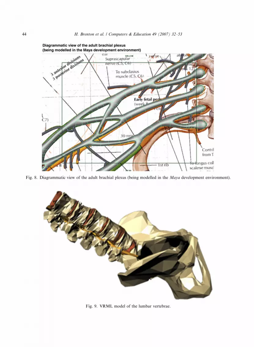
Fig. 8. Diagrammatic view of the adult brachial plexus (being modelled in the Maya development environment).
Fig. 9. VRML model of the lumbar vertebrae.
44 H. Brenton et al. / Computers & Education 49 (2007) 32–53

Fig. 10. T4 vertebrae. (1) Polygon model built with a visible human website http://diwww.epfl.ch/w3lsp/publications/
gigaserver/tvhssaws.html (2) Sub-division model created in Maya 1. Polygonal render of vertebrae 2. sub-division
render of vertebrae.
Fig. 11. Subdivision surface model of the lumbar vertebrae.
H. Brenton et al. / Computers & Education 49 (2007) 32–53 45
4.6.2. Modelling the nervesThe nerves have been modelled in Maya with reference to textbook diagrams and the visible
human dataset (Figs. 7 and 8). Once modelling of the upper vertebrae is complete, the nerves willbe added.
4.6.3. Future work: classroom delivery using a web browser, X3D and H-ANIMWe aim to use X3D (X3D, 2004) and H-ANIM (H-Anim, 2004) to deliver high quality spline-
based models that can be interacted with over the Internet in real time. X3D is an XML-based

46 H. Brenton et al. / Computers & Education 49 (2007) 32–53
successor to the VRML 97 standard, H-ANIM is a standard for representing three-dimensionalhumanoids. We plan to evaluate the model within existing didactic teaching, with an anatomydemonstrator using it as a teaching aid in the classroom.
The specifications for the finished resource are still in the planning stages. The tool will includean interactive X3D model of the adult brachial plexus with annotated pop-up labels explainingstructure and function. This will be supplemented by teaching material such as high qualitypre-rendered subdivision quicktime animations, two-dimensional flash diagrams, photographsof prosections, and links to relevant websites and academic papers.
4.7. Web3D resource 2: the embryological development of the brachial plexus
The embryological development of the brachial plexus is the subject of a lecture given by a Pro-fessor of anatomy at Imperial College, London. The lecture represents an original approach to thesubject and it forms the �storyboard� for the creation of the electronic learning resource. Embryo-logy can give a profound insight into adult functioning, but with increasingly less time devoted toanatomy teaching it has almost disappeared from some courses (Heylings, 2002).
4.7.1. Work in progress: segmentation and modelling
The nascent nervous system is too delicate to image using traditional segmentation techniques.Consequently we are using the vector tools in Macromedia Fireworks to trace the outline of agrowing limb from a series of human embryo MRIs showing 10 key �Carnegie� stages of embry-onic growth between 28 and 54 days (Fig. 12).
Fig. 12. MRIs showing segmented cross-sections of a human embryo limb bud.
H. Brenton et al. / Computers & Education 49 (2007) 32–53 47
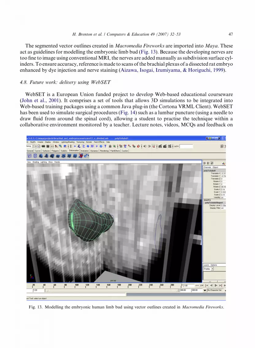
The segmented vector outlines created in Macromedia Fireworks are imported into Maya. Theseact as guidelines for modelling the embryonic limb bud (Fig. 13). Because the developing nerves aretoo fine to image using conventional MRI, the nerves are added manually as subdivision surface cyl-inders. To ensure accuracy, reference is made to scans of the brachial plexus of a dissected rat embryoenhanced by dye injection and nerve staining (Aizawa, Isogai, Izumiyama, & Horiguchi, 1999).
4.8. Future work: delivery using WebSET
WebSET is a European Union funded project to develop Web-based educational courseware(John et al., 2001). It comprises a set of tools that allows 3D simulations to be integrated intoWeb-based training packages using a common Java plug-in (the Cortona VRML Client). WebSEThas been used to simulate surgical procedures (Fig. 14) such as a lumbar puncture (using a needle todraw fluid from around the spinal cord), allowing a student to practise the technique within acollaborative environment monitored by a teacher. Lecture notes, videos, MCQs and feedback on
Fig. 13. Modelling the embryonic human limb bud using vector outlines created in Macromedia Fireworks.

Fig. 14. WebSET courseware showing online psysiology courseware and simulation of surgical procedures.
48 H. Brenton et al. / Computers & Education 49 (2007) 32–53
performance metrics supplement the 3D simulation. WebSET has been evaluated and shown toplace students higher up the learning curve towards surgical competence (Moorthy et al., 2003).
4.8.1. Collaborative environment
Undergraduate medical students require information about the structure of the body but thispropositional knowledge needs to be learned within an appropriate educational context. Modelsin themselves are of little educational or professional value without a carefully considered frame-work for delivery.
Medical virtual environments (Vidal et al., 2004) offer opportunities for collaborative learningwhich may be usefully applied to Web3D anatomy resources. Chittaro and Ranon (2007) arguethat virtual collaborative environments provide valuable first-person substitutes for real experienceand encourage �spontaneous knowledge acquisition that requires less cognitive effort than tradi-tional educational practices�. They draw upon constructivist theories to demonstrate how Web3Dtechnologies can enhance online collaboration, giving as an example the way a group Brazilianschool children work together, actively construct and learn about their own virtual world.

H. Brenton et al. / Computers & Education 49 (2007) 32–53 49
Engagement in this type of complex, personally meaningful task, is in marked distinction to theweak opportunities for interactivity and collaboration in the current generation of Virtual Learn-ing Environments (VLEs), which reinforce behaviourist models of learning (Bonk, Wisher, & Lee,2004). Text-based discussion boards, email and chatrooms may have spawned a new linguisticmedium (Crystal, 2001) but they lack the richness of perceptual cues and opportunities for team-work found within modern 3D environments.
5. Discussion and evaluation strategy
Although the brachial plexus project is still in the early stages of development, the work wehave completed thus far suggests that we can gain a significant improvement in the quality ofthree-dimensional models using Maya. We are planning to use a combination of X3D and H-Anim for the delivery of the adult brachial plexus model, allowing sophisticated animation andmanipulation of high quality spline-based models. We are currently evaluating a commercialX3D/H-anim authoring tool called VizX3D (VizX3D, 2004) to test its functionality and abilityto integrate with Maya.
We intend to conduct a thorough evaluation of the finished system in the next teaching cycle.Our working hypothesis is that introducing the 3D model into existing teaching will improve stu-dents� knowledge and retention of the brachial plexus in accordance with these learningobjectives:
� An ability to memorise the three-dimensional structure of the brachial plexus;� An understanding of how the brachial plexus relates to the peripheral nervous system and the
anatomy of the upper limb;� An ability to clinically evaluate how nerve injuries affect the upper limb;� A good understanding of the formation of the brachial plexus and how the nerves innervate the
whole limb.
5.1. Adult resource
Three groups of undergraduate students will be pre-assessed with multiple choice and short an-swer questions to test their existing anatomical knowledge of the brachial plexus. Group one willthen receive traditional didactic teaching. Group two will then receive traditional didactic teach-ing supplemented by the 3D model. Group three will have access only to the Web3D tool and thesupplemental teaching material on the website. Each group will then be post-assessed to determineany effect that the 3D tool had upon their learning. A questionnaire will also be completed by eachstudent to obtain qualitative feedback.
5.2. Embryological model
The embryological model will be used to test retention of knowledge and the suitability of thetool for revision. Three groups of undergraduate students will be pre-assessed with multiple choiceand short answer questions to test their existing anatomical knowledge of the brachial plexus.
50 H. Brenton et al. / Computers & Education 49 (2007) 32–53
� Group one will then use the tool for self-directed study;� Group two will use the tool within a collaborative environment with one hour �virtual super-
vision� by an anatomy teacher;� Group three will act as a control. They will not have access to the tool and will revise the topic
with traditional revision techniques;� The three groups will then be post-assessed to determine any effect that the tool had upon their
learning.
6. Future work
6.1. Haptics
Jastrow and Vollrath (2002) state that cadaveric dissection is the �only means of experiencingthe consistency of organs and tissues�. This is challengeable on two counts, firstly because the stor-age and fixation of cadavers alters the color and texture of tissues being dissected. Secondly be-cause �haptic� (Hale & Stanney, 2004) technologies allow the simulation of touch and have thepotential to accurately recreate the elasticity and consistency of living tissue. Haptics requiresforce-feedback devices, such as the Phantom (Phantom, 2004), which has recently reduced in price.Temkin, Acosta, Hatfield, Onal, and Tong (2002) have created three-dimensional anatomymodels which can be palpated over the Internet.
6.2. ‘Intelligent’ models
Most anatomical 3D models are �dumb� in the sense that the functioning of organs and limbshas been manually animated by a human. There are some exceptions, such as Garner and Pandy(2001), who created a computer simulation of the major articulations and contractions of bones,muscles and tendons in the upper limb and shoulder. The film industry has been a prime force inthe development of musculoskeletal simulations. Special effects companies such as Weta Digital(Weta, 2004) are primarily concerned with the realistic deformation of skin, on characters suchas cave trolls, but the tools they have developed are of potential benefit to anatomical modelling.Absolute Character Tools (CG Character, 2004) is one of the first commercially available tools formuscle simulation on a standard PC. Three-dimensional environments allow the physical embodi-ment of forces that are invisible in the real world. Abstract concepts, such as chemical bonds ormoney, can be given a perceptible representation with which users can interact. It is feasible thatfuture anatomy models will include �intelligent tutoring systems� (Brusilovsky, 1999) that will sim-ulate and teach complex abstract physiological and biochemical processes.
6.3. Open source models
The visible human project is an excellent resource for two-dimensional images, but the processof segmentation and reconstruction into 3D models is time-consuming and costly. The medicalcommunity would benefit from an open source library of pre-built anatomic structures whichcould be used for non-commercial educational purposes.
H. Brenton et al. / Computers & Education 49 (2007) 32–53 51
6.4. MedX3D standard
The Web3D Medical Working Group is developing an interoperable standard called MedX3D forrepresenting human anatomy (www.web3d.org/applications/medical/index.html). We will monitorthis emerging standard and consider the benefits of including it in the final version of the courseware.
6.5. Online computer games
Three-dimensional Massively Multiplayer Online games such as Star Wars Galaxies (Starwars,2004) highlight the paucity of interaction and collaboration within text-based VLEs. Every daythousands of players simultaneously engage in complex tasks and problem-solving activities with-in fluctuating groups linked by complex bonds of fealty, allegiance and shared localised codes ofconduct. Players are encouraged to become guides, initiating newcomers into the etiquette of acomplex virtual community of practice by providing mentorship, help and remediation. Thesetypes of computer games provide a �proof of concept� for the next generation of educationalcollaborative 3D environments.
6.6. Postgraduate study
We plan to extend the brachial plexus model to include animated effects of disease and injuryupon the plexus (brachial plexopathy). This will provide information useful to surgical and radi-ology postgraduates.
7. Conclusion
Anatomy teaching is undergoing significant changes due to time constraints, limited availabilityof cadaveric specimens and advances in computer-assisted learning. Web3D offers many potentialbenefits, most notably the ability to simulate the spatial relationships between anatomical struc-tures. However, more research needs to be done to evaluate these resources before they are intro-duced into the undergraduate medical curriculum.
Acknowledgments
We wish to thank all the members of the WebSET consortium. We would also like to thankPrimal Pictures (www.primalpictures.com) for permission to use the screenshot from the Interac-tive Thorax and Abdomen CD-ROM.
References
Ackerman, J. United States National Library of Medicine, The Visible Human Project. URL: http://www.nlm.nih.gov/
research/visible/visible_human.html (last accessed: 18-03-2005).

52 H. Brenton et al. / Computers & Education 49 (2007) 32–53
Aizawa, Y., Isogai, S., Izumiyama, M., & Horiguchi, M. (1999). Morphogenesis of the primary arterial trunks of the
forelimb in the rat embryos: the trunks originate from the lateral surface of the dorsal aorta independently of the
intersegmental arteries. Anatomical Embryology (Berl), 200, 573–584.
Aziz, M. A., McKenzie, J. C., Wilson, J. S., Cowie, R. J., Ayeni, S. A., & Dunn, B. K. (2002). The human cadaver in the
age of biomedical informatics. Anatomical Records, 269, 20–32.
Bartz, D., Mayer, D., Fischer, J., Ley, S., Del Rio, A., Thust, S., et al. (2004). Hybrid segmentation and exploration of
the human lungs. In Proceedings of IEEE Visualization 2003.
Bessaud, J., Hersch, R. D. Visible Human website at the Ecole Polytechnique Federale de Lausanne. URL: http://
diwww.epfl.ch/w3lsp/publications/gigaserver/tvhssaws.html (last accessed: 18-03-2005).
Betts, A., Saifuddin, A., Marone, P., Wolman, R., Lambert, S., (2004). Sport�s Injuries: The shoulder (CD-ROM),
Primal Pictures.
Bonk, C., Wisher, R., & Lee, J. (2004). Moderating learner-centered e-learning: problems and solutions, benefits and
implications. In Online collaborative Learning: Theory and Practice. Idea Group Inc..
Brusilovsky, P. (1999). Adaptive and Intelligent Technologies for Web-based Education. Kuntzkiche Intellligenz,
Special Issue on Intelligent Systems and Teleteaching.
CG Character website. URL: http://www.cgcharacter.com/index.php (last accessed: 18-03-2005).
Chittaro, L., Ranon, R. (2007). Web3D Technologies in learning, education and training: motivations, issues,
opportunities. Computers & Education, 49(1), 3–18.
Crystal, D. (2001). Language and the Internet. Cambridge: Cambridge University Press.
Dodd, A., Riding, M., John, N. (2004). Building Realistic Virtual Environments using Java and VRML. In Third Irish
workshop on computer graphics, 2002, pp. 53–61.
Ellis, H. (2001). Teaching in the dissecting room. Clinical Anatomy, 14, 149–151.
Garner, B. A., & Pandy, M. G. (2001). Musculoskeletal model of the upper limb based on the visible human male
dataset. Computational Methods in Biomechanics and Biomedical Engineering, 4, 93–126.
H-Anim Humanoid animation specifications. URL: www.web3d.org/x3d/workgroups/h-anim.html (last accessed: 18-
03-2005).
Hagens, G. (2004). Gunther Von Hagens Bodyworlds anatomical exhibition of real human bodies. URL:
www.koerperwelten.de/en/pages/gunther_von_hagens.asp (last accessed: 18-03-2005).
Hale, K. S., & Stanney, K. M. (2004). Deriving haptic design guidelines from human physiological, psychophysical, and
neurological foundations. IEEE Computational and Graphical Application, 24, 33–39.
Hallgren, R. C., Parkhurst, P. E., Monson, C. L., & Crewe, N. M. (2002). An interactive, Web-based tool for learning
anatomic landmarks. Acad. Med., 77, 263–265.
Heylings, D. J. (2002). Anatomy 1999–2000: the curriculum, who teaches it and how? Medical Education, 36, 702–710.
James, D., Purkayastha, S., Paraskevas, P., Shafiq, O., Darzi, A., & Athanasiou, T. (2004). Anatomy: the future
teaching of undergraduates. Hospital Medicine, 65(11), 681–685.
Jastrow, H., & Hollinderbaumer, A. (2004). On the use and value of new media and how medical students assess their
effectiveness in learning anatomy. Anatomical Records, 280, 20–29.
Jastrow, H., & Vollrath, L. (2002). Anatomy online: presentation of a detailed WWW atlas of human gross anatomy –
reference for medical education. Clinical Anatomy, 15, 402–408.
John, N. W., Riding, M., Phillips, N. I., Mackay, S., Steineke, L., Fontaine, B., et al. (2001). Web-based surgical
educational tools. Studies on Health Technology and Information, 81, 212–217.
Lorensen, W., Cline, H. (2004). Marching cubes: A high resolution 3D surface construction algorithm. In Proceedings
of the 14th annual conference on computer graphics and interactive techniques, pp. 163–169.
Maya Alias/Wavefront PDF file listing Maya functionality. URL: www.alias.com/eng/products-services/maya/file/
maya6_features_in_detail.pdf (last accessed: 18-03-2005).
McLachlan, J. C., Bligh, J., Bradley, P., & Searle, J. (2004). Teaching anatomy without cadavers. Medical Education,
38, 418–424.
Mitchell, B. S., & Stephens, C. R. (2004). Teaching anatomy as a multimedia experience. Medical Education, 38,
911–912.
Moorthy, K., Jiwanji, M., Shah, J., Bello, F., Munz, Y., & Darzi, A. (2003). Validation of a Web-based training tool for
lumbar puncture. Studies on Health and Technological Information, 94, 219–225.

H. Brenton et al. / Computers & Education 49 (2007) 32–53 53
Nunn, S. (2004). Learning Embryology Through Using Plasticine (LETUP) Exercises. In Association for Medical
Education in Europe, Edinburgh 2004 (abstracts of proceedings).
Park, A., Schwartz, R. W., Witzke, D. B., Roth, J. S., Mastrangelo, M., Birch, D. W., et al. (2001). A pilot study of
new approaches to teaching anatomy and pathology. Surgical Endoscopy, 15, 245–250.
Parker, L. M. (2002). Anatomical dissection: Why are we cutting it out? Dissection in undergraduate teaching. ANZ
Journal of Surgery, 72, 910–912.
Phantom Phantom Haptic Device. URL: www.sensable.com (last accessed: 18-03-2005).
Porter, R. (1999). Renaissance. In Greatest benefit to mankind: a medical history of humanity (pp. 163–200). Fontana
Press.
Schroeder, W., Martin, K., Lorensen, B. (2004). The Visualization Toolkit An Object-Oriented Approach To 3D
Graphics.
Starwars Galaxies Website. URL: http://starwarsgalaxies.station.sony.com (last accessed: 18-03-2005).
Temkin, B., Acosta, E., Hatfield, P., Onal, E., & Tong, A. (2002). Web-based three-dimensional Virtual Body
Structures: W3D-VBS. Journal of the American Medical Information Association, 9, 425–436.
Vidal, F., Bello, F., Brodlie, K., Gould, D., Phillips, R., & Avis, N. (2004). Principles and applications of medical
virtual environments. In Proceedings of Eurographics 2004.
VizX3D website. URL: http://www.vizx3d.com (last accessed: 18-03-2005).
Ward, A. (2002). Primal 3D Interactive Series. Acupuncture Medicine, 20, 51–52.
Warren, J. (2002). Subdivision methods for geometric design: a constructive approach. San Francisco; London.
Weta Digital Website. URL: www.wetadigital.com (last accessed: 18-03-2005).
X3D Specifications 2004. URL: www.web3d.org/x3d/specifications/index.html (last accessed: 18-03-2005).
Zhang, S. X., Heng, P. A., Liu, Z. J., Tan, L. W., Qiu, M. G., Li, Q. Y., et al. (2003). Creation of the Chinese visible
human data set. Anatomical Records B New Anat., 275, 190–195.
Zhong, S. Z. (2003). Scientific significance and prospective application of digitized virtual human. Di Yi.Jun.Yi.Da.
Xue.Xue.Bao., 23, 193–195.