urine analysis
TRANSCRIPT
URINE ANALYSIS
Dr. SANJIV KUMAR
A critical examination of urine is one of the most important of all veterinary laboratory
diagnostic procedures. This body fluid is easily obtained from most species of animals. Urine is
not only altered by disease occurring in the kidneys but many extra renal conditions produce
changes that may be of diagnostic significance.
COLLECTION AND PRESERVATION OF URINE
Requirements:
-Chemically clean opaque glass vials/disposable plastic containers along with appropriate rack.
-Sterilized catheters.
-Syringe and needle.
-Preservatives-Toluene/formalin/thymol/chloroform/boric acid/ metaphosphoric acid.
Procedure:
i. Collect the urine sample from animals as it voids. It is advisable to clean the vulva to avoid the
contamination.
ii.An early morning sample from house trained pets is of more diagnostic significance.
iii.Collect midstream urine sample as first stream is contaminated with cellular debris, leukocyte
exudates flushed from urethra.
iv.At least 4 ounce (28.34 ×4 ml) of urine must be collected.
v.For bacterial cultures and sensitivity testing, sample should be collected in sterile container
using aseptic precautions either using catheter or directly from bladder in small animals e.g.
dog/cats.
vi.Collection is advised before any fluid consumption (dilutes the urine)/ meals (sample likely to
have more sugar and protein) than morning sample.
vii.Examine the sample as early as possible. Delay will increase alkalinity of urine (urea,
ammonia) and formed elements (cells and casts) degenerates.
Preservation of urine:
-Refrigeration (urine is suitable for examination upto 2-3 hrs). Bring sample to room
temperature before examination.
- Where refrigeration is not available, preserve the samples using preservatives as follows:
Toluene-Antimicrobial/good for ketone bodies estimation) which preserves urine for 24 hours.
Thymol-Add a small crystal/5-10 ml of 10% solution in isopropyl alcohol, Antimicrobial,
preserve urine for 24hr but renders urine unsatisfactory for protein (false positive reaction),
glucose, ketone bodies, phosphates, magnesium and phenol.
Formalin- 1 drop conc. Formalin per ounce of urine. Best for microscopic examination
(preserve cast and cellular elements), Antimicrobial but renders urine unsuitable for indican,
albumin (false positive with Heller’s test) and glucose.
Chloroform- Add 5 ml of urine sample, preserve for 24hr (antimicrobial but renders urine
unsuitable for glucose and microscopic examinations).
Boric acid-Add 1 gm for urine sample, preserve for 24 hr but interfere with glucose and uric
acid tests.
Others-Metaphosphoric acid (10% aqueous) in 1:5 ratio to urine sample for vita. C preservation,
camphor (small piece) and chloral hydrate (reduces copper) can also be used for preservation.
URINE EXAMINATION
The examination of urine can be divided in to three parts-
(I) Physical /visual examination of urine.
(II) Chemical examination of urine.
(III) Microscopical examination.
(IV) Cultural examination.
PHYSICAL EXAMINATION OF URINE
(1) Volume - Record the amount of urine produced per day by normal animal. It is inversely
related to specific gravity i.e. high volume with low specific gravity and low volume with high
specific gravity exception to it are diabetes mellitus (high volume and high specific gravity, due
to glycosuria) and terminal nephritis (low volume and low specific gravity).
(A) Increased volume: (Polyuria)
i. Physiological (Transient) - Increased water intake, administration of parentral fluids, increased
thirst, corticosteroid and ACTH administration.
ii. Pathological: Chronic progressive renal failure, acute renal failure, diabetes mellitus, primary
renal glycosuria, diabtes insipidus (ADH deficiency), renal Diabetes insipidus renal cortical
hypoplasia, sever cortical hypoplasia, severe renal amyloidsis, chronic pyelonephritis, pyometra,
hyperadrenocorticism and generalized liver disease.
(B) Decreased volume: (Oligouria)
i. Physiological:(transient) decreased water intake, excessive environmental temperature,
panting, dehydration and exercise.
ii. Pathological:-Acute renal failure, fever, chronic primary renal failure, oedema (circulatory
dysfunction), urinary passage obstruction, reduced (marked) blood pressure.
(2) Colour- Record the colour of urine taking in a sample in a clean transparent tube.
Normal colour is yellow/amber /wheat straw coloured and is related to concentration of
urochromes in Urine. Acidic urine is darker than alkaline urines. Blood can also be a
contaminant that gets into the urine unintentionally during collection, such as from hemorrhoids
etc. The depth of urine color is also a crude indicator of urine concentration:
i. Pale yellow or colorless urine indicates dilute urine where lots of water is being excreted.
ii. Dark yellow urine indicates concentrated urine and the excretion of waste products in a
smaller quantity of water, such as is seen with the first morning urine, with dehydration, and
during a fever.
iii. Yellow brown/greenish yellow: Icterus- Green (biliverdin), Yellow brown (bilirubin and
Urobilin)
iv. Red, Port wine/brown: Porphyrinuria, Haemoglobiuria, Haematuria.
vi. Brown to brownish black: Azoturia (myoglobin), Methaemoglobinuria, Melanin, phenol
derivatives, Indican, Bile in Large Amounts.
vii. Green colour: Methylene Blue (urinary antiseptics), Bile (biliverdin), Acriflavin, Diazan
viii. Red to pink colour: Phenothiazine (colourless which turns pink on exposure to air)
Phenolphthlein, Phenolsufothlein, Naphthalene, Pyridium, Neoprontosil.
ix. Milky colour: Chyle, Milk, Marked contamination with petroleum, Normal horse urine
(CaCO3).
(3) Odour- Normal order is urinoid.
Abnormal odours:-
Medicated odour:-Due to oil of turpentine, menthol, curbebs, copaiba and sandal wood oil.
Ammonical odour:-Due to decomposition of protein.
Fruity/sweetish odour:- Acetone/ ketone bodies
Putrid odour:-Due to H2S gas from decomposition and cystinuria
Faecal odour:-Contamination with faeces and E.coli
(4) Transparency -observe in a test tube against light and record as i. clear, ii. Cloudy (slight,
very) iii. Sediment (slight, moderate and heavy). Normal urine is clear but transparent except
in horse, in which it is thick and turbid due to excess of calcium carbonate crystals/mucus.
Substances that cause cloudiness but that are not considered unhealthy include mucus, sperm and
prostatic fluid, cells from the skin, normal urine crystals, and contaminants.
Abnormal Transparency
Cloudy- One standing due to epithelial cells, blood (Hb), leukocytes, Bacteria, mucus, crystals
of Calcium carbonate, amorphous Phosphates.
Sediment- Amorphous urates (whitish/pinkish acidic urine), Amorphous phosphate (white
alkaline), Heavy whitish sediment (pus) and reddish brown smoky sediment (blood).
(5) Foam test - shake the urine in a test tube and record as follows:
i. Normal (slight white foam)
ii. Proteinuria (excess white foam)
iii. Bile/bile pigment (green, yellow, yellow brown foam)
iv. Hemoglobinuria (red to brown colour).
CHEMICAL EXAMINATION OF URINE
To perform the chemical examination, most clinical laboratories use commercially prepared test
strips.
Specific Gravity
A physical characteristics i.e. a measure of urine concentration. There are no “abnormal" specific
gravity values. Knowing the urine concentration one can decide if the urine specimen they are
evaluating is the best one to detect a particular substance. For example, if they are looking for
very small amounts of protein, a concentrated morning urine specimen would be the best sample.
It can be measured by:
(1) By using refractometer--expensive but more accurate.
(2) Exton immiscible balance method –not popular and require hydrometer.
(3) Urinometer method- It is cheap and inexpensive and routinely followed.
pH
There is "abnormal" pH values. Any condition that produces acid or base in the body such as
acidosis or alkalosis, or the ingestion of acidic or basic foods, can directly affect urine pH.
Abnormal reaction:-
Alkaline urine:- Excessive vegetable diet, cystitis, urinary retention, rapid absorption of
transudates, alkalosis (metabolic/respiratory), on standing alkaline (urea-ammonia) and alkaline
therapy (sodium bicarbonate, sodium /potassium citrate/acetate/lactate and potassium nitrate).
Acidic urine: - Diet with excess protein starvation, fever, acidosis, prolonged muscular activity
and administration of acidic salts like sodium acid chloride ammonium chloride, sodium acid
phosphate and calcium chloride.
Protein
Normally, there will not be detectable quantities of albumin in the urine. When urine protein is
elevated, you have a condition called proteinuria. Albumin is smaller than most other proteins
and is typically the first protein that is seen in the urine when kidney dysfunction begins to
develop. The proteinuria can be detected by heat test, heller’s test, Robert’s test, Sulphosalicylic
acid test or Albustix reagent.
The conditions that can produce proteinuria include:
Physiological: Exercise, convulsions, emotional, stress, excessive protein diet and first few days
of life.
Pathological:
Prerenal:-Non-renal causes such as Bence-Jones proteinuria, hemoglobinuria and myglobinuria.
Renal:- Increased permeability of glomerulus, impaired absorption of protein, blood/exudates
of renal origin- renal neoplasm, acute nephritis, glomerulonepritis, nephrosis (chemical
poisoning), amyloidosis, pyelonephritis and polycystic kidneys.
Post-renal:-Due to contamination of urine in urinary track and it can be due to improper
catheterization, inflammatory excudate seen in pyelitis, ureteritis, cystitis, urethritis and
urolithiasis.
Extra urinary causes: Contamination with blood/genital tract (Prepucial/vaginal secretions and
prostatitis) and chronic passive congestion of kidneys (cardiac insufficiency, liver diseases,
febrile reaction bacterial endocarditis, neoplasm, emboli and increased abdominal pressure from
ascetic fluid/tumors).
Glucose
Glucose is normally not present in urine. When glucose is present, the condition is called
glucosuria is may be either due to an excessively high glucose concentration in the blood or a
reduction in the “renal threshold.” It can be tested by-Benedict’s test, Fehling’s test or Clinitest
reagent tablet.
Glycosuria and hyperglycemia:-Excessive carbohydrate diet, fluid administration containing
glucose/fructose, increased secretion/administration of epinephrine, hyperadrenocorticism,
pancreatic disorder, diabetes mellitus, liver diseases, hyperpituitarism, hyperparathyroidism,
increased intracranial pressure due to-tumor, haemorrhage, encephalitis, fracture and
enterotoxemia (sheep).
Renal glycosuria but no hyperglycemia: Defective tubular reabsorption.
False positive reaction for glucose: due to large number of drugs routinely administered e.g.-
antibiotics –penicillin, streptomycin, terramycin and choloramphenicol, lactose/pentose/reducing
sugar, ascorbic acid, morphine, salicylates, chloral hydrate and formaldehyde.
Ketones
Ketones (acetone, acetoacetic acid and beta hydroxy butyric acid) are not normally found in the
urine. They are intermediate products of fat metabolism. They can form when an individual does
not eat enough carbohydrates (for example, in cases of starvation or high-protein diets) or when

an individual’s body cannot use carbohydrates properly. When carbohydrates are not available,
the body metabolizes fat instead to get the energy it needs to keep functioning. Ketonuria can be
detected by Rothera’s nitroprusside test.
Dog & Cats:-Diabetes mellitus, high fever and starvation (in puppies & kittens)
Cattle & buffaloes:-High producing animals, early lactation, improper feeding/anorexia due to
one/another reason and milk fever (prolonged.)
Sheep:-Pregnancy toxemia, multiple lambs/twin lambs, pregnancy (Hypopglycemia)
Non specific:-High fat diet, acidosis, starvation/fasting impaired liver function endocrine
disorders.
Blood
Presence of blood in the urine is known as hematuria while that of haemoglobin is known as
haemoglobinuria. The small number of RBCs normally present in urine usually result in a
"negative" test. However, when the number of RBCs increases, they are detected as a "positive"
test result. Even small increases in the amount of RBCs in urine can be significant. Benzidine
test and orthotolidine test are used to detect blood in urine. Sometimes a chemical test for blood
in the urine is negative, but the microscopic examination shows increased numbers of RBCs.
When this happens, test the sample for ascorbic acid (vitamin C), because vitamin C has been
known to interfere with the accuracy of urine blood test results, causing them to be falsely low or
falsely negative.
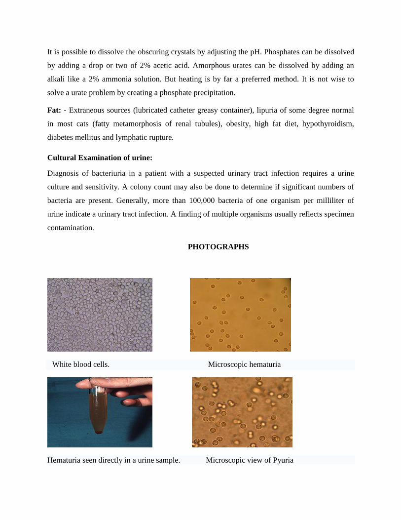
Haematuria (urine reddish- cloudy, clear with sediment on standing and contains intact RBCs):
Acute nephritis, nephrosis, urolithiasis, pyelonephritis, pyelitis, ureteritis, cystitis neoplasms of
uro-genital tract, estrus, post-partum in females, passive congestion of kidneys, renal infarction,
parasites (Dictophyma renale, Capillaria plica, Dirofillaria immitis larvae), Thrombocytopenia,
sweet Clover poisoning shock and toxemia due to Cu, Hg, As & thallium.
Myoglobinuria (urine brown/coffee coloured and no intact RBCs): Azoturia (horses), mechnical
trauma high tension electric shock and snake venoms.
Haemoglobinuria (Urine port-wine/brown/coffee coloured translucent uniform color without
sediment & no intact RBCs): Due to excessive haemolysis of erythrocytes and observed in
condition viz. leptospirosis, piroplasmosis, phenothiazine therapy, toxaemia due to Cu, Hg and
sulphonamides, post parturient haemoglobinuria, bacillary haemoglobinuria (Clostridium
haemolyticum), following severe burns, haemolytic disease of newborns e.g. icterus neonatrum,
erythroblastosis fetails, Azoturia (Myoglobinuria) and plant toxicity e.g. Broom savin
Colchicium frosted turnips.
Bile Pigments
Bilirubin is not present in the urine of normal, healthy individuals. It becomes a component of
bile, a fluid that is secreted into the intestines to aid in food digestion.
Presence of bilirubin in urine can be detected by: i. Foam test ii. Gmelin test iii. Fouchet’s test
iv. Icotest (reagent tablets)
Physiological-Threshold of bilirubin in dog is low, so normal urine in dog contains some
bilirubin.
Pathological-It is observed in condition viz. obstructive jaundice, hepatocellular disease e.g.
leptospirosis infection canine hepatitis cirrhosis toxicities hepatic neoplasm and in haemolytic
jaundice (it is not present unless liver damage)
Indican
Indican is derived from indole, a product of putrefaction of protein. Present in high concentration
in urine of herbivores. It can be determined by Obermeyer’s test.
Not much clinical significance and is absent/traces in dogs cats. Increased amounts observed viz.
gastritis, constipation indigestion enteritis intestinal obstructions diet rich in protein and
decomposition of protein in other parts of body like large abscess, empyema and peritonitis.
Other tests not routinely done are:
Calcium-Precipitate in urine as insoluble calcium oxalates. It is detected by Sulkowitch test.
Normal calcium concentration in urine of Dog -2.1(0.2-7.6) MEq/L, Man- 7.5(1.4-13)MEq/l.
Decreased:
Serum calcium level below Renal threshold of 7.5 mg/dl- Bovine hypocalcemia (not Useful as
no indication of Serum calcium level by Sulkowitch test), canine Puerperal tetany,
hypoparathyroidism and osteomalacia.
Increased:
Serum calcium level above 10.5 mg /dl after administration of calcium solution, renal
osteodystrophy, hyperthyroidism and hypervitaminosis, Puerperal tetany, hypoparathyroidism
and osteomalacia.
Urobilinogen- normally present in urine in low concentrations. It is detected by urobilistrix.
Positive test results help detect liver diseases such as hepatitis and cirrhosis and conditions
associated with increased RBC destruction. When urine urobilinogen is low or absent in a patient
with urine bilirubin and/or signs of liver dysfunction, it can indicate the presence of hepatic or
biliary obstruction. False positive reaction occurs with indole, bile, nitrates, sulfonamide and
formalin.
Substance Normal patient Hemolytic disease Hepatic disease Biliary obstruction
Urine uribilinogen Normal High High Low
Urine bilirubin Negative Negative Positive Positive
Leukocyte esterase - An enzyme present in most white blood cells (WBCs). Normally, a few
white blood cells are present in urine and this test is negative. When the number of WBCs in
urine increases significantly, this screening test will become positive. The most common cause
for WBCs in urine (leukocyturia) is a bacterial urinary tract infection, such as a bladder or
kidney infection.
Nitrite- Based upon the fact that many bacteria can convert nitrate to nitrite in your urine.
Normally the urinary tract and urine are free of bacteria. When bacteria find their way into the
urinary tract, they can cause a urinary tract infection (UTI). A positive nitrite test result can
indicate a UTI. However, since not all bacteria are capable of converting nitrate to nitrite, you
can still have a UTI despite a negative nitrite test.
MICROSCOPIC EXAMINATION OF URINE
It provides important information useful for both diagnosis and prognosis. Microscopic
examination of urine sediment is usually performed in addition to routine procedures. The
specimen used for microscopic examination should be as fresh as possible.
Pour five to ten milliliters of midstream urine into a conical bottom test tube. Centrifuge for
about five minutes. Decant the supernatant and resuspend the sediment in the residual urine that
clings to the bottom of the tube by tapping the tube against a hard surface several times. Place a
drop of the resusupended sediment on a glass microscope slide and place a coverslip over the
drop and examine under the microscope. Although commercial stains are available to highlight
cellular elements, examination of unstained urine is usually adequate. Too much light makes the
cellular and crystalline elements harder to see. Scan the slide under low power to locate areas of
interest. Look for casts just inside the perimeter of the cover slip. Then switch to high dry
magnification and examine ten random fields in the central part of the coverslip. Count the
numbers of red cells and white cells in each. If the field is covered with cells, report as "TNTC"
(too numerous to count) or "packed." Add to the report an estimate of bacteria density, any casts
seen and other structures noted.
Clinical Significance:
In healthy individual, the urine contains small numbers of cells and other formed elements from
the entire urinary tract, and epithelial cells from the kidney, ureter, bladder, and urethra. In renal
disease, the urine often contains increased numbers of substances discharged from an organ that
is otherwise accessible only by biopsy or surgery. A microscopic examination of urine sediment
detects the presence and amounts of i. Organized sediments like blood cells, Bacteria and yeast,
Casts, Epithelial cells, mucous threads, Parasites, Spermatozoa and ii. Unorganized sediments
like Crystals and fat droplets.
Organized sediments
Red Blood Cells -Normally, 2-3 per high power field (HPF). RBCs may be differentiated from
white cells by putting a drop of 5% acetic acid to the urine sediment. It disintegrates any red
cells, but it does not affect the white cells. RBC in urine is called hematuria. It may be due to
glomerulonephritis, Tumors, Kidney trauma, Renal infarcts, Acute tubular necrosis, Upper and
lower urinary tract infections, Nephrotoxins, Traumatic catheterization, Passage of renal stones,
Physical stress.
White Blood Cells-Normally low (0 to 3 leukocytes per high-power field). More than 3 cells per
high-power field probably indicate disease somewhere in the urinary tract.
Pyuria is the presence of 4 or more neutrophils (pus cells) per high power field indicating UTI.
Sterile pyuria is listed as a side-effect from some medications such as paracetamol.
Eosinophiluria - associated with allergic interstitial nephritis, atheroembolic disease.
Epithelial Cells - Normally, a few epithelial cells can be found in the urine sediment. In urinary
tract conditions such as infections, inflammation, and malignancies, more epithelial cells are
present. Determining the kinds of cells present helps the health care provider pinpoint where the
condition is located. For example, a bladder infection may result in large numbers of transitional
epithelial cells in urine sediment. Epithelial cells are usually reported as "few," "moderate," or
"many" present per low power field (LPF).
Microorganisms (bacteria, trichomonads, yeast) - Microorganisms are usually reported as
"none," "few," "moderate," or "many" present per high power field (HPF). In female and rarely
in male, yeast can also be present in urine. Trichomonads are parasites that may be found in the
urine of women or men (rarely). If these are found during a urinalysis, then follow-up testing for
Trichomonas species may be performed to look for a vaginal infection.
Mucus and Mucus threads:- Normal in horse urine and in other animals indicate irritation of
urethera and contamination of urine with genital secretions.
Parasites:-Parasitic ova ( swine kidney worm- Stephanurus dantatus, giant kidney worm of dog-
Dictophyma renale, bladder worm of dog, cat and fox- Capillaria plica, and Dirofilaria immitis,
Microfilaria) and protozoa- contaminants from fecal secretion e.g. Trichomonads and Giardia.
Spermatozoa:-common in male dog urine and no clinical importance indicate contamination
with semen.
Casts - These urinary sediments are formed by coagulation of albuminous material in the kidney
tubules. Casts are cylindrical (cylinduria) and vary in diameter. The sides are parallel, and the
ends are usually rounded. Casts in the urine always indicate some form of kidney disorder and
should always be reported. If casts are present in large numbers, the urine is almost sure to be
positive for albumin. The numbers of casts are reported as "number and type seen per low power
field (LPF)".
There are different types of casts. They are as follows:
Hyaline casts are the most frequently occurring casts in urine, even in mildest renal
disease. They are colorless, homogeneous, transparent, and usually have rounded ends.
Uncommon in alkaline urine.
Red cell casts indicate renal hematuria. Red cell casts may appear brown to almost
colorless and are usually diagnostic of glomerular disease.
Granular casts almost always indicate significant renal disease. Granular casts that
contain fine granules may appear grey or pale yellow in color. Common in domestic
animals.
Epithelial casts are rarely seen in urine because renal disease that primarily affects the
tubules is infrequent. Epithelial casts may be arranged in parallel rows or haphazardly.
Waxy casts result from the degeneration of granular casts. Waxy casts have been found in
patients with severe chronic renal failure, malignant hypertension, and diabetic disease of
the kidney. Waxy casts appear yellow, grey, or colorless. They frequently occur as short,
broad casts, with blunt or broken ends, and often have cracked or serrated edges.
Fatty casts are seen when there is fatty degeneration of the tubular epithelium, as in
degenerative tubular disease. Fatty casts also result from lupus and toxic renal poisoning.
A typical fatty cast contains both large and small fat droplets. The small fat droplets are
yellowish-brown in color.
Unorganized sediment
Crystals – Are waste chemicals that can form crystals, solid forms of a particular substance, in
the urine in case of insufficient water intake, high urine temperature, Infection etc. Crystals are
identified by their shape, color, and by the urine pH. They may be small, sand-like particles with
no specific shape (amorphous) or have specific shapes, such as needle-like. Crystals are
considered "normal" if they are from solutes that are typically found in the urine. Some examples
of crystals that can be found in the urine of healthy individuals include: amorphous urates,
crystalline uric acid, calcium oxalates, amorphous phosphates and calcium carbonate. Some
crystals are found exclusively in acid urine, others are found exclusively in alkaline urine. If the
crystals are from solutes that are not normally in the urine, they are considered "abnormal."
Abnormal crystals may indicate an abnormal metabolic process. Some of these include: Cystine,
Tyrosine and Leucine.
When crystals form as urine is being made in the kidney, they may group together to form
kidney "stones" or calculi. These stones can become lodged in the kidney itself or in the ureters,
tubes that pass the urine from kidney to the bladder, causing extreme pain. Medications, drugs,
and x-ray dye can also crystallize in urine.
CLINICAL SIGNIFICANCE:
Except for the cystine crystals and a few others, the majority of crystals found in the urinary
sediment are of limited clinical value. It is tempting to associate crystals with a risk of
urolithiasis, but the majority of patients with a crystalluria do not have and will not develop
kidney stones.
In many cases, the presence of crystals is a pest to the microscopic examination. The elimination
of these crystals can be made by gently heating the specimen at 37°C. To attain complete
dissolution, it is preferable to heat the whole specimen. Once decanted, it is often impossible to
dissolve the bulk in the small remaining volume.
It is possible to dissolve the obscuring crystals by adjusting the pH. Phosphates can be dissolved
by adding a drop or two of 2% acetic acid. Amorphous urates can be dissolved by adding an
alkali like a 2% ammonia solution. But heating is by far a preferred method. It is not wise to
solve a urate problem by creating a phosphate precipitation.
Fat: - Extraneous sources (lubricated catheter greasy container), lipuria of some degree normal
in most cats (fatty metamorphosis of renal tubules), obesity, high fat diet, hypothyroidism,
diabetes mellitus and lymphatic rupture.
Cultural Examination of urine:
Diagnosis of bacteriuria in a patient with a suspected urinary tract infection requires a urine
culture and sensitivity. A colony count may also be done to determine if significant numbers of
bacteria are present. Generally, more than 100,000 bacteria of one organism per milliliter of
urine indicate a urinary tract infection. A finding of multiple organisms usually reflects specimen
contamination.
PHOTOGRAPHS
White blood cells. Microscopic hematuria
Hematuria seen directly in a urine sample. Microscopic view of Pyuria
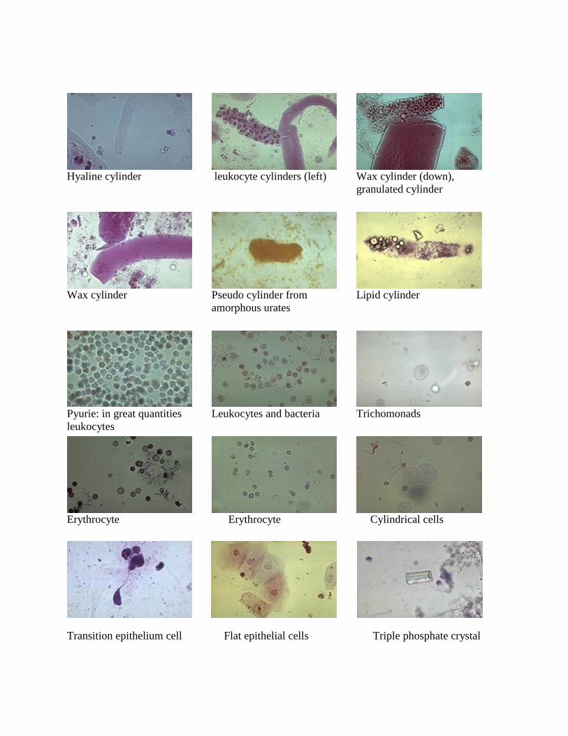
Hyaline cylinder
leukocyte cylinders (left)
Wax cylinder (down),
granulated cylinder
Wax cylinder
Pseudo cylinder from
amorphous urates
Lipid cylinder
Pyurie: in great quantities
leukocytes
Leukocytes and bacteria
Trichomonads
Erythrocyte
Erythrocyte
Cylindrical cells
Transition epithelium cell Flat epithelial cells Triple phosphate crystal

Calcium oxalate crystals Calcium phosphate crystals Urine acid crystals