unscheduled expression of cdc25b in s-phase leads to replicative stress and dna damage
TRANSCRIPT
RESEARCH Open Access
Unscheduled expression of CDC25B in S-phaseleads to replicative stress and DNA damageBéatrix Bugler1,2, Estelle Schmitt1,2,4, Bernadette Aressy1,2, Bernard Ducommun1,2,3*
Abstract
Background: CDC25B phosphatase is a cell cycle regulator that plays a critical role in checkpoint control. Up-regulation of CDC25B expression has been documented in a variety of human cancers, however, the relationshipswith the alteration of the molecular mechanisms that lead to oncogenesis still remain unclear. To address this issuewe have investigated, in model cell lines, the consequences of unscheduled and elevated CDC25B levels.
Results: We report that increased CDC25B expression leads to DNA damage in the absence of genotoxictreatment. H2AX phosphorylation is detected in S-phase cells and requires active replication. We also report thatCDC25B expression impairs DNA replication and results in an increased recruitment of the CDC45 replication factoronto chromatin. Finally, we observed chromosomal aberrations that are also enhanced upon CDC25B expression.
Conclusion: Overall, our results demonstrate that a moderate and unscheduled increase in CDC25B level, asobserved in a number of human tumours, is sufficient to overcome the S-phase checkpoint efficiency thus leadingto replicative stress and genomic instability.
BackgroundMembers of the CDC25 phosphatase family (CDC25A,B and C) regulate cell cycle transitions through depho-sphorylation of their substrates the CDK-Cyclin com-plexes. As ultimate targets of the DNA damageactivated pathway, they also play a critical role in thefate of the cells in response to injury [1,2]. The currentlyemerging picture suggests that all three CDC25 phos-phatases probably act at various stages of the cell cycledepending on the presence of the specific CDK/Cyclincomplexes. Thus, CDC25B has been proposed to partici-pate in the control of S-phase entry since specific anti-sense RNA is able to block HeLa cell replication [3] andis involved in the initiation centrosome duplication cyclein S-phase [4]. Conversely, CDC25A has been shown toplay an activating role during mitosis (for review [5]).Elevated expression of CDC25B has been documented
in a growing list of human cancers [2] suggesting apotential role in the alteration of molecular processesleading to oncogenesis. The mechanisms by which theCDC25B level becomes deregulated in tumours remainsunclear but it does not appear that the overexpression
results from gene amplification or rearrangement.CDC25B expression can be regulated at the transcrip-tional [6,7], translational and post-translational levels[8,9]. During the cell cycle, CDC25B levels begin toincrease from mid-S-phase, they peak during the G2-Mtransition and decrease in mitosis [3]. In contrast withCDC25C, CDC25B was shown to be unstable with a 30-minute half-life, its degradation being proteasomedependent [8-10]. The timing of the transition betweeneach phase of the cell cycle must be strictly respected tomaintain genomic stability. As far as CDC25B is con-cerned, its degradation by the proteasome pathway and/or inactivation by cytoplasmic sequestration appears tobe essential to prevent activation of CDK-cyclin com-plexes and to avoid checkpoint overcome.Very little is known about the mechanisms by which
increased CDC25B expression contributes to the onco-genesis process. It has been shown that overexpressionof CDC25B results in checkpoint bypasss and prematureentry into mitosis [11,12]. We also recently reportedthat moderate CDC25B expression is sufficient to allowbypass of a G2/M checkpoint activated by DNA damage,thus resulting in increased sensitivity to genotoxics andincreased mutagenesis [11]. Accordingly, it has beenproposed that after DNA damage CDC25B
* Correspondence: [email protected]é de Toulouse, LBCMCP, 118 route de Narbonne, F-31062 Toulouse,France
Bugler et al. Molecular Cancer 2010, 9:29http://www.molecular-cancer.com/content/9/1/29
© 2010 Bugler et al; licensee BioMed Central Ltd. This is an Open Access article distributed under the terms of the Creative CommonsAttribution License (http://creativecommons.org/licenses/by/2.0), which permits unrestricted use, distribution, and reproduction inany medium, provided the original work is properly cited.

accumulation [13] triggers the train of the molecularevents leading to checkpoint recovery and progressionin mitosis [14].However, as mentioned above all three CDC25 phos-
phatases have been shown to be involved in the controlof CDK-cyclin activities at the G1-S transition and in S-phase [15-17]. It is therefore tempting to speculate thatin addition to critically perturbing the G2-M checkpoint,elevated and unscheduled levels of one of these phos-phatases to an extent similar to that observed in humantumours might also have deleterious effects on the otherkey transitions.In this study we have investigated cell cycle progres-
sion in response to unscheduled expression of CDC25Band found dramatic effects during DNA replication lead-ing to replicative stress and genomic instability. Theseresults emphasize the relevance of the study of itsexpression in human tumours and shed light on itspotential role in oncogenesis.
ResultsCDC25B unscheduled expression and progression in S-phaseTo examine the impact of unscheduled CDC25B expres-sion on cell cycle progression during S-phase we used aU2OS cell line conditionally expressing an Ha epitope-tagged CDC25B protein under the control of the tetra-cycline promoter [18]. We first examined cell cycle pro-gression after synchronization by a double thymidineblock and release in cells expressing Ha-CDC25B ornot. Cell cycle distribution was determined by flow cyto-metry analyses and is shown in figure 1A as the percen-tage of cells in S and G2-M phase. Progression in thecell cycle appeared similar in both populations with apeak of S-phase cells at 6-7 hours. However, we noticedthat an elevated level of CDC25B-expressing cells wasalready in S-phase immediately after thymidine blockrelease and/or showed uncompleted DNA replicationwhile a majority initiated the G2 phase. Similar observa-tions were also made in cells expressing CDC25B thathad been synchronized by nocodazole treatment andmitotic shake off and release with 23% of BrdU incor-poration in U2OS-CDC25B cells versus 17% in U2OScells, (Figure 1B). These observations could reflect pre-mature entry into S-phase with subsequent perturbationof entry into mitosis as suggested by the flow cytometryanalysis.We thus examined the duration of S-phase in cells
expressing Ha-CDC25B or not. These cells were BrdUlabeled then chased with thymidine and collected at var-ious times for flow cytometry analysis of BrdU positivity.Nocodazole treatment was used during the experimentto stop progression into mitosis. As shown in figure S1,Additional file 1, BrdU positivity was increased at the
Figure 1 Overexpression of CDC25B alters progression in S-phase but not replication duration. (A) U2OS cells conditionallyexpressing Ha-CDC25B were synchronized by double thymidineblock (Materials and Methods). At the indicated time after release,Ha-CDC25B induced cells (U2OS CDC25B) or not (U2OS) wereharvested and the cell cycle distribution was monitored by flowcytometry after propidium iodide staining (PI). A western blotanalysis with monoclonal anti-Ha antibodies shows the Ha-CDC25Blevel in the induced U2OS CDC25B cells. (B) U2OS cells conditionallyexpressing Ha-CDC25B were synchronized in mitosis by nocodazoletreatment (100 nM, 17 h). Mitotic cells were recovered by shake offat 0 h and grown in drug free medium. Expression of CDC25B wasachieved by tetracycline removal at the time of release (0 h). Atindicated times after CDC25B induction, cells were labeled withBrdU (30 μM, 15 min) and the percentage of BrdU positive cells wasdetermined by flow cytometry analysis.
Bugler et al. Molecular Cancer 2010, 9:29http://www.molecular-cancer.com/content/9/1/29
Page 2 of 12

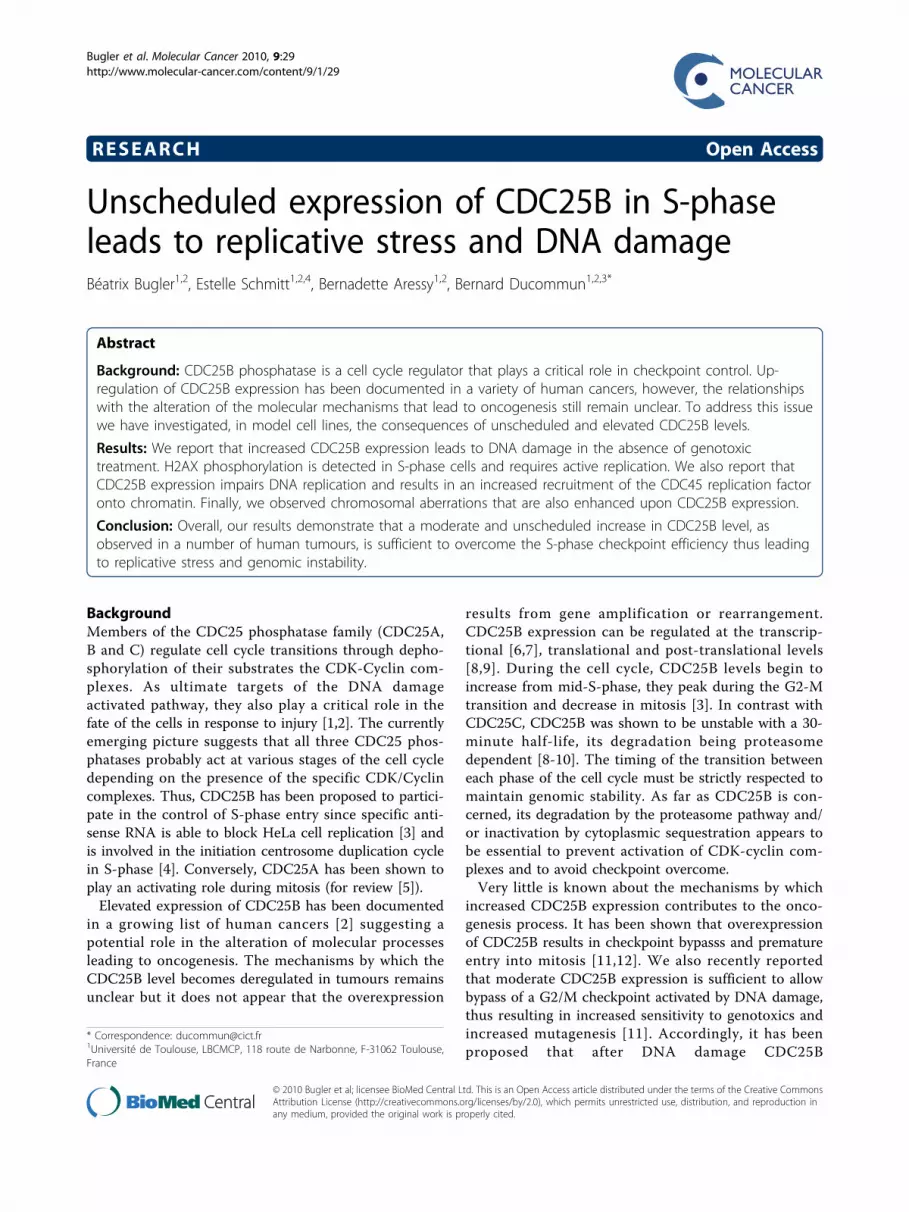
beginning of the U2OS-CDC25B S phase, however overtime S-phase appeared identical in both cell populationsindicating that S-phase duration was similar. Togetherwith previous reports [16], these results suggest thatunscheduled CDC25B expression results in a prematureentry into S phase without impact on the duration ofDNA replication but with possible consequences on itsregulation and on its fidelity.Elevated CDC25B expression in S-phase induces DNAdamageWe next examined the possible consequences ofunscheduled CDC25B expression on the occurrence ofreplication-linked DNA damage. With this aim, we usedimmunofluorescence microscopy to monitor g-H2AXstaining, a sensitive and early marker of DNA injury. Asshown in figure 2A (left panel) the U2OS cells expressingHa-CDC25B displayed a strong positive g-H2AX staining.This positivity was also observed by western blot on totalextract of cells in S-phase after synchronisation by noco-dazole block and release, but was never observed inU2OS cells that do not express CDC25B (Figure 2A rightpanel). To examine the relationship between S-phase andthe occurrence of DNA damage, we performed immuno-fluorescence after double staining with g-H2AX andBrdU of U2OS cells expressing CDC25B or not. Asreported in figure 2B, g-H2AX staining was found to belargely associated with BrdU incorporating cells. Flowcytometry analysis of cell cycle distribution confirmedthat while the overall percentage of cells displaying a g-H2AX positivity was about 8% (Figure 2C, left panel),most of the U2OS-CDC25B cells displaying DNAdamage were in S-phase with nearly 60% of g-H2AXlabeling in that phase of the cell cycle (right panel, Figure2C). In contrast a very low staining level was observed inU2OS cells as shown in the scatter plots.In order to confirm this observation in a cellular con-
text in which the unscheduled expression of CDC25B islimited to a level frequently observed in many tumourcell lines, we made use of HCT116 cells that were engi-neered to stably express a moderate level of Ha-CDC25B. As shown in Figure 2D this expression is lim-ited to about two-fold in HCT116 CDC25B+ (centralpanel) while in contrast a much higher expression levelis achieved in U2OS cells. HCT116 and HCT116CDC25B+ were synchronised by thymidine block andprocessed to immunofluorescence detection after 3 h ofrelease. A g-H2AX staining was observed in most of theHCT116 cells expressing Ha-CDC25B while a negligiblesignal was observed in the parental cell line. This obser-vation was confirmed by the quantification of the g-H2AX fluorescence as shown in the right panel of thefigure 2D.These observations were specific for CDC25B, as they
were not observed in U2OS cells conditionally
expressing CDC25C (see Figure S2, Additional file 2).Thus, our results suggest a specific role for unscheduledexpression of CDC25B in the induction of DNA damageduring S-phase.Replication is required for g-H2AX labeling in cellsexpressing elevated levels of CDC25BAs 60% of the cells displaying g-H2AX staining were inS phase, we explored whether active DNA replicationwas necessary to observe DNA damage upon unsched-uled expression of CDC25B. Asynchronous U2OS cellswere induced to express Ha-CDC25B and treated at thesame time with the DNA polymerase inhibitor aphidico-lin to inhibit replication while increasing CDC25Bexpression. After 20 hours the drug was removed toresume cell cycle and the levels of g-H2AX and BrdUincorporation were monitored by flow cytometry at eachindicated time after induction of CDC25B expression.As shown in figure 3A, at the time of release from theaphidicolin block, cells were mainly arrested in G1 with-out BrdU incorporation and did not present any g-H2AX positivity. By contrast, when the cell cycle wasresumed by aphidicolin removal, progressive phosphory-lation of g-H2AX was clearly detected in U2OS-CDC25B by immunofluorescence staining and flowcytometry 3 and 6 hours after release, and paralleledBrdU incorporation. This positivity was not observed inthe control U2OS cells population that did notexpressed CDC25B. Moreover as shown in figure 3B,treatment with the CDK inhibitor roscovitin (10 μM) atthe time of induction of CDC25B expression, resultedafter 17 h in only 3% of g-H2AX positivity while 11% ofg-H2AX positivity was observed when the cells weretreated 4 h hours after the induction of CDC25B expres-sion. These data suggest a correlation between the ele-vated level of CDC25B and its consequence on CDK2activity, replication unwinding and g-H2AX labeling.DNA damage was obvious as early as 3 hours after
aphidicolin block release and g-H2AX positivity was notfound to be associated with condensed, fragmented ormicronucleated morphology, indicating that the DNAdamage observed could not result from CDC25B-depen-dent mitotic catastrophe and subsequent apoptosis (seealso figure S3A, Additional file 3).Furthermore, when U2OS cells were synchronized in
mitosis and released in Ha-CDC25B induction condi-tions, g-H2AX labeling was detected only 13 h after syn-chronization when the cells entered S-phase, while Ha-CDC25B positive cells were already detected 6 hoursbefore. Thus, despite expression of CDC25B during G1-phase, DNA damage occurred only during DNA replica-tion and long before entry into mitosis (Figure S3B,Additional file 3).Overall, these results indicate that DNA replication is
required to observe g-H2AX labeling upon unscheduled
Bugler et al. Molecular Cancer 2010, 9:29http://www.molecular-cancer.com/content/9/1/29
Page 3 of 12
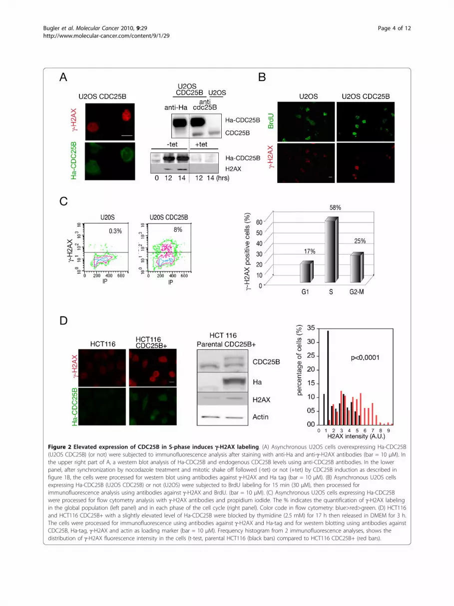
Figure 2 Elevated expression of CDC25B in S-phase induces g-H2AX labeling. (A) Asynchronous U2OS cells overexpressing Ha-CDC25B(U2OS CDC25B) (or not) were subjected to immunofluorescence analysis after staining with anti-Ha and anti-g-H2AX antibodies (bar = 10 μM). Inthe upper right part of A, a western blot analysis of Ha-CDC25B and endogenous CDC25B levels using anti-CDC25B antibodies. In the lowerpanel, after synchronization by nocodazole treatment and mitotic shake off followed (-tet) or not (+tet) by CDC25B induction as described infigure 1B, the cells were processed for western blot using antibodies against g-H2AX and Ha tag (bar = 10 μM). (B) Asynchronous U2OS cellsexpressing Ha-CDC25B (U2OS CDC25B) or not (U2OS) were subjected to BrdU labeling for 15 min (30 μM), then processed forimmunofluorescence analysis using antibodies against g-H2AX and BrdU. (bar = 10 μM). (C) Asynchronous U2OS cells expressing Ha-CDC25Bwere processed for flow cytometry analysis with g-H2AX antibodies and propidium iodide. The % indicates the quantification of g-H2AX labelingin the global population (left panel) and in each phase of the cell cycle (right panel). Color code in flow cytometry: blue>red>green. (D) HCT116and HCT116 CDC25B+ with a slightly elevated level of Ha-CDC25B were blocked by thymidine (2.5 mM) for 17 h then released in DMEM for 3 h.The cells were processed for immunofluorescence using antibodies against g-H2AX and Ha-tag and for western blotting using antibodies againstCDC25B, Ha-tag, g-H2AX and actin as loading marker (bar = 10 μM). Frequency histogram from 2 immunofluorescence analyses, shows thedistribution of g-H2AX fluorescence intensity in the cells (t-test, parental HCT116 (black bars) compared to HCT116 CDC25B+ (red bars).
Bugler et al. Molecular Cancer 2010, 9:29http://www.molecular-cancer.com/content/9/1/29
Page 4 of 12

expression of CDC25B and strongly suggest that DNAdamage is associated with replication stress and defectsin the initiation and/or progression of replication forks.Elevated levels of CDC25B cause increased CDC45recruitment on chromatinIt is well known that the initiation factor CDC45requires the combined activation of the cyclin-depen-dent kinase CDK and the Dbf4-dependent kinase DDKto initiate replication firing of the inactive pre-replica-tion complexes [19]. As CDK2-cyclinA is a bona-fidesubstrate for CDC25B, the likely enhanced activation ofCDK2 by elevated levels of the phosphatase could resultin increased phosphorylation of CDC45 resulting in therecruitment of this factor on the pre-replication com-plexes. To test this hypothesis, we measured the amountof CDC45 associated with the chromatin-bound fractionafter DNase treatment in U2OS cells expressing elevatedlevels of CDC25B. The cells were harvested 3 h afterrelease from thymidine block to enrich in S-phase cellsand limit premature entry into mitosis due to CDC25Boverexpression [16]. As shown in Figure 4A, the ele-vated and unscheduled expression of CDC25B resultedin a significant increase of chromatin-associated CDC45,whereas the CDC45 level recovered in the soluble frac-tion was not significantly affected. Orc2 binding was notmodified by CDC25B level modulation and constitutesan internal standard. As predicted this suggests also aCDC25B involvement in the activation but not in the
licensing of replication. We next examined whetherDNA damage induced by unscheduled CDC25B expres-sion was dependent on the activity of CDC45. With thisaim, CDC45 expression was invalidated in U2OS cellsexpressing CDC25B by RNA interference and g-H2AXwas monitored by western blot. As depicted in figure4B, DNA damage revealed by g-H2AX labeling was sig-nificantly reduced in CDC45-depleted cells while nochanges were observed in untransfected cells or in cellstransfected with scrambled siRNA. Indeed, no DNAdamage was detected in U2OS cells that did not expressCDC25B.These results strongly support the hypothesis that ele-
vated and unscheduled activity of CDC25B is responsi-ble for abnormal CDK2-cyclin activation and thesubsequent phosphorylation of CDC45. This wouldresult in the deregulation of its recruitment on the repli-cation complexes that could likely account for theobserved replication stress and the subsequent DNAdamage [20-22].Elevated level of CDC25B impairs replication forkprogressionTo gain insight into the mechanism by which unsched-uled CDC25B expression could promote replicationstress we examined the progression of replication forksin cells expressing or not CDC25B. With this aim, thethymidine analogs CldU and IdU were successivelyincorporated into DNA (see methods) and fluorescence
Figure 3 Replication is required for g-H2AX labeling in cells expressing elevated levels of CDC25B. (A) U2OS cells were induced tooverexpress Ha-CDC25B by tetracycline removal for 20 hours together with aphidicoline (2 μg/ml). The cells were incubated with BrdU (30 μM)for 15 min before harvesting at the indicated time (0, 3 h and 6 h) after release and staining with antibodies against g-H2AX forimmunofluorescence and rat BrdU and mouse g-H2AX together with propidium iodide for flow cytometry analysis (bar = 10 μM). The indicatedpercentage corresponds to g-H2AX and BrdU positive cells. (B) The CDK inhibitor roscovitin (10 μM) was added at the time of CDC25B inductionin U2OS cells (0 h) or 4 h hours after the beginning of induction. A 17 h after CDC25B induction, flow cytometry was achieved after stainingwith g-H2AX antibodies and propidium iodide. The indicated percentage corresponds to g-H2AX positive cells.
Bugler et al. Molecular Cancer 2010, 9:29http://www.molecular-cancer.com/content/9/1/29
Page 5 of 12
microscopy was used to visualize, in each of the replica-tion foci, the corresponding labeling detected with anti-bodies to CldU (green) and IdU (red) (Figure 5). Asdemonstrated by others [23], the DNA replication pro-gression is inversely proportional to the colocalization ofthe two markers, the larger the overlapping areas of theCldU and IdU foci, the slower the fork migrates andvice versa. This analysis was performed in U2OS cellsconditionally expressing CDC25B (Figure 5A) and inHCT116 cells expressing CDC25B (Figure 5B) that weresynchronized by thymidine block and released for 2hours to enrich the S-phase population. As shown, therelative colocalization areas of CldU-IdU were signifi-cantly more elevated in both cell types, indicating a sig-nificant perturbation of the fork progression likely dueto fork stalling upon CDC25B expression.
To confirm that this observation in HCT116 CDC25B+ cells was totally dependent on CDC25B expression,we invalidated its expression by RNA interference usingsiRNA against CDC25B that has already been validated[11]. As presented in figure 5C, while scrambled siRNAwas inefficient, the reduction of CDC25B expressionwith a specific siRNA led to a significant lowering of theoverlapping CldU-IdU areas reflecting an increase infork progression. These data demonstrate a clear rela-tionship between unscheduled expression of CDC25Band deregulation of fork progression. This replicativestress is likely due to the abnormal CDC45 recruitmenton replication complexes.Elevated levels of CDC25B cause chromosome instabilityThe ability of abnormal and unscheduled increasedlevels of CDC25B to promote replication stress resulting
Figure 4 Elevated level of CDC25B causes increased CDC45 recruitment on chromatin. (A) U2OS cells conditionally expressing CDC25Bwere induced (U2OS CDC25B) or not (U2OS) in the presence of 2.5 mM thymidine for 17 h then released from the cell cycle block bythymidine removal. After 3 h, the cells were harvested and processed for western blot analysis of two chromatin extracts, the detergent-solublefraction (Sol. Chrm.) and the DNase1 soluble fraction (DNase). The CDC45 protein level was monitored using Orc2 as loading marker. A typicalwestern blot of one of the four independent experiments used for quantification is shown. * indicates a p value < 0.01 (t-test where U2OSCDC25B is compared to U2OS). (B) U2OS conditionally expressing CDC25B were transfected with CDC45 or control siRNA (Scr) for 28 h or nottransfected (NT) then treated with 2.5 mM thymidine for 17 h in the absence of tetracycline to induce CDC25B expression. Cells were releasedfrom thymidine block by thymidine removal from the media. After 3 h, total extracts were processed for western blot analysis using anti-Ha,anti-CDC45, anti-g-H2AX and anti-actin antibodies as loading controls. The CDC45 antibody was already tested for its specificity in [46]. Non-induced U2OS are shown as controls (U2OS).
Bugler et al. Molecular Cancer 2010, 9:29http://www.molecular-cancer.com/content/9/1/29
Page 6 of 12

Figure 5 Elevated level of CDC25B causes perturbation of S-phase progression. (A) U2OS were induced to express Ha-CDC25B (U2OSCDC25B) or not (U2OS) then incubated in the presence of 2.5 mM thymidine for 17 h and released. Two hours after release, replication siteswere pulse labeled for 30 min with CldU then for 30 min with IdU. After immunostaining with antibodies specific for CldU and IdU, theoverlapping foci areas were quantified in each isolated cell and the results were expressed as the colocalization ratio (about 50 cells wereanalyzed). The insert shown in the central panel of this figure displays representative labeling with CldU and IdU and the typical merge figurethat supports colocalization quantification using Image J software. (B) HCT116 cells expressing (CDC25B+) or not (parental) elevated levels ofCDC25B (see figure 2) were processed after thymidine treatment as in (A). A total of 100 to 150 cells were analyzed. (C) HCT116 expressingelevated levels of CDC25B (HCT CDC25B+) were transfected with a CDC25B siRNA (siCDC25B) or a control siRNA (siScr) for 24 h then treated asin A and B. As controls, untransfected HCT116 CDC25B+ (NT) and parental cells (ctr). *** indicates a p value < 0.0001. A total of 130 to 170 cellswere analyzed.
Bugler et al. Molecular Cancer 2010, 9:29http://www.molecular-cancer.com/content/9/1/29
Page 7 of 12

from a decrease of fork progression, prompted us toanalyze this chromosome feature.We examined chromosomal aberrations in metaphase
spreads that were prepared using U2OS cells expressingCDC25B after colcemid treatment. The frequencies ofchromatid and chromosome aberrations such as gapsand breaks were respectively 1.2% and 0.6% in U2OScells whereas they rose to 2.7% and 1.6% in U2OS cellsexpressing CDC25B (Figure 6A). As illustrated in Figure6B, a typical spreading of metaphase CDC25B-expres-sing U20S cells revealed gaps, breaks and joined chro-mosomes illustrating the chromosomal aberrations thatwere detected.
DiscussionIn this study, we show that a moderate and unscheduledincrease in CDC25B protein level, comparable to theincreased level that has been reported to be observed inhuman tumours, has a critical incidence during S phasethrough the generation of replication defects. We firstdemonstrate that abnormal level of CDC25B expressionresults in DNA damage essentially occurring in replicat-ing cells. This observation is reminiscent of the prema-ture activation of cyclin E- and cyclin A-dependentkinase observed upon CDC25A overexpression [15]. Italso recalls the effect of ectopic expression of a constitu-tively active CDK mutant that causes DNA damage spe-cifically in S-phase. Furthermore, chemical inhibition ofCDK-cyclin can reverse the DNA damage observed inconditional Chk1 knockdown ES cells [24]. Enhancedactivation of CDK2 by elevated levels of the phosphataseCDC25B has already been shown [25,26], and overex-pression of CDC25B was able to overcome the unrepli-cated DNA checkpoint [16]. Chk1 therefore appears to
be critical in controlling initiation of replication andelongation and probably acts through the modulation ofCDC25 phosphatase activity [21,22,27]. One likelyhypothesis to explain our observations would be that byweakening the role of Chk1, elevated and unscheduledexpression of CDC25B in G1 phase would compromisethe checkpoint relative to the S phase and lead toabnormal activation of CDK-cyclin activity associated toDNA replication. This effect is consistent with Chk1haplo-insufficiency observed in some Chk1 dependentphenotypes with accumulation of DNA damage duringreplication and failure to restrain mitotic entry [28,29].CDK-cyclin complexes play an essential role in regulat-ing the activity of replication factors such as Cdc6, Cdt1and CDC45 (reviewed in [30]) as well as in chromatindecondensation by phosphorylation of histone H1 togain access to DNA in S phase [31]. Here we report anincreased loading of the key replication factor CDC45during S phase, upon elevated and unscheduled expres-sion of CDC25B and a reversion of the DNA damagethat was correlated to the specific depletion of CDC45.CDC45 is CDK-dependent for its activity on the chro-matin and is required for origin unwinding and for theloading of the replicative polymerases [19,32]. As bind-ing of CDC45 to chromatin is rate limiting for DNAreplication, the CDC45 active form constitutes one ofthe critical regulator for the activation of pre-replicationcomplexes [33] and increased loading of CDC45 in theabsence of CDC25 regulation by Chk1 has already beencorrelated to replication stress [20-22]. Thus, anincrease of CDC25B expression albeit to a minor extentclose to physiological variations as observed in theHCT116 CDC25B+ cells, could phenocopy a Chk1depletion leading to inappropriate cell cycle transition,
Figure 6 Elevated level of CDC25B induces significant chromosomal aberrations. (A) U2OS were induced by tetracycline removal toexpress Ha-CDC25B (CDC25B) or not (U2OS) during 24 h then chromosomal breaks, gaps and fusions were quantified on metaphase spreads. (B)Representative metaphase spreads with breaks (arrow 1), gaps (arrow 2) and fusions (arrow 3).
Bugler et al. Molecular Cancer 2010, 9:29http://www.molecular-cancer.com/content/9/1/29
Page 8 of 12
DNA replication stress and accumulation of DNAdamage.Although S-phase duration was not changed, we also
observed a decrease in the replication rate upon expres-sion of CDC25B and we demonstrated that depletion ofits expression was sufficient to rescue a normal progres-sion. As the replication rate is inversely correlated withthe density of active origins [34,35], an attractive expla-nation for the occurrence of DNA damage in CDC25Bexpressing cells would be the activation of unscheduledand unstable replication origins [36,37]. Shortening theinter origin distance induced by the formation of newactive origins could increase DNA torsion stress whichcould in turn promote stalled and collapsed forks thusleading to double strand breaks of DNA and a slow-down of fork progression [38]. In contrast with otheroncogenes CDC25B deregulation leads to replicativestress in the absence of detectable re-replication andprobably through the activation of new replication ori-gins as already observed after Myc deregulation [39].We also report an increase in numbers of chromoso-
mal aberrations such as gaps, breaks and joined chro-mosomes that illustrates the deleterious consequences ofelevated CDC25B expression during S-phase and itspotential role in genomic instability. In line with thisobservation, we previously reported that HCT116 cells,expressing elevated levels of CDC25B, displayed an ele-vated mutation rate compared to the parental cell line[11]. CDC25A overexpression in primary human epithe-lial cells was also previously shown to promote genomicinstability at common fragile sites, thus accounting forthe oncogenic consequences of its increased expressionin human tumours [40]. In the case of CDC25B, it hasbeen thought that as a regulator of the G2-M transition,this phosphatase did not act at the G1-S transition andin S-phase, and that the oncogenic properties associatedwith its overexpression in tumours could be related toG2-M checkpoint bypass and unscheduled entry intomitosis. Our findings demonstrate that this vision wasincomplete. It appears that CDC25B expression must betightly controlled and particularly in S phase, anyunscheduled increase in its nuclear expression leadingto replication stress and checkpoint control deficiency.Interestingly, CDC25B is mostly nuclear in G1 phase ofunperturbed HeLa cells and gradually moves to thecytoplasm as cells progress to S phase depending on thepresence of Cyclin B1 [16] or on the p38 mitogen acti-vated protein kinase activation suggesting a regulationin response to various types of cellular stress [41]. Itsability to be down regulated by p53 ([42] and personalcommunication), well-known for its frequent inactiva-tion in tumours, its in vitro transforming potential [43]and its ability to promote unscheduled entry into S-phase constitute essential features for the contribution
of CDC25B to oncogenesis according to the proposedinduced senescence model (for review [44]).
ConclusionOur findings indicate that unscheduled and moderateexpression of CDC25B during S-phase is sufficient toinduce replicative stress and genomic instability. Sinceabnormal expression of CDC25B has been found innumerous cancers (reviewed in [2,45]) our results pro-vide new insights into the molecular mechanisms of theinvolvement of this phosphatase in tumorigenesis.
MethodsCell culture and transfectionU2OS conditionally expressing Ha-CDC25B3 (B3 iso-form) cells were grown as previously described [18].Cells were synchronized and induced for CDC25B atthe G1-S transition by a double thymidine block as fol-lows: 16 h of treatment with 2.5 mM thymidine and 5μg/ml tetracycline to repress the promotor, then 16 hrelease followed by the second thymidine block for 17 hwithout tetracycline to induce CDC25B. Cells were syn-chronized at the G2-M transition by nocodazole (100nM, 17 h) with 5 μg/ml tetracycline then released, sha-ken off to retrieve mitotic cells and induced for Ha-CDC25B in the absence of tetracycline. HCT116 p53-/-clones expressing elevated levels of CDC25B were gen-erated and grown as previously described [11].A previously validated siRNA for CDC25B with the
following sequence 5’AGACUGCAGAUACCCCUAU-3’was used. Human CDC45 siRNA pool was purchasedfrom Santa Cruz (CA). Cells were electrotransfectedusing AMAXA nucleofector following the manufac-turer’s instructions for HCT116 and U2OS cells.ImmunofluorescenceMouse anti-phospho Ser139 g-H2AX (clone JBW 301,Upstate Biotechnology, Lake Placid, NY), rabbit anti-phospho Ser139 g-H2AX (Upstate Biotechnology),mouse anti-Ha tag (clone Ha.11 Covance), rabbit anti-phospho H3-Ser210 (Upstate Biotechnology), rat anti-BrdU (clone BU1/75 Serotec), mouse anti-BrdU (BectonDickinson), rabbit CDC25B antibody (C-20. Santa Cruz,CA), mouse anti-actin (Chemicon, Temecula, CA), rab-bit anti-CDC45 (ref. 20685. Santa Cruz). Mouse rabbitand rat anti-IgG Alexa 488 and 594 for immunofluores-cence (Molecular Probes, Invitrogen), rabbit and mouseanti-HRP antibodies (Cell Signalling).Cells cultured on glass coverslips were processed as
previously described then incubated with rabbit anti-g-H2AX and mouse anti-Ha tag or rabbit anti-phosphoH3 Ser210 and mouse anti-phospho g-H2AX followedby rabbit and mouse Alexa secondary antibody staining[12]. Cells were mounted in Vectashield anti-fademounting medium and visualized using a DM6000
Bugler et al. Molecular Cancer 2010, 9:29http://www.molecular-cancer.com/content/9/1/29
Page 9 of 12
microscope (Leica, Wetzlar, Germany). For BrdU stain-ing, cells were incubated with 30 μM BrdU (Calbio-chem) for 15 min and fixed with 3.7% formaldehyde for10 min. The cells were processed as described in [23]with some modifications: they were washed with PBSand incubated with methanol for 5 min at -20°C thentreated with PBS/0.5%Triton ×100/0.02% SDS for 30min at room temperature. DNA was denatured usingfreshly prepared 1.5 M HCl, then neutralized by washingwith 0.1 M sodium borate (pH 8.5) and PBS. To blocknon-specific binding, cells were incubated in 5%PBS-BSA, 30 min to overnight at 4°C then submitted to anti-g-H2AX or anti-BrdU for 1 h then two washes with PBSfollowed by mouse anti-IgG Alexa 594 and rat anti-IgGAlexa 488 respectively.Replication focus detection with CldU and IdU was
performed on U2OS or HCT116 cells blocked by thymi-dine (2.5 mM) for 17 h then released in DMEM for twohours. Cells were incubated in medium containing 100μM CldU (Sigma, St Quentin, France) for 30 min then100 μM IdU for the last 30 minutes after washing withhot medium. IdU incorporation was stopped with med-ium containing thymidine (1 mM) then cells were fixedwith cold 70% ethanol. They were treated with 100%methanol at -20°C for 5 min, washed twice with PBSthen incubated in 1.5 M HCl for 20 min. After twowashes with PBS, they were incubated in 0.5%Tween20/0.25%BSA/5% fetal veal serum/PBS/(TBS) for30 min in a humid box. Incubation in the primary anti-body rat anti-BrdU against CldU and mouse anti-BrdUagainst IdU in TBS for 2 hours was followed by anti-ratIgG Alexa 594 and anti-mouse IgG Alexa 488 in TBSrespectively. Cells were washed twice in 0.5% Tween/PBS then mounted in Vectashield solution and visua-lized using a DM 6000 microscope. Pictures wereacquired with MetaMorph software, keeping the sameintensities for each fluorescent dye for all the pictures ofthe same assay and the signals were measured usingImageJ software. IdU-CldU colocalization was quantifiedfrom the merge picture by dividing the colocalizationarea by the total area for each nucleus and the non-parametric Welch T corrected test was used to analysethe data.Flow cytometryCells were processed as previously described with mouseanti-phospho Ser139 g-H2AX, followed by mouse anti-IgG Alexa 488 [12]. DNA was stained with propidiumiodide (10 μg/ml) in the presence of RNase (5 μg/ml)and analyses were done on a FACScan flow cytometer(Cell Quest, Becton Dickinson, Mountain View, CA).For BrdU incorporation assay, the cells were incubatedwith 30 μM BrdU (Calbiochem) for 15 min, fixed asabove then DNA was denatured by freshly prepared 1.5M HCl, then neutralized by 0.1 M sodium borate (pH
8.5) followed by PBS. After washing in 1%PBS-BSA, ratanti-BrdU was added for 2 h together with mouse anti-phospho g-H2AX then two PBS washes followed by ratanti-IgG Alexa 488 and mouse anti-IgG Alexa 594staining.Chromatin fractionation and Western BlottingU2OS cells were synchronized and induced for CDC25Bexpression at the G1-S transition by a simple thymidineblock (2.5 mM, 17 h). After 3 h of thymidine release,the cells were harvested and resuspended in buffer A(10 mM HEPES pH 7.5, 1.5 mM MgCl2, 10 mM KCl,10% glycerol, 0.34 M sucrose, protease inhibitors (cock-tail-Complete, Roche), 10 min on ice. EDTA 10 mMwas added for 30 min and this chromatin fractionobtained after centrifugation (12 000 g, 3 min, 4°C)represented the soluble fraction. The pellets werewashed twice in buffer A and incubated 30 min at RTwith 2000 U/ml DNaseI (Roche) and a further 30 minat 4°C with 0.5 M NaCl. The DNase solubilized chroma-tin fraction was obtained after centrifugation (12000 g, 3min, 4°C).Chromatin fractions and whole protein extracts were
electrophoresed on a 4%-12% SDS gradient gel (Invitro-gen, Carlsbad, CA) and analysed by Western Blotting.For protein quantification, pictures were acquired with aBioimaging Systems, Syngene Camera and the signalsmeasured using ImageJ software.Metaphase chromosomes spreadsU2OS cells were induced for CDC25B or not for 24 hrsat which point Colcemid (0.1 μg/mL; Gibco) was addedfor the last 3 h to accumulate mitotic cells prior to tryp-sinisation, centrifugation, resuspension in PBS, centrifu-gation and swelling in hypotonic (50 mM) KCl solutionfor 25 min at RT. A fixation solution of 100% ethanol/acetic acid (3:1) was added and the cells were centri-fuged, rinsed twice in ethanol/acetic acid before spread-ing on slides and being left to dry. Chromosomes werestained with 0.05 μg/ml DAPI/PBS (Sigma) for 10 minthen washed with several changes of PBS and mountedwith mounting medium (Dakocytomation) prior tomicroscopy. About 30 spreads were scored for statisticaldata.
Additional file 1: Analysis of S phase duration. Asynchronous cellsoverexpressing CDC25B (U2OS CDC25B) or not (U20S) were treated withnocodazole (200 nM) all along the assay. The cells were pulse labeledwith BrdU (30 μM, 15 min) then BrdU was replaced by thymidine (1mM). The cells were collected at the indicated times and immunostainedwith anti BrdU antibodies. The percentage of BrdU positive cells in Sphase was determined by flow cytometry analysis. 100% correspond tothe cell population before chase. As an example, the percentages of cellsin S phase at 0 h and 10 h after thymidine chase were measured asshown in the two lower plots.Click here for file[ http://www.biomedcentral.com/content/supplementary/1476-4598-9-29-S1.PDF ]
Bugler et al. Molecular Cancer 2010, 9:29http://www.molecular-cancer.com/content/9/1/29
Page 10 of 12

Additional file 2: Analysis of g-H2AX staining in overexpressingCDC25C U2OS cells. Asynchronous U2OS cells conditionallyoverexpressing Ha-CDC25B or Ha-CDC25C by tertracycline removal (+)[47] for 17 h were processed for flow cytometry analysis with g-H2AXantibodies and propidium iodide. The results indicate the percentage ofg-H2AX positive cells in interphase (G1-S-G2) and mitosis.Click here for file[ http://www.biomedcentral.com/content/supplementary/1476-4598-9-29-S2.PDF ]
Additional file 3: Analysis of g-H2AX staining during the cell cycle.U2OS cells conditionally expressing Ha-CDC25B were synchronized inmitosis by nocodazole treatment (100 nM, 17 h) as in figure 1B. Atindicated times after CDC25B induction, cells were processed forimmunofluorescence analysis using rabbit antibodies against g-H2AXtogether with antibodies against phosphorylated histone H3 (panel A) orwith anti-Ha to detect CDC25B (panel B).Click here for file[ http://www.biomedcentral.com/content/supplementary/1476-4598-9-29-S3.PDF ]
AcknowledgementsWe gratefully acknowledge Marie-Jeanne Pillaire for advice, technical supportand critical reading of the manuscript. ES was a recipient of a post-doctoralfellowship from the Fonds pour la Recherche (Québec). This work wassupported by C.N.R.S, l’Université Paul Sabatier, la région Midi-Pyrénées,l’Institut National du Cancer, the Cancéropôle Grand Sud-Ouest and la LigueNationale Contre le Cancer (Equipe labellisée 2008).
Author details1Université de Toulouse, LBCMCP, 118 route de Narbonne, F-31062 Toulouse,France. 2CNRS, LBCMCP-UMR5088, F-31062 Toulouse, France. 3CHU Purpan,TSA 40031, F-31059 Toulouse, France. 4Notre Dame Hospital and MontrealCancer Institute, Montreal H2L 4 M1, Canada.
Authors’ contributionsBB designed, carried out the experiments and drafted the manuscript. ESperformed the double block thymidine synchronisation experiment. BAconstructed the HCT116 CDC25B+ cell line. BD supervised the project andfinalised the manuscript. All authors have read and approved the finalmanuscript.
Competing interestsThe authors declare that they have no competing interests.
Received: 30 September 2009Accepted: 4 February 2010 Published: 4 February 2010
References1. Aressy B, Ducommun B: Cell cycle control by the CDC25 phosphatases.
Anticancer Agents Med Chem 2008, 8:818-824.2. Boutros R, Lobjois V, Ducommun B: CDC25 phosphatases in cancer cells:
key players? Good targets?. Nat Rev Cancer 2007, 7:495-507.3. Garner-Hamrick PA, Fisher C: Antisense phosphorothioate
oligonucleotides specifically down-regulate cdc25B causing S-phasedelay and persistent antiproliferative effects. Int J Cancer 1998,76:720-728.
4. Boutros R, Lobjois V, Ducommun B: CDC25B involvement in thecentrosome duplication cycle and in microtubule nucleation. Cancer Res2007, 67:11557-11564.
5. Malumbres M, Barbacid M: Cell cycle, CDKs and cancer: a changingparadigm. Nat Rev Cancer 2009, 9:153-166.
6. Galaktionov K, Chen X, Beach D: Cdc25 cell-cycle phosphatase as a targetof c-myc. Nature 1996, 382:511-517.
7. Wang IC, Chen YJ, Hughes D, Petrovic V, Major ML, Park HJ, Tan Y,Ackerson T, Costa RH: Forkhead box M1 regulates the transcriptionalnetwork of genes essential for mitotic progression and genes encodingthe SCF (Skp2-Cks1) ubiquitin ligase. Mol Cell Biol 2005, 25:10875-10894.
8. Kanemori Y, Uto K, Sagata N: {beta}-TrCP recognizes a previouslyundescribed nonphosphorylated destruction motif in Cdc25A andCdc25B phosphatases. Proc Natl Acad Sci USA 2005, 102:6279-6284.
9. Kieffer I, Lorenzo C, Dozier C, Schmitt E, Ducommun B: Differential mitoticdegradation of the CDC25B phosphatase variants. Oncogene 2007,26:7847-7858.
10. Baldin V, Cans C, Knibiehler M, Ducommun B: Phosphorylation of humanCDC25B phosphatase by CDK1-cyclin A triggers its proteasome-dependent degradation. J Biol Chem 1997, 272:32731-32734.
11. Aressy B, Bugler B, Valette A, Biard D, Ducommun B: Moderate variationsin CDC25B protein levels modulate the response to DNA damagingagents. Cell Cycle 2008, 7:2234-2240.
12. Bugler B, Quaranta M, Aressy B, Brezak MC, Prevost G, Ducommun B:Genotoxic-activated G2-M checkpoint exit is dependent on CDC25Bphosphatase expression. Mol Cancer Ther 2006, 5:1446-1451.
13. Bansal P, Lazo JS: Induction of Cdc25B regulates cell cycle resumptionafter genotoxic stress. Cancer Res 2007, 67:3356-3363.
14. van Vugt MA, Bras A, Medema RH: Polo-like kinase-1 controls recoveryfrom a G2 DNA damage-induced arrest in mammalian cells. Mol Cell2004, 15:799-811.
15. Blomberg I, Hoffmann I: Ectopic expression of Cdc25A accelerates the G(1)/S transition and leads to premature activation of cyclin E- and cyclinA-dependent kinases. Mol Cell Biol 1999, 19:6183-6194.
16. Karlsson C, Katich S, Hagting A, Hoffmann I, Pines J: Cdc25B and Cdc25Cdiffer markedly in their properties as initiators of mitosis. J Cell Biol 1999,146:573-584.
17. Turowski P, Franckhauser C, Morris MC, Vaglio P, Fernandez A, Lamb NJ:Functional cdc25C dual-specificity phosphatase is required for S-phaseentry in human cells. Mol Biol Cell 2003, 14:2984-2998.
18. Davezac N, Baldin V, Gabrielli B, Forrest A, Theis-Febvre N, Yashida M,Ducommun B: Regulation of CDC25B phosphatases subcellularlocalization. Oncogene 2000, 19:2179-2185.
19. Zou L, Stillman B: Assembly of a complex containing Cdc45p, replicationprotein A, and Mcm2p at replication origins controlled by S-phasecyclin-dependent kinases and Cdc7p-Dbf4p kinase. Mol Cell Biol 2000,20:3086-3096.
20. Rodriguez R, Gagou ME, Meuth M: Apoptosis induced by replicationinhibitors in Chk1-depleted cells is dependent upon the helicasecofactor Cdc45. Cell Death Differ 2008, 15:889-898.
21. Shechter D, Costanzo V, Gautier J: ATR and ATM regulate the timing ofDNA replication origin firing. Nat Cell Biol 2004, 6:648-655.
22. Syljuasen RG, Sorensen CS, Hansen LT, Fugger K, Lundin C, Johansson F,Helleday T, Sehested M, Lukas J, Bartek J: Inhibition of human Chk1 causesincreased initiation of DNA replication, phosphorylation of ATR targets,and DNA breakage. Mol Cell Biol 2005, 25:3553-3562.
23. Seiler JA, Conti C, Syed A, Aladjem MI, Pommier Y: The intra-S-phasecheckpoint affects both DNA replication initiation and elongation:single-cell and -DNA fiber analyses. Mol Cell Biol 2007, 27:5806-5818.
24. Niida H, Tsuge S, Katsuno Y, Konishi A, Takeda N, Nakanishi M: Depletion ofChk1 leads to premature activation of Cdc2-cyclin B and mitoticcatastrophe. J Biol Chem 2005, 280:39246-39252.
25. Lindqvist A, Kallstrom H, Lundgren A, Barsoum E, Rosenthal CK: Cdc25Bcooperates with Cdc25A to induce mitosis but has a unique role inactivating cyclin B1-Cdk1 at the centrosome. J Cell Biol 2005, 171:35-45.
26. Varmeh S, Manfredi JJ: Inappropriate activation of cyclin-dependentkinases by the phosphatase Cdc25b results in premature mitotic entryand triggers a p53-dependent checkpoint. J Biol Chem 2009,284:9475-9488.
27. Feijoo C, Hall-Jackson C, Wu R, Jenkins D, Leitch J, Gilbert DM, Smythe C:Activation of mammalian Chk1 during DNA replication arrest: a role forChk1 in the intra-S phase checkpoint monitoring replication originfiring. J Cell Biol 2001, 154:913-923.
28. Lam MH, Liu Q, Elledge SJ, Rosen JM: Chk1 is haploinsufficient formultiple functions critical to tumor suppression. Cancer Cell 2004, 6:45-59.
29. Wilsker D, Petermann E, Helleday T, Bunz F: Essential function of Chk1 canbe uncoupled from DNA damage checkpoint and replication control.Proc Natl Acad Sci USA 2008, 105:20752-20757.
30. Takeda DY, Dutta A: DNA replication and progression through S phase.Oncogene 2005, 24:2827-2843.
Bugler et al. Molecular Cancer 2010, 9:29http://www.molecular-cancer.com/content/9/1/29
Page 11 of 12

31. Alexandrow MG, Hamlin JL: Chromatin decondensation in S-phaseinvolves recruitment of Cdk2 by Cdc45 and histone H1 phosphorylation.J Cell Biol 2005, 168:875-886.
32. Masuda T, Mimura S, Takisawa H: CDK- and Cdc45-dependent priming ofthe MCM complex on chromatin during S-phase in Xenopus eggextracts: possible activation of MCM helicase by association with Cdc45.Genes Cells 2003, 8:145-161.
33. Edwards MC, Tutter AV, Cvetic C, Gilbert CH, Prokhorova TA, Walter JC:MCM2-7 complexes bind chromatin in a distributed pattern surroundingthe origin recognition complex in Xenopus egg extracts. J Biol Chem2002, 277:33049-33057.
34. Conti C, Sacca B, Herrick J, Lalou C, Pommier Y, Bensimon A: Replicationfork velocities at adjacent replication origins are coordinately modifiedduring DNA replication in human cells. Mol Biol Cell 2007, 18:3059-3067.
35. Petermann E, Maya-Mendoza A, Zachos G, Gillespie DA, Jackson DA,Caldecott KW: Chk1 requirement for high global rates of replication forkprogression during normal vertebrate S phase. Mol Cell Biol 2006,26:3319-3326.
36. Maya-Mendoza A, Petermann E, Gillespie DA, Caldecott KW, Jackson DA:Chk1 regulates the density of active replication origins during thevertebrate S phase. Embo J 2007, 26:2719-2731.
37. Woodward AM, Gohler T, Luciani MG, Oehlmann M, Ge X, Gartner A,Jackson DA, Blow JJ: Excess Mcm2-7 license dormant origins ofreplication that can be used under conditions of replicative stress. J CellBiol 2006, 173:673-683.
38. Conti C, Seiler JA, Pommier Y: The mammalian DNA replicationelongation checkpoint: implication of Chk1 and relationship with originfiring as determined by single DNA molecule and single cell analyses.Cell Cycle 2007, 6:2760-2767.
39. Dominguez-Sola D, Ying CY, Grandori C, Ruggiero L, Chen B, Li M,Galloway DA, Gu W, Gautier J, Dalla-Favera R: Non-transcriptional controlof DNA replication by c-Myc. Nature 2007, 448:445-451.
40. Cangi MG, Piccinin S, Pecciarini L, Talarico A, Dal Cin E, Grassi S, Grizzo A,Maestro R, Doglioni C: Constitutive overexpression of CDC25A in primaryhuman mammary epithelial cells results in both defective DNA damageresponse and chromosomal breaks at fragile sites. Int J Cancer 2008,123:1466-1471.
41. Lindqvist A, Kallstrom H, Karlsson Rosenthal C: Characterisation of Cdc25Blocalisation and nuclear export during the cell cycle and in response tostress. J Cell Sci 2004, 117:4979-4990.
42. Scian MJ, Carchman EH, Mohanraj L, Stagliano KE, Anderson MA, Deb D,Crane BM, Kiyono T, Windle B, Deb SP, Deb S: Wild-type p53 and p73negatively regulate expression of proliferation related genes. Oncogene2008, 27:2583-2593.
43. Galaktionov K, Lee AK, Eckstein J, Draetta G, Meckler J, Loda M, Beach D:CDC25 phosphatases as potential human oncogenes. Science 1995,269:1575-1577.
44. Halazonetis TD, Gorgoulis VG, Bartek J: An oncogene-induced DNAdamage model for cancer development. Science 2008, 319:1352-1355.
45. Kristjansdottir K, Rudolph J: Cdc25 phosphatases and cancer. Chem Biol2004, 11:1043-1051.
46. Bauerschmidt C, Pollok S, Kremmer E, Nasheuer HP, Grosse F: Interactionsof human Cdc45 with the Mcm2-7 complex, the GINS complex, andDNA polymerases delta and epsilon during S phase. Genes Cells 2007,12:745-758.
47. Esmenjaud-Mailhat C, Lobjois V, Froment C, Golsteyn RM, Monsarrat B,Ducommun B: Phosphorylation of CDC25C at S263 controls itsintracellular localisation. FEBS Lett 2007, 581:3979-3985.
doi:10.1186/1476-4598-9-29Cite this article as: Bugler et al.: Unscheduled expression of CDC25B inS-phase leads to replicative stress and DNA damage. Molecular Cancer2010 9:29.
Submit your next manuscript to BioMed Centraland take full advantage of:
• Convenient online submission
• Thorough peer review
• No space constraints or color figure charges
• Immediate publication on acceptance
• Inclusion in PubMed, CAS, Scopus and Google Scholar
• Research which is freely available for redistribution
Submit your manuscript at www.biomedcentral.com/submit
Bugler et al. Molecular Cancer 2010, 9:29http://www.molecular-cancer.com/content/9/1/29
Page 12 of 12