unravelling the functional biomechanics of dental features and tooth wear
TRANSCRIPT
Unravelling the Functional Biomechanics of DentalFeatures and Tooth WearStefano Benazzi1*, Huynh Nhu Nguyen1, Ottmar Kullmer2, Jean-Jacques Hublin1
1 Department of Human Evolution, Max Planck Institute for Evolutionary Anthropology, Leipzig, Germany, 2 Department of Palaeoanthropology and MesselResearch, Senckenberg Research Institute, Frankfurt am Main, Germany
Abstract
Most of the morphological features recognized in hominin teeth, particularly the topography of the occlusal surface,are generally interpreted as an evolutionary functional adaptation for mechanical food processing. In this respect, wecan also expect that the general architecture of a tooth reflects a response to withstand the high stresses producedduring masticatory loadings. Here we use an engineering approach, finite element analysis (FEA), with an advancedloading concept derived from individual occlusal wear information to evaluate whether some dental traits usuallyfound in hominin and extant great ape molars, such as the trigonid crest, the entoconid-hypoconulid crest and theprotostylid have important biomechanical implications. For this purpose, FEA was applied to 3D digital models ofthree Gorilla gorilla lower second molars (M2) differing in wear stages. Our results show that in unworn and slightlyworn M2s tensile stresses concentrate in the grooves of the occlusal surface. In such condition, the trigonid and theentoconid-hypoconulid crests act to reinforce the crown locally against stresses produced along the mesiodistalgroove. Similarly, the protostylid is shaped like a buttress to suffer the high tensile stresses concentrated in the deepbuccal groove. These dental traits are less functional in the worn M2, because tensile stresses decreasephysiologically in the crown with progressing wear due to the enlargement of antagonistic contact areas and changesin loading direction from oblique to nearly parallel direction to the dental axis. This suggests that the wear processmight have a crucial influence in the evolution and structural adaptation of molars enabling to endure bite stressesand reduce tooth failure throughout the lifetime of an individual.
Citation: Benazzi S, Nguyen HN, Kullmer O, Hublin J (2013) Unravelling the Functional Biomechanics of Dental Features and Tooth Wear. PLoS ONE8(7): e69990. doi:10.1371/journal.pone.0069990
Editor: David Carrier, University of Utah, United States of America
Received March 26, 2013; Accepted June 13, 2013; Published July 23, 2013
Copyright: © 2013 Benazzi et al. This is an open-access article distributed under the terms of the Creative Commons Attribution License, which permitsunrestricted use, distribution, and reproduction in any medium, provided the original author and source are credited.
Funding: The Occlusal Fingerprint Analyser software programming is financed by the Deutsche Forschungsgemeinschaft (DFG, German ResearchFoundation). This is publication no. 54 of the DFG Research Unit 771 "Function and performance enhancement in the mammalian dentition – phylogeneticand ontogenetic impact on the masticatory apparatus." The funders had no role in study design, data collection and analysis, decision to publish, orpreparation of the manuscript.
Competing interests: The authors have declared that no competing interests exist.
* E-mail: [email protected]
Introduction
Since decades scholars have focused their attention on themorphology of the occlusal surface of human and non-humanprimate teeth to gain insight on the food items each species ismore adapted to process and to improve our understanding ofearly hominin diets and dietary adaptations [1–5]. Even thougha certain amount of within-species variability in the food itemsconsumed cannot be excluded, it is acknowledged that cuspswith steeply inclined slopes are well suited to generate shear-cutting forces, suggesting a diet of both soft and ductilefoodstuffs; conversely, rounded (or blunt) cusps are well suitedfor crushing, indicating a diet of hard, brittle foods [4,6,7].
However, during food processing teeth must solve anotherequally important function, namely they should be designed toresist failure while distributing forces produced duringmasticatory loading to their supporting structures [8–12]. These
two main functions, food diminution and resilience to failure, acttogether in the occlusal part of the chewing cycle, the powerstroke, during which food is comminuted and tooth-to-toothcontacts occur. Accordingly, beside the well-known variation indental occlusal topography (i.e., steep or blunt cusps), whichevolved to improve mechanical efficiency for food reduction,there should be other dental morphological features thatsimultaneously evolved to withstand occlusal loads.
Results from fracture mechanics suggest that, at least ingreat apes, dental material properties are less likely to be ofconcern than dental morphology (both internal and externalarchitecture) in the load-bearing capacity of the teeth [13,14].With regard to the internal architecture, the enamel thicknessmight be an example of such adaptation, as thick enamelallows both to increase wear resistance and to withstandand/or dissipate high masticatory loads [15–19]. Dentalbiomechanics suggest also that the arrangement of crystals
PLOS ONE | www.plosone.org 1 July 2013 | Volume 8 | Issue 7 | e69990
-J
within each enamel rod, enamel prism interweaving(decussation) and self-healing processes (growing fissuresfilled with organic fluids) are designed to better arrest crackgrowth in thick enamel driven by extended use or overloading[11,20–24].
Less attention, however, has been devoted to understandwhether the external geometry of the teeth might optimizeresilience on stress distribution. In an attempt to interpret theload-dissipation behavior of great ape molars, Macho andSpears [12] used two-dimensional (2D) finite element analysis(FEA) to suggest that modifications of the occlusal topographyare more responsible for efficient load dissipation thanincreasing enamel thickness by 100%, which ultimatelyreduces maximum tensile stresses by only 15%. Magne andBelser [25] used the same approach (2D FEA) to evaluate thebiomechanical behavior of opposing human molars in differentload-cases. They observed that high stress levels wereconcentrated in the central groove of maxillary molars, and thatenamel bridges and crests might reduce tensile stress locally,thus protecting crown biomechanics. Lucas and colleagues [17]suggested that the cingulum (a ridge encircling the base of atooth) might be functional important to protect the neck of thetooth from margin cracks driven by tensile stresses. Indeed,margin cracks begin at the base of the enamel (at or near thecervix) and extend longitudinally toward the occlusal surface[7,15,26], and are a source of failure both in real and ceramicdental crowns [27]. Finally, Anderson and colleagues [28] usedFEA in cone shape “teeth” to show that cingula structure mightindeed be important to reduce tensile strains in the enamel.
Despite these works, little is known about the functionalbiomechanics of external dental features, mainly because all ofthe above studies have much simplified the complex three-dimensional (3D) geometry of the tooth either using sections(2D approach), or modeling the tooth as cone shape-like, orapplying unrealistic occlusal loadings.
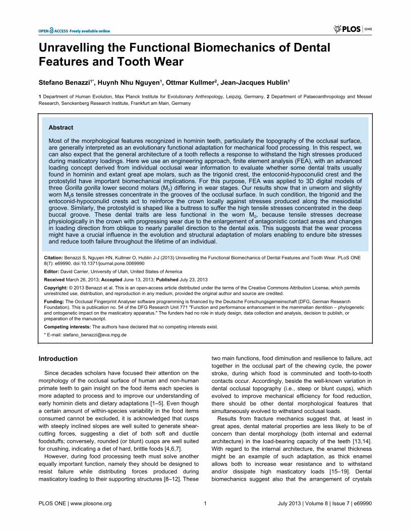
In this pilot study we used 3D FEA [29] with a newlydeveloped advanced loading concept derived from individualocclusal wear information [8,30–32] to test whether somedental traits usually found in hominin and extant great apelower molars, such as the trigonid crest pattern, the entoconid-hypoconulid crest and the protostylid (crest feature on thebuccal wall of the crown, normally associated with the buccalgroove [33,34]), might represent evolutionary responses toocclusal loadings (Figure 1). As African apes represent goodmodels for understanding dental functional morphology in earlyhominins [35], we compared maximum principal stresses in 3Ddigital models of three Gorilla gorilla lower left second molars(LM2) during maximum intercuspation tooth-to-tooth contact,which might be more damaging to the tooth crown than food-tooth contacts because of increased localized stresses. Theabove mentioned dental traits (trigonid crest pattern, theentoconid-hypoconulid crest and the protostylid) are wellexpressed in gorilla molars, which are also characterized by tallcusps, long shearing crests and relatively thin enamel [36],presumably an adaptation to folivorous diet [37]. As our threegorilla LM 2s differ in wear stages, we also aim to evaluate theeffects of a reduced relief through wear on the stressdistribution. Finally, a digital simulation was carried out to
assess whether interrupting the continuity of the crests (thetrigonid and the entoconid-hypoconulid crest, respectively)might affect the pattern of stress distribution.
Materials and Methods
Three Gorilla gorilla female skulls from the Museum fürNaturkunde, Humboldt Universität, Berlin, Germany wereselected for 3D FEA. The three specimens (ID = ZMB-31435,ZMB-31626 and ZMB-83551, respectively) were selected bothbecause of their complete dentition and because their LM2
differing in wear stage (after Smith [38]): ZMB-31435 = wearstage 1; ZMB-31626 = wear stage 3; ZMB-83551 = wear stage4. We obtained permission from the Museum für Naturkunde(Humboldt Universität, Berlin) to micro-CT scan the skulls atthe Bundesanstalt für Materialforschung und –prüfung, Berlin,Germany (scan parameters: 160kV, 150µA, 1.0mm copperfilter, and 2400 views per rotation). Volume data werereconstructed using isometric voxels ranging between 61 and65µm.
Figure 1. Digital reconstruction of the gorilla specimenZMB-31626 (lower left second molar – LM2). The threedental traits examined in this study (protostylid, trigonid crest,entoconid-hypoconulid crest) are highlighted both in the crown(top) and in the enamel-dentine junction (bottom).doi: 10.1371/journal.pone.0069990.g001
The Functional Biomechanics of Dental Features
PLOS ONE | www.plosone.org 2 July 2013 | Volume 8 | Issue 7 | e69990
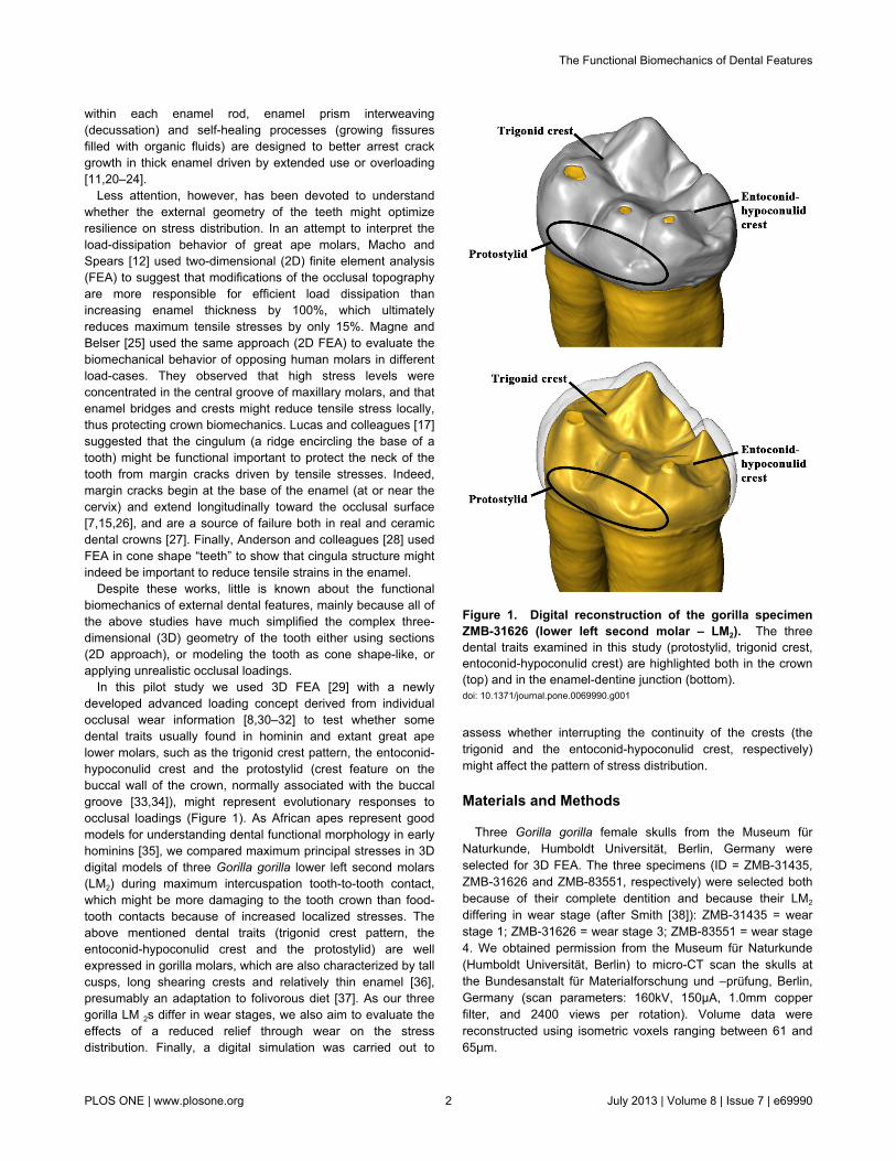
To reduce the size of the digital models, we cut themandibles distally to the socket of the lower left first molar(LM1) and mesially to the socket of the lower left third molar(LM3). Consequently, we considered only the bone tissuessurrounding the LM2. Segmentation of the LM2 dental tissues(enamel, dentine and pulp chamber) and its supporting dentaltissues (periodontal ligament - PDL, trabecular and corticalbone) was carried out in Avizo 7 (Visualization Sciences GroupInc.) (Figure 2A). For LM1-LM2, which were used to assess theocclusal contacts with LM2 (two-body interactions), only theexternal surface of the teeth was segmented. The finalrefinement of the digital models was carried out in RapidformXOR2 (IN, US Technology, Inc., Seoul, Korea).
As described in previous contributions [8,30,32], the dentalsurface models of lower and upper molars were imported intothe Occlusal Fingerprint Analyser (OFA) software to recognizethe contact areas on the LM2 with the antagonistic teeth duringthe power stroke. The contact areas were automaticallyselected by the software, thus informing on the position whereocclusal loads should be applied (red areas in Figure 2B seealso Video S1-S3). In order to compare the pattern of stressdistribution of the three gorilla specimens, the maximumintercuspation contact situation was selected. With regard tothe loading direction, it has been already suggested, formaximum intercuspation, to apply perpendicular loads to thecontact areas [8,30,39].
The surface models were then imported into HyperWorksSoftware (Altair Engineering, Inc.), where volumetric meshes(for enamel, dentine, pulp, PDL, cortical and trabecular boneshown in Figure 2C) were created using 10-nodes tetrahedral
elements (Table S1). Information for material properties suchas the elastic modulus -E, and the Poisson’s ratio werecollected from the literature [13,40–43] and summarized inTable 1. All the biological materials represented in the modelswere considered homogeneous, linearly elastic and isotropic.
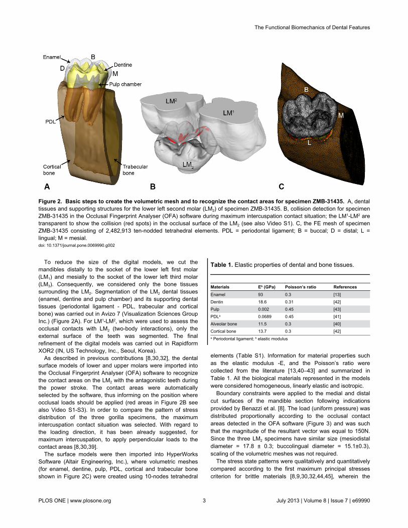
Boundary constraints were applied to the medial and distalcut surfaces of the mandible section following indicationsprovided by Benazzi et al. [8]. The load (uniform pressure) wasdistributed proportionally according to the occlusal contactareas detected in the OFA software (Figure 3) and was suchthat the magnitude of the resultant vector was equal to 150N.Since the three LM2 specimens have similar size (mesiodistaldiameter = 17.8 ± 0.3; buccolingual diameter = 15.1±0.3),scaling of the volumetric meshes was not required.
The stress state patterns were qualitatively and quantitativelycompared according to the first maximum principal stressescriterion for brittle materials [8,9,30,32,44,45], wherein the
Table 1. Elastic properties of dental and bone tissues.
Materials Eb (GPa) Poisson’s ratio ReferencesEnamel 93 0.3 [13]Dentin 18.6 0.31 [42]Pulp 0.002 0.45 [43]PDLa 0.0689 0.45 [41]Alveolar bone 11.5 0.3 [40]Cortical bone 13.7 0.3 [42]a Periodontal ligament; b elastic modulus
Figure 2. Basic steps to create the volumetric mesh and to recognize the contact areas for specimen ZMB-31435. A, dentaltissues and supporting structures for the lower left second molar (LM2) of specimen ZMB-31435. B, collision detection for specimenZMB-31435 in the Occlusal Fingerprint Analyser (OFA) software during maximum intercuspation contact situation; the LM1-LM2 aretransparent to show the collision (red spots) in the occlusal surface of the LM2 (see also Video S1). C, the FE mesh of specimenZMB-31435 consisting of 2,482,913 ten-nodded tetrahedral elements. PDL = periodontal ligament; B = buccal; D = distal; L =lingual; M = mesial.doi: 10.1371/journal.pone.0069990.g002
The Functional Biomechanics of Dental Features
PLOS ONE | www.plosone.org 3 July 2013 | Volume 8 | Issue 7 | e69990
stresses inform about tensile behaviour in specific sites of thevolumetric meshes.
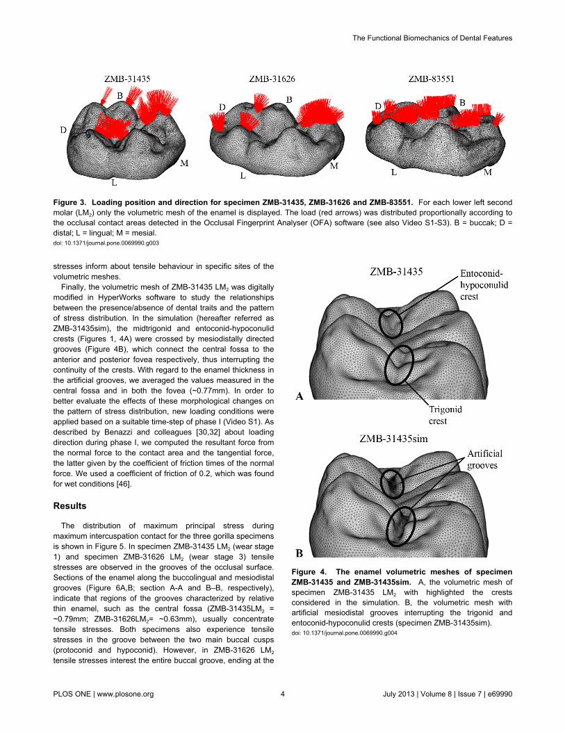
Finally, the volumetric mesh of ZMB-31435 LM2 was digitallymodified in HyperWorks software to study the relationshipsbetween the presence/absence of dental traits and the patternof stress distribution. In the simulation (hereafter referred asZMB-31435sim), the midtrigonid and entoconid-hypoconulidcrests (Figures 1, 4A) were crossed by mesiodistally directedgrooves (Figure 4B), which connect the central fossa to theanterior and posterior fovea respectively, thus interrupting thecontinuity of the crests. With regard to the enamel thickness inthe artificial grooves, we averaged the values measured in thecentral fossa and in both the fovea (~0.77mm). In order tobetter evaluate the effects of these morphological changes onthe pattern of stress distribution, new loading conditions wereapplied based on a suitable time-step of phase I (Video S1). Asdescribed by Benazzi and colleagues [30,32] about loadingdirection during phase I, we computed the resultant force fromthe normal force to the contact area and the tangential force,the latter given by the coefficient of friction times of the normalforce. We used a coefficient of friction of 0.2, which was foundfor wet conditions [46].
Results
The distribution of maximum principal stress duringmaximum intercuspation contact for the three gorilla specimensis shown in Figure 5. In specimen ZMB-31435 LM2 (wear stage1) and specimen ZMB-31626 LM2 (wear stage 3) tensilestresses are observed in the grooves of the occlusal surface.Sections of the enamel along the buccolingual and mesiodistalgrooves (Figure 6A,B; section A-A and B–B, respectively),indicate that regions of the grooves characterized by relativethin enamel, such as the central fossa (ZMB-31435LM2 =~0.79mm; ZMB-31626LM2= ~0.63mm), usually concentratetensile stresses. Both specimens also experience tensilestresses in the groove between the two main buccal cusps(protoconid and hypoconid). However, in ZMB-31626 LM2
tensile stresses interest the entire buccal groove, ending at the
Figure 4. The enamel volumetric meshes of specimenZMB-31435 and ZMB-31435sim. A, the volumetric mesh ofspecimen ZMB-31435 LM2 with highlighted the crestsconsidered in the simulation. B, the volumetric mesh withartificial mesiodistal grooves interrupting the trigonid andentoconid-hypoconulid crests (specimen ZMB-31435sim).doi: 10.1371/journal.pone.0069990.g004
Figure 3. Loading position and direction for specimen ZMB-31435, ZMB-31626 and ZMB-83551. For each lower left secondmolar (LM2) only the volumetric mesh of the enamel is displayed. The load (red arrows) was distributed proportionally according tothe occlusal contact areas detected in the Occlusal Fingerprint Analyser (OFA) software (see also Video S1-S3). B = buccak; D =distal; L = lingual; M = mesial.doi: 10.1371/journal.pone.0069990.g003
The Functional Biomechanics of Dental Features
PLOS ONE | www.plosone.org 4 July 2013 | Volume 8 | Issue 7 | e69990
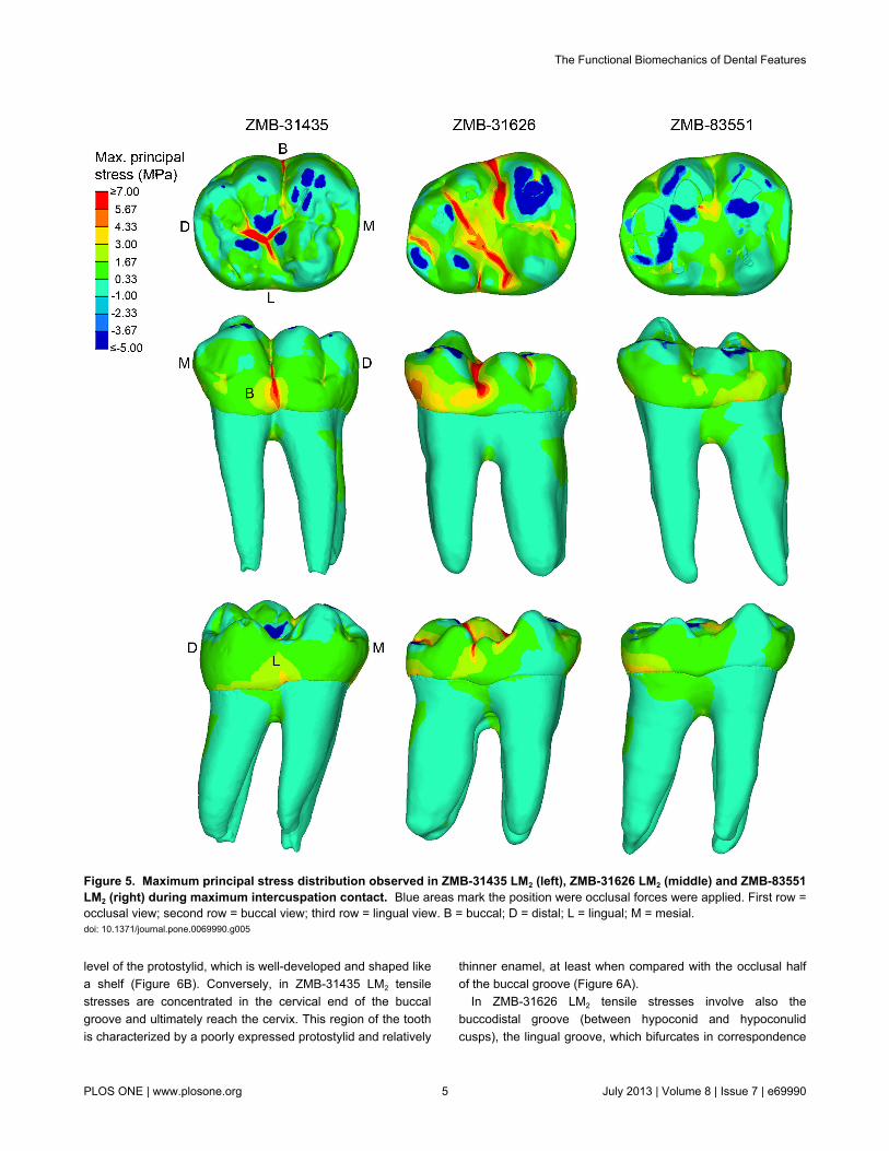
level of the protostylid, which is well-developed and shaped likea shelf (Figure 6B). Conversely, in ZMB-31435 LM2 tensilestresses are concentrated in the cervical end of the buccalgroove and ultimately reach the cervix. This region of the toothis characterized by a poorly expressed protostylid and relatively
thinner enamel, at least when compared with the occlusal halfof the buccal groove (Figure 6A).
In ZMB-31626 LM2 tensile stresses involve also thebuccodistal groove (between hypoconid and hypoconulidcusps), the lingual groove, which bifurcates in correspondence
Figure 5. Maximum principal stress distribution observed in ZMB-31435 LM2 (left), ZMB-31626 LM2 (middle) and ZMB-83551LM2 (right) during maximum intercuspation contact. Blue areas mark the position were occlusal forces were applied. First row =occlusal view; second row = buccal view; third row = lingual view. B = buccal; D = distal; L = lingual; M = mesial.doi: 10.1371/journal.pone.0069990.g005
The Functional Biomechanics of Dental Features
PLOS ONE | www.plosone.org 5 July 2013 | Volume 8 | Issue 7 | e69990
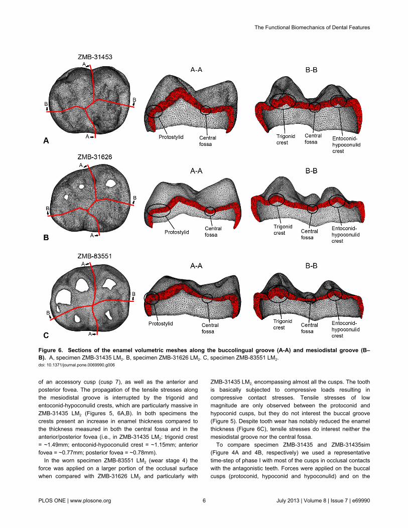
of an accessory cusp (cusp 7), as well as the anterior andposterior fovea. The propagation of the tensile stresses alongthe mesiodistal groove is interrupted by the trigonid andentoconid-hypoconulid crests, which are particularly massive inZMB-31435 LM2 (Figures 5, 6A,B). In both specimens thecrests present an increase in enamel thickness compared tothe thickness measured in both the central fossa and in theanterior/posterior fovea (i.e., in ZMB-31435 LM2: trigonid crest= ~1.49mm; entoconid-hypoconulid crest = ~1.15mm; anteriorfovea = ~0.77mm; posterior fovea = ~0.78mm).
In the worn specimen ZMB-83551 LM2 (wear stage 4) theforce was applied on a larger portion of the occlusal surfacewhen compared with ZMB-31626 LM2 and particularly with
ZMB-31435 LM2, encompassing almost all the cusps. The toothis basically subjected to compressive loads resulting incompressive contact stresses. Tensile stresses of lowmagnitude are only observed between the protoconid andhypoconid cusps, but they do not interest the buccal groove(Figure 5). Despite tooth wear has notably reduced the enamelthickness (Figure 6C), tensile stresses do interest neither themesiodistal groove nor the central fossa.
To compare specimen ZMB-31435 and ZMB-31435sim(Figure 4A and 4B, respectively) we used a representativetime-step of phase I with most of the cusps in occlusal contactswith the antagonistic teeth. Forces were applied on the buccalcusps (protoconid, hypoconid and hypoconulid) and on the
Figure 6. Sections of the enamel volumetric meshes along the buccolingual groove (A-A) and mesiodistal groove (B–B). A, specimen ZMB-31435 LM2. B, specimen ZMB-31626 LM2. C, specimen ZMB-83551 LM2.doi: 10.1371/journal.pone.0069990.g006
The Functional Biomechanics of Dental Features
PLOS ONE | www.plosone.org 6 July 2013 | Volume 8 | Issue 7 | e69990
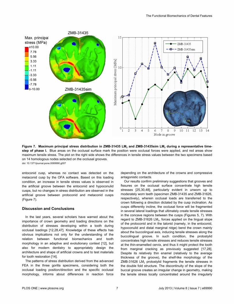
entoconid cusp, whereas no contact was detected on themetaconid cusp by the OFA software. Based on this loadingcondition, an increase in tensile stress values is observed inthe artificial groove between the entoconid and hypoconulidcusps, but no changes in stress distribution are observed in theartificial groove between protoconid and metaconid cusps(Figure 7).
Discussion and Conclusions
In the last years, several scholars have warned about theimportance of crown geometry and loading directions on thedistribution of stresses developing within a tooth duringocclusal loadings [12,28,47]. Knowledge of these effects hasobvious implications not only for the understanding of therelation between functional biomechanics and toothmorphology in an adaptive and evolutionary context [12], butalso for modern dentistry to appropriately design thearchitecture and shape of artificial crowns and to test materialsfor tooth restoration [14].
The patterns of stress distribution derived from the advancedFEA in the three gorilla specimens, considering both theocclusal loading position/direction and the specific occlusalmorphology, informs about differences in reaction force
depending on the architecture of the crowns and compressiveantagonistic contacts.
Our results confirm preliminary suggestions that grooves andfissures on the occlusal surface concentrate high tensilestresses [25,30,48], particularly evident in unworn up tomoderately worn teeth (specimen ZMB-31435 and ZMB-31626,respectively), wherein occlusal loads are transferred to thecrown following a direction dictated by the cusp inclination. Ascusps differently incline, the occlusal force will be fragmentedin several lateral loadings that ultimately create tensile stressesin the concave regions between the cusps (Figures 5, 7). Withregard to ZMB-31626 LM2, forces applied on the lingual slopeof the protoconid and in the talonid (namely, in the entoconid,hypoconulid and distal marginal ridge) bend the crown mainlyabout the buccolingual axis, inducing tensile stresses along thebuccolingual groove. In such condition, the protostylidconcentrates high tensile stresses and reduces tensile stressesat the thin-enamelled cervix, and thus it might protect the toothfrom marginal cracking as previously suggested [17,28].Despite its relatively thin enamel (relatively to the averagethickness of the groove), the shelf-like morphology of theZMB-31626 LM2 protostylid fragments the tensile stresses inthe double fold structure. The discontinuity of the slope of thebuccal groove creates an irregular change in geometry, makingthe tensile stress locally concentrated around the irregularly
Figure 7. Maximum principal stress distribution in ZMB-31435 LM2 and ZMB-31435sim LM2 during a representative time-step of phase I. Blue areas on the occlusal surface mark the position were occlusal forces were applied, and red areas showmaximum tensile stress. The plot on the right side shows the differences in tensile stress values between the two specimens basedon 14 homologous nodes selected on the occlusal grooves.doi: 10.1371/journal.pone.0069990.g007
The Functional Biomechanics of Dental Features
PLOS ONE | www.plosone.org 7 July 2013 | Volume 8 | Issue 7 | e69990
geometrical area, which is relatively far away from the cervix.Specimen ZMB-31435 LM2 confirms that the protostylid is adental feature subjected to tensile stresses, but it alsoemphasizes that poorly expressed protostylids are less suitableto reduce tensile stresses at the cervix. Most of the tensilestresses observed in ZMB-31435 LM2 during maximumintercuspation are concentrated in the central fossa (Figure 5),and we interpret that the trigonid crest and the entoconid-hypoconulid crest reinforce the crown against stressesproduced along the mesiodistal groove. This is also evident inthe slightly more worn specimen ZMB-31626 LM2, whereintensile stresses occur not only in the buccolingual grooves, butalso in the anterior and posterior fovea, hence beside the twocrests. Results of the simulation during phase I shown in Figure7 underline our assumptions, but they also emphasize thatpresence/absence of a crest has only local effects, aspreviously suggested by Magne and Belser [25]. The occlusalload applied to ZMB-31435sim LM2, without trigonid andentoconid-hypoconulid crests, was merely concentrated in thetalonid region, with obvious increase in the tensile stressvalues on the simulated distal groove, but without any effect onthe simulated mesial groove. Therefore, it is conceivable thatseveral dental features work in concert to reduce locallydangerous effects of high tensile stresses. In some cases, assuggested by Macho and Spears [12] and as observed in theprotostylid of specimen ZMB-31626 LM2, the presence and itscharacteristic expression may be responsible to withstandstress distribution. However, in other circumstances theinteraction between the external topography and the internalarchitecture, i.e., the thickened enamel in the crest, probablyplays an important role (Figure 6). Similar roles can besupposed also for other occlusal features along the border ofthe crown, such as the mesial and distal marginal ridges andthe lingual crest (the crest between the lingual cusps,metaconid and entoconid). Much more works are needed toexplore the biomechanical effects of dental features, and thecorrespondence between external and internal architecture andits functional implications.
In worn teeth, dental features such as the trigonid crest andthe entoconid-hypoconulid crest are not exempted from toothwear. However, the progressive deterioration of these featuresoccurs in concert with morphological alterations of the entireocclusal surface, which ultimately reduces locally directedstresses and improves the dispersion of occlusal forces.Indeed, the buccal cusps of the worn ZMB-83551 LM2
specimen are lower and flatter than in ZMB-31626 LM2 andparticularly in ZMB-31435 LM2, so that the load directionschange from oblique to nearly parallel direction to the dentalaxis (Figure 3). Moreover, as the contact areas increased innumber and extension, the occlusal force per unit of surfacearea decreases. As a result, the tensile stresses in the crowndecrease meaningfully. Similar conclusions have beenemphasized in restorative dentistry, where it has beenobserved that reduction of cusp height reduces tensile stressvalues (i.e., [49]). It is also worthwhile to note that the lowtensile stresses observed in the occlusal surface of ZMB-83551LM2, wherein tooth wear has notably reduced the enamelthickness and has partially removed and flattened both the
occlusal grooves and the central fossa (Figure 6C), furthersuggest that the occlusal topography might be more importantfor efficient load dissipation than the enamel thickness,supporting previous assumptions by Macho and Spears [12].To summarize, we do not suggest that it is better to have acompletely worn and flat occlusal surface, because there is nodoubt that occlusal reliefs are important for food processing [6].We observe, however, that a reduction in cusp’s steepness dueto tooth wear reduces tensile stresses in the crown, and thisdecrease might be useful when morphological features suchus, i.e., crests and ridges, grooves, enamel thickness, crownheight, have been either completely removed or heavilyreduced by tooth wear. We suggest that a strong interactionsubsists among dental morphology, occlusal load and toothwear. Some dental features might be useful to suffer hightensile stresses in unworn or moderately worn teeth, whereinthe occlusal load is applied in relatively small contact areasalong cusp slopes, generating non-axial loadings. However, adecrease in tensile stresses due to tooth wear makesbuttresses-like features less important to compensate loads.
Therefore, as suggested in a recent contribution [50], thewear process with its loss of dental tissue and the reduction ofthe occlusal relief might have had a crucial influence in theevolutionary adaptation of teeth, augmenting to endure specificstresses in advanced periods of an individual’s lifetime. Itseems that we observe an evolutionary compromise, and toothevolution and dental biomechanics can only be understood, ifwe further investigate tooth function in respect to the dynamicchanges of tooth structures during the lifespan of individuals.
Maybe in other extant and extinct hominoid primates, dentalfeatures such as the protostylid, the trigonid and the entoconid-hypoconulid crests represent plesiomorphic traits that do notprovide any functional advantage for the tooth, while otherfeatures not considered in this study (e.g., crenulated occlusalsurface, complexity of occlusal grooves pattern, bulging ofcusp’s buccal wall, or the Carabelli cusp and oblique crest inthe upper molar) might be also important to reinforce thecrown, limiting dangerous effect of high tensile stresses due toocclusal loadings.
Finally, it is important to underline some limits of our FEanalysis that should be addressed in future works. We haveattributed isotropic property to the enamel, but it has beensuggested that enamel should be considered anisotropic [24].We have investigated only a static occlusal loading condition.Even though we are confident about our results (we havechosen the most critical scenarios with maximum individualtooth-tooth contacts), further works should also consider kineticloading conditions during tooth-food-tooth contacts, which mayprovide a more realistic picture of the stress distribution in thetooth. Moreover, we have considered only three specimensdue to the efforts required to develop the FE models. Eventhough we do believe these specimens are morphologicallyrepresentative, we emphasize that the results described fromthis pilot work should be extended considering not only otherextant hominoid species, but also hominin fossil species. It iswell known that fossil African hominin taxa such asaustralopiths differ from extant African apes in having thickerenamel and generally lower and blunt molar reliefs [4,51],
The Functional Biomechanics of Dental Features
PLOS ONE | www.plosone.org 8 July 2013 | Volume 8 | Issue 7 | e69990
suggesting that less tensile stresses occur in maximumintercuspation. Since early Homo specimens showintermediate occlusal reliefs and surface sloping betweenGorilla and Pan [4], we expect also an intermediate tensilestress distribution, depending on the expression of edges andgrooves on their occlusal surfaces. However, even if weassume that such features (as well as other morphologicaltraits such as accessory cusps, complex groove/fissurepatterns, general crown height and flaring, cusp sizeproportion) contribute to withstand occlusal loads, moreinvestigations are required for a better understanding of thebiomechanical behavior and the evolution of hominin dentalfeatures.
Supporting Information
Table S1. Numbers of nodes and tetrahedral elements foreach specimen. (DOC)
Video S1. Simulation of the individual occlusal “powerstroke” of specimen ZMB-31435 applying the OcclusalFingerprint Analyser (OFA) software. The OFA calculates arelief-guided pathway of antagonistic tooth rows from collisiondetection, deflection and break-free algorithms for user-definedtimesteps. The contact areas of maximum intercuspation havebeen chosen for applying loads in the FE models.
(MP4)
Video S2. Simulation of the individual occlusal “powerstroke” of specimen ZMB-31626 applying the OcclusalFingerprint Analyser (OFA) software. The OFA calculates arelief-guided pathway of antagonistic tooth rows from collisiondetection, deflection and break-free algorithms for user-definedtimesteps. The contact areas of maximum intercuspation havebeen chosen for applying loads in the FE models.(MP4)
Video S3. Simulation of the individual occlusal “powerstroke” of specimen ZMB-83551 applying the OcclusalFingerprint Analyser (OFA) software. The OFA calculates arelief-guided pathway of antagonistic tooth rows from collisiondetection, deflection and break-free algorithms for user-definedtimesteps. The contact areas of maximum intercuspation havebeen chosen for applying loads in the FE models.(MP4)
Author Contributions
Conceived and designed the experiments: SB. Performed theexperiments: SB HNN OK. Analyzed the data: SB HNN OK.Contributed reagents/materials/analysis tools: SB HNN OKJJH. Wrote the manuscript: SB HNN OK JJH.
References
1. Berthaume M, Grosse IR, Patel ND, Strait DS, Wood S et al. (2010)The effect of early hominin occlusal morphology on the fracturing ofhard food items. Anat Rec 293: 594-606. doi:10.1002/ar.21130.
2. Kay RF (1975) The functional adaptations of primate molar teeth. Am JPhys Anthropol 43: 195-216. doi:10.1002/ajpa.1330430207. PubMed:810034.
3. Scott RS, Ungar PS, Bergstrom TS, Brown CA, Grine FE et al. (2005)Dental microwear texture analysis shows within-species diet variabilityin fossil hominins. Nature 436: 693-695. doi:10.1038/nature03822.PubMed: 16079844.
4. Ungar P (2004) Dental topography and diets of Australopithecusafarensis and early Homo. J Hum Evol 46: 605-622. doi:10.1016/j.jhevol.2004.03.004. PubMed: 15120268.
5. Ungar PS, Sponheimer M (2011) The diets of early hominins. Science334: 190-193. doi:10.1126/science.1207701. PubMed: 21998380.
6. Kay RF, Hiiemae KM (1974) Jaw movement and tooth use in recentand fossil primates. Am J Phys Anthropol 40: 227-256. doi:10.1002/ajpa.1330400210. PubMed: 4815136.
7. Lawn BR, Lee JJW (2009) Analysis of fracture and deformation modesin teeth subjected to occlusal loading. Acta Biomaterialia 5: 2213-2221.doi:10.1016/j.actbio.2009.02.001. PubMed: 19268644.
8. Benazzi S, Kullmer O, Grosse IR, Weber GW (2012) Briefcommunication: Comparing loading scenarios in lower first molarsupporting bone structure using 3D finite element analysis. Am J PhysAnthropol 147: 128-134. doi:10.1002/ajpa.21607. PubMed: 21952986.
9. Field C, Li Q, Li W, Swain M (2010) Biomechanical response inmandibular bone due to mastication loading on 3-unit fixed partialdentures. J Dent Biomech 1: 1-11.
10. Lucas P (2004) Dental functional morphology: how teeth work.Cambridge: Cambridge University Press.
11. Macho GA, Shimizu D (2010) Kinematic parameters inferred fromenamel microstructure: new insights into the diet of Australopithecusanamensis. J Hum Evol 58: 23-32. doi:10.1016/j.jhevol.2009.07.004.PubMed: 19783029.
12. Macho GA, Spears IR (1999) Effects of loading on the biomechanical[correction of biochemical] behavior of molars of Homo, Pan, andPongo. Am J Phys Anthropol 109: 211-227. doi:10.1002/(SICI)1096-8644(199906)109:2. PubMed: 10378459.
13. Lee JJW, Morris D, Constantino PJ, Lucas PW, Smith TM et al. (2010)Properties of tooth enamel in great apes. Acta Biomaterialia 6:4560-4565. doi:10.1016/j.actbio.2010.07.023. PubMed: 20656077.
14. Sornsuwan T, Ellakwa A, Swain MV (2011) Occlusal geometricalconsiderations in all-ceramic pre-molar crown failure testing. DentMater 27: 1127-1134. doi:10.1016/j.dental.2011.08.005. PubMed:21908033.
15. Barani A, Keown AJ, Bush MB, Lee JJW, Chai H et al. (2011)Mechanics of longitudinal cracks in tooth enamel. Acta Biomaterialia 7:2285-2292. doi:10.1016/j.actbio.2011.01.038. PubMed: 21296195.
16. Constantino PJ, Lucas PW, Lee JJW, Lawn BR (2009) The influence offallback foods on great ape tooth enamel. Am J Phys Anthropol 140:653-660. doi:10.1002/ajpa.21096. PubMed: 19890852.
17. Lucas P, Constantino P, Wood B, Lawn B (2008) Dental enamel as adietary indicator in mammals. BioEssays 30: 374-385
18. Schwartz GT (2000) Taxonomic and functional aspects of thepatterning of enamel thickness distribution in extant large-bodiedhominoids. Am J Phys Anthropol 111: 221-244. doi:10.1002/(SICI)1096-8644(200002)111:2. PubMed: 10640949.
19. Vogel ER, van Woerden JT, Lucas PW, Utami Atmoko SS, van SchaikCP et al. (2008) Functional ecology and evolution of hominoid molarenamel thickness: Pan troglodytes schweinfurthii and Pongo pygmaeuswurmbii. J Hum Evol 55: 60-74. doi:10.1016/j.jhevol.2007.12.005.PubMed: 18243275.
20. Bajaj D, Arola D (2009) Role of prism decussation on fatigue crackgrowth and fracture of human enamel. Acta Biomaterialia 5: 3045-3056.doi:10.1016/j.actbio.2009.04.013. PubMed: 19433137.
21. Chai H, Lee JJW, Constantino PJ, Lucas PW, Lawn BR (2009)Remarkable resilience of teeth. Proc Natl Acad Sci U S A 106:7289-7293. doi:10.1073/pnas.0902466106. PubMed: 19365079.
22. Lee JJW, Constantino PJ, Lucas PW, Lawn BR (2011) Fracture inteeth-a diagnostic for inferring bite force and tooth function. Biol Rev86: 959-974. doi:10.1111/j.1469-185X.2011.00181.x. PubMed:21507194.
23. Myoung S, Lee J, Constantino P, Lucas P, Chai H et al. (2009)Morphology and fracture of enamel. J Biomech 42: 1947-1951. doi:10.1016/j.jbiomech.2009.05.013. PubMed: 19559438.
24. Spears IR, van Noort R, Crompton RH, Cardew GE, Howard IC (1993)The effects of enamel anisotropy on the distribution of stress in a tooth.
The Functional Biomechanics of Dental Features
PLOS ONE | www.plosone.org 9 July 2013 | Volume 8 | Issue 7 | e69990
J Dent Res 72: 1526-1531. doi:10.1177/00220345930720111101.PubMed: 8227704.
25. Magne P, Belser UC (2002) Rationalization of shape and related stressdistribution in posterior teeth: a finite element study using nonlinearcontact analysis. Int J Periodontics Restorative Dent 22: 425-433.PubMed: 12449302.
26. Chai H, Lee JJW, Kwon JY, Lucas PW, Lawn BR (2009) A simplemodel for enamel fracture from margin cracks. Acta Biomaterialia 5:1663-1667. doi:10.1016/j.actbio.2008.11.007. PubMed: 19269906.
27. Qasim T, Ford C, Bush MB, Hu X, Malament KA et al. (2007) Marginfailures in brittle dome structures: relevance to failure of dental crowns.J Biomed Mater Res B Appl Biomater 80: 78-85. PubMed: 16615075.
28. Anderson PS, Gill PG, Rayfield EJ (2011) Modeling the effects ofcingula structure on strain patterns and potential fracture in toothenamel. J Morphol 272: 50-65. doi:10.1002/jmor.10896. PubMed:20960463.
29. Zienkiewicz OC, Taylor RL, Zhu JZ (2005) The Finite Element Method:Its Basis and Fundamentals. Oxford: Elsevier. 752pp.
30. Benazzi S, Kullmer O, Grosse IR, Weber GW (2011) Using occlusalwear information and finite element analysis to investigate stressdistributions in human molars. J of Anat 219: 259-272. doi:10.1111/j.1469-7580.2011.01396.x.
31. Kullmer O, Schulz D, Benazzi S (2012) An experimental approach toevaluate the correspondence between wear facet position and occlusalmovements. Anat Rec 295: 846-852. doi:10.1002/ar.22440.
32. Benazzi S, Grosse IR, Gruppioni G, Weber GW, Kullmer O (2013)Comparison of occlusal loading conditions in a lower second premolarusing three-dimensional finite element analysis. Clin Oral Investig. doi:10.1007/s00784-013-0973-8.
33. Hlusko LJ (2004) Protostylid variation in Australopithecus. J Hum Evol46: 579-594. doi:10.1016/j.jhevol.2004.03.003. PubMed: 15120266.
34. Skinner MM, Wood BA, Hublin JJ (2009) Protostylid expression at theenamel-dentine junction and enamel surface of mandibular molars ofParanthropus robustus and Australopithecus africanus. J Hum Evol 56:76-85. doi:10.1016/j.jhevol.2008.08.021. PubMed: 18986683.
35. Ungar PS, M’Kirera F (2003) A solution to the worn tooth conundrum inprimate functional anatomy. Proc Natl Acad Sci U S A 100: 3874-3877.doi:10.1073/pnas.0637016100. PubMed: 12634426.
36. Kono RT (2004) Molar enamel thickness and distribution patterns inextant great apes and humans: New insights based on a 3-dimensionalwhole crown perspective. Anthropol Sci 112: 121-146. doi:10.1537/ase.03106.
37. Fleagle GJ (1999) Primate Adaptation and Evolution. San Diego:Academic Press.
38. Smith BH (1984) Patterns of molar wear in hunter-gatherers andagriculturalists. Am J Phys Anthropol 63: 39-56. doi:10.1002/ajpa.1330630107. PubMed: 6422767.
39. Hattori Y, Satoh C, Kunieda T, Endoh R, Hisamatsu H et al. (2009) Biteforces and their resultants during forceful intercuspal clenching in
humans. J Biomech 42: 1533-1538. doi:10.1016/j.jbiomech.2009.03.040. PubMed: 19446816.
40. Dejak B, Mlotkowski A, Romanowicz M (2007) Strength estimation ofdifferent designs of ceramic inlays and onlays in molars based on theTsai-Wu failure criterion. J Prosthet Dent 98: 89-100. doi:10.1016/S0022-3913(07)60042-0. PubMed: 17692590.
41. Holmes DC, Diaz-Arnold AM, Leary JM (1996) Influence of postdimension on stress distribution in dentin. J Prosthet Dent 75: 140-147.doi:10.1016/S0022-3913(96)90090-6. PubMed: 8667271.
42. Ko CC, Chu CS, Chung KH, Lee MC (1992) Effects of posts on dentinstress distribution in pulpless teeth. J Prosthet Dent 68: 421-427. doi:10.1016/0022-3913(92)90404-X. PubMed: 1432755.
43. Rubin C, Krishnamurthy N, Capilouto E, Yi H (1983) Stress analysis ofthe human tooth using a three-dimensional finite element model. J DentRes 62: 82-86. doi:10.1177/00220345830620021701. PubMed:6571871.
44. Cheng YY, Li JY, Fok SL, Cheung WL, Chow TW (2010) 3D FEA ofhigh-performance polyethylene fiber reinforced maxillary dentures.Dent Mater 26: e211-e219. doi:10.1016/j.dental.2010.05.002. PubMed:20542552.
45. Hasegawa A, Shinya A, Nakasone Y, Lassila LV, Vallittu PK et al.(2010) Development of 3D CAD/FEM Analysis System for NaturalTeeth and Jaw Bone Constructed from X-Ray CT Images. Int JBiomater.
46. Li H, Zhou ZR (2001) Wear behavior of human teeth in dry and artificialsaliva conditions. Wear 249: 980-984. doi:10.1016/S0043-1648(01)00835-3.
47. Sornsuwan T, Swain MV (2012) The effect of margin thickness, degreeof convergence and bonding interlayer on the marginal failure of glass-simulated all-ceramic crowns. Acta Biomater 8: 4426-4437. doi:10.1016/j.actbio.2012.08.006. PubMed: 22902822.
48. Wang M, Mehta N (2013) A possible biomechanical role of occlusalcusp-fossa contact relationships. J Oral Rehabil 40: 69-79. doi:10.1111/j.1365-2842.2012.02333.x. PubMed: 22882571.
49. Chang YH, Lin WH, Kuo WC, Chang CY, Lin CL (2009) Mechanicalinteractions of cuspal-coverage designs and cement thickness in acusp-replacing ceramic premolar restoration: A finite element study.Med Biol Eng Comput 47: 367-374. doi:10.1007/s11517-008-0379-y.PubMed: 18679734.
50. Benazzi S, Nguyen HN, Schulz D, Grosse IR, Gruppioni G et al. (2013)The Evolutionary Paradox of Tooth Wear: Simply Destruction orInevitable Adaptation? PLOS ONE 8: e62263. doi:10.1371/journal.pone.0062263. PubMed: 23638020.
51. Kay RF (1985) Dental Evidence for the Diet of Australopithecus. AnnuRev Anthropol 14: 315-341. doi:10.1146/annurev.an.14.100185.001531.
The Functional Biomechanics of Dental Features
PLOS ONE | www.plosone.org 10 July 2013 | Volume 8 | Issue 7 | e69990