unique bone histology in partial large bone shafts from aust cliff (england, upper triassic): an...
TRANSCRIPT

This is a PDF file of the manuscript
that has been accepted for publication.
This file will be reviewed by the authors and editors
before the paper is published in its final form.
Please note that during the production process errors
may be discovered which could affect the content.
All legal disclaimers that apply to the journal pertain.
1
Unique bone histology in partial large bone shafts from Aust Cliff (England, Upper Triassic): an early independent experiment in gigantism RAGNA REDELSTORFF, P. MARTIN SANDER, AND PETER M. GALTON Redelstorff , R., Sander, P.M., and Galton, P.M. 201X. Unique bone histology in partial large bone shafts from Aust Cliff (England, Upper Triassic): an early independent experiment in gigantism. Acta Palaeontologica Polonica 5X (X): xxx-xxx. http://dx.doi.org/10.4202/app.2012.0073 Copyright © 201X R. Redelstorff et al. This is an open-access article distributed under the terms of the Creative Commons Attribution License, which permits unrestricted use, distribution, and reproduction in any medium, provided the original author and source are credited. Two giant partial bone shafts, possible femora, from the Rhaetian Bone Bed (Upper Triassic) of Aust Cliff in SW England continue to conceal their origin. The most striking characteristic of these bones is their size, showing that dinosaur-like gigantism had already evolved by the Late Triassic. Based on their characteristic, columnar shaft morphology, Galton (2005) suggested they came from a prosauropod or stegosaur. The bone histology of both specimens is very similar: the cortex is always rather thin, not exceeding 10 mm, and is of fibrolamellar type with longitudinal primary osteons. The primary osteons show a rather unusual feature, the development of a secondary osteon inside the primary one. The bone surface in both specimens shows open vascular canals, suggesting that the animals were still growing at the time of death, but an external fundamental system (EFS) is visible in the outermost cortex of specimen BRSMG Cb3870. The external cortex shows dense growth marks, but their annual nature is difficult to ascertain. The bones are probably dinosaurian, as indicated by the fibrolamellar bone, and possibly belong to an unknown basal sauropodomorph lineage. Alternatively, some very large pseudosuchians may have evolved fibrolamellar bone independently as an adaptation for reaching giant size. Key words: Reptilia, Dinosauria, bone histology, fibrolamellar bone, primary osteon, secondary osteon, Upper Triassic, England. Ragna Redelstorff [[email protected]], Zoology Department, University of Cape Town, Private Bag X3, Rondebosch, 7701, Cape Town, South Africa; P. Martin Sander [[email protected]], Steinmann Institute for Geology, Mineralogy and Palaeontology, University of Bonn, Nussallee 8, 53115 Bonn, Germany; Peter M. Galton [[email protected]] College of Naturopathic Medicine, University of Bridgeport, Bridgeport, Connecticut, USA; home address: 1065 Vintage Drive, 94572 Rio Vista, California, USA. Received 9 July 2012, accepted 10 November 2012, available online 14 November 2012.
2
Introduction
Identification of fossils can be challenging, especially if only isolated bones or, even worse,
incomplete isolated bone fragments are preserved. Such fragments can be significant if they
are of an unusual size and/or shape. In total, five partial shafts of large long bones have been
found in the historically important Rhaetic Bone Bed near the base of the Westbury
Formation (Upper Triassic) at Aust Cliff near Bristol, SW England (Storrs 1994; Galton
2005). The Westbury Formation was deposited in a shallow marine sedimentary environment
during a transgressive period of the Westbury Sea, as indicated by finds of a rich fish fauna
mainly represented by teeth (summarised by Storrs 1994). Rhaetian fissure fills and cave
deposits are also known from SW England, indicating nearby landmasses that were inhabited
by large terrestrial animals (e.g. Galton 1998, 2007; Whiteside and Marshall 2008). This is
indicated by the find of the basal sauropodomorph Camelotia borealis represented by a partial
skeleton (femur length of 1008 mm) discovered near the base of Westbury Formation of
nearby Wedmore, Somerset (Galton 1998). This skeleton was identified as a basal
sauropodomorph (melanorosaurid prosauropod), but is now regarded as a basal sauropod
(Yates 2007, 2010).
Two of the long bone shafts were destroyed in November, 1940 but, based on the
original unillustrated descriptions (Stutchbury 1850; Sanders 1876; Reynolds 1946), they
were tentatively assigned by Galton (2005) to Dinosauria incertae sedis and Camelotia
borealis. Because of the large size and columnar form of the bone shafts, Galton (2005)
considered only dinosaurian bones in his comparison. Of the surviving elements, Galton
(2005) assigned one to Dinosauria incertae sedis and the other two tentatively to Stegosauria,
because of their distinctive columnar shape with an oval cross-section, the width being greater
transversely than anteroposteriorly. If this is correct, these specimens would push the fossil
record of stegosaurs back into the Late Triassic.

3
Currently, the earliest evidence for stegosaurs are the footprints and trackways of
Deltapodus brodricki from the Middle Jurassic (Aalenian) of Yorkshire, NE England (Whyte
and Romano 2001; Whyte et al. 2007). The earliest skeletal evidence consists of isolated
bones from the Lower and Middle Bathonian of Oxfordshire and Gloucestershire, England
(Galton and Powell 1983) and associated bones from the Bathonian of western Siberia
(Averianov and Krasnolutskii 2009). Articulated skeletons are known of Huayangosaurus
taibaii from the Bathonian-Callovian of China (Zhou 1984; Sereno and Dong 1992;
Maidment et al. 2006) and of “Lexovisaurus durobrivensis” from the early-middle Callovian
near, Peterborough, England (Galton 1985; now Loricatosaurus priscus, Maidment et al.
2008).
Butler et al. (2006) noted that the Aust Cliff bones lack any stegosaurian
synapomorphies, a result of their being weathered fragments, as well as the absence of Early
Jurassic stegosaurs, and the small size of all known Triassic ornithischians. They concluded
that, although the bones “probably represent fragmentary femora of large dinosaurs (possibly
sauropods)”, they cannot be identified “with confidence beyond Reptilia indet.” (Butler et al.
2006: p. 627). Irmis et al. (2007: p. 15) argued that extensive cancellous bone or trabeculae
does not provide a phylogenetic signal because it can relate to biomechanics and life history,
and he identified the bones as indeterminate Tetrapoda. Maidment et al. (2008: p. 385), who
regarded them as indeterminate reptiles, also noted the presence of other large reptiles, such
as thecodontians, in the Late Triassic. As regards the absence of Early Jurassic Stegosauria,
the group would have been present at least in the Toarcian as indicated by the earliest
occurrence of its sister group, the Ankylosauria (Thompson et al., 2012), the first record of
which is a nodosaurid represented by a collection of 30 associated plates of four types from
the upper unit of the Kota Formation of the Pranhita-Godavari Valley, India (Nath et al.,

4
2002; for stratigraphy see Bandyopadhyay and Sengupta, 2006; Bandyopadhyay et al., 2010;
pers. comms. Yadagiri, Bandyopadhyay).
While these views may be correct from the point of view of a synapomorphy-based
identification, certain taxa such as dicynodonts can be excluded from consideration because
they do not have a straight shaft in any long bone. Also the marine reptiles of the Latest
Triassic (ichthyosaurs and plesiosaurs) can be excluded from consideration because they do
not reach anywhere near the size of the Aust Cliff bone shafts, do not have long straight shafts
in their long bones, and differ completely in their histology.
Institutional Abbreviations. —BRSMG, Bristol City Museum and Art Gallery, Bristol, UK.
Material and Methods
The two large partial bones (BRSMG Cb3869 and BRSMG Cb3870) sampled by us were
collected from the Rhaetic Bone Bed of the Westbury Formation at Aust Cliff, SW England.
Judging from their straight, columnar shape and large size (Table 1), the specimens most
likely represent midshafts of femora (Galton 2005). Measurements are given in Table 1.
Camelotia borealis was not accessible for histological sampling because it is known only
from the holotype femur.
We sampled both bone shafts using the core drilling method described in Sander
(2000) and Stein and Sander (2009). The sampling location in BRSMG Cb3870 and Cb3869
was determined by the area of the shaft showing the seemingly best preserved bone surface
(Fig. 1A-B, respectively). To obtain the thickest cortex possible, i.e. the optimal growth
record, from a core sample, long bones need to be sampled exactly at midshaft (Sander 2000),
but this could not be controlled sufficiently in the two Aust Cliff specimens. The drill cores
5
were processed into thin sections and studied with a Leica DMLP microscope at University of
Bonn and a Nikon Eclipse E200 Microscope at University of Cape Town.
Results
Notably, both samples show a very similar but rather unusual bone histology (Fig. 2A-B),
suggesting that they pertain to the same taxon. The cortex is thin in BRSMG Cb3870, not
exceeding 10 mm, and surrounds a large medullary area filled with an extensive web of
trabeculae (Fig. 2A), while in BRSMG Cb3869, the cortex is about three times thicker (Fig.
2B). The primary bone is fibrolamellar, consisting of primary osteons in a matrix of woven-
fibred bone. Primary osteons are oriented exclusively longitudinally. They are relatively
immature in that the vascular canal remains large, and there are only a few lamellae of
centripetally deposited bone. Both samples show one striking feature, however, that only
becomes apparent upon close inspection, which is that, towards the deeper cortex, many of
the primary osteons harbour a secondary osteon (Fig. 3A-D). These secondary osteons may
also be termed immature in that only a few lamellae are present (arrow in Fig. 3A-D). The
secondary osteon is clearly separated from the primary osteon surrounding it by a resorption
line, which appears slightly undulating and, unlike a LAG (line of arrested growth), cuts into
structures of the previously deposited tissue of the primary osteon (Fig. 3A-D). Erosion
cavities decrease in size towards the bone surface (white arrow in Fig. 3E) and, in most cases,
show an at least partial thin lining of lamellar bone (black arrow in Fig. 3E).
In BRSMG Cb3870, an almost avascular outermost cortex is visible (Figs. 2A, 3F),
which still retains a few vascular canals opening to the surface. Just below the bone surface,
the specimen shows 10 or 11 distinctive and closely spaced growth marks that form a
characteristic convoluted pattern because of their incorporation of the superficial blood
vessels during growth (Fig. 3G). These LAGs may represent a rather thick external

6
fundamental system (EFS; Fig. 3G), but this is difficult to determine because of the thinness
of the cortex. Alternatively, they may represent slow growth in a zonal pattern. There are 30
or 31 LAGs in total (Fig. 3F), including the 10 or 11 below the bone surface (Fig. 3G), but the
annual nature of these LAGs is difficult to evaluate because of their close spacing.
BRSMG Cb3869 represents a younger individual. The drill bit did not penetrate the
entire cortex, the recovered thickness being about 10 mm. However, the inner to mid cortex
appears cancellous due to numerous erosion cavities (Fig. 2B), suggesting that it was not
much thicker. Erosion cavities are scarce and unfilled in the outer cortex (arrows in Fig. 3H)
and more frequent in the inner cortex. Here, the cavities are either filled by a thin line of
lamellar bone or remain unfilled. The secondary osteons inside the primary osteons are filled
with lamellar bone only up to half of their diameter (Fig. 3H), giving them the characteristic
immature appearance as noted above. The outermost cortex is less vascularised with simple
longitudinal vascular canals, some of which are open to the bone surface, suggesting that the
animal was still growing slowly at the time of death. There appears to be an annulus in the
outermost cortex, which is quite broad and has a stripey appearance, similar to an
accumulation of growth marks, and may thus be interpreted as an EFS (Fig. 3H). Growth
marks are much less distinctive in BRSMG Cb3869 than in BRSMG Cb3870. Up to possibly
13 or 14 annuli (arrows in Fig. 2B) occur in the mid to outer cortex of this sample (number 14
may be an EFS).
Based on their bone histology, the two partial femoral shafts are likely from the same
species, one being somatically mature and the other younger.
Discussion
Life history variation.—Based on shaft circumference, the histologically younger BRSMG
Cb3869 is slightly larger than histologically older BRSMG Cb3870 (Table 1). However,

7
circumference measurements may be in error in the latter because extensive pre-burial erosion
and/or weathering resulted in partial loss of the cortex. The differences between the two
specimens may, however, also be a result of different growth histories. BRSMG Cb3869
formed annuli instead of LAGs, i.e. growth was slowed down periodically but not arrested at
any time as far as the record shows in the unremodelled primary bone. Continuous, although
not even, growth may impact on the specimen’s terminal size, e.g. periodically arrested
growth results in smaller body size than periodically slowed down growth. This is supported
by the distinctly thicker cortex in the younger, annuli-forming specimen BRSMG Cb3869.
The presence of LAGs in the one and of annuli in the other specimen may be (1) an ecological
signal, (2) sexual dimorphism or (3) intraspecific variation.
Regarding hypothesis (1), BRSMG Cb3869 may have experienced more agreeable,
consistent weather conditions than BRSMG Cb3870. Wikelski and Thom (2000) described
changes in the growth history of an individual of the marine iguana Amblyrhynchus christatus
from Galapagos, the original body length of which decreased about 20% in only two years as
a result of low availability of food during “El Nino” events and resumed growing after
environmental conditions normalised.
Regarding hypothesis (2), female reproduction requires a vast amount of energy and,
thus, growth may be arrested during reproduction periods. Egg-shelling requires additional
calcium, which in crocodilians is resorbed from the bones (e.g. Wink and Elsey 1986; Wink
et al. 1987; Schweitzer et al. 2007), hence the resorption (erosion) cavities. The latter are
indeed extensive in the LAG-forming specimen BRSMG Cb3870, implying a possible female
sex. Linking LAGs with reproduction would imply annual reproduction cycles, which does
occur in wild crocodiles but their reproduction frequency is usually rather variable from
annually to every 2-5 years (Joanen and McNease 1971; Lance 1989; Kofron 1990).

8
Uniqueness of the bone tissue of the Aust Cliff specimens.—The cortical bone histology of
the long bone shafts from Aust Cliff is unlike any described in the literature so far. Since the
histology of both bones is the same, showing the same peculiarities, they very likely pertain to
the same taxon. While the cortex of both bones is rather thin in the sections, this represents
only a minimum thickness because of the poorly controlled sampling location. However,
Galton (2005) pointed out the rather thin cortex of the Aust Cliff shafts apparent in the
terminal fracture surfaces. Only serial sectioning or µCT imaging would resolve the true
cortex thickness of these bones.
The uniqueness of the histology lies in the development of the primary and secondary
osteons, with the latter developed within the former (Fig. 3A-D). Normal secondary osteons,
while commonly following preexisting primary vascularity, cut across existing primary
osteon boundaries and other existing structures. This is because the cutting cone of normal
secondary osteons is roughly the size of primary osteons and thus obliterates the primary
osteon. Cutting across primary osteons is only rarely the case in the Aust Cliff bones where
the cementing line of the secondary osteon is within the lamellae of the primary osteon. In
fact, the cementing line only becomes apparent at close inspection with the compound
microcope, using higher magnifications, a nearly closed diaphragm, and a condensor. The
cutting cone of the secondary osteons must have been distinctly smaller than usual, and
osteoclast activity was influenced by the preexisting lamellae of the primary osteon. In fact,
we are unsure whether to call these structures proper secondary osteons or, more
descriptively, primary osteons with a two-phase centripetal deposition.
A clue to understanding this remodelling pattern may be the numerous resorption
cavities deeper in the cortex, which show a similar thin lining of lamellar bone as the
secondary osteons (Fig. 3E). The secondary osteons inside the primary ones may be part of
the same remodelling process acting in the inner cortex, except that it was less extensive in

9
the outer cortex, resulting in only a little resorption before redeposition commenced again
(Fig. 4). During a later resorption phase, the unfilled resorption cavities were possibly formed.
For the lack of modern examples of the peculiar tissue of the Aust Cliff bone shafts, its exact
mode of formation remains difficult to understand.
Histological comparison with other Late Triassic large tetrapods.—Based on morphology
and microanatomy, i.e. the large size and the straight shaft of the partial femora and their thin
cortex, Galton (2005) suggested affinity of the giant Aust Cliff bones with two types of
dinosaurs, namely prosauropods or stegosaurs. A similarly straight shaft is also found in
Sauropoda, which had been excluded by Galton (2005) based on their thicker cortex, and in
large pseudosuchian archosaurs (Niedźwiedzki et al. 2012). As noted above, the apparent
thinness of the cortex may be an artefact of the location of sampling sites and fracture planes.
Thus, we will enter into a closer comparison of the histology of the Aust Cliff specimens with
these four taxa, namely Pseudosuchia, basal Sauropodomorpha, Sauropoda, and Stegosauria.
The histological diversity of Triassic crurotarsan archosaurs has been described
comprehensively by de Ricqlès et al. (2003, 2008), including large-bodied forms such as
phytosaurs and rauisuchians. Longitudinal vascular canals, primary and secondary osteons as
well as numerous growth marks are shared with the Aust Cliff bones. A modification of the
fibro-lamellar bone complex, which is characterised by not highly developed woven bone,
occurs in the deep cortex, i.e. during early development, of some archosaurs (de Ricqlès et al.
2003, 2008). Among basal sauropodomorphs, the histology of Plateosauridae such as
Plateosaurus (Klein and Sander 2007) and Massospondylus (Chinsamy-Turan 2005) is well
known and differs from that of the Aust Cliff specimens in having laminar fibrolamellar bone
and few secondary osteons. The histology of other basal sauropodomorphs is similar to that of
Plateosauridae (e.g. de Ricqlès 1968; Chinsamy 1993; Cherry 2002; Klein and Sander 2007).

10
The Aust Cliff bones are also unlike Sauropoda, which uniformly have laminar fibrolamellar
bone usually lacking growth marks (Sander 2000; Klein and Sander 2008; Sander et al. 2011).
This histology is even seen in the earliest sauropods such as the large cf. Isanosaurus from the
Late Triassic of Thailand (Buffetaut et al. 2000, 2002; Sander et al. 2004). Finally, stegosaur
bone histology resembles that of the Aust Cliff bones in having fibrolamellar bone with
predominantly longitudinal primary osteons (Redelstorff and Sander 2009; Hayashi et al.
2009), but stegosaurs lack the peculiar secondary osteons inside the primary osteons and
show normal secondary osteons instead.
The Aust Cliff bones simply do not offer a good match with any previously described
histologies. The presence of fibrolamellar bone would suggest dinosaurian affinity, possibly
with an unknown lineage of sauropodomorphs. Alternatively, very large pseudosuchian
archosaurs may have had a bone histology different from that described for other non-
dinosaurian archosaurs, particularly the smaller rauisuchians, by de Ricqlès et al. (2003,
2008). Pseudosuchian affinities of the Aust Cliff bones have gained in credibility because a
very large one (femur length ca. 700 mm) has recently been described from Poland, Smok
wawelski (Niedźwiedzki et al. 2012), that occurs in sediments only slightly older than the
Westbury Formation. Previously, Late Triassic non-dinosaurian archosaurs of such large size
(up to 6-7 m), with pillar-like hind limbs, had been known only from Argentina (Bonaparte
1981). Very large size in such non-dinosaurian archosaurs could have evolved by an increase
in growth rate, resulting in the fibrolamellar bone seen in the Aust Cliff specimens. Testing
this hypothesis would require histological sampling of this material. To narrow down the
affinities of the large Aust Cliff bones, we furthermore hope that the Camelotia material can
be histologically sampled in the future.
Conclusions

11
The unique bone histology of the two sampled partial femoral shafts from the Rhaetian
Westbury Formation at Aust Cliff, England, encompasses fibrolamellar bone with
longitudinal primary osteons, most of which are modified by secondary remodelling. Growth
marks are common as either LAGs or annuli, indicating cyclical growth. In the older
specimen, BRSMG CB3870, LAGs become gradually more narrow-spaced towards the bone
surface, where they form an EFS. The bone tissue of the younger but slightly larger specimen,
BRSMG Cb3869, contains annuli rather than LAGs. This variation in type of growth marks in
combination with body size may indicate stronger and weaker interruptions of growth as a
response to ecological variation or sexual dimorphism.
Based on the geological age, the bone size and the straight shaft, the affinity of the Aust
Cliff bones can only be sought among certain dinosaurs (basal sauropodomorphs, Sauropoda
and Stegosauria) or large pseudosuchian archosaurs. However, all sauropodomorphs have a
laminar vascular architecture, pseudosuchians lack fibrolamellar bone, and stegosaurs, while
having longitudinal primary osteons, lack the secondary modification seen in the Aust Cliff
bones. In addition, the fossil record of the latter only begins in the Middle Jurassic, at least 25
Ma years later than the age of the Westbury Formation. While it is clear that the Aust Cliff
femora represent an early experiment in a graviportal stance and gigantism, it is unclear at
present in which (presumably archosaurian) lineage this experiment took place.
Acknowledgements
We thank Roger Vaughan at the Bristol City Museum and Art Gallery, UK, for collection
access and permission to sample the specimens. P.M.G. thanks S. Bandyopadhyay (Indian
Statistical Institute, Kolkata, India) and D. Yadagiri (Geological Survey, Hyderabad, India)
for information concerning the Kota ankylosaur. This study was partly funded by the DFG
(contribution number xx of the DFG Research Unit 533, ‘‘Biology of the Sauropod
12
Dinosaurs: The Evolution of Gigantism’’). We would also like to thank the reviewers and the
editor M.J. Benton for helpful comments on and improvement of the manuscript.
References
Averianov, A.D. and Krasnolutskii, S.A. 2009. Stegosaur remains from the Middle Jurassic of West Siberia. Proceedings of the Zoological Institute, Russian Academy of Sciences 313: 153–167. Bandyopadhyay, S. and Sengupta, D.P. 2006. Vertebrate faunal turnover during the Triassic-Jurassic transition: an Indian scenario. New Mexico Museum of Natural History & Science Bulletin 37: 77–85. Bandyopadhyay, S., Gillette, D.D., Ray, S. and Sengupta, D.P. 2010. Osteology of Barapasaurus tagorei (Dinosauria: Sauropoda) from the Early Jurassic of India. Palaeontology 53: 533-569. http://dx.doi.org/10.1111/j.1475-4983.2010.00933.x Bonaparte, J.F. 1981. Descripción de 'Fasolasuchus tenax' y su significado en la sistematica y evolución de los Thecodontia. Revista del Museo Argentino de Ciencias Naturales "Bernardino Rivadavia" 3: 55–101. Buffetaut, E., Suteethorn, V., Cuny, G., Tong, H., Le Loeuff, J., Khansubha, S., and Jongautchariyakul, S. 2000. The earliest known sauropod dinosaur. Nature 407: 72–74. http://dx.doi.org/10.1038/35024060 PMid:10993074 Buffetaut, E., Suteethorn, V., Le Loeuff, J., Cuny, G., Tong, H., and Khansubha, S. 2002. The first giant dinosaurs: a large sauropod from the Late Triassic of Thailand. Comptes Rendus Paleovol 1: 103–109. http://dx.doi.org/10.1016/S1631-0683(02)00019-2 Butler, R.J., Porro, L.B., and Heckert, A.B. 2006. A supposed heterodontosaurid tooth from the Rhaetian of Switzerland and a reassessment of the European Late Triassic record of Ornithischia (Dinosauria). Neues Jahrbuch für Geologie und Paläontologie, Monatshefte 2006: 613–633. Cherry, C. 2002. Bone histology of the primitive dinosaur, Thecodontosaurus antiquus. Unpublished Masters Thesis, University of Bristol. Chinsamy, A. 1993. Bone histology and growth trajectory of the prosauropod dinosaur Massospondylus carinatus (Owen). Modern Geology 18: 319–329.
13
Chinsamy-Turan, A. 2005. The microstructure of dinosaur bones: deciphering biology through fine scale techniques. Johns Hopkins University Press, Baltimore, p. 195. de Ricqlès, A. 1968. Recherches paléohistologiques sur les os longs des tetrapodes. I. Origine du tissu osseux plexiforme des dinosauriens sauropodes. Annales de Paléontologie 54: 133–145. de Ricqlès, A., Padian, K., and Horner, J.R. 2003. On the bone histology of some Triassic pseudosuchian archosaurs and related taxa. Annales de Paléontologie 89: 67–101. http://dx.doi.org/10.1016/S0753-3969(03)00005-3 de Ricqlès, A., Padian, K., Knoll, F. and Horner, J.R. 2008. On the origin of high growth rates in archosaurs: Complementary histological studies on Triassic archosauriforms and the problem of a "phylogenetic signal" in bone histology. Annales de Paléontologie 94: 57–76. http://dx.doi.org/10.1016/j.annpal.2008.03.002 Galton, P.M. 1985. British plated dinosaurs (Ornithischia, Stegosauridae). Journal of Vertebrate Paleontology 5: 211–254. http://dx.doi.org/10.1080/02724634.1985.10011859 Galton, P.M. 1998. Saurischian dinosaurs from the Upper Triassic of England: Camelotia (Prosauropoda, Melanorosauridae) and Avalonianus (Theropoda, ?Carnosauria). Palaeontographica, Abteilung A 250: 155–172. Galton, P.M. 2005. Bones of large dinosaurs (Prosauropoda and Stegosauria) from the Rhaetic Bone Bed (Upper Triassic) of Aust Cliff, southwest England. Revue de Paléobiologie 24: 51–74. Galton, P.M. 2007. Notes on the remains of archosaurian reptiles, mostly basal sauropodomorph dinosaurs, from the 1834 fissure fill (Rhaetian, Upper Triassic) at Clifton in Bristol, southwest England. Revue de Paléobiologie 26: 501–591. Galton, P.M. and Powell, H.P. 1983. Stegosaurian dinosaurs from the Bathonian (Middle Jurassic) of England, the earliest record of the family Stegosauridae. Géobios 16: 219–229. http://dx.doi.org/10.1016/S0016-6995(83)80020-5 Hayashi, S., Carpenter, K., and Suzuki, D. 2009. Different growth patterns between the skeleton and osteoderms of Stegosaurus (Ornithischia: Thyreophora). Journal of Vertebrate Paleontology 29: 123–131. http://dx.doi.org/10.1080/02724634.2009.10010366 Irmis, R.B., Parker, W.G., Nesbitt, S.J., and Liu, J. 2007. Early ornithischian dinosaurs: the Triassic record. Historical Biology 19: 3–22. http://dx.doi.org/10.1080/08912960600719988 Joanen, T. and McNease, L. 1971. Propagation of the American alligator in captivity. Proceedings of the Annual Conference, Southeastern Association of Game and Fish

14
Commissioners 25: 106–116. Klein, N. and Sander, P.M. 2007. Bone histology and growth of the prosauropod Plateosaurus engelhardti MEYER, 1837 from the Norian bonebeds of Trossingen (Germany) and Frick (Switzerland). Special Papers in Palaeontology 77: 169–206. Klein, N. and Sander, P.M. 2008. Ontogenetic stages in the long bone histology of sauropod dinosaurs. Paleobiology 34: 248–264. http://dx.doi.org/10.1666/0094-8373(2008)034[0247:OSITLB]2.0.CO;2 Kofron, C.P. 1990. The reproductive cycle of the Nile crocodile (Crocodylus niloticus). Journal of Zoology 221: 477–488. http://dx.doi.org/10.1111/j.1469-7998.1990.tb04014.x Lance, V.A. 1989. Reproductive cycle of the American alligator. American Zoologist 29: 999–1018. Maidment, S.C.R., Wei, G. and Norman, D.B. 2006. Re-description of the postcranial skeleton of the Middle Jurassic stegosaur Huayangosaurus taibaii. Journal of Vertebrate Paleontology 26: 944–956. http://dx.doi.org/10.1671/0272-4634(2006)26[944:ROTPSO]2.0.CO;2 Maidment, S.C.R., Norman, D.B., Barrett, P.M., and Upchurch, P. 2008. Systematics and phylogeny of Stegosauria (Dinosauria: Ornithischia). Journal of Systematic Palaeontology 6: 367–407. http://dx.doi.org/10.1017/S1477201908002459 Nath, T.T., Yadagiri, P. and Moitra, A.L. 2002. First record of armoured dinosaur from the Lower Jurassic Kota Formation, Pranhita-Godavari Valley, Andhra Pradesh. Journal of Geological Society of India 59: 575-577. Redelstorff, R. and Sander, P.M. 2009. Long and girdle bone histology of Stegosaurus: implications for growth and life history. Journal of Vertebrate Paleontology 29: 1087–1099. http://dx.doi.org/10.1671/039.029.0420 Reynolds, S.H. 1946. The Aust section. Proceedings of the Cotteswold Naturalists' Field Club 29: 29–39. Sander, P.M. 2000. Long bone histology of the Tendaguru sauropods: Implications for growth and biology. Paleobiology 26: 466–488. http://dx.doi.org/10.1666/0094-8373(2000)026<0466:LHOTTS>2.0.CO;2 Sander, P.M., Klein, N., Buffetaut, E., Cuny, G., Suteethorn, V., and Le Loeuff, J. 2004. Adaptive radiation in sauropod dinosaurs: bone histology indicates rapid evolution of giant body size through acceleration. Organisms, Diversity & Evolution 4: 165–173. http://dx.doi.org/10.1016/j.ode.2003.12.002

15
Sander, P.M., Christian, A., Clauss, M., Fechner, R., Gee, C.T., Griebeler, E.-M., Gunga, H.-C., Hummel, J., Mallison, H., Perry, S.F., Preuschoft, H., Rauhut, O.W.M., Remes, K., Tütken, T., Wings, O., and Witzel, U. 2011. Biology of the sauropod dinosaurs: the evolution of gigantism. Biological Reviews 86: 117–155. http://dx.doi.org/10.1111/j.1469-185X.2010.00137.x PMid:21251189 PMCid:3045712 Sanders, W. 1876. On certain large bones in Rhaetic beds at Aust Cliff, near Bristol. Annual Report of the British Association for the Advancement of Science, Transactions of the Sections 1875 45: 80–81. Schweitzer, M.H., Elsey, R.M., Dacke, C.G., Horner, J.R., and Lamm, E-T. 2007. Do egg-laying crocodilian (Alligator mississippiensis) archosaurs form medullary bone? Bone 40: 1152–1158. http://dx.doi.org/10.1016/j.bone.2006.10.029 PMid:17223615 Sereno, P.C. and Dong, Z. 1992. The skull of the basal stegosaur Huayangosaurus taibaii and a cladistic analysis of Stegosauria. Journal of Vertebrate Paleontology 12: 318–343. http://dx.doi.org/10.1080/02724634.1992.10011463 Stein, K. and Sander, P.M. 2009. Histological core drilling: a less destructive method for studying bone histology. In: M.A. Brown, J.F. Kane, and W.G. Parker (eds.), Methods In Fossil Preparation: 69–80. Proceedings of the First Annual Fossil Preparation and Collections Symposium. Storrs, G.W. 1994. Fossil vertebrate faunas of the British Rhaetian (latest Triassic). Zoological Journal of the Linnean Society 112: 217–259. http://dx.doi.org/10.1111/j.1096-3642.1994.tb00319.x Stutchbury, S. 1850. On a large cylindrical bone found by Mr. Thompson in the "Bone-bed" of Aust Cliff, on the Severn. Annual Report of the British Association for the Advancement of Science, Transactions of the Sections 1849 19: 67. Thompson, R.S., Parish, J.C., Maidment, S.C.R. and Barrett, P.M. 2012. Phylogeny of the ankylosaurian dinosaurs (Ornithischia: Thyreophora). Journal of Systematic Palaeontology 10: 301-312. http://dx.doi.org/10.1080/14772019.2011.569091 Whiteside, D.I. and Marshall, J.E.A. 2008. The age, fauna and palaeoenvironment of the Late Triassic fissure deposits of Tytherington, South Gloucestershire, UK. Geological Magazine 145: 105–147. http://dx.doi.org/10.1017/S0016756807003925 Whyte, M.A. and Romano, M. 2001. Probable stegosaurian dinosaur tracks from the Saltwick Formation (Middle Jurassic) of Yorkshire, England. Proceedings of the

16
Geologists' Association 112: 45–54. Whyte, M.A., Romano, M., and Elvidge, D.J. 2007. Reconstruction of Middle Jurassic dinosaur-dominated communities from the vertebrate ichnofauna of the Cleveland Basin of Yorkshire, UK. Ichnos 14: 117–129. http://dx.doi.org/10.1080/10420940601010802 Wikelski, M. and Thom, C. 2000. Marine iguanas shrink to survive "El Nino". Nature 403: 37–38. http://dx.doi.org/10.1038/47396 PMid:10638740 Wink, C.S. and Elsey, R.M. 1986. Changes in femoral morphology during egg-laying in Alligator mississippiensis. Journal of Morphology 189: 83–188. http://dx.doi.org/10.1002/jmor.1051890208 Wink, C.S., Elsey, R.M. and Mill, E.M. 1987. Changes in femoral robusticity and porosity during the reproductive cycle of the female alligator (Alligator mississippiensis). Journal of Morphology 93: 317–321. http://dx.doi.org/10.1002/jmor.1051930309 Yates, A.M. 2007. The first complete skull of the Triassic dinosaur Melanorosaurus Haughton (Sauropodomorpha: Anchisauria). Special Papers in Palaeontology 77: 9–55. Yates, A.M. 2010. A revision of the problematic sauropodomorph dinosaurs from Manchester, Connecticut and the status of Anchisaurus Marsh. Palaeontology 53: 739–752. http://dx.doi.org/10.1111/j.1475-4983.2010.00952.x Zhou, S.W. 1984. The Middle Jurassic dinosaurian fauna from Dashanpu, Zigong, Sichuan. Volume 2: Stegosaurs. Chongquing: Sichuan Scientific and Technological Publishing House, Sichuan, 51 p. [Chinese, 1–45; English, 45–51]
17
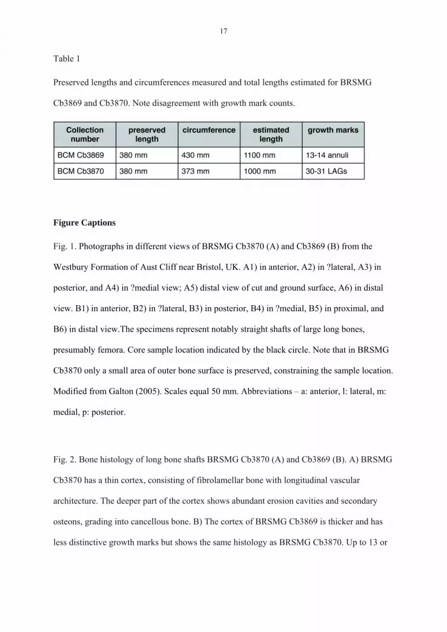
Table 1
Preserved lengths and circumferences measured and total lengths estimated for BRSMG
Cb3869 and Cb3870. Note disagreement with growth mark counts.
Figure Captions
Fig. 1. Photographs in different views of BRSMG Cb3870 (A) and Cb3869 (B) from the
Westbury Formation of Aust Cliff near Bristol, UK. A1) in anterior, A2) in ?lateral, A3) in
posterior, and A4) in ?medial view; A5) distal view of cut and ground surface, A6) in distal
view. B1) in anterior, B2) in ?lateral, B3) in posterior, B4) in ?medial, B5) in proximal, and
B6) in distal view.The specimens represent notably straight shafts of large long bones,
presumably femora. Core sample location indicated by the black circle. Note that in BRSMG
Cb3870 only a small area of outer bone surface is preserved, constraining the sample location.
Modified from Galton (2005). Scales equal 50 mm. Abbreviations – a: anterior, l: lateral, m:
medial, p: posterior.
Fig. 2. Bone histology of long bone shafts BRSMG Cb3870 (A) and Cb3869 (B). A) BRSMG
Cb3870 has a thin cortex, consisting of fibrolamellar bone with longitudinal vascular
architecture. The deeper part of the cortex shows abundant erosion cavities and secondary
osteons, grading into cancellous bone. B) The cortex of BRSMG Cb3869 is thicker and has
less distinctive growth marks but shows the same histology as BRSMG Cb3870. Up to 13 or

18
14 annuli (arrows) occur in the middle and outer cortex of the primary compact bone. The
deep cortex becomes increasingly cancellous as a result of abundant large erosion cavities.
Fig. 3. Detailed bone histology of the femoral shafts. A) BRSMG Cb3869: Primary osteon is
modified by a secondary osteon that developed inside the primary one. White arrow indicates
lamellae deposited in the secondary osteon. B) BRSMG Cb3869: this close-up of a secondary
osteon within a primary osteon clearly shows a resorption line. White arrow indicates
lamellae deposited in the secondary osteon. C) BRSMG Cb3870: Resorption can cut through
the lamellae of the primary osteon (discordant) or follow them (concordant). Black arrows
indicate lamellae deposited in the secondary osteon. D) Close-up of (C): The resorption line
of the secondary osteon clearly cuts through the tissue of the primary osteon. Black arrow
indicates lamellae deposited in the secondary osteon. E) Erosion cavities can show a thin
lining of lamellar bone (black arrow) or none at all (white arrow). Image in polarised light. F)
Up to 19 LAGs (white arrows) occur in the primary cortex of BRSMG Cb3870,
complemented by 11 or 12 in the external fundamental system (G). H) The outermost cortex
of BRSMG Cb3869 is poorly vascularized and contains unfilled erosion cavities (arrows).
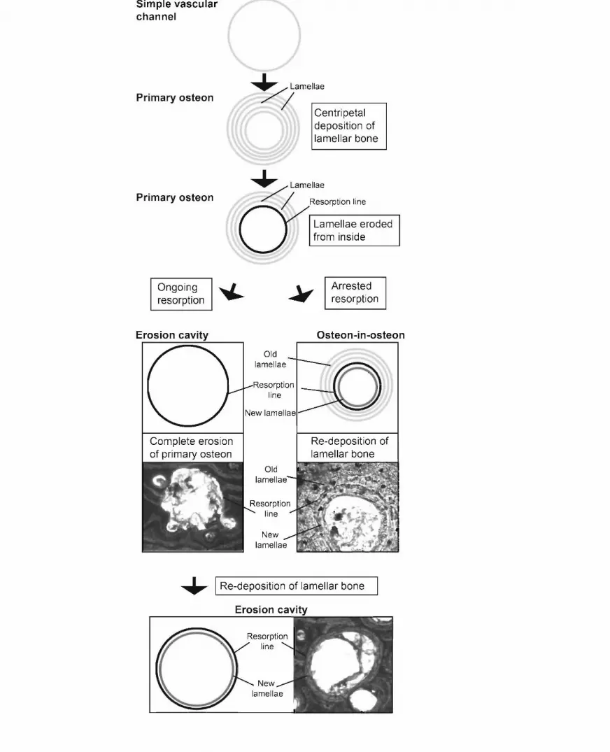
Fig. 4. Flow diagram explaining the peculiar patterns of remodelling seen in the Aust Cliff
bones BRSMG Cb3869 and Cb3870. In a simple vascular canal, lamellar bone is deposited
centripetally forming a primary osteon. In the Aust Cliff shafts, the inner lamellae of this
primary osteon are later resorbed from the inside. When erosion stops before the entire
primary osteon is resorbed, leaving a resorption line within the primary osteon, new lamellae
can be deposited and a secondary osteon forms within the primary one. With ongoing
resorption, an erosion cavity forms, the size of which exceeds the one of the former primary
osteon. When resorption stops, deposition of lamellar bone can resume.