two g i1 rate-modifying mutations act in concert to allow receptor-independent, steady-state...
TRANSCRIPT

Two Gαi1 Rate-Modifying Mutations Act in Concert to AllowReceptor-Independent, Steady-State Measurements of RGSProtein Activity
THOMAS ZIELINSKI1,§, ADAM J. KIMPLE2,§, STEPHANIE Q. HUTSELL3, MARK D. KOEFF1,DAVID P. SIDEROVSKI2,4,5, and ROBERT G. LOWERY1
1BellBrook Labs 5500 Nobel Drive, Suite 250, Madison, WI 537112Department of Pharmacology, The University of North Carolina at Chapel Hill, Chapel Hill, NC27599-73653Department of Biochemistry & Biophysics, The University of North Carolina at Chapel Hill,Chapel Hill, NC 27599-73654Lineberger Comprehensive Cancer Center, The University of North Carolina at Chapel Hill,Chapel Hill, NC 27599-73655UNC Neuroscience Center, The University of North Carolina at Chapel Hill, Chapel Hill, NC27599-7365
AbstractRGS proteins are critical modulators of G protein-coupled receptor (GPCR) signaling given theirability to deactivate Gα subunits via “GTPase-accelerating protein” (GAP) activity. Theirselectivity for specific GPCRs makes them attractive therapeutic targets. However, measuringGAP activity is complicated by slow GDP release from Gα and lack of solution-phase assays fordetecting free GDP in the presence of excess GTP. To overcome these hurdles, we developed aGαi1 mutant with increased GDP dissociation and decreased GTP hydrolysis, enabling detectionof GAP activity using steady-state GTP hydrolysis. Gαi1(R178M/A326S) GTPase activity wasstimulated 6~12 fold by RGS proteins known to act on Gαi subunits, and not affected by thoseunable to act on Gαi, demonstrating that the Gα/RGS domain interaction selectivity was notaltered by mutation. Gαi1(R178M/A326S) interacted with RGS proteins with expected bindingspecificity and affinities. To enable non-radioactive, homogenous detection of RGS protein effectson Gαi1(R178M/A326S), we developed a Transcreener® fluorescence polarization immunoassaybased on a monoclonal antibody that recognizes GDP with greater than 100-fold selectivity overGTP. Combining Gαi1(R178M/A326S) with a homogenous, fluorescence-based GDP detectionassay provides a facile means to explore the targeting of RGS proteins as a new approach forselective modulation of GPCR signaling.
KeywordsFluorescence polarization; GDP detection; regulators of G-protein signaling; surface plasmonresonance
Address correspondence to: David P. Siderovski, PhD, UNC-Chapel Hill, 4073 Genetics Medicine Bldg, 120 Mason Farm Road,CB#7365, Chapel Hill, NC 27599-7365. Tel: 919-843-9363 Fax: 919-966-5640 [email protected];. Robert G. Lowery, PhD,BellBrook Labs, 5500 Nobel Drive, Suite 250, Madison, WI 53711. Tel: 608-227-4501 Fax: [email protected]..§T.Z. and A.J.K. are co-first authors who contributed equally to this work.
NIH Public AccessAuthor ManuscriptJ Biomol Screen. Author manuscript; available in PMC 2010 November 1.
Published in final edited form as:J Biomol Screen. 2009 December ; 14(10): 1195–1206. doi:10.1177/1087057109347473.
NIH
-PA Author Manuscript
NIH
-PA Author Manuscript
NIH
-PA Author Manuscript

INTRODUCTIONThe standard model of GPCR signal transduction had long been restricted to a three-component system: receptor, G-protein, and effector 1. The seven-transmembrane domainreceptor is coupled to a membrane-associated heterotrimeric complex composed of a GTP-hydrolyzing Gα subunit and a Gβγ dimeric partner. Agonist-induced conformationalchanges enhance the guanine nucleotide exchange activity of the receptor, leading to therelease of GDP (and subsequent binding of GTP) by the Gα subunit. On binding GTP,conformational changes within the three `switch' regions of Gα allow the release of Gβγ.Separated Gα·GTP and Gβγ subunits are then free to propagate intracellular signaling viadiverse effectors 2. The intrinsic GTP hydrolysis (GTPase) activity of Gα resets the cycle byforming Gα·GDP which has low affinity for effectors but high affinity for Gβγ. In this way,the inactive, GDP-bound heterotrimer (Gα·GDP/Gβγ) is reformed and capable once again tointeract with activated receptor.
Based on this cycle of receptor-catalyzed GTP exchange and intrinsic GTP hydrolysis byGα, the duration of heterotrimeric G-protein signaling is thought to be controlled by thelifetime of the Gα subunit in its GTP-bound state. After the establishment of this basicmodel 1, RGS proteins (“regulators of G-protein signaling”) were subsequently discovered3–5 to bind Gα subunits (via a conserved ~120 amino-acid RGS domain) and dramaticallyaccelerate their intrinsic GTPase activity 6, thereby attenuating heterotrimer-linkedsignaling. Nearly 40 human proteins contain at least one RGS domain, with many of theseproteins (e.g., RGS4, RGS16) serving as GTPase-accelerating proteins (GAPs) for Gαi/osubunits, yet others such as RGS2 and p115-RhoGEF being particularly attuned to Gαq/11and Gα12/13 substrates, respectively 7 . The discovery of this superfamily of Gα-directedGAPs resolved apparent timing paradoxes between observed rapid physiological responsesmediated by GPCRs and the slow hydrolysis activity of the cognate G-proteins seen in vitro.Thus, in this capacity as negative regulators of GPCR signal transduction, the RGS proteinspresent themselves as excellent potential drug discovery targets 7. For example,pharmacological inhibition of RGS domain GAP activity should lead to prolonged signalingfrom G-proteins activated by agonist-bound GPCRs.
The most direct way to detect RGS protein function is by measuring the increased GTPaseactivity exhibited by its target Gα protein. However, accurate in vitro measurements of Gα-catalyzed GTP hydrolysis are difficult to obtain without laborious biochemicalreconstitutions with purified Gβγ and an activated GPCR (e.g., ref. 8). In the absence ofGPCR-mediated nucleotide exchange, it is GDP release (rather than GTP hydrolysis) that isthe rate-limiting step in the Gα nucleotide cycle 9. Thus, to examine the effect of an RGSprotein in accelerating GTP hydrolysis by an isolated Gα subunit in vitro, a single round ofhydrolysis of radiolabelled GTP is usually performed (a.k.a. the “single-turnover GTPaseassay”; ref. 6). This standard assay for measuring RGS domain-mediated GAP activity islow-throughput and requires discrete steps of [γ-32P]GTP loading onto Gα, protein reactantadmixture (with addition of the critical cofactor Mg2+ to initiate hydrolysis), isolation (indiscrete time intervals) of released [32P]phosphate with activated charcoal precipitation andcentrifugation, and finally scintillation counting. We have described an alternative single-turnover GTPase assay 10 using a coumarin-labeled, phosphate-binding protein to facilitatefluorescence-based detection of inorganic phosphate production; however, this methoddemands stringent controls on multiple experimental steps to eliminate phosphatecontaminants that interfere with the detection of GTPase activity. Such convoluted protocolsof inorganic phosphate detection are difficult for the non-specialist and especially not suitedfor high-throughput screening (HTS) of large compound libraries for RGS domaininhibitors. We and others have reported alternative, fluorescence-based strategies fordetecting the binding between RGS protein and Gα substrate 11–13, but none has
ZIELINSKI et al. Page 2
J Biomol Screen. Author manuscript; available in PMC 2010 November 1.
NIH
-PA Author Manuscript
NIH
-PA Author Manuscript
NIH
-PA Author Manuscript

specifically facilitated a discrete endpoint measurement of RGS domain-mediated GAPactivity per se.
In order to develop a facile steady-state GTPase assay for RGS domain GAP activity, wefirst set out to increase the spontaneous GDP release rate of Gα (koff(GDP)) while alsodecreasing its intrinsic rate of GTP hydrolysis (kcat(GTPase)), thereby allowing detection of atleast a five-fold enhancement of steady-state GTP hydrolysis by RGS proteins to provide anadequate signal-to-noise ratio. Gαi1 and closely related Gα proteins have been the focus ofextensive structure/function studies 14–17, and point mutations that affect both koff(GDP) andkcat(GTPase) without affecting functional interaction with the RGS domain have beenidentified previously 15–18 (e.g., Fig. 1A). Two of the most striking Gα mutations havebeen made to the highly-conserved active-site arginine (R178C; ref. 15), which causes a~100-fold reduction in GTPase activity, and to the alanine residue within the conservedTCAT loop that contacts the guanine ring (A326S; ref. 16), which results in a ~25-foldincrease in koff(GDP) relative to wildtype yet an identical kcat(GTPase).
To detect RGS protein-accelerated GTPase activity, we adapted a monoclonal antibody andfluorescent tracer, previously developed for the Transcreener ADP assay 19, for selectiveimmunodetection of GDP with a fluorescence polarization readout. Measurement of GTPaseactivity using this Transcreener GDP assay overcomes the signal-to-noise limitations ofphosphate detection methods and has been validated as a robust HTS method in the case ofADP detection for kinases and ATPases 20–22. Moreover, because it is a catalytic assayrather than a substrate binding assay, it should enable detection of all types of modulators ofRGS protein GAP activity, including those that bind at allosteric sites and affect RGSprotein catalytic activity without directly targeting the RGS domain Gα binding-site 23.
In this present study, we tested multiple point-mutant Gαi1 proteins with increased GDPdissociation and/or decreased GTP hydrolysis rates for their ability to enable detection ofRGS domain GAP activity using a steady-state GTPase assay format (i.e., multiple roundsof turnover of GTP to GDP). Coupling one of these variants, Gαi1(R178M/A326S), to theTranscreener GDP detection system has not only allowed facile detection of RGS proteinGAP activity, but was useful in helping establish (along with surface plasmon resonancespectroscopy) that the mutant Gαi1 interacted with RGS proteins with the same specificityand affinity as the wildtype Gαi1 protein.
MATERIALS AND METHODSChemicals and assay materials
GDP and GTP were purchased from USB Corp. (Cleveland, OH). The monoclonal antibodyand tracer used for GDP detection were developed at BellBrook Labs (Madison, WI) asdescribed 19, with the tracer comprising ADP conjugated to Alexa Fluor 633 (Invitrogen/Molecular Probes). Unless otherwise specified, all additional reagents were of the highestquality obtainable from Sigma (St. Louis, MO) or Fisher Scientific (Hampton, NH).
Protein expression and purificationWildtype, full-length human Gαi1 and various RGS proteins used in these studies wereexpressed in E. coli and purified as previously described 24. Gαi1 point mutants werecreated using PCR-based site-directed mutagenesis (QuikChange® II, Stratagene; La Jolla,CA) on the wildtype pProEXHTb-Gαi1 expression vector; mutagenesis primers weredesigned using Stratagene's QuikChange primer-design program and synthesized/PAGE-purified by Sigma-Genosys. All mutant constructs were sequence verified at FunctionalBiosciences LLC (Madison, WI) before protein expression, purification, concentration,quantitation, and cryopreservation using established protocols 10,24.
ZIELINSKI et al. Page 3
J Biomol Screen. Author manuscript; available in PMC 2010 November 1.
NIH
-PA Author Manuscript
NIH
-PA Author Manuscript
NIH
-PA Author Manuscript

Radiolabeled nucleotide binding and single turnover GTPase assaysAssessments of spontaneous GDP release and single-turnover GTP hydrolysis rates bywildtype and mutant Gαi1 subunits, using measurements of [35S]GTPγS binding and[γ-32P]GTP hydrolysis respectively, were conducted exactly as previously described 24,25.Briefly, for [35S]GTPγS binding by 100 nM of Gαi1 subunits at 20 °C, timed aliquots wereremoved, filtered through nitrocellulose, and washed four times with 10 ml of wash bufferbefore scintillation counting. Assays were conducted in duplicate, counts were subtractedfrom analogous reactions in “non-specific binding” buffer 24, and normalized data plotted asmean ± S.E.M. For single-turnover [γ-32P]GTP hydrolysis assays, Gαi1 subunits (100 nM)were pre-bound to [γ-32P]GTP in the absence of Mg2+ for 10 minutes at 30 °C. Reactionswere then initiated by the addition of 10 mM MgCl2 (final concentration) and the productionof 32Pi was measured by activated charcoal filtration and liquid scintillation counting 9,25.Initial rates obtained by data analysis using GraphPad Prism (La Jolla, CA).
Radiolabeled nucleotide steady-state GTPase assaysAssessments of steady-state [γ-32P]GTP hydrolysis rates by wildtype and mutant Gαi1subunits were conducted essentially as previously described 26. Briefly, Gαi1 protein wasdiluted to 50 nM in a buffer containing 50 mM Tris pH 7.5, 100 mM NaCl, 0.05% C12E10, 1mM DTT, 5 mM EDTA, 10 mM MgCl2, and 5 μg/ml BSA. Assays were initiated with theaddition of [γ-32P]GTP (and RGS4 if used), aliquots stopped at indicated time intervals, andfree [γ-32P]Pi quantified as previously described 26.
Transcreener GDP assaysStandard curves and GTPase reactions were both run at 30 °C in kinetic mode on a TecanSafire2 multiwell reader in Corning® 384-well black round-bottom low-volume polystyrenenon-binding surface microplates (Part # 3676). Fluorescence polarization was read using635 nm excitation (20 flashes per well) and 670 nm emission. A free tracer reference was setto 20 mP by adjusting the photomultiplier tubes, and buffer containing GDP antibody alonewas used as a blank for sample and reference wells. EC50 and EC85 values, Hill slopes, andcurves were generated by GraphPad Prism (La Jolla, CA). Unless otherwise indicated,reactions were run in 20 mM Tris 7.5 pH, 1 mM EDTA, 10 mM MgCl2, 10 μM GTP, 8 μg/ml GDP antibody, and 2 nM tracer in a final 20 μl volume. GDP antibody was used at aconcentration 85% of the amount required for saturated binding to tracer (i.e., the EC85).Where shown, polarization data was converted to the amount of GDP produced usingstandard curves. Reaction rates were then determined in GraphPad Prism using linearregression to estimate slope. For GTPase and GAP assays, reactions were started with theaddition of GTP with or without RGS protein.
Compound interference testTo assess the robustness of the Transcreener GDP assay for practical screening applications,we performed a control screen using the GenPlus library of 960 bioactive molecules fromMicrosource Discovery Systems, many of which are approved drugs. GDP assay reagents(as denoted above) were added to duplicate wells containing 10 μM compound and either 10μM GTP to mimic no-enzyme control reactions or 9 μM GTP plus 1 μM GDP to mimiccompleted enzyme reactions in 1% DMSO.
Pilot screen and counterscreen of GenPlus LibraryScreens of the GenPlus library with the Transcreener GDP assay (10 μM final compoundconcentration) for modulators of RGS4 GAP activity on Gαi1(R178M/A326S), as well as fornon-specific modulators of intrinsic GTPase activity of Gαi1(R178M/A326S) alone, wereconducted as mentioned above with the following changes. GTPase reactions containing 50
ZIELINSKI et al. Page 4
J Biomol Screen. Author manuscript; available in PMC 2010 November 1.
NIH
-PA Author Manuscript
NIH
-PA Author Manuscript
NIH
-PA Author Manuscript
nM Gαi1(R178M/A326S) with or without 250 nM RGS4 were run in Corning® 384-wellmicroplates at 30 °C in 20 mM Tris pH 7.5, 1 mM EDTA, 10 mM MgCl2, 10 μM GTP, 12μg/μl GDP antibody, 2 nM tracer, and 0.5% DMSO (v/v) in a final volume of 20 μl.Fluorescence polarization was read at 60, 90, 120, and 180 minutes of elapsed reaction timeon a Tecan Safire2 multiwell reader as described above.
Surface plasmon resonance (SPR) spectroscopyOptical detection of surface plasmon resonance (SPR) was performed using a BIAcore 3000(GE Healthcare; Piscataway, NJ). Wildtype and mutant Gαi1 proteins were immobilizedonto nickel-nitrilotriacetic acid SPR sensor chips (GE Healthcare) by hexahistidine tag-mediated capture-coupling as previously described 27. Affinities of RGS proteins forimmobilized Gαi1 proteins were obtained from dose-response sensorgrams usingequilibrium saturation binding analyses as previously described 24.
RESULTS AND DISCUSSIONProfiling multiple Gαi1 point-mutations for nucleotide cycling rate alterations
Using PCR-based site-directed mutagenesis, we created several amino-acid substitutions atvarious positions within Gαi1 known to affect koff(GDP) and/or kcat(GTPase) (e.g., Fig. 1).These mutants included: aspartate, serine, or threonine replacing Ala-326; cysteine, lysine,or methionine replacing Arg-178; alanine, serine, or valine replacing Thr-181; singlemutants K192A and F336A; and double mutants K192A/F336A, R178C/A326S, R178C/A326T, R178M/A326S, R178C/A326T, and T181A/A326S. Note that multiple differentsubstitutions were made at several sites, including amino-acids intended to be more or lessdisruptive than the original reported mutation. For instance, R178K and R178M were testedas more conservative substitutions at the catalytic arginine position relative to the originalR178C variant; it was thought that either of these alternative substitutions might result in asmaller decrease in kcat(GTPase) than the cysteine replacement which reduces kcat(GTPase) bytwo orders of magnitude 15. While the R178C mutation leads to a substantial decrease inkcat(GTPase), Berman et al. 6 have shown that the single-turnover GTPase rate of this Gαmutant can still be increased by RGS domain-mediated GAP activity, whereas the moreconventional GTPase-crippling mutation of Q204L renders Gαi1 truly dead in terms ofresponsiveness to RGS proteins. Thus, the Gαi1(Q204L) mutant was not pursued in thisstudy.
Gαi1 mutants were initially profiled for enhanced GDP release and/or reduced GTPase ratesufficient to see a change in steady-state GTP hydrolysis upon RGS protein addition. Thisinitial profiling led us to focus on two positions: Arg-178 and Ala-326. Binding of the non-hydrolyzable GTP analog, [35S]GTPγS, to Gαi1·GDP was used to measure the rate of GDPdissociation (e.g., Fig. 1B); the prevailing assumption for Gα subunits is that kon for[35S]GTPγS binding is much more rapid than koff(GDP) 28. Single turnover GTP hydrolysismeasuring 32Pireleased from Gα-bound [γ-32P]GTP – an assay format which is not rate-limited by GDP dissociation 9 – was used to assess intrinsic kcat rates for the Gαi1 mutants(e.g., Figure 1C).
As expected, Gαi1 variants with mutation to the active-site catalytic residue Arg-178 hadvery low or undetectable levels of GTP hydrolysis, whereas Gαi1(A326S), the singlemutation reported to only affect GDP dissociation, had a GTPase rate similar to wildtypeGαi1 (Fig. 1C–D). [35S]GTPγS binding assays showed that two variants with mutations onlyat the catalytic site, R178M and R178C, had GDP dissociation rates similar to wildtypeGαi1, whereas introduction of the A326S mutation, either alone or in combination withR178C, caused a three-fold acceleration in GDP dissociation (Fig. 1B,D). When the A326Smutation was combined with methionine at Arg-178 (instead of cysteine), the GDP
ZIELINSKI et al. Page 5
J Biomol Screen. Author manuscript; available in PMC 2010 November 1.
NIH
-PA Author Manuscript
NIH
-PA Author Manuscript
NIH
-PA Author Manuscript

dissociation rate increased more than ten-fold over wildtype: from 0.008 min−1 to 0.130min−1 (Fig. 1D). We currently do not have a precise structural explanation for why theparticular combination of R178M and A326S mutations results in more rapid GDP releasethan the single A326S mutation alone; it is not an additive effect, since the singly-mutatedGαi1(R178M) variant exhibits wildtype GDP dissociation (Fig. 1B). It is interesting to notethat Posner and colleagues, when reporting the crystal structure of the Gαi1(A326S) mutant16, suggested the presence of an indirect interaction between the Arg-178 and Ser-326residues (via contacts with nucleotide and Gly-45), thereby providing a possible mechanismfor the functional interaction we have observed here between the R178M and A326Smutations.
Combined action of two Gαi1 mutations allows steady-state measurement of GAP activityWith the R178M/A326S mutant of Gαi1 demonstrating the largest change in GDP releaserate of all mutants tested, we next examined whether this particular Gαi1 variant would beaffected by RGS domain-mediated GAP activity in steady-state [γ-32P]GTP hydrolysisassays. Addition of purified RGS4 protein to the Gαi1(R178M/A326S) variant (in thepresence of free [γ-32P]GTP and Mg2+) resulted in a dramatic increase in [32P]Pi detectedover time. In contrast, there was no effect of RGS4 on wildtype Gαi1 in this steady-stateassay (Fig. 2A vs B), as expected given the original report by Berman et al. 6.
Development of a Transcreener GDP assayThe Transcreener platform relies upon highly selective antibodies for detection ofnucleotides produced in enzyme reactions 29. To allow measurement of RGS protein-mediated acceleration of steady-state GTP hydrolysis in a homogenous format withoutradioactivity, a Transcreener assay for GDP was developed (Figure 3A) using a competitivefluorescence polarization immunoassay format. For this method, a recently-developedmonoclonal antibody that recognizes GDP with >100-fold higher affinity than GTP 19 isadded to the reaction, along with a fluorescent tracer which binds to the antibody with highaffinity. When no free, unlabeled GDP is present in the reaction, the fluorescent tracerremains antibody-bound and exhibits a high polarization given its high apparent molecularweight. GDP produced in the reaction displaces the tracer from the antibody, therebyreducing its apparent molecular weight, increasing its rotational motion, and thus reducingthe degree of polarization of emitted light. A similar Transcreener assay has been widelyused for detection of ADP produced by kinases and other ATP-hydrolyzing proteins (e.g.,refs. 20–22; reviewed in 29).
Figure 3B shows typical fluorescence polarization standard curves mimicking theconversion of GTP to GDP by a GTPase. An important aspect of flexibility for a GTPaseassay is the ability to accommodate a range of initial GTP concentrations, so that diverseenzymes and screening strategies can be employed; therefore, these studies were performedusing different GTP concentrations of 1, 10 and 100 μM. Because the antibody cross-reactsto some degree with GTP, its concentration must be increased as higher GTP concentrationsare used, in order to buffer for the total guanine nucleotide pool. Thus, for this analysis, theEC85 concentrations of monoclonal antibody were first determined in the presence of theindicated initial GTP concentrations (2.2, 12 and 64 μg/ml Ab for 1, 10 and 100 μM GTP,respectively) and the standard curves for GTP to GDP conversion were performed with 16replicates at those antibody concentrations. At a GDP concentration equivalent to 10%conversion of GTP, which is generally considered to be well within the initial velocityregion, polarization shifts of 108, 134, and 148 mP were observed for the 1, 10, and 100 μMGTP concentration curves, respectively (Fig. 3B). Acceptable Z'-factor values (ref. 30) ofgreater than 0.5 were observed down to 2% conversion for the two higher initial GTPconcentrations, and to 5% for the 1 μM initial GTP curve (Fig. 3C), suggesting that the
ZIELINSKI et al. Page 6
J Biomol Screen. Author manuscript; available in PMC 2010 November 1.
NIH
-PA Author Manuscript
NIH
-PA Author Manuscript
NIH
-PA Author Manuscript

Transcreener GDP assay should be capable of very robust detection of GTPase enzymeinitial velocity over at least a 100-fold range of initial GTP concentration.
To assess the potential for compound interference with the Transcreener GDP assay readout,we performed a control screen (Fig. 3D) with the GenPlus library of 960 bioactivemolecules, many of which are approved drugs. This control screen was done underconditions mimicking 10% conversion to GDP for a GTPase reaction run at 10 μM initialGTP concentration. All wells were run in duplicate. The vast majority of the compoundsclustered very tightly around the means for the 10 μM GTP and the 9 μM GTP/1 μM GDPconditions; the Z'-factor for the no-compound controls in this screen was 0.93. There wereonly three compounds in the control screen that caused the signal to vary more than threestandard deviations from the mean: dirithromycin, metazolamide, and lonidamin (Figure3D). There is no obvious structural similarity between them nor are any of them similar instructure to guanine nucleotide. These data suggests that compound interference with theTranscreener GDP assay readout will be minimal.
FP-based detection of RGS protein GAP activity is dependent on two rate-alteringmutations
Having validated the utility of the Transcreener assay in detecting GDP in the presence ofGTP, we next tested its use in measuring RGS protein GAP activity on several rate-alteredGαi1 variants (Figure 4). In these experiments, the Gαi1 proteins were incubated with andwithout the well-characterized, Gαi-directed RGS protein RGS4 31 in the presence of theTranscreener GDP assay reagents, and plates were read at intervals starting at 15 minutes.The change in the absolute value of polarization at each time-point (ΔmPt = | mPt(Gαi1) −mPt(no Gαi1) |) was plotted over a time-course of 6 hours; in addition, the plotted change inpolarization that occurred in the linear region (over the first hour) was converted to GDPformation using standard curves (akin to those of Figure 3B) and normalized to the amountof Gαi1 protein present in the reaction, with the resultant initial rates of GTP hydrolysiscalculated from these data shown in Fig. 4E.
The two Gαi1 variants with single mutations at the catalytic arginine only, R178C orR178M, each had lower steady-state GTPase activity than wildtype Gαi1 and, like wildtype,were unaffected by RGS4 (GAP factors of 0.9 and 1.2, respectively; Fig. 4E). These resultsare expected because their steady-state GTPase rate is limited by slow GDP dissociation.Conversely, the A326S variant exhibited a much higher steady-state GTPase rate thanwildtype, as expected from its higher koff(GDP) (Fig. 1); however, its steady-state GTPaserate was unaffected by RGS4 (GAP ratio of 1.1; Fig. 4E), presumably because a further rateincrease in GTPase is limited by koff(GDP). Most importantly, the two double mutants,R178C/A326S and R178M/A326S, had very low basal steady-state GTPase activities thatbecame demonstrably higher in the presence of RGS4 (e.g., Fig. 4A–B); the GAP effect onGαi1(R178M/A326S) was greater than with the Gαi1(R178C/A326S) variant (GAP factorsof 6.5 and 3.6, respectively; Fig. 4E). Given its high GAP factor response in both the steady-state [γ-32P]GTP hydrolysis assay (Fig. 2) and the Transcreener GDP assay (Fig. 4), theGαi1(R178M/A326S) variant was used in subsequent analyses.
Gαi1(R178M/A326S) interacts with RGS proteins with same affinity and specificity aswildtype
A possible concern about the use of a mutated Gα protein for RGS protein GAP assays isthat the mutation(s) could disrupt the global fold of Gα or, at the very least, affect thedisposition of the switch regions and other surface contact points to which RGS proteinsinteract 24,31, thereby altering the normal affinity and specificity that RGS proteins showfor their various Gα substrates. The two point mutations of R178M and A326S are interior
ZIELINSKI et al. Page 7
J Biomol Screen. Author manuscript; available in PMC 2010 November 1.
NIH
-PA Author Manuscript
NIH
-PA Author Manuscript
NIH
-PA Author Manuscript

to the guanine nucleotide binding pocket (Figure 5), but could nevertheless affect the RGSdomain interaction surface.
To test for this possibility, we used SPR to compare the binding interactions of RGS2 andRGS16 with wildtype Gαi1 versus the Gαi1(R178M/A326S) variant. Multiple previousstudies 8,24,32 have established that RGS2, a potent GAP for Gαq, does not interact withwildtype Gαi1 in vitro; this same lack of interaction was observed with the Gαi1(R178M/A326S) mutant (data not shown). Conversely, RGS16 is known to be a Gαi-interacting RGSprotein 24, and was found by SPR to bind equivalently to immobilized wildtype Gαi1 andGαi1(R178M/A326S) proteins (Fig. 6). This equivalence included RGS16 only interactingwith high affinity to the Gα subunits in their transition state-mimetic form (namely, Gα incomplex with GDP and aluminum tetrafluoride 31). These binding results suggest that nolong-range perturbations have been made to the RGS domain interaction sites on Gαi1 by thetwo rate-altering mutations of R178M and A326S.
Using the Transcreener GDP assay, we performed an additional test of the Gαi1(R178M/A326S) variant to control for any unintended changes the two point mutations could haveengendered within Gα to alter its interaction specificity with various RGS proteins. With thesame RGS protein spectrum used in the SPR binding experiments, we found that RGS2(highly selective for Gαq over Gαi substrates) had no effect on increasing steady-stateGTPase activity of Gαi1(R178M/A326S), whereas RGS16 increased steady-state GTPaseactivity 12-fold over the basal rate (Fig. 7).
Pilot screen for inhibitors of RGS4 GAP activity on Gαi1(R178M/A326S)Given the robust performance of the Transcreener GDP assay in the control screen forpotential compound interference (Fig. 3D) and evidence that the two mutations to Gαi1affected neither the affinity nor specificity of the Gα/RGS domain interaction (Figs. 6 and7), we proceeded to a pilot screen with Gαi1(R178M/A326S) and RGS4 using the GenPluslibrary of 960 bioactive molecules (Fig. 8). The screening window was first optimized byvarying the concentrations of Gα and RGS protein inputs; at 50 nM Gαi1(R178M/A326S)and 250 nM RGS4, a maximal signal to background difference of 112 mP units wasobtained after 120 minutes of elapsed reaction time before FP measurement. The thiol-reactive compound CCG-4986 was used in the screen as a positive control for RGS4inhibition 13,33. The GenPlus library screen was conducted with Gαi1(R178M/A326S) andRGS4 (Fig. 8A); a separate counterscreen of the library was performed with Gαi1(R178M/A326S) and no RGS4 (Fig. 8B) to identify compounds having either non-specific effects ormodifying Gαi1 GTPase activity (rather than RGS4 GAP activity per se).
Z'-factors of 0.60, 0.83, 0.83, and 0.82 were obtained at 60, 90, 120, and 180 minuteselapsed reaction time, as calculated based on data from control wells containing eitherGαi1(R178M/A326S) only or Gαi1(R178M/A326S) plus RGS4. Note that these Z'-factorvalues reflect only the RGS4-dependent increase in GTPase activity, and not the totalobserved GTPase activity relative to no-enzyme controls. The Z-factor for the GenPluslibrary screen at the 120 minute time point (shown in Fig. 8A) was 0.73, which wascalculated by excluding values from wells containing hit compounds. Of the 960 compoundsin the GenPlus library, 17 compounds were initially considered hits in the RGS4/Gα screen:i.e., those data points that fell outside the μ±3σ range. However, ten of these 17 hits alsoresulted in a greater than ±3σ change in the mean signal within the Gα-only counterscreenand thus were excluded because these compounds are likely either affecting the Gα subunitor otherwise interfering with the assay. Thus the RGS4-specific hit rate was 0.7%: sevencompounds from the 960 compound library exhibited an modulatory effect on GDPproduction that was specific to RGS4-stimulated GTPase activity (Fig. 8A vs 8B). Sixcompounds (id # 62, 63, 244, 413, 524, 812) exhibited an RGS4-specific inhibitory effect
ZIELINSKI et al. Page 8
J Biomol Screen. Author manuscript; available in PMC 2010 November 1.
NIH
-PA Author Manuscript
NIH
-PA Author Manuscript
NIH
-PA Author Manuscript

(i.e., a change in polarization greater than the [mean + 3 S.D.] signal threshold) and onecompound (#596) exhibited an RGS4-specific activating effect on GDP production (i.e., achange in polarization less than the [mean − 3 S.D.] signal threshold). This hit rate may beartificially high in this pilot screen given that the collection of compounds surveyed(GenPlus library) is not a diverse sampling of chemical space but a collection of US,European, and Japanese approved drugs and other bioactive compounds. As expected 13,33,the thiol-reactive RGS4 inhibitor CCG-4986 consistently exhibited inhibition of RGS4-stimulated GDP production (Fig. 8A).
To our knowledge, the combined use of a GDP detection assay with a rate-altered Gαsubunit represents a unique strategy to the detection of RGS protein GAP activity. Eventhough the two primary components of Gα catalysis, GTP hydrolysis rate and productrelease, were altered significantly by mutation, the resultant Gα subunits still served asfunctional substrates for the GTPase-accelerating activity of RGS proteins. Using thisdouble mutation strategy to develop a steady-state RGS protein GAP assay that is easy forthe non-specialist to perform, and is well-suited for HTS, removes a major technical barrierpreventing the exploration of RGS proteins as therapeutic targets. Moreover, Gαi1 is asubstrate for the GAP activity of several RGS protein family members 24 in addition tothose we have tested here; thus, the reagents and methods that we have described shouldhave broad applicability across the protein family. Employing the rate-altered Gαi1(R178M/A326S) mutant in a homogeneous, end-point-based, enzymatic HTS assay will not only beuseful in screening for RGS protein inhibitors but, unlike existing assays based on the RGSdomain/Gα binding interaction 11–13, this enzymatic assay should also facilitateidentification of small molecule activators of RGS domain-mediated GAP activity. The lipidmoiety phosphatidylinositol-3,4,5-trisphophate (PIP3) has been shown to bind to, andthereby inhibit in an allosteric fashion, the GAP activity of select RGS domains such as thatof RGS4 23; Ca2+/calmodulin reverses this PIP3.-mediated inhibition by competing for thePIP3-binding site 23. A small molecule targeting this site of allosteric modulation over RGSdomain GAP activity could potentially be quite valuable therapeutically inpathophysiological situations which may arise from a loss of RGS protein activity, such asRGS2 in hypertension 34 and RGS4 in schizophrenia 35.
AcknowledgmentsThanks to Drs. Christopher Johnston and Francis Willard (UNC) for initial discussions regarding rate-altering Gαmutations, and Dr. Steve Hayes (BellBrook Labs) for discussion and critical appraisal of the manuscript. Work atBellBrook Labs was supported by NIH SBIR grant R43 NS059082 and work in the Siderovski lab was funded byNIH grant R01 GM082892. A.J.K. acknowledges early support from NIH training grant T32 GM008719 andcurrent support from NIH fellowship F30 MH074266.
REFERENCES1. Gilman AG. G proteins: transducers of receptor-generated signals. Annu Rev Biochem. 1987;
56:615–649. [PubMed: 3113327]2. McCudden CR, Hains MD, Kimple RJ, Siderovski DP, Willard FS. G-protein signaling: back to the
future. Cell Mol Life Sci. 2005; 62:551–577. [PubMed: 15747061]3. Siderovski DP, Hessel A, Chung S, Mak TW, Tyers M. A new family of regulators of G-protein-
coupled receptors? Curr Biol. 1996; 6:211–212. [PubMed: 8673468]4. Koelle MR, Horvitz HR. EGL-10 regulates G protein signaling in the C. elegans nervous system and
shares a conserved domain with many mammalian proteins. Cell. 1996; 84:115–125. [PubMed:8548815]
5. Druey KM, Blumer KJ, Kang VH, Kehrl JH. Inhibition of G-protein-mediated MAP kinaseactivation by a new mammalian gene family. Nature. 1996; 379:742–746. [PubMed: 8602223]
ZIELINSKI et al. Page 9
J Biomol Screen. Author manuscript; available in PMC 2010 November 1.
NIH
-PA Author Manuscript
NIH
-PA Author Manuscript
NIH
-PA Author Manuscript

6. Berman DM, Wilkie TM, Gilman AG. GAIP and RGS4 are GTPase-activating proteins for the Gisubfamily of G protein alpha subunits. Cell. 1996; 86:445–452. [PubMed: 8756726]
7. Neubig RR, Siderovski DP. Regulators of G-protein signalling as new central nervous system drugtargets. Nat Rev Drug Discov. 2002; 1:187–197. [PubMed: 12120503]
8. Ingi T, Krumins AM, Chidiac P, Brothers GM, Chung S, Snow BE, Barnes CA, Lanahan AA,Siderovski DP, Ross EM, Gilman AG, Worley PF. Dynamic regulation of RGS2 suggests a novelmechanism in G-protein signaling and neuronal plasticity. J Neurosci. 1998; 18:7178–7188.[PubMed: 9736641]
9. Ross EM. Quantitative assays for GTPase-activating proteins. Methods Enzymol. 2002; 344:601–617. [PubMed: 11771414]
10. Willard FS, Siderovski DP. Purification and in vitro functional analysis of the Arabidopsis thalianaregulator of G-protein signaling-1. Methods Enzymol. 2004; 389:320–338. [PubMed: 15313574]
11. Willard FS, Kimple RJ, Kimple AJ, Johnston CA, Siderovski DP. Fluorescence-based assays forRGS box function. Methods Enzymol. 2004; 389:56–71. [PubMed: 15313559]
12. Willard FS, Kimple AJ, Johnston CA, Siderovski DP. A direct fluorescence-based assay for RGSdomain GTPase accelerating activity. Anal Biochem. 2005; 340:341–351. [PubMed: 15840508]
13. Roman DL, Talbot JN, Roof RA, Sunahara RK, Traynor JR, Neubig RR. Identification of small-molecule inhibitors of RGS4 using a high-throughput flow cytometry protein interaction assay.Mol Pharmacol. 2007; 71:169–175. [PubMed: 17012620]
14. Sondek J, Lambright DG, Noel JP, Hamm HE, Sigler PB. GTPase mechanism of Gproteins fromthe 1.7-A crystal structure of transducin alpha-GDP-AIF-4. Nature. 1994; 372:276–279. [PubMed:7969474]
15. Coleman DE, Berghuis AM, Lee E, Linder ME, Gilman AG, Sprang SR. Structures of activeconformations of Gi alpha 1 and the mechanism of GTP hydrolysis. Science. 1994; 265:1405–1412. [PubMed: 8073283]
16. Posner BA, Mixon MB, Wall MA, Sprang SR, Gilman AG. The A326S mutant of Gialpha1 as anapproximation of the receptor-bound state. J Biol Chem. 1998; 273:21752–21758. [PubMed:9705312]
17. Nishina H, Nimota K, Kukimoto I, Maehama T, Takahashi K, Hoshino S, Kanaho Y, Katada T.Significance of Thr182 in the nucleotide-exchange and GTP-hydrolysis reactions of the alphasubunit of GTP-binding protein Gi2. J Biochem (Tokyo). 1995; 118:1083–1089. [PubMed:8749330]
18. Marin EP, Krishna AG, Sakmar TP. Rapid activation of transducin by mutations distant from thenucleotide-binding site: evidence for a mechanistic model of receptor-catalyzed nucleotideexchange by G proteins. J Biol Chem. 2001; 276:27400–27405. [PubMed: 11356823]
19. Kleman-Leyer KM, Klink TA, Kopp AL, Westermeyer TA, Koeff MD, Larson BR, Worzella TJ,van de Kar SAT, Zaman GJR, Hornberg JJ, Lowery RG. Characterization and Optimization of aRed-shifted Fluorescence Polarization ADP Detection Assay. Assay and Drug DevelopmentTechnologies. 2009; 7:56–67. [PubMed: 19187009]
20. Huss KL, Blonigen PE, Campbell RM. Development of a Transcreener kinase assay for proteinkinase A and demonstration of concordance of data with a filter-binding assay format. J BiomolScreen. 2007; 12:578–584. [PubMed: 17409274]
21. Klink TA, Kleman-Leyer KM, Kopp A, Westermeyer TA, Lowery RG. Evaluating PI3 kinaseisoforms using Transcreener ADP assays. J Biomol Screen. 2008; 13:476–485. [PubMed:18566477]
22. Liu Y, Zalameda L, Kim KW, Wang M, McCarter JD. Discovery of acetyl-coenzyme Acarboxylase 2 inhibitors: comparison of a fluorescence intensity-based phosphate assay and afluorescence polarization-based ADP Assay for high-throughput screening. Assay Drug DevTechnol. 2007; 5:225–235. [PubMed: 17477831]
23. Popov SG, Krishna UM, Falck JR, Wilkie TM. Ca2+/Calmodulin reverses phosphatidylinositol3,4, 5-trisphosphate-dependent inhibition of regulators of G protein-signaling GTPase-activatingprotein activity. J Biol Chem. 2000; 275:18962–18968. [PubMed: 10747990]
24. Soundararajan M, Willard FS, Kimple AJ, Turnbull AP, Ball LJ, Schoch GA, Gileadi C, FedorovOY, Dowler EF, Higman VA, Hutsell SQ, Sundstrom M, Doyle DA, Siderovski DP. Structural
ZIELINSKI et al. Page 10
J Biomol Screen. Author manuscript; available in PMC 2010 November 1.
NIH
-PA Author Manuscript
NIH
-PA Author Manuscript
NIH
-PA Author Manuscript

diversity in the RGS domain and its interaction with heterotrimeric G protein alpha-subunits. ProcNatl Acad Sci U S A. 2008; 105:6457–6462. [PubMed: 18434541]
25. Johnston CA, Lobanova ES, Shavkunov AS, Low J, Ramer JK, Blaesius R, Fredericks Z, WillardFS, Kuhlman B, Arshavsky VY, Siderovski DP. Minimal determinants for binding activated Galpha from the structure of a G alpha(i1)-peptide dimer. Biochemistry. 2006; 45:11390–11400.[PubMed: 16981699]
26. Afshar K, Willard FS, Colombo K, Johnston CA, McCudden CR, Siderovski DP, Gonczy P. RIC-8is required for GPR-1/2-dependent Galpha function during asymmetric division of C. elegansembryos. Cell. 2004; 119:219–230. [PubMed: 15479639]
27. Willard FS, Siderovski DP. Covalent immobilization of histidine-tagged proteins for surfaceplasmon resonance. Anal Biochem. 2006; 353:147–149. [PubMed: 16620750]
28. Ferguson KM, Higashijima T, Smigel MD, Gilman AG. The influence of bound GDP on thekinetics of guanine nucleotide binding to G proteins. J Biol Chem. 1986; 261:7393–7399.[PubMed: 3086311]
29. Lowery RG, Kleman-Leyer K. Transcreener: screening enzymes involved in covalent regulation.Expert Opin Ther Targets. 2006; 10:179–190. [PubMed: 16441236]
30. Zhang JH, Chung TD, Oldenburg KR. A Simple Statistical Parameter for Use in Evaluation andValidation of High Throughput Screening Assays. J Biomol Screen. 1999; 4:67–73. [PubMed:10838414]
31. Tesmer JJ, Berman DM, Gilman AG, Sprang SR. Structure of RGS4 bound to AlF4--activated G(ialpha1): stabilization of the transition state for GTP hydrolysis. Cell. 1997; 89:251–261. [PubMed:9108480]
32. Heximer SP, Watson N, Linder ME, Blumer KJ, Hepler JR. RGS2/G0S8 is a selective inhibitor ofGqalpha function. Proc Natl Acad Sci U S A. 1997; 94:14389–14393. [PubMed: 9405622]
33. Kimple AJ, Willard FS, Giguere PM, Johnston CA, Mocanu V, Siderovski DP. The RGS proteininhibitor CCG-4986 is a covalent modifier of the RGS4 Galpha-interaction face. Biochim BiophysActa. 2007; 1774:1213–1220. [PubMed: 17660054]
34. Heximer SP, Knutsen RH, Sun X, Kaltenbronn KM, Rhee MH, Peng N, Oliveira-dos-Santos A,Penninger JM, Muslin AJ, Steinberg TH, Wyss JM, Mecham RP, Blumer KJ. Hypertension andprolonged vasoconstrictor signaling in RGS2-deficient mice. J Clin Invest. 2003; 111:445–452.[PubMed: 12588882]
35. Mirnics K, Middleton FA, Stanwood GD, Lewis DA, Levitt P. Disease-specific changes inregulator of G-protein signaling 4 (RGS4) expression in schizophrenia. Mol Psychiatry. 2001;6:293–301. [PubMed: 11326297]
ZIELINSKI et al. Page 11
J Biomol Screen. Author manuscript; available in PMC 2010 November 1.
NIH
-PA Author Manuscript
NIH
-PA Author Manuscript
NIH
-PA Author Manuscript

Figure 1. Increased GDP release and decreased GTP hydrolysis of the Gαi1(R178M/A326S)mutant compared to wildtype Gαi1 and single point-mutants, as measured by [35S]GTPγSbinding and single-turnover [γ-32P]GTP hydrolysis, respectively(A) Point mutations to Gαi-family subunits previously reported in the literature 15–18 toalter intrinsic GTP hydrolysis and GDP dissociation rates. (B) Binding of [35S]GTPγS towildtype or indicated mutant Gαi1 subunits. (C) Single-turnover GTP hydrolysis activities ofwildtype or indicated mutant Gαi1 subunits. (D) Initial rates of GTP binding and hydrolysisfor the Gαi1(R178M/A326S) mutant, as well as other Gαi1 point mutants derived from datasimilar to that of panels B and C.
ZIELINSKI et al. Page 12
J Biomol Screen. Author manuscript; available in PMC 2010 November 1.
NIH
-PA Author Manuscript
NIH
-PA Author Manuscript
NIH
-PA Author Manuscript

Figure 2. RGS4 GAP activity is observed as an increase in steady-state GTP hydrolysis only forthe rate-altered Gαi1(R178M/A326S) variantTime courses of steady-state [γ-32P]GTP hydrolysis by 50 nM Gαi1(R178M/A326S) mutant(A) or 50 nM wildtype Gαi1 (B) in the presence or absence of 250 nM RGS4 at 20°C.Results are the mean (± S.E.M.) of duplicate samples.
ZIELINSKI et al. Page 13
J Biomol Screen. Author manuscript; available in PMC 2010 November 1.
NIH
-PA Author Manuscript
NIH
-PA Author Manuscript
NIH
-PA Author Manuscript
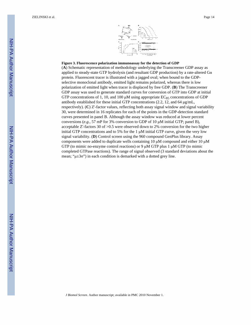
Figure 3. Fluorescence polarization immunoassay for the detection of GDP(A) Schematic representation of methodology underlying the Transcreener GDP assay asapplied to steady-state GTP hydrolysis (and resultant GDP production) by a rate-altered Gαprotein. Fluorescent tracer is illustrated with a jagged oval; when bound to the GDP-selective monoclonal antibody, emitted light remains polarized, whereas there is lowpolarization of emitted light when tracer is displaced by free GDP. (B) The TranscreenerGDP assay was used to generate standard curves for conversion of GTP into GDP at initialGTP concentrations of 1, 10, and 100 μM using appropriate EC85 concentrations of GDPantibody established for these initial GTP concentrations (2.2, 12, and 64 μg/mL,respectively). (C) Z'-factor values, reflecting both assay signal window and signal variability30, were determined in 16 replicates for each of the points in the GDP-detection standardcurves presented in panel B. Although the assay window was reduced at lower percentconversions (e.g., 57 mP for 3% conversion to GDP of 10 μM initial GTP; panel B),acceptable Z'-factors 30 of >0.5 were observed down to 2% conversion for the two higherinitial GTP concentrations and to 5% for the 1 μM initial GTP curve, given the very lowsignal variability. (D) Control screen using the 960 compound GenPlus library. Assaycomponents were added to duplicate wells containing 10 μM compound and either 10 μMGTP (to mimic no-enzyme control reactions) or 9 μM GTP plus 1 μM GTP (to mimiccompleted GTPase reactions). The range of signal observed (3 standard deviations about themean; “μ±3σ”) in each condition is demarked with a dotted grey line.
ZIELINSKI et al. Page 14
J Biomol Screen. Author manuscript; available in PMC 2010 November 1.
NIH
-PA Author Manuscript
NIH
-PA Author Manuscript
NIH
-PA Author Manuscript

Figure 4. RGS4 increases the steady-state GTPase activity of Gαi1(R178M/A326S) but notwildtype Gαi1, as measured using the Transcreener GDP assay and reported in absolute changein polarization (left panels) and GDP produced per Gα protein in reaction (right panels)R178M/A326S double-mutant (A,B) and wildtype (C,D) Gαi1 proteins were present at 50nM final concentration. Dashed lines represent reactions conducted in the presence, andsolid lines (“mock”) in the absence, of 250 nM RGS4 protein. (A,C) Change in polarization(ΔmP) at each time-point for indicated Gαi1 protein was calculated as ΔmP = |mP(Gαi1) −mP(no Gαi1)|. (B,D) Data from panels A and C were converted to GDP produced per mol ofinput Gαi1 using previously established standard curves for GDP detection in the presence ofGTP (e.g., Figure 3B). (E) Summary of initial rates obtained by the Transcreener GDP assayfor each Gαi1 mutant tested. “GAP factor” is defined as the ratio between steady-stateGTPase rate in the presence of RGS protein and steady-state GTPase rate in the absence ofRGS protein.
ZIELINSKI et al. Page 15
J Biomol Screen. Author manuscript; available in PMC 2010 November 1.
NIH
-PA Author Manuscript
NIH
-PA Author Manuscript
NIH
-PA Author Manuscript

Figure 5. Structural features of the RGS16/Gαi1·GDP·AlF −4 complex highlighting the locationsof Arg-178 and Ala-326 residue positions mutated in the Gαi1(R178M/A326S) variantThe RGS16/Gα complex (PDB id 2IK8; ref. 24), was rendered using PyMOL with theRGS16 RGS domain in orange and Gαi1 protein in blue, respectively. Gαi1 switch regionsare depicted in grey; switches one and two (SI, SII) are visible in the foreground, whereasswitch three is in the background and thus unlabeled. GDP is shown in magenta, the AlF ‒4ion is red, and Mg2+ ion is depicted as a yellow sphere. Residues arginine-178 andalanine-326 are rendered as `sticks' in green with CPK atomic coloring (nitrogen = blue,oxygen = red).
ZIELINSKI et al. Page 16
J Biomol Screen. Author manuscript; available in PMC 2010 November 1.
NIH
-PA Author Manuscript
NIH
-PA Author Manuscript
NIH
-PA Author Manuscript

Figure 6. RGS16 binds equivalently to wildtype Gαi1 and the rate-altered G αi1 (R178M/A326S)mutant(A,B) Sensorgrams derived from 600 second injections of various concentrations (3 nM to10 μM) of RGS16 over SPR biosensors of immobilized (A) wildtype Gαi1·GDP·AlF ‒4 or(B) Gαi1(R178M/A326S)·GDP·AlF ‡4. SPR experiments were also conducted with bothGαi1 subunits in their inactive, GDP-bound state (data not shown). (C,D) Resultantsensorgrams were used in equilibrium saturation binding analyses (as previously described24) to derive dissociation constants (KD values). RGS16 bound to wildtypeGαi1·GDP·AlF −4 with a dissociation constant of 124 nM (95% C.I. of 76–174 nM; panelC), whereas RGS16 bound to Gαi1(R178M/A326S)·GDP·AlF −4 with a dissociationconstant of 115 nM (64–166 nM; panel D). Note that interactions were not observed (foreither Gα subunit) when the Gα was GDP-bound (as expected; refs. 24,31), nor when RGS2was injected (data not shown).
ZIELINSKI et al. Page 17
J Biomol Screen. Author manuscript; available in PMC 2010 November 1.
NIH
-PA Author Manuscript
NIH
-PA Author Manuscript
NIH
-PA Author Manuscript

Figure 7. The steady-state GTPase activity of Gαi1(R178M/A326S) is increased by RGS4 andRGS16, but not by the Gαq-selective RGS2Transcreener GDP assays were performed as in Figure 4, using 250 nM of the indicatedRGS protein. Moles of GDP produced per mol input Gαi1(R178M/A326S) protein were firstplotted over time using GraphPad Prism and linear regression performed to determinesteady-state GTPase rates. Presented bar graph denotes GAP factors derived from thesesteady-state GTPase rates.
ZIELINSKI et al. Page 18
J Biomol Screen. Author manuscript; available in PMC 2010 November 1.
NIH
-PA Author Manuscript
NIH
-PA Author Manuscript
NIH
-PA Author Manuscript

Figure 8. Pilot screen and counterscreen using the 960 compound GenPlus libraryTranscreener GDP assay components were added to wells containing 50 nM Gαi1 (R178M/A326S) with (panel A) or without (panel B) 250 nM RGS4 protein and either 10 μMcompound (in 0.5% [v/v] final concentration of DMSO), 150 μM of reactive RGS4 inhibitorCCG-4986, or 0.5% DMSO only, as indicated in the legends. The range of signal observed(three standard deviations [σ] about the mean [μ]) is denoted by the dashed lines for the 960compound library screen using RGS4 and Gαi1 (R178M/A326S) (black; panel A, coefficientof variation [CV%] = 8.8%) and library counterscreen using Gαi1 (R178M/A326S) alone(gray; panel B, CV% = 3.0%). Data in panel A was obtained at 120 minutes of elapsedreaction time; data in panel B was obtained after 210 minutes of elapsed reaction time, giventhe slower GTPase (and GDP production) rate of Gαi1 (R178M/A326S) in the absence ofRGS4 GAP activity.
ZIELINSKI et al. Page 19
J Biomol Screen. Author manuscript; available in PMC 2010 November 1.
NIH
-PA Author Manuscript
NIH
-PA Author Manuscript
NIH
-PA Author Manuscript